Broadband interferometric subtraction of optical fields - Attoworld
←
→
Page content transcription
If your browser does not render page correctly, please read the page content below
Vol. 27, No. 3 | 4 Feb 2019 | OPTICS EXPRESS 2432

Broadband interferometric subtraction of
optical fields
T. BUBERL,1 P. SULZER,2 A. LEITENSTORFER,2 F. KRAUSZ,1,3 AND I.
PUPEZA1,3,*
1
  Max Planck Institute of Quantum Optics, Hans-Kopfermann-Straße, 1 85748, Garching, Germany
2
  Department of Physics and Center for Applied Photonics, University of Konstanz, 78457, Konstanz,
Germany
3
  Ludwig-Maximilians-Universität München, Am Coulombwall 1, 85748, Garching, Germany
*
  ioachim.pupeza@mpq.mpg.de

Abstract: We present a Mach-Zehnder-like interferometer capable of simultaneous super-
octave (950 – 2100 nm) destructive interference with an intensity extinction of 4 × 10⁻⁴.
Achromatic nulling is achieved by unbalancing the number of Fresnel reflections off optically
denser media in the two interferometer arms. With a methane gas sample in one
interferometer arm, we isolate the coherent molecular vibrational emission from the
broadband, impulsive excitation and quantitatively examine the potential improvement in
detectable concentration, compared to direct transmission geometry. The novel concept will
benefit sensing applications requiring high detection sensitivity and dynamic range, including
time-domain and frequency-domain spectroscopy.

© 2019 Optical Society of America under the terms of the OSA Open Access Publishing Agreement

1. Introduction
In traditional spectroscopic techniques, the signal associated with the process under scrutiny
manifests as a change of the radiation source intensity (or field). This imposes two main
limitations on the smallest detectable signal: Firstly, its magnitude is lower-bound by excess
source noise. Secondly, resolving a small change of a comparatively large signal requires
high-dynamic-range detection.
    In the field of Fourier transform infrared (FTIR) spectroscopy, the idea of a dual-beam
approach to overcome part of these limitations was first realized by Bar-Lev [1] in 1966. He
utilized both the interferogram and the anti-interferogram beam produced by an FTIR
spectrometer and measured the resulting difference signal with a balanced detector. Later
works inserted a reference and a sample into each beam and focused both outputs on a single
detector [2,3], which results in the cancellation of the common AC-components of the
opposing interferograms. In doing so, the effect of excess source noise on the detection limit
can be in principle suppressed down to the shot-noise level of the source [4]. Because
sequential measurements of sample and reference signal become unnecessary, the
measurement time and systematic errors are reduced and the dynamic range requirements of
the digitization electronics are relaxed [5]. However, none of the various dual-beam FTIR
schemes [1–6], addressed the issue of the limited dynamic range of the detector because the
DC-component of the interferograms was always present. In contrast, optically subtracting a
reference signal before detection spatially isolates the sought-for signal, disclosing it in front
of a quasi-zero background. This can be realized by an interferometer in whose arms the
propagation of light differs precisely by the process under scrutiny [7]. Ideally, in the
interferometer port combining both arms with opposing phase, only the (miniscule)
differences do not interfere destructively [8]. Thereby, the cancellation of the – usually orders
of magnitude stronger – reference signal facilitates excitation intensities far above the
saturation limit of the detector. Thus, the amplitude of the detected sample response can
always be raised above the detector and shot noise levels. In particular, interferometric,

#355896                                                               https://doi.org/10.1364/OE.27.002432
Journal © 2019                           Received 21 Dec 2018; accepted 26 Dec 2018; published 25 Jan 2019
Vol. 27, No. 3 | 4 Feb 2019 | OPTICS EXPRESS 2433
optical subtraction facilitates the direct comparison of two samples by increasing the visibility
of slight differences between their spectra.
To adapt interferometric, optical subtraction for broadband frequency-domain and
ultrafast, time-domain spectroscopy (which by definition is broadband) the challenge of
simultaneous cancellation of super-octave spectra has to be addressed. In this paper, we
present a Mach-Zehnder-type interferometer, with an unprecedented combination of broad
bandwidth (950 nm - 2100 nm) and achromatic intensity suppression (4 × 10⁻⁴). Compared to
the more complex approaches developed, e.g., for the direct observation of extrasolar planets
[9], our concept for achromatic nulling solely relies on the combination of Fresnel reflections
off boundaries between media with different refractive indices. In a proof-of-principle
experiment of differential molecular fingerprinting, we spatially isolate the resonant response
of a molecular sample to an impulsive, linear excitation from the instantaneous sample
response, carrying negligible fingerprint information.
A phase shift between two interferometer arms can be achieved by increasing the length
of one arm with respect to the other. The acquired phase shift Δφ is given by:
Δl
Δϕ = 2π , (1)
λ
where Δl is the difference in optical path length and λ the wavelength. For broadband
destructive interference, a wavelength-independent phase shift of π between the interfering
electro-magnetic fields is necessary. As Eq. (1) shows, the phase shift due to mutual delaying
however, is always wavelength dependent.
Several solutions for achromatic phase shifting have been developed in the field of
observational astronomy [10]. For instance, a pair of mirror-symmetric periscopes can be
used to geometrically implement a phase shift [11], the Gouy phase shift introduced by an
additional focus in one arm can be exploited [12], and a pair of right-angle Fresnel rhombs
acts as achromatic quarter-wave plates [13]. However, all these concepts are either technically
complex or not suitable for ultrashort-pulse applications. Here, we adapt a simple concept,
employed by Hayden et al. [14] for spectral absorption and dispersion measurements of a
liquid sample with a tunable narrowband laser, to broadband, achromatic nulling.
2. Interferometric setup
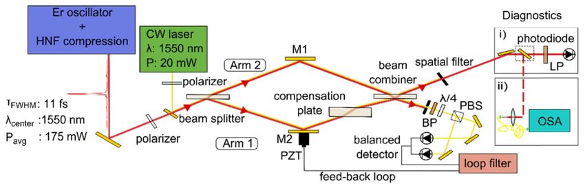
The experimental setup is sketched in Fig. 1. Apart from two adaptations, our setup is
equivalent to a Mach-Zehnder interferometer with uncoated surfaces for beam splitting and
combining. In the latter, an achromatic phase difference of π between the interferometer arms
is caused by the fundamental property of light waves undergoing no phase change when
reflected off a boundary to a medium with lower refractive index. However, light propagating
to the destructive port of such an interferometer is reflected three times in one arm and
transmitted twice and reflected once in the other arm, which leads to imperfect cancellation
for broadband radiation due to refractive index changes. Therefore, here we combine an equal
number of reflections and transmissions in each arm in the destructive port. For
achromaticity, the resulting additional material passage in arm 1 is exactly compensated for
by adding a window twice as thick as the beam combiner in arm 2. In the upper output port of
the interferometer in Fig. 1, the interfering light has opposing phase because the light is
reflected off the first surface of the beam splitter in arm 1 and off the second surface of the
beam combiner in arm 2.
The interferometer employs plane, 3-mm thick borosilicate crown glass (N-BK7)
windows for beam splitting and combining. The substrate thickness and material were chosen
to provide reasonable spatial separation between the reflection off the first and the second
surface for spatially filtering of undesired multiple reflections. The angle of incidence on the
windows is 60°, providing a reflectivity of approximately 18% for s-polarized light which is
close to the optimum splitting ratio of 20:80 for maximized power in the destructive port (seeVol. 27, No. 3 | 4 Feb 2019 | OPTICS EXPRESS 2434
Appendix). For symmetry reasons, small pointing fluctuations of the incident beam do not
affect the extinction. For stabilization of the optical path difference (OPD), the length of arm
1 is controlled via a feed-back loop acting on a piezo-electric transducer (PZT). A Hänsch-
Couillaud error signal [15] is generated using an auxiliary laser which exactly follows the
beam path of the main laser, except for an offset in beam height. The input polarization of the
auxiliary laser is linear with 45° rotation with respect to the propagation plane, so that both
arms acquire a polarization change in the constructive port (two transmissions in arm 2 and
two reflections in arm 1).
Our experiments were carried out with a super-octave spectrum generated by a
femtosecond Er:fiber laser system. The commercial oscillator-amplifier combination operates
at a repetition rate of 56 MHz. The 90-fs output is coupled into a highly nonlinear fiber
assembly, and spectrally broadened. Temporal compression results in a pulse duration of 11
fs spanning a spectrum between 950 nm and 2100 nm with 175 mW of average power (for
details see [16]).
It is noteworthy that our interferometer design is not specific to ultrashort, pulsed laser
sources and can in general be adapted to any kind of light source (including, e.g., thermal
sources) if the transmissive optics are selected according to the desired wavelength range.
Fig. 1. Mach-Zehnder-type interferometer: Light entering the interferometer is partially
reflected and partially transmitted at the first surface of the beam splitter. The reflected part
passes through the compensation plate and is partially reflected off the first surface of the
beam combiner. The transmitted part is partially reflected off the second surface of the beam
combiner. The destructive port of the interferometer is sent to the diagnostics, whereas the
constructive port is used to generate a Hänsch-Couillaud error signal from an auxiliary laser.
Through the interferometer we propagated 11-fs input pulses spanning from 950 nm to 2100
nm, generated from an erbium fiber laser system. BP: Band pass filter, FWHM = 12 nm at
1550 nm. PBS: Polarizing beam splitter. LP: 950 nm longpass filter. OSA: Optical spectrum
analyzer.
3. Theoretical limitations of interferometric extinction
Besides equal arm length, the alignment of the optical components within the interferometer
is critical for achromatic nulling. Ideally, all optical surfaces should be parallel to each other
and substrate thicknesses should match to minimize the influence of transverse misalignment,
and unbalance of intensity and dispersion. Figure 2(a) shows the calculated influence of
angular misalignment on the spatial overlap of both 30-cm-long arms after recombination,
which identically translates into a degradation of the extinction ratio (see Appendix).
According to our calculations, a misalignment of any optical element has the same
quantitative influence on the extinction ratio. For longer arm length, the alignment sensitivity
increases. With the precision of commercially available motorized kinematic mounts (1 µrad)
an extinction ratio of 6 × 10⁻⁶ for a central wavelength of 1550 nm and 6 × 10⁻⁷ for 10-µm
central wavelength is in principle attainable if only the spatial overlap is considered.
In addition to the alignment, the extinction ratio is influenced by dispersive effects owing
to substrate thickness variations. Figure 2(b) shows that the influence of substrate thickness
mismatch decreases for increasing pulse duration. The shaded orange area corresponds toVol. 27, No. 3 | 4 Feb 2019 | OPTICS EXPRESS 2435
thickness tolerances typically specified by manufacturers. If the worst combination of
thickness mismatch (0.9 mm between arm 1 and 2) is considered, the extinction ratio for 10-fs
input pulses is limited to 2 × 10⁻⁴.
Fig. 2. (a) Simulated decrease of extinction ratio for misalignment of two different optical
elements in the interferometer at 1550 nm and 10 µm central wavelength. (b) Simulated
decrease of extinction ratio depending on the difference in substrate thickness of beam splitter
and beam combiner for different input pulse durations. Shaded area: typical thickness
tolerances specified by the manufacturer.
4. Experimental interferometric extinction
In the experiment, the performance of the interferometer was characterized by recording the
signal at the destructive port with a photodiode (PD) while applying a saw-tooth voltage to
the PZT to scan the OPD through zero. Far from OPD = 0 a calibrated neutral density filter
was applied to ensure a linear response of the PD. Ideally, the interfering fields E1 and E2 are
identical and the following equality holds:
2 2
I con = E1 + E2 = 2 E1 = 4 I1 (2)
where Icon is the intensity of the ideal constructive interference and I1 is the intensity of one
arm. We measured the voltage corresponding to the intensity of one arm and divided the
recorded signal during destructive interference by four times this value. The resulting
extinction ratio reaches a minimum of 4.2 × 10⁻⁴. This value is in good agreement with the
computed values given the limited precision of manual alignment and the presence of air
fluctuations in our experiment.
It is worth noting that broadband constructive interference cannot be reached in the port
designed for destructive interference, because a modification of the OPD will result in a
wavelength-dependent phase difference between the two interferometer arms.
Figure 3(a) shows a comparison of the locked and the free-running PD signal at the
destructive port of the interferometer on a timescale of 30 s. In the locked state, the root mean
square (RMS) value of the extinction ratio is 6.1 × 10⁻⁴. Comparing the standard deviation of
the free-running and the locked PD signal reveals a reduction of the fluctuations of the
extinction ratio by almost one order of magnitude from 6.4 × 10⁻⁴ in the free-running to 7.2 ×
10−5 in the locked state. The discrepancy between the values of the extinction ratio in the
scanned and in the locked state is most likely caused by differing air fluctuations in the two
arms of the interferometer, which cannot be removed by the 1-dimensional PZT lock.
To investigate the wavelength dependence of the extinction, the stabilized signal was sent
to an optical spectrum analyzer (OSA). The power spectral densities (PSD) of the destructive
and the ideal constructive interference (defined as four times the PSD from a single arm) are
depicted in Fig. 3(b). The RMS value of the spectrally resolved extinction ratio of 6.2 × 10⁻⁴
is in excellent agreement with that of the spectrally integrated extinction ratio determined
with the PD.Vol. 27, No. 3 | 4 Feb 2019 | OPTICS EXPRESS 2436
Fig. 3. (a) Extinction ratio measured with a photo diode for the free-running interferometer and
the interferometer locked via the Hänsch-Couillaud error signal. (b) Spectrally resolved
constructive and destructive interference. The modulation of the destructive interference
spectrum at constant frequency corresponds to the thickness of the beam splitter/ combiner and
can be explained by imperfect spatial filtering of multiple reflections in the substrate(s). The
dashed orange curve shows the extinction ratio calculated by integrating the PSD in steps of 40
nm and dividing the destructive by the constructive PSD. The integration avoids extrema due
to non-coinciding modulations in the two signals arising from the fact that the destructive
interference lies only slightly above the detector noise of the OSA and therefore shows
stronger fluctuations than the constructive interference.
5. Sensitivity improvement for differential spectroscopy
In the following, we discuss the potential afforded by this interferometric extinction for
spectroscopic applications. To quantitatively investigate the advantages of the differential
measurement over the direct one, regarding dynamic range and detection sensitivity, in the
following we derive expressions for the signal strength (i.e. power in a given spectral
element) associated purely with the light-matter interaction and for the noise background, for
the direct and the differential measurement case, in analogy to [14]. In the direct
measurement, for each spectral element the power attenuation in a molecular sample is given
by Beer’s law:
P( S , dir ) = P0 e− A (3)
where P0 is the power recorded by the detector without sample, PS,dir is the power recorded
with sample, and A is the attenuation which is linearly proportional to the sample thickness,
the absorption cross section, and the concentration. For simplicity, we do not include the
complex molecular response here assuming that the considered concentrations are so small
that the influence of phase effects is negligible with respect to the extinction ratio. Note, that
in our experiment the “reference power” P0 is that of the laser beam, after propagation along
the empty sample arm when the other arm is blocked, and constitutes the background in the
direct measurement. Furthermore, the two power values refer to the nominal levels, i.e. do not
account for noise. Then, the difference between P0 and PS,dir yields a signal which can be
associated purely with the light-matter interaction in the sample arm, which we henceforth
refer to as “molecular signal” PM,dir:
P( M , dir ) = P0 − PS , dir = P0 (1 − e − A ). (4)
This signal vanishes in the limit A → 0 . Furthermore, in the above equation the background
P0 can be interpreted as P0 = lim( PS , dir ) , which holds in a more general sense. The amplitudes
A→ 0
E of the electric fields involved are connected to the measured powers P via the equalityVol. 27, No. 3 | 4 Feb 2019 | OPTICS EXPRESS 2437
E = c P , with some proportionality constant c. Thus, for the electric field amplitude ES,dir
the following equation can be derived:
E( S , dir ) = c PS , dir = E0 e− A / 2 , (5)
where E0 = c P0 . To model the differential measurement, we consider the overlap of the
electric fields E1 and E2 in the two interferometer arms, with opposing phase and a small
difference δ in field amplitude describing the imperfect extinction. According to the definition
of the extinction ratio Δ, the unbalance factor δ is given by δ = 4Δ . In the following, we
account for the unbalance by reducing the amplitude of E1 with respect to the ideal case.
Thus, after transmission through the sample, the field amplitude E1 reads:
E1 = E0 (1 − δ ) e− A / 2 (6)
and the reference field amplitude E2 is E0. The electric field amplitude at the destructive port
of the interferometer ES,diff is given by:
ES , diff = E2 − E1 (7)
and the power PS,diff arriving at the detector is:
1 2
ES , diff = P0 (1 − (1 − δ ) e − A / 2 ) .
2
PS , diff = 2
(8)
c
In analogy to the argumentation for the direct signal, the molecular signal in the differential
measurement is given by:
PM , diff = PS , diff − lim ( PS , diff )
A→ 0
)
(9)
(
= P0 (1 − (1 − δ ) e − A / 2 ) − δ 2 .
2
For both cases – direct and differential measurement – to determine the weakest detectable
attenuation A, the molecular signals, as derived in Eq. (4) and Eq. (9), need to be, compared
to the respective noise powers. For modeling the latter, we consider intensity noise of the
laser source PL, shot noise PSN, and detector noise Pdet as uncorrelated contributions to the
total noise power PN [8]:
PN = PL2 + PSN2 + Pdet2 . (10)
The total noise power PN,dir in the direct measurement is given by:
PS , dir h ν
(σP ) + ( Pdet ) ,
2 2
PN , dir = S , dir + (11)
T
where σ is the relative intensity noise of the source (in our experiment 0.21%, determined as
the relative standard deviation of 70 measurements of P0 with the OSA at 1645 nm), h is
Planck’s constant, ν is the optical frequency, and T is the measurement time for the respective
spectral element. Accordingly, the total noise power PN,diff is given by:
Ps , diff h ν
(σP ) + ( Pdet ) .
2 2
PN , diff = S , diff + (12)
TVol. 27, No. 3 | 4 Feb 2019 | OPTICS EXPRESS 2438
Here, we assume an ideal lock, i.e. that the mean extinction ratio Δ is maintained over the
entire measurement time. Note that the shot noise in our classical, linear optical system is
proportional to the square root of the number of photons arriving at the detector.
To be detectable, the molecular signal has to be larger than the total noise power, which
leads to the following inequalities:
PM , dir > PN , dir (13)
and
PM , diff > PN , diff . (14)
Setting the lhs equal to the rhs in Eq. (13) and Eq. (14) and solving for the attenuation A
reveals the smallest detectable attenuation in the direct and differential case depending on the
input power, the extinction ratio, and the relative intensity noise. Note that in this derivation
we accounted for the interferometer unbalancing by reducing the electric field E1 in the
sample arm by the factor (1-δ), see Eq. (6). A similar derivation can be performed by
reducing E2. While this leads to a slightly different strength of the molecular signal, this
difference is negligible for most practical cases.
Figure 4(a) shows the calculated limit of detection (LOD) defined as the minimum
detectable attenuation for increasing power per spectral element in the direct and differential
configuration for an extinction ratio of 6 × 10⁻⁴, considering a 10−3-nm broad spectral element
centered at 1545 nm and a measurement time of 10 ms for this spectral element. The
magnitude of the different noise contributions to the LOD is also shown in Fig. 4(a). In
general, the influence of detector noise and shot noise on the LOD decreases with increasing
power, i.e., number of photons, but the influence of intensity noise is constant. Thus, for all
configurations, the reachable LOD is ultimately limited by the intensity noise. However,
detector saturation sets a significantly stronger constraint on increasing the power in the direct
configuration, whereas in differential measurements the power reaching the detector
predominantly consists of signal specific to the light-matter interaction under scrutiny. In the
latter case, the incident power can exceed the saturation limit of the detector by orders of
magnitude.
Fig. 4. (a) Calculated limit of detection for increasing power per spectral element in the direct
and the differential configuration for an extinction ratio of 6 × 10⁻⁴ and a constant relative
intensity noise (RIN) of 0.2%. SN: shot noise. (b) Ratio of the direct and the differential LOD
for four different extinction ratios in the intensity-noise-limited regime (red crosses) and 1/
4Δ for comparison (dashed black line).
Figure 4(b) shows the ratio of the direct and the differential LOD for four different
extinction ratios in the intensity-noise-limited regime. The improvement towards the direct
measurement approaches 1/ 4Δ (dashed black line) for decreasing extinction ratio. TheVol. 27, No. 3 | 4 Feb 2019 | OPTICS EXPRESS 2439
coincidence of the two shot-noise-limited LOD curves in Fig. 4(a) confirms the fundamental
limit imposed by linear spectroscopy with classical light.
At our current input power, the optimum LOD is expected to be detector-noise limited,
and a factor of 20 better than in the direct measurement. Characterizing the RIN of the
destructive interference by calculating the standard deviation of 70 measurements with the
OSA, however, yields a value of 8%, which is more than one order of magnitude higher than
the 0.21% measured for the RIN in the direct measurement. The additional fluctuations of the
destructive interference must be caused by interferometer instabilities which cannot be
compensated for by our simple active locking scheme. Consequently, with the current
combination of a beam path in air, a one-dimensional PZT lock and a slow OSA for spectral
measurements, we are not able to experimentally demonstrate an increased sensitivity in
differential spectroscopy. However, in future implementations, the influence of these residual
fluctuations on the LOD can be mitigated – or even completely removed – by a mechanically
more stable design, by evacuating the interferometer environment, by an improved active
stabilization scheme and/or by fast acquisition of spectra such as, e.g., in dual-comb
configuration [17,18].
6. Differential molecular fingerprinting
For a proof-of-principle differential molecular fingerprinting measurement, we introduced
methane molecules in one arm of the interferometer. To this end, we placed a rudimentary gas
cell into one interferometer arm and, for balancing Fresnel reflections and dispersion, two
windows equivalent to those of the cell in the other arm. With ambient air in the gas cell the
extinction ratio was not notably affected. Filling the 8-cm long gas cell in the sample arm
with methane at an estimated pressure of roughly 1 bar resulted in an additional phase
difference between the two interferometer arms due to the non-resonant interaction with the
methane molecules. The group delay was compensated by geometrically adjusting the OPD.
Fig. 5. Methane resonances within the spectral coverage of the utilized OSA, in direct and
differential measurement configuration, at ~1 bar. For comparison, the inverted theory curve
for the differential measurement is shown.
Figure 5 shows the resonances in the 2ν3 vibrational overtone band of methane within the
spectral coverage of the utilized OSA, measured in the interferometric, differential
configuration as well as in a direct configuration, i.e. with the reference arm of the
interferometer blocked. The theory curve based on Eq. (9) and absorption data from HITRAN
[19] shows good qualitative agreement with the measurement. Due to the absence of a
suitable pressure gauge and the limited spectral resolution of the OSA, in the following we
explain the experimental observations in a qualitative picture rather than quantitatively. Up to
a small fraction of energy transferred to molecular vibrations, the instantaneous response of
the methane sample is identical to the excitation.Thus, in the differential measurement the
instantaneous response destructively interferes with the excitation pulse in the reference arm
and the differential signal mainly contains the isolated, resonant response emitted by the
vibrationally excited methane molecules [20]. However, in the direct measurement, theVol. 27, No. 3 | 4 Feb 2019 | OPTICS EXPRESS 2440
interference of the instantaneous and the phase-shifted, resonant sample response manifests
itself as a depletion of the PSD at the resonance frequencies (i.e. absorption in classical
spectroscopy).
7. Conclusion and outlook
We showed an unprecedented combination of interferometric deep nulling and broad
bandwidth with a Mach-Zehnder-like interferometer. Optimizing the quality of the optics, and
utilizing high-precision, kinematic mounts for alignment will push the extinction ratio
towards the theoretical limit. Improving the mechanical stability, operating the system in
vacuum and enhancing the active path length stabilization scheme will reduce residual
fluctuations of the destructive interference. In combination with higher input powers and a
better extinction ratio, this promises an improvement of the LOD by at least two orders of
magnitude as compared to the direct measurement.
Our proof of principle demonstrates the suitability of the novel concept for a variety of
applications. For instance, optical subtraction combined with frequency-comb spectroscopy
[21–23] will yield an unprecedented combination of sensitivity and spectral resolution.
Furthermore, time-resolved detection of the difference signal [17,18,24–27] promises fully
background-free detection of molecular fingerprints, by spatial and temporal separation of the
latter from an impulsive excitation.
Finally, the techniques presented here might both benefit, and profit from related
interferometer work. For instance, the broadband extinction might be useful in Mach-
Zehnder-type white-light interferometry, and technical solutions from gravitational wave
detectors [28] might help improve the stability of the interferometer.
8. Appendix
8.1 Optimum splitting ratio
We define the optimum power-splitting ratio at the beam splitter (and combiner) as the value
that maximizes the power propagating through each arm (with the other arm blocked) to the
destructive port. This optimization criterion maximizes the signal related to the light-matter
interaction under scrutiny, in the destructive interference arm. In the case of uncoated optics,
the power transmission T and reflection R at a material-air interface is defined by the Fresnel
equations for the refractive index of the material, the angle of incidence (AOI), and
polarization. For our interferometer geometry, the following equation describes the intensities
I1 and I2 in the interferometer arms 1 and 2 – respectively – reaching the difference port while
the other arm is blocked:
I1 = I 0 (TT T RT ) = I 2 = I 0 ( RT T T T ) = I 0 RT 4 , (15)
where I0 is the intensity entering the interferometer. Eq. (15) assumes that the external AOI on
all optics are identical, resulting in identical transmission and reflection. Neglecting
absorption, R = 1-T holds and one can easily calculate that I1 and I2 are maximized for R =
0.2 and T = 0.8. For N-BK7 this splitting ratio is achieved at an AOI of 62.5° for s-polarized
light.
8.2 Influence of misalignment
To analyze the influence of misalignment of optical elements on the extinction ratio, we built
a geometrical optics model of the interferometer using ray transfer matrix analysis. Angular
misalignments of the interferometer were included by extending the ABCD-matrices to 3x3-
matrices [29]. The observed quantity is the spatial overlap of the two arms after
recombination, which is mathematically expressed by the overlap integral over the
normalized complex transverse eld distributions of both arms [30]. The overlap integral ofVol. 27, No. 3 | 4 Feb 2019 | OPTICS EXPRESS 2441
two Gaussian beams for a certain angular misalignment normalized to the overlap integral of
two perfectly overlapping Gaussian beams (i.e. with equal beam parameters and no
misalignment) yields the extinction ratio given by 1 minus the aforementioned normalized
overlap integral.
8.3 Influence of beam splitter thickness mismatch
Ideally, the beam splitter and beam combiner should have the exact same thickness and the
compensation plate should have exactly twice this thickness. We calculated the influence of
deviations from this ideal case on the extinction ratio by comparing the intensities in the
destructive and constructive interference arms. The intensity of the destructive interference is
that of the difference of the electric fields of the input pulse dispersed by 9 mm borosilicate
crown glass (N-BK7) and by 9 mm + Δd N-BK7 where Δd is the thickness
tolerance/mismatch. The intensity of the constructive interference is that of the sum of the
electric fields of the input pulse dispersed by 9 mm N-BK7 and by 9 mm + Δd N-BK7. The
ratio of these two intensities is the extinction ratio plotted in Fig. 2(b).
References
1. H. Bar-Lev, “A dual-beam infrared interferometer-spectrometer,” Infrared Phys. 7(2), 93–98 (1967).
2. H. R. Chandrasekhar, L. Genzel, and J. Kuhl, “Double-beam fourier spectroscopy with interferometric
background compensation,” Opt. Commun. 17(1), 106–110 (1976).
3. D. Kuehl and P. R. Griffiths, “Dual-Beam Fourier Transform Infrared Spectrometer,” Anal. Chem. 50(3), 418–
422 (1978).
4. V. V. Goncharov and G. E. Hall, “Broadband laser enhanced dual-beam interferometry,” Opt. Lett. 37(12),
2406–2408 (2012).
5. D. L. Beduhn and R. L. White, “Advantages of Dual-Beam Interferometry in Fourier Transform Infrared
Spectrometry,” Appl. Spectrosc. 40(5), 628–632 (1986).
6. L. Genzel and J. Kuhl, “A new version of a Michelson interferometer for Fourier transform infrared
spectroscopy,” Infrared Phys. 18(2), 113–120 (1978).
7. J. P. Dakin, H. O. Edwards, and B. H. Weigl, “Progress with optical gas sensors using correlation spectroscopy,”
Sens. Actuators B Chem. 29(1), 87–93 (1995).
8. Z. Guan, M. Lewander, and S. Svanberg, “Quasi zero-background tunable diode laser absorption spectroscopy
employing a balanced Michelson interferometer,” Opt. Express 16(26), 21714–21720 (2008).
9. R. Bracewell, “Detecting nonsolar planets by spinning infrared interferometer,” Nature 274(5673), 780–781
(1978).
10. P. Gabor, A study of the performance of a nulling interferometer, Université Paris Sud - Paris XI, Paris, (2009).
11. E. Serabyn and M. M. Colavita, “Fully symmetric nulling beam combiners,” Appl. Opt. 40(10), 1668–1671
(2001).
12. J. Gay and Y. Rabbia, “An interferometric method for coronography”, C. R. Acad. Sci. Paris 322(3), (1996).
13. D. Mawet, C. Hanot, C. Lenaers, P. Riaud, D. Defrère, D. Vandormael, J. Loicq, K. Fleury, J.-Y. Plesseria, J.
Surdej, and S. Habraken, “Fresnel rhombs as achromatic phase shifters for infrared nulling interferometry,” Opt.
Express 15(20), 12850–12865 (2007).
14. J. Hayden, S. Hugger, F. Fuchs, and B. Lendl, “A quantum cascade laser-based Mach–Zehnder interferometer
for chemical sensing employing molecular absorption and dispersion,” Appl. Phys. B 124(2), 29 (2018).
15. T. W. Hänsch and B. Couillaud, “Laser frequency stabilization by polarization spectroscopy of a reflecting
reference cavity,” Opt. Commun. 35(3), 441–444 (1980).
16. D. Brida, G. Krauss, A. Sell, and A. Leitenstorfer, “UltrabroadbandEr:fiber lasers,” Laser Photonics Rev. 8(3),
409–428 (2014).
17. A. Muraviev, V. O. Smolski, Z. E. Loparo, and K. L. Vodopyanov, “Massively parallel sensing of trace
molecules and their isotopologues with broadband subharmonic mid-infrared frequency combs,” Nat. Photonics
12(4), 209–214 (2018).
18. I. Coddington, W. C. Swann, and N. R. Newbury, “Time-domain spectroscopy of molecular free-induction
decay in the infrared,” Opt. Lett. 35(9), 1395–1397 (2010).
19. I. E. Gordon, L. S. Rothman, C. Hill, R. V. Kochanov, Y. Tan, P. F. Bernath, M. Birk, V. Boudon, A.
Campargue, K. V. Chance, B. J. Drouin, J.-M. Flaud, R. R. Gamache, J. T. Hodges, D. Jacquemart, V. I.
Perevalov, A. Perrin, K. P. Shine, M.-A. H. Smith, J. Tennyson, G. C. Toon, H. Tran, V. G. Tyuterev, A. Barbe,
A. G. Császár, V. M. Devi, T. Furtenbacher, J. J. Harrison, J.-M. Hartmann, A. Jolly, T. J. Johnson, T. Karman,
I. Kleiner, A. A. Kyuberis, J. Loos, O. M. Lyulin, S. T. Massie, S. N. Mikhailenko, N. Moazzen-Ahmadi, H. S.
P. Müller, O. V. Naumenko, A. V. Nikitin, O. L. Polyansky, M. Rey, M. Rotger, S. W. Sharpe, K. Sung, E.
Starikova, S. A. Tashkun, J. V. Auwera, G. Wagner, J. Wilzewski, P. Wcisło, S. Yu, and E. J. Zak, “The
HITRAN2016 Molecular Spectroscopic Database,” J. Quant. Spectrosc. Radiat. Transf. 203, 3–69 (2017).Vol. 27, No. 3 | 4 Feb 2019 | OPTICS EXPRESS 2442
20. A. Laubereau and W. Kaiser, “Vibrational dynamics of liquids and solids investigated by picosecond light
pulses,” Rev. Mod. Phys. 50(3), 607–665 (1978).
21. T. Udem, R. Holzwarth, and T. W. Hänsch, “Optical frequency metrology,” Nature 416(6877), 233–237 (2002).
22. J. Ye and S. T. Cundiff, “Femtosecond optical frequency comb: principle, operation and applications”,
Femtosecond Optical Frequency Comb Technology (Springer, 2005).
23. S. A. Diddams, L. Hollberg, and V. Mbele, “Molecular fingerprinting with the resolved modes of a femtosecond
laser frequency comb,” Nature 445(7128), 627–630 (2007).
24. M. Tonouchi, “Cutting-edge terahertz technology,” Nat. Photonics 1(2), 97–105 (2007).
25. A. A. Lanin, A. A. Voronin, A. B. Fedotov, and A. M. Zheltikov, “Time-domain spectroscopy in the mid-
infrared,” Sci. Rep. 4(1), 6670 (2015).
26. I. Pupeza, M. Huber, W. Schweinberger, M. Trubetskov, S. A. Hussain, L. Vamos, O. Pronin, F. Habel, V.
Pervak, N. Karpowicz, M. Zigman, and F. Krausz in 2017 European Conference on Lasers and Electro-Optics –
European Quantum Electronics Conference (Optical Society of America, 2017), “Field-Resolved Spectroscopy
in the Molecular Fingerprint Region”, paper CH-2_4.
27. M. Huber, W. Schweineberger, M. Trubetskov, S. A. Hussein, O. Pronin, E. Fill, A. Apolonski, M. Zigman, and
F. Krausz, I. Pupeza in 2017 European Conference on Lasers and Electro-Optics – European Quantum
Electronics Conference (Optical Society of America, 2017), “Detection sensitivity of field-resolved spectroscopy
in the molecular fingerprint region”, paper CH-P_4.
28. B. P. Abbott, R. Abbott, R. Adhikari, P. Ajith, B. Allen, G. Allen, R. S. Amin, S. B. Anderson, W. G. Anderson,
M. A. Arain, M. Araya, H. Armandula, P. Armor, Y. Aso, S. Aston, P. Aufmuth, C. Aulbert, S. Babak, P. Baker,
S. Ballmer, C. Barker, D. Barker, B. Barr, P. Barriga, L. Barsotti, M. A. Barton, I. Bartos, R. Bassiri, M.
Bastarrika, B. Behnke, M. Benacquista, J. Betzwieser, P. T. Beyersdorf, I. A. Bilenko, G. Billingsley, R. Biswas,
E. Black, J. K. Blackburn, L. Blackburn, D. Blair, B. Bland, T. P. Bodiya, L. Bogue, R. Bork, V. Boschi, S.
Bose, P. R. Brady, V. B. Braginsky, J. E. Brau, D. O. Bridges, M. Brinkmann, A. F. Brooks, D. A. Brown, A.
Brummit, G. Brunet, A. Bullington, A. Buonanno, O. Burmeister, R. L. Byer, L. Cadonati, J. B. Camp, J.
Cannizzo, K. C. Cannon, J. Cao, L. Cardenas, S. Caride, G. Castaldi, S. Caudill, M. Cavaglià, C. Cepeda, T.
Chalermsongsak, E. Chalkley, P. Charlton, S. Chatterji, S. Chelkowski, Y. Chen, N. Christensen, C. T. Y.
Chung, D. Clark, J. Clark, J. H. Clayton, T. Cokelaer, C. N. Colacino, R. Conte, D. Cook, T. R. C. Corbitt, N.
Cornish, D. Coward, D. C. Coyne, J. D. E. Creighton, T. D. Creighton, A. M. Cruise, R. M. Culter, A. Cumming,
L. Cunningham, S. L. Danilishin, K. Danzmann, B. Daudert, G. Davies, E. J. Daw, D. DeBra, J. Degallaix, V.
Dergachev, S. Desai, R. DeSalvo, S. Dhurandhar, M. Díaz, A. Dietz, F. Donovan, K. L. Dooley, E. E. Doomes,
R. W. P. Drever, J. Dueck, I. Duke, J.-C. Dumas, J. G. Dwyer, C. Echols, M. Edgar, A. Effler, P. Ehrens, E.
Espinoza, T. Etzel, M. Evans, T. Evans, S. Fairhurst, Y. Faltas, Y. Fan, D. Fazi, H. Fehrmenn, L. S. Finn, K.
Flasch, S. Foley, C. Forrest, N. Fotopoulos, A. Franzen, M. Frede, M. Frei, Z. Frei, A. Freise, R. Frey, T. Fricke,
P. Fritschel, V. V. Frolov, M. Fyffe, V. Galdi, J. A. Garofoli, I. Gholami, J. A. Giaime, S. Giampanis, K. D.
Giardina, K. Goda, E. Goetz, L. M. Goggin, G. González, M. L. Gorodetsky, S. Goßler, R. Gouaty, A. Grant, S.
Gras, C. Gray, M. Gray, R. J. S. Greenhalgh, A. M. Gretarsson, F. Grimaldi, R. Grosso, H. Grote, S. Grunewald,
M. Guenther, E. K. Gustafson, R. Gustafson, B. Hage, J. M. Hallam, D. Hammer, G. D. Hammond, C. Hanna, J.
Hanson, J. Harms, G. M. Harry, I. W. Harry, E. D. Harstad, K. Haughian, K. Hayama, J. Heefner, I. S. Heng, A.
Heptonstall, M. Hewitson, S. Hild, E. Hirose, D. Hoak, K. A. Hodge, K. Holt, D. J. Hosken, J. Hough, D.
Hoyland, B. Hughey, S. H. Huttner, D. R. Ingram, T. Isogai, M. Ito, A. Ivanov, B. Johnson, W. W. Johnson, D. I.
Jones, G. Jones, R. Jones, L. Ju, P. Kalmus, V. Kalogera, S. Kandhasamy, J. Kanner, D. Kasprzyk, E.
Katsavounidis, K. Kawabe, S. Kawamura, F. Kawazoe, W. Kells, D. G. Keppel, A. Khalaidovski, F. Y. Khalili,
R. Khan, E. Khazanov, P. King, J. S. Kissel, S. Klimenko, K. Kokeyama, V. Kondrashov, R. Kopparapu, S.
Koranda, D. Kozak, B. Krishnan, R. Kumar, P. Kwee, P. K. Lam, M. Landry, B. Lantz, A. Lazzarini, H. Lei, M.
Lei, N. Leindecker, I. Leonor, C. Li, H. Lin, P. E. Lindquist, T. B. Littenberg, N. A. Lockerbie, D. Lodhia, M.
Longo, M. Lormand, P. Lu, M. Lubinski, A. Lucianetti, H. Lück, B. Machenschalk, M. MacInnis, M.
Mageswaran, K. Mailand, I. Mandel, V. Mandic, S. Márka, Z. Márka, A. Markosyan, J. Markowitz, E. Maros, I.
W. Martin, R. M. Martin, J. N. Marx, K. Mason, F. Matichard, L. Matone, R. A. Matzner, N. Mavalvala, R.
McCarthy, D. E. McClelland, S. C. McGuire, M. McHugh, G. McIntyre, D. J. A. McKechan, K. McKenzie, M.
Mehmet, A. Melatos, A. C. Melissinos, D. F. Menéndez, G. Mendell, R. A. Mercer, S. Meshkov, C. Messenger,
M. S. Meyer, J. Miller, J. Minelli, Y. Mino, V. P. Mitrofanov, G. Mitselmakher, R. Mittleman, O. Miyakawa, B.
Moe, S. D. Mohanty, S. R. P. Mohapatra, G. Moreno, T. Morioka, K. Mors, K. Mossavi, C. MowLowry, G.
Mueller, H. Müller-Ebhardt, D. Muhammad, S. Mukherjee, H. Mukhopadhyay, A. Mullavey, J. Munch, P. G.
Murray, E. Myers, J. Myers, T. Nash, J. Nelson, G. Newton, A. Nishizawa, K. Numata, J. O’Dell, B. O’Reilly,
R. O’Shaughnessy, E. Ochsner, G. H. Ogin, D. J. Ottaway, R. S. Ottens, H. Overmier, B. J. Owen, Y. Pan, C.
Pankow, M. A. Papa, V. Parameshwaraiah, P. Patel, M. Pedraza, S. Penn, A. Perraca, V. Pierro, I. M. Pinto, M.
Pitkin, H. J. Pletsch, M. V. Plissi, F. Postiglione, M. Principe, R. Prix, L. Prokhorov, O. Punken, V. Quetschke,
F. J. Raab, D. S. Rabeling, H. Radkins, P. Raffai, Z. Raics, N. Rainer, M. Rakhmanov, V. Raymond, C. M. Reed,
T. Reed, H. Rehbein, S. Reid, D. H. Reitze, R. Riesen, K. Riles, B. Rivera, P. Roberts, N. A. Robertson, C.
Robinson, E. L. Robinson, S. Roddy, C. Röver, J. Rollins, J. D. Romano, J. H. Romie, S. Rowan, A. Rüdiger, P.
Russell, K. Ryan, S. Sakata, L. S. de la Jordana, V. Sandberg, V. Sannibale, L. Santamaría, S. Saraf, P. Sarin, B.
S. Sathyaprakash, S. Sato, M. Satterthwaite, P. R. Saulson, R. Savage, P. Savov, M. Scanlan, R. Schilling, R.
Schnabel, R. Schofield, B. Schulz, B. F. Schutz, P. Schwinberg, J. Scott, S. M. Scott, A. C. Searle, B. Sears, F.
Seifert, D. Sellers, A. S. Sengupta, A. Sergeev, B. Shapiro, P. Shawhan, D. H. Shoemaker, A. Sibley, X.Vol. 27, No. 3 | 4 Feb 2019 | OPTICS EXPRESS 2443
Siemens, D. Sigg, S. Sinha, A. M. Sintes, B. J. J. Slagmolen, J. Slutsky, J. R. Smith, M. R. Smith, N. D. Smith,
K. Somiya, B. Sorazu, A. Stein, L. C. Stein, S. Steplewski, A. Stochino, R. Stone, K. A. Strain, S. Strigin, A.
Stroeer, A. L. Stuver, T. Z. Summerscales, K.-X. Sun, M. Sung, P. J. Sutton, G. P. Szokoly, D. Talukder, L.
Tang, D. B. Tanner, S. P. Tarabrin, J. R. Taylor, R. Taylor, J. Thacker, K. A. Thorne, A. Thüring, K. V.
Tokmakov, C. Torres, C. Torrie, G. Traylor, M. Trias, D. Ugolini, J. Ulmen, K. Urbanek, H. Vahlbruch, M.
Vallisneri, C. V. D. Broeck, M. V. van der Sluys, A. A. van Veggel, S. Vass, R. Vaulin, A. Vecchio, J. Veitch, P.
Veitch, C. Veltkamp, A. Villar, C. Vorvick, S. P. Vyachanin, S. J. Waldman, L. Wallace, R. L. Ward, A.
Weidner, M. Weinert, A. J. Weinstein, R. Weiss, L. Wen, S. Wen, K. Wette, J. T. Whelan, S. E. Whitcomb, B. F.
Whiting, C. Wilkinson, P. A. Willems, H. R. Williams, L. Williams, B. Willke, I. Wilmut, L. Winkelmann, W.
Winkler, C. C. Wipf, A. G. Wiseman, G. Woan, R. Wooley, J. Worden, W. Wu, I. Yakushin, H. Yamamoto, Z.
Yan, S. Yoshida, M. Zanolin, J. Zhang, L. Zhang, C. Zhao, N. Zotov, M. E. Zucker, H. Mühlen, and J. Zweizig,
“LIGO: the Laser Interferometer Gravitational-Wave Observatory,” Rep. Prog. Phys. 72(7), 76901 (2009).
29. H. Kogelnik and T. Li, “Laser beams and resonators,” Appl. Opt. 5(10), 1550–1567 (1966).
30. W. B. Joyce and B. C. DeLoach, “Alignment of Gaussian beams,” Appl. Opt. 23(23), 4187–4196 (1984).You can also read