Case Report Esophageal Perforation following Accidental Ingestion of a Razor Blade
←
→
Page content transcription
If your browser does not render page correctly, please read the page content below
Hindawi
Case Reports in Surgery
Volume 2022, Article ID 1974147, 4 pages
https://doi.org/10.1155/2022/1974147

Case Report
Esophageal Perforation following Accidental Ingestion of a
Razor Blade

          Suraj Shrestha ,1 Ranjan Sapkota ,2 Suraj Bhatta,1 Sanjeev Kharel ,1
          Bibek Man Shrestha ,1 and Aakriti Sharma2
          1
              Maharajgunj Medical Campus, Institute of Medicine, Kathmandu, Nepal
          2
              Department of Cardio-Thoracic and Vascular Surgery, Manmohan Cardio-Thoracic Vascular and Transplant Center,
              Kathmandu, Nepal

          Correspondence should be addressed to Ranjan Sapkota; ranjansapkota@gmail.com

          Received 11 October 2021; Accepted 3 March 2022; Published 17 March 2022

          Academic Editor: Paola De Nardi

          Copyright © 2022 Suraj Shrestha et al. This is an open access article distributed under the Creative Commons Attribution License,
          which permits unrestricted use, distribution, and reproduction in any medium, provided the original work is properly cited.

          Background. Ingestion of sharp foreign bodies is uncommon and often underreported. It can present with esophageal perforation
          which is a life-threatening complication requiring prompt diagnosis and management. Case Presentation. We report a case of
          accidental ingestion of a razor blade in a chronic alcoholic who presented with hematemesis after an esophageal perforation,
          the diagnosis of which was confirmed by radiology. Conclusion. Early recognition of esophageal perforation is crucial for early
          intervention. Proper history taking and radiological investigations are a key to reaching a diagnosis.

1. Background                                                          following probable ingestion of a foreign body under the
                                                                       influence of alcohol. The patient was apparently unaware
Ingestion of foreign bodies (FB) is common in children, while          of the incident and timing of the ingestion. There was no
in adults it is commonly seen among those with psychiatric             history of fever, loss of consciousness, body discoloration,
disorders, mental retardation, prisoners, and alcoholics [1].          significant weight loss, dysphagia, and similar events in the
The ingestion of a sharp foreign body like a razor blade can           past. He had been consuming approximately 1.5 liters of
be catastrophic and may be associated with numerous com-               homemade alcohol every day for the last 40 years. There is
plications such as perforation, mediastinitis, subcutaneous            no history of intake of psychoactive substances and of any
emphysema, aspiration, mediastinal abscess, bleeding, com-             documented psychiatric illness.
plete bowel obstruction, and pressure necrosis with subse-                 On examination, the patient was ill-looking, alert, and
quent aortic/tracheal fistula formation. This mandates a                afebrile with a blood pressure of 110/80 mmHg, pulse rate
prompt diagnosis and urgent removal of these objects [2].              of 100 beats per minute, respiratory rate of 28 per minute,
    Reports of ingestion of sharp objects, more specifically            and SpO2 of 82% in room air with right intercostal drain
razor blades, are rare in the literature. Herein, we report a          and nasogastric tube in situ draining blood mixed contents.
case of a chronic alcoholic who accidentally ingested a razor          On chest auscultation, air entry was reduced bilaterally with-
blade and presented with esophageal perforation.                       out any added sound. The abdomen was soft, and examina-
                                                                       tion of other systems was unremarkable.
2. Case Presentation                                                       Chest X-ray revealed pneumomediastinum, pneumoper-
                                                                       icardium, and right-sided pleural effusion with a tube in the
A 58-year-old male was referred to our center with a two-              right chest. Contrast-enhanced computed tomography
day history of acute anterior chest pain radiating to the              (CECT) of the chest and abdomen revealed a 5 cm × 2 cm
shoulder, dysphagia, and multiple episodes of hematemesis              double-edged shaving razor blade in the midesophagus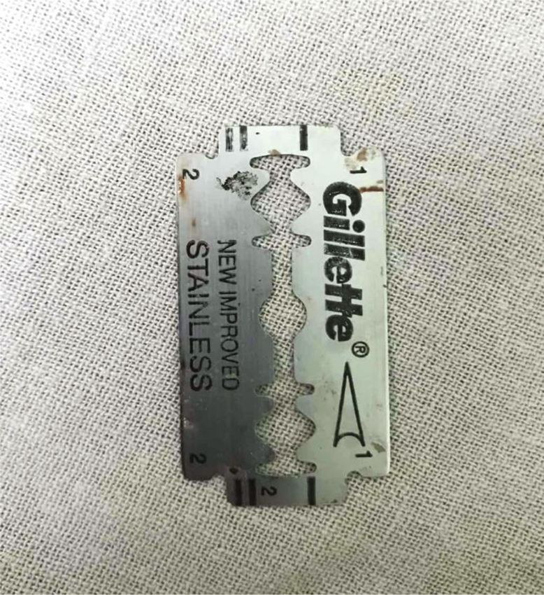
2                                                                                                           Case Reports in Surgery

                                         (a)                                              (b)

Figure 1: CT chest. (a) Sharp razor blade (arrow) in the mediastinum with pneumomediastinum and moderately large right pleural effusion.
(b) Reconstructed image shows the whole blade; apparently, the NG tube traveled through the mediastinum.

                                                                     3. Discussion
                                                                     Foreign body ingestion is not an uncommon entity. Individ-
                                                                     uals under the influence of drugs and/or alcohol often pres-
                                                                     ent to the emergency after ingesting multiple foreign bodies.
                                                                     The ingestion tends to be spontaneous, and frequently,
                                                                     patients do not remember swallowing the object [3]. Our
                                                                     patient was too unaware of the incident.
                                                                         One-third of foreign bodies retained in the gastrointesti-
                                                                     nal tract are present in the esophagus [4]. The duodenal
                                                                     loop, duodenojejunal junction, ileocecal valve, and appendix
                                                                     are other potential areas where it can get lodged due to ana-
                                                                     tomical narrowing or acute angulations [5]. These ingested
                                                                     sharp objects that are retained in the esophagus carry the
                                                                     risk of perforating the esophagus leading to acute mediasti-
                                                                     nitis, acute bleeding, or rarely abscess or fistula formation,
                                                                     which may even result in death and are thus considered by
                                                                     far the most dangerous [6, 7].
                                                                         The common presenting complaint in esophageal perfo-
                                                                     ration is pain in the neck, chest, abdomen, or shoulder,
            Figure 2: The razor blade after removal.                 depending upon the location of the perforation. Episodes
                                                                     of forceful or repeated vomiting may precede pain. In about
                                                                     25% of the patients, pain is followed by vomiting and dys-
causing a 45 mm perforation at its lateral aspect at T6-8 level      pnea. Dyspnea is more common in thoracic esophageal per-
with extensive pneumomediastinum and surgical emphy-                 forations. The presence of the Mackler’s triad, a history of
sema in the neck along with bilateral pleural effusions               forceful emesis, subxiphoid chest pain, and subcutaneous
(Figure 1).                                                          emphysema often suggests acute esophageal rupture [8].
    With a diagnosis of razor blade ingestion causing tho-           Our patient had chest pain, dysphagia, hematemesis, and
racic esophageal perforation, a decision was taken to pursue         subcutaneous emphysema in the neck.
operative management. The patient underwent a right-sided                For those who ingested FB accidentally under the influ-
posterolateral thoracotomy. The blade was removed intact, the        ence of alcohol as in our case, medical evaluation can be diffi-
pleural cavity was cleared of pus and debris (Figure 2). The         cult as they often cannot provide a reliable history [9]. Because
mediastinum and the pleural cavity were thoroughly washed,           of the aforementioned dreadful complications, patients with
and the chest closed with a drain. The left neck was explored        suspected esophageal perforation should be regarded as criti-
and the esophagus was exteriorized (Figure 3). A feeding jeju-       cally ill. Early recognition with timely intervention is crucial
nostomy (FJ) was done via a small laparotomy. After an ICU           for early recovery.
stay of 3 days, he recovered well. At discharge, he was self-            Chest and abdominal X-rays can elicit a radiopaque FB-
ambulating, receiving approximately 1500 Kcal diet per day           like metallic material among suspected esophageal FB [10].
via FJ and off oxygen. A gastric pull-up operation has been           However, it was not easily visualized in the X-ray of our
planned in about six weeks’ time.                                    patient. As relevant to our case, if a patient is unable to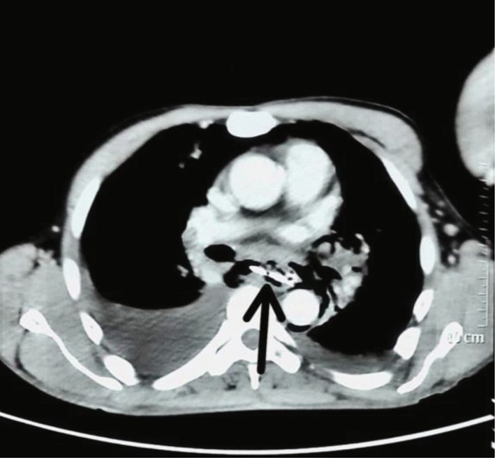
Case Reports in Surgery 3
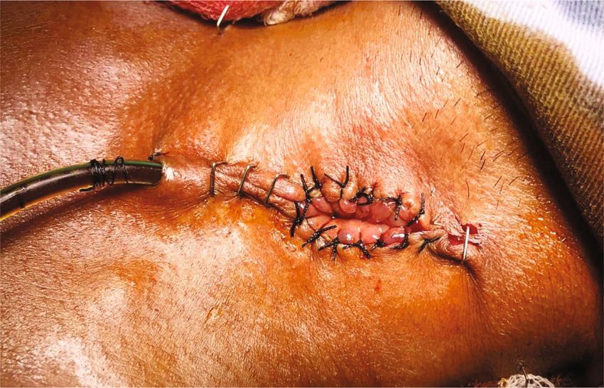
Figure 3: Exteriorized esophagus.
provide a satisfactory history and chest X-ray is inconclu- Abbreviations
sive, other modalities of diagnosis like CT scan and diagnos-
tic endoscopy are generally the preferred modalities. Pleural CECT: Contrast-enhanced computed tomography
effusion, pneumomediastinum, subcutaneous emphysema, FB: Foreign body
hydrothorax, and pneumothorax are some other indirect FJ: Feeding jejunostomy
signs of esophageal injury that aid in the diagnosis [11]. ICU: Intensive care unit.
Apart from demonstrable perforation, our patient also had
a bilateral pleural collection, pneumomediastinum, and sub- Data Availability
cutaneous emphysema in the neck.
Of all the FB ingested, about 20% need endoscopic All the necessary information are provided within the case
extraction and surgical interventions are required in only report.
less than 1% of presentations while the remaining pass
through the gastrointestinal tract uneventfully [12]. How- Consent
ever, with the ingestion of sharp FBs, the need for surgical
intervention is as high as 35% as their complications are Written informed consent was obtained from the patient’s
high and if retained in the esophagus can be life- family for publication of this case report and accompanying
threatening [4, 13]. Thus, extraction of the foreign body as images.
soon as the diagnosis is made is mandatory [4]. As esopha-
geal perforation is a thoracic emergency, surgical treatment Conflicts of Interest
is the rule. Hence, aggressive operative intervention remains
The authors declare that they have no competing interests.
the mainstay for the treatment [14, 15]. Further, extraction
of the foreign body, enteric without oral feeding, antibiotics,
and drainage of collections constitute the treatment strategy Authors’ Contributions
[16]. The exclusion and diversion of the esophagus promote
Ranjan Sapkota (RS) and Aakriti Sharma (AS) are involved in
healing in addition to minimizing the risk of further con-
the study concept, data collection, and surgical therapy for the
tamination and infection [17]. Our patient was kept nil
patient. Suraj Shrestha (SS) and Suraj Bhatta (SB) are involved
orally after suspected esophageal perforation, underwent
in writing-original draft preparation. Bibek Man Shrestha, SS,
emergency thoracotomy with FB removal along with esoph-
SB, and Sanjeev Kharel (SK) are involved in editing and writ-
ageal diversion and insertion of an FJ.
ing. RS and AS are involved in reviewing the manuscript. All
In addition to the medical and surgical treatment, psy-
the authors read and approved the final manuscript.
chiatric evaluations are necessary in cases of intentional
and/or repeat foreign body ingestion [18]. Our patient was
evaluated for alcohol dependence syndrome while no other Supplementary Materials
psychiatric illness was found. CARE-checklist. (Supplementary Materials)
4. Conclusion References
Ingestion of sharp objects causing esophageal perforation is [1] W. A. Webb, “Management of foreign bodies of the upper gas-
a life-threatening condition. Prompt diagnosis is imperative trointestinal tract: update,” Gastrointestinal Endoscopy, vol. 41,
for favorable results. no. 1, pp. 39–51, 1995.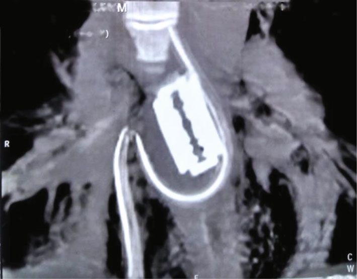
4                                                                       Case Reports in Surgery

 [2] S. O. Ikenberry, T. L. Jue, M. A. Anderson et al., “Management
     of ingested foreign bodies and food impactions,” Gastrointesti-
     nal Endoscopy, vol. 73, no. 6, pp. 1085–1091, 2011.
 [3] N. Koyuncu, S. Yilmaz, and S. Soysal, “An unusual cause of
     chest pain: foreign body in the oesophagus,” Emergency Medi-
     cine Journal, vol. 24, no. 1, article e1, 2007.
 [4] D. Weissberg and Y. Refaely, “Foreign bodies in the esopha-
     gus,” The Annals of Thoracic Surgery, vol. 84, no. 6,
     pp. 1854–1857, 2007.
 [5] T. B. Hunter and M. S. Taljanovic, “Foreign bodies,” Radio-
     graphics, vol. 23, no. 3, pp. 731–757, 2003.
 [6] R. S. Miller, J. Paul Willging, M. J. Rutter, and K. Rookkapan,
     “Chronic esophageal foreign bodies in pediatric patients: a ret-
     rospective review,” International Journal of Pediatric Otorhi-
     nolaryngology, vol. 68, no. 3, pp. 265–272, 2004.
 [7] R. R. Bloom, P. H. Nakano, S. W. Gray, and J. E. Skandalakis,
     “Foreign bodies of the gastrointestinal tract,” The American
     Surgeon, vol. 52, no. 11, pp. 618–621, 1986.
 [8] S. M. Griffin, P. J. Lamb, J. Shenfine, D. L. Richardson,
     D. Karat, and N. Hayes, “Spontaneous rupture of the oesoph-
     agus,” The British Journal of Surgery, vol. 95, no. 9, pp. 1115–
     1120, 2008.
 [9] M. Birk, P. Bauerfeind, P. H. Deprez et al., “Removal of foreign
     bodies in the upper gastrointestinal tract in adults: European
     Society of Gastrointestinal Endoscopy (ESGE) clinical guide-
     line,” Endoscopy, vol. 48, no. 5, pp. 489–496, 2016.
[10] M. Wai Pak, W. Chung Lee, H. Kwok Fung, and C. A. van Has-
     selt, “A prospective study of foreign-body ingestion in 311
     children,” International Journal of Pediatric Otorhinolaryngol-
     ogy, vol. 58, no. 1, pp. 37–45, 2001.
[11] C. M. Vial and R. I. Whyte, “Boerhaave's syndrome: diagnosis
     and treatment,” The Surgical Clinics of North America, vol. 85,
     no. 3, pp. 515–524, 2005.
[12] P. Ambe, S. A. Weber, M. Schauer, and W. T. Knoefel, “Swal-
     lowed foreign bodies in adults,” Deutsches Ärzteblatt Interna-
     tional, vol. 109, no. 50, pp. 869–875, 2012.
[13] R. Palta, A. Sahota, A. Bemarki, P. Salama, N. Simpson, and
     L. Laine, “Foreign-body ingestion: characteristics and out-
     comes in a lower socioeconomic population with predomi-
     nantly intentional ingestion,” Gastrointestinal Endoscopy,
     vol. 69, no. 3, pp. 426–433, 2009.
[14] K. Athanassiadi, M. Gerazounis, E. Metaxas, and N. Kalantzi,
     “Management of esophageal foreign bodies: a retrospective
     review of 400 cases1,” European Journal of Cardio-Thoracic
     Surgery, vol. 21, no. 4, pp. 653–656, 2002.
[15] B. L. Bufkin, J. I. Miller Jr., and K. A. Mansour, “Esophageal
     perforation: emphasis on management,” The Annals of Tho-
     racic Surgery, vol. 61, no. 5, pp. 1447–1452, 1996.
[16] R. R. Naidoo and A. A. Reddi, “Chronic retained foreign bod-
     ies in the esophagus,” The Annals of Thoracic Surgery, vol. 77,
     no. 6, pp. 2218–2220, 2004.
[17] P. D. Kiernan, J. Rhee, L. Collazo, V. Hetrick, B. Vaughan, and
     P. Graling, “Complete esophageal diversion: a simplified, easily
     reversible technique,” Journal of the American College of Sur-
     geons, vol. 200, no. 5, 2005.
[18] C. Palese and F. H. Al-Kawas, “Repeat intentional foreign body
     ingestion: the importance of a multidisciplinary approach,”
     Gastroenterology & Hepatology, vol. 8, no. 7, pp. 485-486,
     2012.
You can also read