Cerebral venous thrombosis: a spectrum of imaging findings
←
→
Page content transcription
If your browser does not render page correctly, please read the page content below

Singapore Med J 2021; 62(12): 630-635
Pictorial Essay
                                                                                                                        https://doi.org/10.11622/smedj.2021235

CMEArticle
Cerebral venous thrombosis: a spectrum of imaging
findings
                                                            Shuliang Ge1,       FRCR, MBBS,    Jinhang Wen1,         MBBS, FRCR,   Pin Lin Kei1,     MBChB, FRCR

INTRODUCTION                                                                        of the brain. The superior sagittal sinus, inferior sagittal sinus and
Cerebral venous thrombosis (CVT) is uncommon compared to                            straight sinus converge at the torcular herophili, deep into the
cerebral arterial disease, and its initial clinical manifestations                  occipital protuberance. This drains into the bilateral transverse
are often non-specific. The most common presenting symptom                          sinuses, sigmoid sinuses and internal jugular veins.
is headache, while other clinical manifestations include focal                           The superficial cerebral (cortical) veins are located in the
neurological deficit, seizures, nausea and/or vomiting, and altered                 subarachnoid space and drain into the nearest dural venous sinus,
mental status.(1) Unenhanced head computed tomography (CT)                          either directly or via anastomotic pathways. The vein of Trolard
is usually the first-line investigation of choice for these patients.               (superior anastomotic vein) connects the superficial middle
However, the most striking findings on imaging can mimic other                      cerebral veins with the superior sagittal sinus, while the vein of
pathologies, leading to delayed or wrong diagnoses. As such, a                      Labbé (inferior anastomotic vein) connects the superficial middle
good understanding of the pathophysiology, anatomy and myriad                       cerebral veins with the transverse sinus.
imaging findings of CVT is pivotal in making a timely and accurate                       The deep venous system drains the thalami, septum
diagnosis. An early diagnosis enables early institution of appropriate              pellucidum, basal ganglia, upper brainstem and deep white
treatment, resulting in significantly improved patient morbidity and                matter of the parietal, temporal and frontal lobes. The internal
mortality.(2) In this article, we illustrate a wide spectrum of CVT cases.          cerebral veins join the basal vein of Rosenthal to form the vein
                                                                                    of Galen, which then joins the inferior sagittal sinus to form the
PATHOPHYSIOLOGY AND RISK FACTORS                                                    straight sinus.
Two main mechanisms that contribute to the clinical presentation                         The superior sagittal sinus is the most commonly affected sinus
of CVT have been proposed.(3) Firstly, CVT can result in increased                  in CVT (62.0%), followed by the transverse and sigmoid sinuses
retrograde pressures in the cerebral capillaries and venules,                       (41.2%–44.7% per side), straight sinus (18.0%), cortical veins
leading to decreased cerebral perfusion, ischaemic injury and                       (17.1%), jugular veins (11.9%) and deep venous system (10.9%).(1)
cytotoxic oedema. Disruption of the blood-brain barrier leads
to vasogenic oedema, while rupture of capillaries and venules                       IMAGING FINDINGS AND CASE
results in parenchymal haemorrhages. Secondly, CVT can result in                    DISCUSSION
impaired cerebrospinal fluid (CSF) absorption, as CSF is typically                  Imaging findings can be broadly divided into direct signs and
absorbed through arachnoid granulations into the superior sagittal                  indirect signs.(8)
sinus. This can lead to increased intracranial pressure.                                One direct sign is a hyperdense thrombus seen on
    Known risk factors for CVT include prothrombotic states,                        unenhanced head CT, which can be a cord-like hyperattenuating
hormonal status (such as oral contraceptive use, pregnancy                          sinus classically seen in the transverse sinus on axial images (cord
and puerperium), infection, chronic inflammatory disease,                           sign) or triangular dense vein that is typically seen in the posterior
malignancy, haematological disorders and trauma.(1) The recent                      superior sagittal sinus (delta or pseudo-delta sign). Another direct
COVID-19 pandemic has also brought considerable attention to                        sign is the loss of flow signal on unenhanced time-of-flight or
the pro-thrombotic tendencies associated with the disease.(4) There                 phase-contrast magnetic resonance (MR) venography sequences.
have been case reports of CVT associated with COVID-19.(5) In our                   Direct visualisation of thrombus as a filling defect may be seen on
tropical climate, especially during the months of May to July,                      contrast-enhanced CT/MR venography. A triangular filling defect
which are usually the hottest months in the year, dehydration is                    within the superior sagittal sinus with surrounding enhancement
also a likely contributing factor, particularly affecting vulnerable                on contrast-enhanced CT/MR venography can produce a classical
groups, including elderly individuals who live alone and                            ‘empty delta sign’.
physically active young adults (such as soldiers). Clinicians and                       Indirect signs largely consist of parenchymal changes, which
radiologists should have a high index of suspicion for CVT when                     include vasogenic or cytotoxic white matter oedema, cortical
approaching a case with these aforementioned risk factors.                          swelling, changes in diffusion-weighted imaging/apparent
                                                                                    diffusion coefficient values, cortical or meningeal enhancement
CEREBRAL VENOUS SYSTEM ANATOMY                                                      and intracranial haemorrhage.(9,10) Thrombosis may also be
Fig. 1 shows the anatomy of the cerebral venous system.(6,7)                        suspected when there is loss of normal venous flow void on
    The dural venous sinuses are venous channels located                            routine T1-weighted (T1-W) and T2-weighted (T2-W) MR imaging
between the dural layers forming the major drainage pathways                        sequences.

Department of Radiology, Ng Teng Fong General Hospital, Singapore
1

Correspondence: Dr Ge Shuliang, Resident, Department of Radiology, Ng Teng Fong General Hospital, 1 Jurong East Street 21, Singapore 609606. shuliang_ge@nuhs.edu.sg

                                                                                                                                                               630
Pictorial Essay

                                                                                                                                   Internal cerebral
                                                                 Cortical vein                                                     vein
      Superior                                               Vein of Trolard
 sagittal sinus                                              (superior                                                             Inferior sagittal sinus
                                                             anastomotic vein)                                                     (not well seen)
       Torcula
      Herophili
                                                                                                                                   Vein of Galen
                                                             Vein of Labbe
   Transverse                                                (inferior
         sinus                                               anastomotic vein)
      Sigmoid
         sinus                                                                                                                     Straight sinus
      Internal
  jugular vein

                                                                                                                                   Basal vein of
                                                                                                                                   Rosenthal

Fig. 1 Maximum-intensity projection reconstructions of three-dimensional phase-contrast magnetic resonance venograms in a 19-year-old woman show
the anatomy of the cerebral venous system. Note the unilateral hypoplasia of the transverse sinus, a normal variant.

                      2a                              2b                                        2c

                                           2d                                      2e

Fig. 2 A 27-year-old man presented with fall and seizure. (a) Unenhanced axial CT image of the head shows an intraparenchymal haematoma in the
left frontal lobe. The case was initially managed as traumatic brain injury. (b & c) On further review, the axial and coronal CT images show hyperdense
cortical veins (arrowhead) and superior sagittal sinus (black arrows). (d & e) Coronal and axial maximum-intensity projection reconstructions from a CT
venogram confirm filling defects in the superior sagittal sinus, extending to the right transverse, sigmoid sinuses and right internal jugular vein (white
arrows), with an ‘empty delta’ appearance.

     Intraparenchymal haemorrhage as a complication of CVT                       vascular territories and close attention to the appearance of the
may sometimes be misinterpreted as haemorrhagic contusions in                    dural venous sinuses allows for a more confident diagnosis of CVT.
patients with concurrent trauma (Fig. 2). Attention should always                    Isolated thrombosis involving the deep venous system
be paid to the dural venous sinuses on unenhanced head CT, as                    may mimic other pathologies affecting the basal ganglia and
a hyperdense acute thrombus may sometimes be seen within the                     thalami (Fig. 4). For example, differential diagnoses of bilateral
cerebral venous sinuses (cord sign, Fig. 2). However, this finding               thalamic lesions include arterial infarction (basilar tip or
has low sensitivity, with a reported frequency of 25%–56% in                     artery of Percheron), infections (such as Japanese encephalitis
cases of confirmed CVT.(10-12) In chronic thrombosis, the clot may               virus, toxoplasmosis), metabolic causes (such as Wernicke
return to normal attenuation and, hence, be difficult to detect.                 encephalopathy and Wilson’s disease), central variant of posterior
     Focal oedema on unenhanced CT may be mistaken for a mass                    reversible encephalopathy syndrome and neoplasms. Deep CVT
lesion (Fig. 3). The presence of restricted diffusion in an atypical             needs to be identified, as it is associated with a poorer prognosis.(1)
territory suggests cytotoxic oedema due to venous rather than                        Dural sinus thrombosis can be chronic on presentation,
arterial infarction. A good understanding of arterial and venous                 with concurrent acute thrombosis in the cortical veins, as in our

631
Pictorial Essay
3a 3b 3c
3d 3e
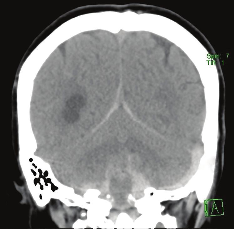
Fig. 3 A 51-year-old woman presented with seizures. (a) Unenhanced axial CT image of the head shows a focal hypodense lesion in the right frontal
lobe, thought to be a mass. Indirect signs of venous infarction are noted, including (b) focal gyriform restricted diffusion on axial diffusion-weighted
MR image; (c) associated small amount of haemorrhage on axial T2-W MR image; and (d) gyriform enhancement on contrast-enhanced axial T1-W MR
image. (e) Axial T2-W MR image shows loss of flow void in the superior sagittal sinus, in keeping with thrombosis (arrow). This was further confirmed
on a subsequent CT venogram (not shown). Further history-taking revealed use of oral contraceptives by the patient during her recent overseas travel.
4a 4b 4c
4d 4e
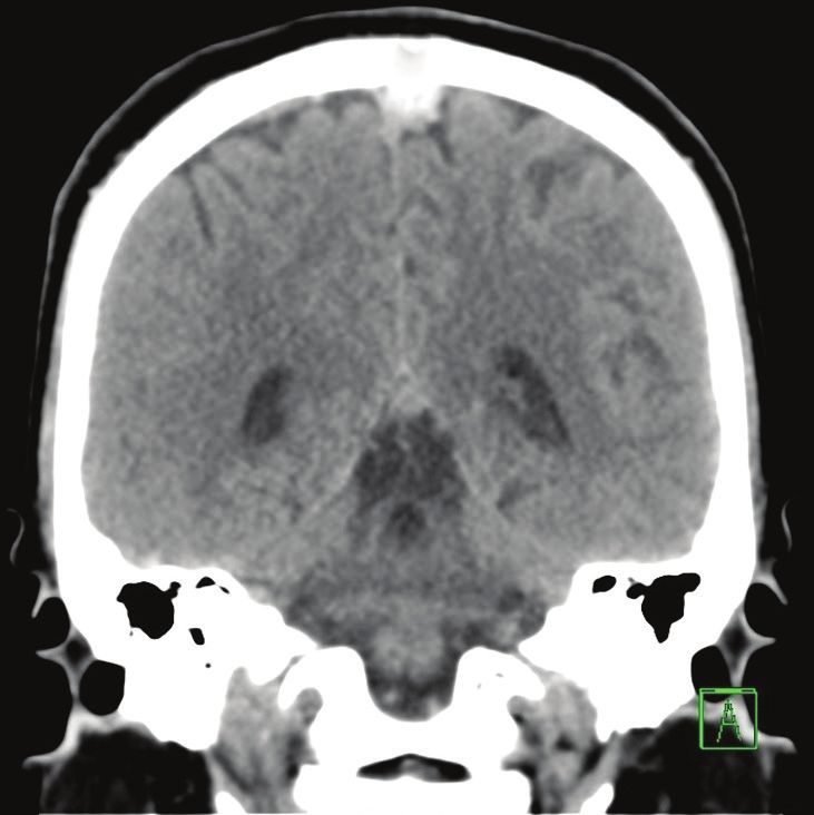
Fig. 4 A 41-year-old woman with a background of long-term oral contraceptive use for nine years presented with progressive drowsiness, giddiness,
vomiting and generalised weakness. (a & b) Unenhanced axial CT images of the head show an acute intraparenchymal haematoma in the parasagittal
left frontal lobe as well as significant oedema in the basal ganglia and thalami (white arrowheads). (c) Sagittal CT image shows hyperdensity within the
inferior sagittal sinus, vein of Galen and straight sinus (arrow). (d) Sagittal maximum-intensity projection reconstruction from CT venography shows loss
of contrast opacification in the deep venous system, inferior sagittal and straight sinuses (black arrowheads). (e) A repeat CT image of the brain obtained
a few days later shows worsening haemorrhage in the affected regions, diffuse subarachnoid and intraventricular haemorrhage as well as worsening
mass effect, subsequently leading to the patient’s death.
632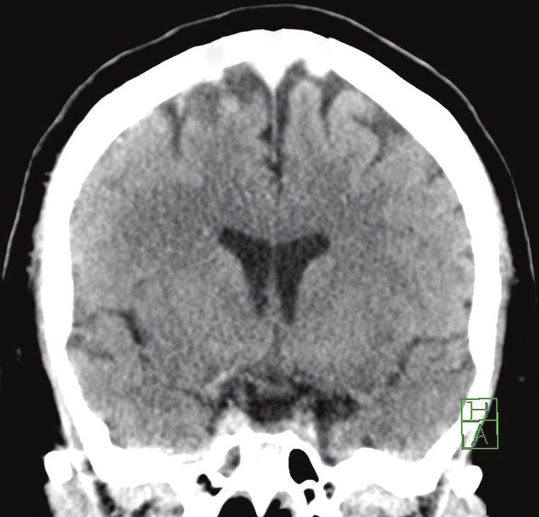
Pictorial Essay

                      5a                                 5b                                      5c

Fig. 5 A 62-year-old man presented with seizures. (a) Axial T2-W MR image shows focal oedema in the right perirolandic cortex (black arrow). (b) Maximum-
intensity projection reconstruction from three-dimensional phase-contrast MR venography subsequently shows loss of flow signal in the anterior two-
thirds of the superior sagittal sinus (arrowheads). (c) Contrast-enhanced axial T1-W MR image shows thin enhancement of the superior sagittal sinus,
suggesting chronic thrombosis. A filling defect is also seen within a cortical vein at the vertex (white arrow), in keeping with acute thrombosis.

         6a                                            6b                                          6c

Fig. 6 A 35-year-old man presented with syncope. (a) Unenhanced axial CT image of the head shows right frontal acute subarachnoid haemorrhage
(arrow). (a & b) Unenhanced axial and coronal CT images show hyperdensity along the superior sagittal sinus and bilateral frontal cortical veins (black
arrowheads). (c) Sagittal maximum intensity projection reconstruction from CT venography confirms filling defect in the anterior two thirds of the
superior sagittal sinus (white arrowheads).

                    7a                                         7b                                       7c

Fig. 7 A 60-year-old woman presented after a fall. (a) Unenhanced coronal head CT image shows subarachnoid haemorrhage, subdural haemorrhage and
hyperdense left sigmoid sinus (black arrow). (b) Unenhanced axial head CT image with reconstructed bone window shows an undisplaced fracture in
the left occipital and temporal bone extending into the left jugular foramen (arrowhead). (c) Maximum intensity projection reconstruction from three-
dimensional phase-contrast MR venogram shows loss of flow signal in the left sigmoid sinus (white arrow).

case (Fig. 5). Chronic thrombi have significant variability in MR             presenting with headache or trauma. Radiologists should pay
signal intensity and appearance on conventional unenhanced                    special attention to dural venous sinuses in cases of convexal
MR venography techniques. These may appear as peripheral                      distribution of subarachnoid haemorrhage.
enhancement with central filling defect, irregular central and                    Trauma may also be a direct cause of CVT. The dural venous
peripheral filling defects with heterogeneous enhancement or                  sinuses must be closely interrogated when there are fractures extending
central enhancing channel on contrast-enhanced MR venography.(13)             to or closely associated with them (Fig. 7). The diagnosis may be
Contrast-enhanced MR venography techniques are shown to be                    confounded by intracranial haemorrhage in the rest of the brain.
superior compared to non-contrast MR techniques such as three-                    Long-term sequelae of CVT include dural arteriovenous fistula
dimensional phase-contrast and two-dimensional time-of-flight.(14,15)         formation (Fig. 8) and increased intracranial pressure.
    Rarely, CVT can result in acute subarachnoid haemorrhage                      Various conditions and variants can render the interpretation
(Fig. 6),(8) which can be confused for other aetiologies in patients          of CVT difficult.(10) Increased attenuation of dural sinuses on

633
Pictorial Essay

                    8a                                             8b                                            8c

Fig. 8 A 66-year-old woman presented with non-ST-elevation myocardial infarction and had convulsive episodes during hospitalisation. (a & b) Axial
T2-W MR images show focal oedema in the right frontal lobe (black arrow) and loss of flow void in the superior sagittal sinus (white arrow). (c) Axial
maximum-intensity projection reconstruction from a follow-up CT venogram eight months later shows persistent thrombosis in the superior sagittal sinus
with abnormal dilated vessels adjacent to the sinus (arrowheads), indicating dural arteriovenous fistula formation.

unenhanced CT may be attributed to dehydration or high                                    Stroke 2004; 35:664-70.
                                                                                      2. Medel R, Monteith SJ, Crowley RW, Dumont AS. A review of therapeutic
haematocrit values, in which case comparison with the blood                               strategies for the management of cerebral venous sinus thrombosis. Neurosurg
pool of the other sinuses and arteries in the brain is imperative.                        Focus 2009; 27:E6.
                                                                                      3. Stam J. Thrombosis of the cerebral veins and sinuses. N Engl J Med 2005;
Subdural or subarachnoid blood products may be mistaken for                               352:1791-8.
a hyperattenuating dural sinus owing to their close proximity.                        4. Klok FA, Kruip MJHA, van der Meer NJM, et al. Confirmation of the high
                                                                                          cumulative incidence of thrombotic complications in critically ill ICU patients
Hypoplasia or aplasia of dural sinuses, commonly seen                                     with COVID-19: an updated analysis. Thromb Res 2020; 191:148-50.
unilaterally in the transverse sinus, may mimic loss of flow signal                   5. Cavalcanti DD, Raz E, Shapiro M, et al. Cerebral venous thrombosis associated
                                                                                          with COVID-19. AJNR Am J Neuroradiol 2020; 41:1370-6.
on unenhanced MR venography, in which case contrast-enhanced
                                                                                      6. Ayanzen RH, Bird CR, Keller PJ, et al. Cerebral MR venography: normal anatomy
CT/MR techniques may be helpful. Arachnoid granulations can                               and potential diagnostic pitfalls. AJNR Am J Neuroradiol 2000; 21:74-8.
                                                                                      7. Farb RI, Scott JN, Willinsky RA, et al. Intracranial venous system: gadolinium-
invaginate into dural sinuses and be mistaken for filling defects,
                                                                                          enhanced three-dimensional MR venography with auto-triggered elliptic
although they typically have rounded shape, CSF density/signal                            centric-ordered sequence--initial experience. Radiology 2003; 226:203-9.
and characteristic locations.                                                         8. Linn J, Brückmann H. Cerebral venous and dural sinus thrombosis: state-of-the-
                                                                                          art imaging. Clin Neuroradiol 2010; 20:25-37.
                                                                                      9. Leach JL, Fortuna RB, Jones BV, Gaskill-Shipley MF. Imaging of cerebral venous
CONCLUSION                                                                                thrombosis: current techniques, spectrum of findings, and diagnostic pitfalls.
                                                                                          Radiographics 2006; 26 Suppl 1:S19-41; discussion S42-3.
CVT, while uncommon, is associated with high morbidity and                            10. Canedo-Antelo M, Baleato-González S, Mosqueira AJ, et al. Radiologic clues
mortality if left untreated. It can be difficult to diagnose initially                    to cerebral venous thrombosis. Radiographics 2019; 39:1611-28.
                                                                                      11. Virapongse C, Cazenave C, Quisling R, Sarwar M, Hunter S. The empty delta
because of its non-specific clinical features and indirect imaging                        sign: frequency and significance in 76 cases of dural sinus thrombosis. Radiology
features, which may be misinterpreted. In the current COVID-19                            1987; 162:779-85.
                                                                                      12. Teasdale E. Cerebral venous thrombosis: making the most of imaging. J R Soc
pandemic, with increasing concerns about its thrombotic                                   Med 2000; 93:234-7.
complications, clinicians and radiologists should be vigilant                         13. Leach JL, Wolujewicz M, Strub WM. Partially recanalized chronic dural sinus
                                                                                          thrombosis: findings on MR imaging, time-of-flight MR venography, and contrast-
when approaching cases in their daily practice. This case series                          enhanced MR venography. AJNR Am J Neuroradiol 2007; 28:782-9.
illustrates a wide range of possible imaging findings of CVT.                         14. Liang L, Korogi Y, Sugahara T, et al. Evaluation of the intracranial dural sinuses
                                                                                          with a 3D contrast-enhanced MP-RAGE sequence: prospective comparison
                                                                                          with 2D-TOF MR venography and digital subtraction angiography. AJNR Am J
REFERENCES                                                                                Neuroradiol 2001; 22:481-92.
1. Ferro JM, Canhão P, Stam J, Bousser MG, Barinagarrementeria F; ISCVT               15. Lettau M, Laible M, Barrows RJ, et al. 3-T contrast-enhanced MR angiography
   Investigators. Prognosis of cerebral vein and dural sinus thrombosis: results of       with parallel imaging in cerebral venous and sinus thrombosis. J Neuroradiol
   the International Study on Cerebral Vein and Dural Sinus Thrombosis (ISCVT).           2011; 38:275-82.

                                                                                                                                                                       634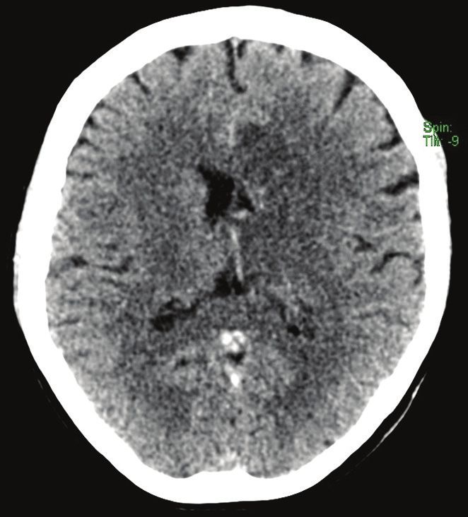
Pictorial Essay

SINGAPORE MEDICAL COUNCIL CATEGORY 3B CME PROGRAMME
(Code SMJ 202112B)

Question 1. Regarding cerebral venous thrombosis:                                                                                                        True  False
(a) The most common clinical presentation is seizure.                                                                                                     □                □
(b) The most common dural venous sinus involved is the superior sagittal sinus.                                                                           □                □
(c) The vein of Galen usually drains directly into the cavernous sinuses.                                                                                 □                □
(d) Head trauma is a risk factor.                                                                                                                         □                □
Question 2. The following are indirect signs of cerebral venous thrombosis:
(a) Restricted diffusion                                                                                                                                  □                □
(b) Vasogenic white matter oedema                                                                                                                         □                □
(c) Filling defect within a sinus on contrast-enhanced magnetic resonance (MR) venography                                                                 □                □
(d) Cortical enhancement                                                                                                                                  □                □
Question 3. The following are direct signs of cerebral venous thrombosis:
(a) Cord sign                                                                                                                                             □                □
(b) Empty delta sign                                                                                                                                      □                □
(c) Intracranial haemorrhage                                                                                                                              □                □
(d) Cerebral infarction                                                                                                                                   □                □
Question 4. Regarding cerebral venous thrombosis:
(a) Fractures extending to the dural venous sinuses can precipitate cerebral venous thrombosis.                                                           □                □
(b) Cerebral venous thrombosis can present with acute convexal subarachnoid haemorrhage.                                                                  □                □
(c) A hyperdense thrombus seen on unenhanced computed tomography is a highly sensitive (> 90%)                                                            □                □
    finding for cerebral venous thrombosis.
(d) Deep cerebral venous thrombosis has a better prognosis than thrombosis of the superficial dural                                                       □                □
    venous sinuses.

Question 5. Regarding imaging of cerebral venous thrombosis:
(a) Japanese encephalitis with bilateral thalamic haemorrhagic lesions can mimic deep cerebral venous                                                     □                □
    thrombosis.
(b) Two-dimensional time-of-flight MR venography is a contrast-enhanced technique.                                                                        □   □
(c) Unenhanced MR venography techniques are superior to contrast-enhanced MR venography in                                                                □   □
    evaluating chronic cerebral venous thrombosis.
(d) One should compare the attenuation of the dural venous sinuses to the rest of the intracranial blood                                                  □                □
    pool before deciding that it is hyperdense.

Doctor’s particulars:
Name in full:___________________________________________ MCR no.:�����������������������������������������������
Specialty: ______________________________________________ Email:��������������������������������������������������

 SUBMISSION INSTRUCTIONS:
 Visit the SMJ website: http://www.smj.org.sg/current-issue and select the appropriate quiz. You will be redirected to the SMA login page.
 For SMA member: (1) Log in with your username and password (if you do not know your password, please click on ‘Forgot your password?’). (2) Select your answers for each
 quiz and click ‘Submit’.
 For non-SMA member: (1) Create an SMJ CME account, or log in with your SMJ CME username and password (for returning users). (2) Make payment of SGD 21.40 (inclusive of
 7% GST) via PayPal to access this month’s quizzes. (3) Select your answers for each quiz and click ‘Submit’.

 RESULTS:
 (1) Answers will be published online in the SMJ February 2022 issue. (2) The MCR numbers of successful candidates will be posted online at the SMJ website by 24 February
 2022. (3) Passing mark is 60%. No mark will be deducted for incorrect answers. (4) The SMJ editorial office will submit the list of successful candidates to the Singapore Medical
 Council. (5) One CME point is awarded for successful candidates. (6) SMC credits CME points according to the month of publication of the CME article (i.e. points awarded for a
 quiz published in the December 2021 issue will be credited for the month of November 2021, even if the deadline is in February 2022).

 Deadline for submission (December 2021 SMJ 3B CME programme): 12 noon, 17 February 2022.

635

You can also read