Characterization of biomaterials and biological materials: a toolbox - Sorbonne Université

Page created by Ann Strickland
 
CONTINUE READING
Characterization of biomaterials and biological materials: a toolbox - Sorbonne Université
Characterization of biomaterials and
                       biological materials: a toolbox

C. Bonhomme – Professor in Chemistry
Laboratoire de Chimie de la Matière Condensée
UMR CNRS 7574 – Sorbonne Université, Paris

christian.bonhomme@upmc.fr
                                                         1
Characterization of biomaterials and biological materials: a toolbox - Sorbonne Université
A specific class of materials

 bio-nanocomposites involving:

 ■ minerals (hydroxyapatite Ca10(PO4)6(OH)2,
 calcium carbonate CaCO3, calcium oxalates       calcite (CaCO3)
 CaC2O4.nH2O…) → eventual polymorphism           … aragonite, vaterite

 ■ organics (proteins…)

 ■ organic/inorganic «interfaces»

  intrinsic structural complexity

                                               collagen
  multiple tools of investigation !                                  2
Characterization of biomaterials and biological materials: a toolbox - Sorbonne Université
Hierarchical materials: from atomic (Å) to macroscopic (cm) description

 pathological calcifications (kidney stones)

a major societal/health problem worldwide
(in France, related costs per year: > 800 millions   $)

  ■ minerals (calcium oxalates,
  phosphates, struvite…)
  ■ proteins
  ■ triglycerids
  ■…

                                      M. Daudon, D. Bazin et al., J. Appl. Crystallogr., 42,109,32009
Characterization of biomaterials and biological materials: a toolbox - Sorbonne Université
The electromagnetic spectrum

                                             keywords / questioning:

                                             ■ local vs long range probe

                                             ■ specific frequency / time domain
                                    noyaux

                          Spin e-            ■ complementarity of techniques

                                             ■ advantages / drawbacks

                                             ■ equipment (lab., synchrotron…)

                                             ■ spectroscopy / diffraction / imaging

                                                                                      4
Characterization of biomaterials and biological materials: a toolbox - Sorbonne Université
Vibrational spectroscopies (IR and Raman diffusion)

  harmonic oscillator
- h2/2m d2/dx2 + V(x) = E 
                                          v
    V(x) = ½ kx2                              v                                 v
                                                                       v

                                                  v

                                                      v

                                                          v                !!

                                                  w = (k/m)1/2
                                                  k : force constant
Ev = (v+1/2) hw    w = (k/m)1/2                   m = m1m2/(m1+m2)
                                                                                    5
Characterization of biomaterials and biological materials: a toolbox - Sorbonne Université
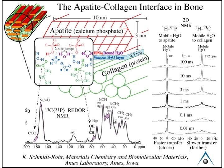
Assignments of IR / Raman modes

  normal modes of vibrations

     ex : H2O                                   characteristic frequencies

         symmetry
         group theory
         active / inactive modes (IR & Raman)
                                                                             6
Characterization of biomaterials and biological materials: a toolbox - Sorbonne Université
Hydroxyapatite, Ca10(PO4)6(OH)2 and carbonates

main mineral in bones, teeth, enamel

      Ca10 (PO4)6 (OH)2

    Mg2+, Zn2+, Na+,K+ …

    SO42-, CO
           CO3
             3 ….
               2
              2-
                               Ca10 (PO4)6 (OH)2

    CO3
    CO3
         2-, F-, Cl-…
        2-                             B   A
                                                   SiO44- or CO32-
                                                                     7
Characterization of biomaterials and biological materials: a toolbox - Sorbonne Université
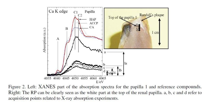
Nuclear Magnetic Resonance (NMR)
                                                  B0 (~10T)
        ^
        m = gh Î                                                           mI=-1/2      DmI =  1

magnetic moment         spin angular momentum
                                                                           mI=+1/2      n0=gB0/2p

                 gyromagnetic ratio
                                                                                Larmor frequency !

        E = - m . B

   Boltzmann equation
   Curie’s law                M                                     My

         N g2 h2 B0 I(I+1)
   M=
             12 p2 kT

                                         Man, Encyclopedia of analytical
                                                                               B1 (RF) at resonance !
                                         chemistry, 2000, 12228.                                 8
Characterization of biomaterials and biological materials: a toolbox - Sorbonne Université
NMR parameters: the chemical shift and J coupling

  interactions
                                                     13C-{1H}:   the case of a steroid

                  chemical shift: d

                  indirect coupling: J

                              F    -

                           B
                    F              F
                          F

                   ex:   19F   spectrum of BF4-

                                       multiplets (J in Hz)
                                       d in ppm
                                                                                         9
Characterization of biomaterials and biological materials: a toolbox - Sorbonne Université
Characterization of hydrated calcium oxalates by 13C solid state NMR

                               1 T.Echigo et al., Mineral. Mag., 2005, 69(1), 77-88
                               2 V.Tazzoli et al., Amer. Mineral., 1980, 65, 327-334
                                                                                          10
 C. Leroy, PhD thesis          3 S. Deganello et al., Amer. Mineral., 1981, 66, 859-865
The structure of interfaces in biological hydroxyapatites

     a model by C. Rey et al., Toulouse

 ■ 2D and 3D experiments
 ■ connectivities and internuclear distances
 ■ towards interfaces !

                                               Y. Wang et al., Nature Mater. 2013
                                                                                    11
Imaging by NMR

  inhomogeneous field : B0 (r)

                                 ex vivo bone sample
                                                       12
 Levitt, Spin dynamics, 2002.
X-ray diffraction

 Thomson’s formula (1856-1940)        periodic crystalline structure

                               P

                                 x

                                         symmetry
   as mP>>me: only the contribution      cell
   of the electrons has to be taken
                                         space group
             into account

                                                                       13
X-ray structure and powder pattern
                                                          powder diffraction
 periodic structure -> diffraction pattern !

       “chemistry”      “periodicity”

    electronic density calculation

                                               single crystal diffraction

     an old presentation: LiCl.C5H5N           ... to proteins !...
                                                                               14
X-ray diffraction: the case of Cl- substituted hydroxyapatite

                                                                15
Identification of phases and crystallinity of powders

                                                        16
Domain of coherence

                                        Scherrer’s law

                      ■ chemical disorder

                      ■ size of the (nano-) particles

                      ■ strain

                                                         17
Neutron diffraction

                      18
Application to hydrated calcium oxalates

  calcium oxalate

   powder
   neutron
   diffraction
                                                 powder XRD
                    improved structural models
                                                              19
Scanning Electron Microscopy and Transmission Electron Microscopy

                                                    ■ morphology
                                                    ■ diffraction patterns
                                                    ■ local chemical analyses

  http://barrett-group.mcgill.ca/tutorials/nanotechnology/nano02.htm

                                                                                20
SEM of bone and derivatives

       cell in a bone matrix

                               bone building blocks

                                                      21
SEM of apatite, calcium oxalates and bioglasses

     micro- HAp

                                            micro- HAp

                   Ca oxalates

      10 mm

       C. Leroy, PhD                     2 mm
                                                                  Ca oxalates
       C. Chan Chang, Master 2

                                         pathological calcifications
                                         (kidney stones & Randall’s plaque)
                                          D. Bazin, M. Daudon

                                                                                22
TEM of synthetic apatite

                                           nano- HAp

                                              50 nm

                                          HAp nanoparticles obtained by
                                          precipitation methods

     Y. Wang et al., Nature Mater. 2013

                                                                          23
Atomic Force Microscopy

                          24
Analysis of surfaces

                       25
Cell on surfaces

                   26
Applications of the synchrotron radiation

 see:

                                            27
A large variety of experiments

                                 see:

                                        28
X ray absorption                   X-ray Absorption Near Edge Spectroscopy
                                   Extended X-ray Absorption Fine Structure

  the (linear) absorption coefficient of X-rays
  by an element                                                          29
X ray absorption          X-ray Absorption Near Edge Spectroscopy
                          Extended X-ray Absorption Fine Structure
   XANES:

   • oxydation state                  EXAFS:
   • site symmetry
                                      • type of ligands
   carefull calibration               • distances
   of the E scale                     • coordination
                                        number
   error bar for
   accurate                           distances: 0.02 Å
   comparison of
   XANES spectra                      CN: 20-25%

                                                                30
Fe containing samples

                        31
Applications to the study of KS

 see:

                                  32
Micro-tomography

                   33
Mechanical properties

                        34
Thermal analyses

                   35
You can also read