Characterization of the RNA dependent RNA polymerase from Chikungunya virus and discovery of a novel ligand as a potential drug candidate
←
→
Page content transcription
If your browser does not render page correctly, please read the page content below

www.nature.com/scientificreports
OPEN Characterization
of the RNA‑dependent RNA
polymerase from Chikungunya
virus and discovery of a novel
ligand as a potential drug
candidate
Marjorie C. L. C. Freire1, Luis G. M. Basso2, Luis F. S. Mendes3, Nathalya C. M. R. Mesquita1,
Melina Mottin4, Rafaela S. Fernandes1, Lucca R. Policastro1, Andre S. Godoy1,
Igor A. Santos5, Uriel E. A. Ruiz5, Icaro P. Caruso6,7, Bruna K. P. Sousa4, Ana C. G. Jardim5,6,
Fabio C. L. Almeida7,8, Laura H. V. G. Gil9, Carolina H. Andrade4 & Glaucius Oliva1*
Chikungunya virus (CHIKV) is the causative agent of Chikungunya fever, an acute febrile and
arthritogenic illness with no effective treatments available. The development of effective therapeutic
strategies could be significantly accelerated with detailed knowledge of the molecular components
behind CHIKV replication. However, drug discovery is hindered by our incomplete understanding of
their main components. The RNA-dependent RNA-polymerase (nsP4-CHIKV) is considered the key
enzyme of the CHIKV replication complex and a suitable target for antiviral therapy. Herein, the nsP4-
CHIKV was extensively characterized through experimental and computational biophysical methods.
In the search for new molecules against CHIKV, a compound designated LabMol-309 was identified
as a strong ligand of the nsp4-CHIKV and mapped to bind to its active site. The antiviral activity of
LabMol-309 was evaluated in cellular-based assays using a CHIKV replicon system and a reporter
virus. In conclusion, this study highlights the biophysical features of nsP4-CHIKV and identifies a new
compound as a promising antiviral agent against CHIKV infection.
The Chikungunya virus (CHIKV) belongs to the Togaviridae family and is the causative agent of Chikungunya
fever. The main transmission route occurs through the bite of infected female mosquitoes of the Aedes sp. Genus.
After CHIKV infection, the proportion of individuals who develop clinical and debilitating symptoms is con-
sidered the highest compared to other arboviruses, with an average of 80% of symptomatic cases1,2. The control
of the mosquito vector remains the best prophylaxis since there are no licensed vaccines or efficient antivirals
available3. In this scenario, the infection caused by CHIKV has a high social impact and constitutes a serious
public health issue3.
1
Institute of Physics of Sao Carlos, University of Sao Paulo, Sao Carlos, SP, Brazil. 2Physical Sciences
Laboratory, State University of Northern Rio de Janeiro Darcy Ribeiro (UENF), Campos dos Goytacazes, RJ,
Brazil. 3Departament of Physics, Ribeirao Preto School of Philosophy, Science and Literature, University of
Sao Paulo, Ribeirao Preto, SP, Brazil. 4Laboratory for Molecular Modeling and Drug Design, Labmol, Faculty of
Pharmacy, Universidade Federal de Goiás, Goiânia, GO, Brazil. 5Institute of Biomedical Sciences, Federal University
of Uberlandia, Uberlandia, MG, Brazil. 6Institute of Biosciences, Humanities and Exact Sciences (Ibilce), Sao Paulo
State University (Unesp), Campus Sao Jose do Rio Preto, Sao Jose do Rio Preto, SP, Brazil. 7Institute of Medical
Biochemistry (IBqM) Leopoldo de Meis, National Center of Nuclear Magnetic Resonance Jiri Jonas, Federal
University of Rio de Janeiro, Rio de Janeiro, RJ, Brazil. 8National Center of Nuclear Magnetic Resonance (CNRMN),
Center of Structural Biology and Bioimaging (CENABIO), Federal University of Rio de Janeiro, Rio de Janeiro, RJ,
Brazil. 9Instituto Aggeu Magalhaes (IAM-FIOCRUZ), Recife, PE, Brazil. *email: oliva@ifsc.usp.br
Scientific Reports | (2022) 12:10601 | https://doi.org/10.1038/s41598-022-14790-x 1
Vol.:(0123456789)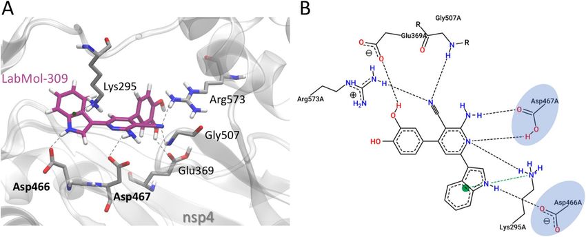
www.nature.com/scientificreports/
Chikungunya fever presents an acute phase characterized by high fever, arthralgia, myalgia, headaches, edema,
periorbital pain and cutaneous rash4,5. Later, some patients progress to the so-called chronic phase, mainly
characterized by persistent arthralgia and musculoskeletal pain for months and even y ears4,6–8. Since there are
no specific antiviral drug treatments, the clinical management targets primarily the relief of symptoms using
analgesics and antipyretics in the acute phase; and non-steroidal anti-inflammatory drugs (NSAIDs) and cor-
ticosteroids in the chronic p hase5.
CHIKV is a spherical, enveloped, and positive single-stranded RNA virus. As a member of the Alphavi-
rus genus, its genome has approximately 12 kb and codes for two distinct polyproteins: non-structural and
structural9. The first one is cleaved and gives rise to four non-structural proteins (nsP1, nsP2, nsP3 and nsP4)
that form the viral replication complex and have functions in the infection process, such as interaction with
host factors9–11. The nsP1 has methyltransferase and guanylyltransferase activities, promoting the capping of
viral RNA. In addition, this protein also anchors the replication complex in the cell m embrane12. The nsP2 is
a multifunctional protein with NTPase and helicase activities in the N-terminal region. Its C-terminal has a
cysteine-protease activity, responsible for processing the non-structural p olyprotein12. The nsP3 is an accessory
protein that recruits host cell factors that participate in and optimize the replicative process13. Finally, nsP4 is
the RNA-dependent RNA polymerase (RdRp), which is considered the key enzyme of the CHIKV replication
complex and acts principally by promoting the synthesis and elongation of viral R NA14.
On the other hand, the structural polyprotein is cleaved, giving rise to five structural proteins—E1, E2, E3,
C and 6 k—which are part of the viral assembly and structure9. The envelope proteins, specially E2 and E1, are
responsible for virus anchoring, receptor interaction and membrane fusion, promoting virus entry in the host
cell15,16. Due to its location on the viral surface, envelope proteins are targets of the humoral immune response
and thus become targets for vaccine development against CHIKV17.
Therefore, due to their vital role in the viral life cycle, non-structural proteins emerge as potential targets for
developing antiviral drugs, aiming to interrupt the replication process and, consequently, the viral e limination18.
Among these proteins, nsP4 is an attractive target due to its central role in viral genome replication, transcription
and genome r epair14,19. Recently, the three-dimensional structures of the RdRp domain of Sindbis (SINV) and
Ross River (RRV) viruses has been experimentally solved20. However, the structure of this domain of CHIKV
(nsP4-CHIKV) has not been reported. This fact generates limitations and challenges when the final goal is apply-
ing structure-based drug design strategies against this target.
Due to the lack of high-resolution structural information on the nsP4-CHIKV and the need to search for
new drugs to treat the infection caused by the virus, we present here a detailed biophysical characterization
of this protein. Size exclusion chromatography coupled with multi-angle light scattering was used to infer the
oligomeric state of the nsp4-CHIKV protein in solution. A high prevalence of ordered helical secondary struc-
tures was observed by circular dichroism, which also showed that the nsP4-CHIKV unfolds under a cooperative
transition during thermal denaturation. The thermal denaturation was further studied using differential scanning
calorimetry, indicating a kinetically controlled process.
Moreover, in the search for ligands for the development of new inhibitors, we selected 12 compounds for an
initial screen using differential scanning fluorimetry experiments. These compounds came from a global collabo-
rative project named O penZika21,22, through the World Community Grid computational network that enabled
massive docking-based virtual screening campaigns for ZIKV proteins as well as other flaviviruses (DENV, YFV,
WNV). One compound, LabMol-309, showed a significant effect on nsP4-CHIKV stability and its interaction
with the protein was confirmed using a sophisticated combination of experimental and computational strategies.
Furthermore, the inhibitory activity of LabMol-309 was studied in cellular-based antiviral assays using a
CHIKV replicon system and a reporter virus, and the results showed that this molecule caused inhibition in
these cellular assays and has the potential to be further evaluated as a CHIKV inhibitor. In this way, this work
provides novel structural features of nsP4-CHIKV and identifies a new compound that interacts with this protein,
generating perspectives in the drug development field to treat the infection caused by CHIKV and potentially
other alphaviruses.
Results
nsP4‑CHIKV purification and SEC‑MALS analysis. The nsP4-CHIKV was bacterially expressed
and purified using chromatography systems, and the purity was confirmed by acrylamide gel electrophoresis
(Supplementary Fig. 1). The nsP4-CHIKV is formed by 492 amino acids and has a theoretical molecular mass
(MM) of 54.54 kDa. This construction covers the entire region of the RNA-dependent RNA polymerase (RdRp)
domain, responsible for the nsP4-CHIKV function and where the catalytic aspartic acid residues (Asp371 and
Asp466) are located23. The Asp466 is in the GDD motif, a highly conserved sequence of viral p
olymerases23.
In order to determine the oligomeric state of nsP4-CHIKV in the working buffer solution, Size Exclusion
Chromatography coupled with Multi-angle Light Scattering (SEC-MALS) was e mployed24. The SEC-MALS data
showed a low polydispersity index and yielded a MM of (60 ± 1)kDa for nsP4-CHIKV (Fig. 1A). The proximity
of the experimental value with the theoretical molecular mass of the protein suggests that the monomeric state
is the most populated oligomer under the evaluated conditions.
Evaluation of nsP4‑CHIKV secondary structure profile. Circular dichroism (CD) spectroscopy was
used to estimate the secondary structure content of nsP4-CHIKV in solution25. The CD spectrum of nsP4-
CHIKV is characteristic of an α-helical rich protein, with two negative minima at 208 and 222 nm and one
positive maximum around 195 nm (Fig. 1B). This profile corroborates with structural features described for viral
polymerases and other polymerase structures solved experimentally23.
Scientific Reports | (2022) 12:10601 | https://doi.org/10.1038/s41598-022-14790-x 2
Vol:.(1234567890)
www.nature.com/scientificreports/
Figure 1. Biophysical characterization of nsP4-CHIKV. (A) SEC-MALS analysis of nsP4-CHIKV in solution.
The proximity of the estimated molecular weight of (60 ± 1) kDa with the molecular weight of the protein
suggests that nsP4-CHIKV is mostly at a monomeric state under the evaluated conditions. (B) Secondary
structure profile of nsP4-CHIKV by CD spectroscopy. The nsP4-CHIKV spectrum suggests the predominance
of helical secondary structures. (C) Thermal stability of nsP4-CHIKV probed by CD spectroscopy. The
ellipticity at 222 nm as a function of temperature for nsP4-CHIKV was recorded at 0.5 °C/min (left) and
1.0 °C/min (right) and transformed to the protein unfolded fraction. Solid lines are best fits to the CD data
using a two-state equilibrium model. The thermodynamic parameters of the protein unfolding transition are
summarized in Table 1. (D) Scan-rate normalized (13 °C/h) DSC data of the irreversible thermal denaturation
of 11.9 μM of nsP4-CHIKV and the corresponding instrumental buffer (50 mM Tris–HCl pH 8,0, 200 mM
NaCl e 5% glycerol) baseline. (E) Excess heat capacity of nsP4-CHIKV at the indicated scan rates obtained after
normalization by protein concentration and subtraction of the buffer baseline. (F) Arrhenius-type plot showing
the scan rate dependence of the nsP4-CHIKV unfolding temperature, Tm. The slope yields the activation energy
for the irreversible denaturation of nsP4-CHIKV.
Rate (°C/min) Tm (°C) ΔHapp (kcal/mol) ΔSapp (cal/mol K)
0.5 41.8 ± 0.2 122 ± 12 387 ± 38
1.0 43.7 ± 0.2 104 ± 8 331 ± 26
Table 1. Thermodynamic parameters of nsP4-CHIKV unfolding by CD spectroscopy. The Tm and the
odel28. The ΔSapp of the unfolding
ΔHapp were determined by fitting the CD data to a two-state equilibrium m
transition at Tm was calculated as ΔSapp = ΔHapp/Tm since ΔG = 0 at Tm.
Dichroweb was used in the quantitative analysis of the CD spectrum26. The best-fit spectra were obtained with
the CDSSRT m ethod27, which displayed an NRMSD of 0.009 for the SP175 database and 0.012 for Sets 4 and 7,
considered the most representative of the secondary structure content of nsP4-CHIKV (Supplementary Table 1).
Secondary structure content estimation from the CD spectrum confirmed the higher percentage of α-helix in the
nsP4-CHIKV (60%). The remaining fractions were: 6.3% of sheets, 9.6% of turns and 24% of disordered structure.
nsP4‑CHIKV thermal stability profile through CD spectroscopy and DSC. The CD technique was
also applied to study the thermal behavior of this protein when subjected to temperature v ariations28. The unfold-
ing transition of nsP4-CHIKV was studied by monitoring the ellipticity at 222 nm as a function of temperature28.
The results showed that the thermal denaturation process occurred cooperatively, exhibiting the transition from
folded to unfolded states in a defined way (Fig. 1C).
The thermodynamic parameters of the nsP4-CHIKV unfolding were obtained by adjusting a two-state equi-
librium model to the experimental data. Changes in the heat capacity were not considered, and the fitting was
performed taking into account the linear changes in pre- and post-transition ellipticity as a function of the
temperature28. Thus, the melting temperature (Tm), the apparent enthalpy change (ΔHapp), and the apparent
entropy change (ΔSapp) for the same protein concentration were obtained at two different heating rates (Table 1).
Scientific Reports | (2022) 12:10601 | https://doi.org/10.1038/s41598-022-14790-x 3
Vol.:(0123456789)
www.nature.com/scientificreports/
Concentration (µM) Rate (°C/h) Tm (°C) ΔHcal (kcal/mol) ΔT1/2 (°C) ΔS (cal/mol) ΔHvH (kcal/mol)
9.3 8 36.3 205 4.8 662 142
11.9 13 37.0 102 4.4 330 151
7.4 33 38.8 86 4.3 275 165
7.4 64 39.8 66 4.5 210 150
Table 2. Thermodynamic parameters associated with the nsP4-CHIKV thermal denaturation by DSC. Tm
represents the temperature where CP reaches its maximum value. ΔHcal was calculated as the area under
the DSC trace. ΔT1/2 corresponds to the linewidth at half the height of the transition peak. The entropy
change at Tm, ΔS, was calculated as ΔS = ΔHcal/Tm. ΔHvH was calculated as 4RTm2Cp,max/ΔHcal. Analyses of the
thermograms were performed with MicroCal Origin software. Uncertainties: Tm (± 0.2 °C), ΔHcal (± 1‒3 kcal/
mol), ΔT1/2 (± 0.2 °C).
The thermodynamic parameters of the nsP4-CHIKV unfolding transition exhibited a dependence on the heat-
ing rate. This result suggests that the irreversible nsP4-CHIKV unfolding transition is kinetically dependent29,30.
The values of the enthalpy and entropy changes obtained from the CD data agree well with those observed for
globular proteins31. The thermal behavior of nsP4-CHIKV was also evaluated using Differential Scanning Calo-
rimetry (DSC)32. Figure 1D shows the temperature-dependence of the heat capacity profile (Cp – sample minus
reference) of nsP4-CHIKV and the instrumental buffer baseline, acquired with a scan rate of 13 °C/h. The protein
Cp was subtracted from the buffer baseline Cp and normalized to the protein concentration.
DSC experiments were performed at different scan rates to investigate its effect on the DSC profile and the
reversibility of the transitions. The temperature- and scan rate dependence of the excess heat capacity profile
of nsP4-CHIKV are illustrated in Fig. 1E. The nsP4-CHIKV undergoes an irreversible thermal denaturation,
and the values of the calorimetric enthalpy change (ΔHcal) obtained from the analysis of the thermograms are
within the range of values observed for other globular p roteins33–36. Moreover, the transition peak shows a clear
scan rate dependence, confirming that all thermodynamic parameters associated with nsP4-CHIKV thermal
denaturation depend upon the heating rate (Table 2), as observed in our previous CD analysis. Except for the
van’t Hoff enthalpy change (ΔHvH), the dependences of the thermodynamic parameters on the heating rate are
markedly non-linear (Table 2 and Supplementary Fig. 2). This feature illustrates the non-equilibrium character
of the protein denaturation process.
The dependence of Tm on the scan rate was used to calculate the kinetic activation energy, Ea, for the irrevers-
ible nsP4-CHIKV thermal denaturation. According to Sanchez-Ruiz et al.30, the Tm shifts induced by different
heating scan rates, ν, for an irreversible two-state process can be modeled by the following equation:
ν AR − RTEa
2
= e m
Tm Ea
where A
is the pre-exponential factor in the Arrhenius equation, and R is the gas constant. Thus, by plotting
2 against 1/T , the apparent activation energy can be determined from the slope of the curve. The
ln ν/Tm m
Arrhenius plot showing the scan rate-dependent changes in the Tm is illustrated in Fig. 1F, from which Ea was
determined as (110 ± 4) kcal/mol.
Evaluation of nsp4‑CHIKV interaction with compounds. In the search for new compounds able to
interact and inhibit the nsP4-CHIKV in solution, an initial experimental screening with a series of 12 com-
pounds (Supplementary Table 2) was selected from the OpenZika p roject22,37 and performed using differential
scanning fluorimetry (DSF or ThermoFluor assay). For nsP4-CHIKV, the Tm in the absence of compounds (only
with DMSO) was 37.7 ± 0.4 °C. Among the compounds, LabMol-309 (Fig. 2A) caused the highest thermal shift
(~ 4 °C), suggesting the occurrence of interaction with nsP4-CHIKV. Therefore, this compound was chosen to
proceed with the other assays.
The interaction between LabMol-309 and nsP4-CHIKV was later analyzed by MicroScale thermophoresis
(MST) and solution nuclear magnetic resonance (NMR). The MST data showed an affinity curve with the occur-
rence of well-defined bound and unbound states (Fig. 2B). From that, the dissociation constant (KD) for the
interaction of nsP4-CHIKV with LabMol-309 was estimated as (6 ± 1) µM.
The interaction of LabMol-309 with nsP4-CHIKV was further evaluated by solution NMR, monitoring the
chemical shift perturbation (CSP). Figure 2C shows the spectra obtained for the compound LabMol-309 in the
presence (red line) and absence of the protein (blue line). These spectra were superimposed, and the chemical
shift differences were identified and mapped according to the respective positions of the proton resonances
(Fig. 2C) previously identified in the LabMol-309 assignment (Supplementary Fig. 3).
Therefore, detecting these chemical shifts perturbations is additional evidence for the interaction between
nsP4-CHIKV and compound LabMol-309, reinforcing the results obtained using DSF and MST.
The nsP4‑CHIKV three‑dimensional model and structural analysis. A combined analysis of sev-
eral biophysical techniques suggests that the nsP4-CHIKV is a monomeric α-helical rich protein capable of
forming a complex with the compound LabMol-309 within a moderate dissociation constant (10−6 M). The
nsP4-CHIKV 3D structural model was obtained using A lphaFold38. The model obtained showed a very high
Scientific Reports | (2022) 12:10601 | https://doi.org/10.1038/s41598-022-14790-x 4
Vol:.(1234567890)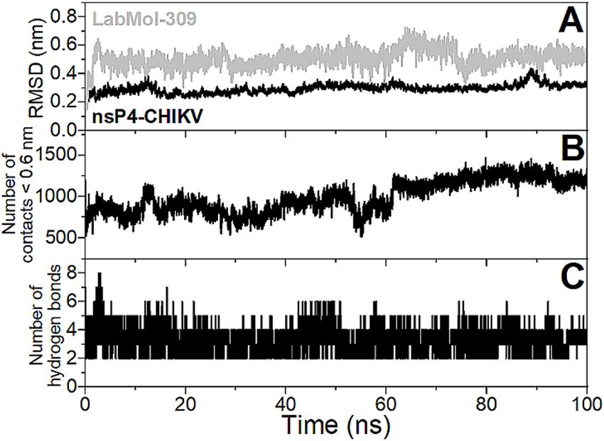
www.nature.com/scientificreports/

                                  Figure 2.  Evaluation of nsp4-CHIKV interaction with the compound LabMol-309. (A) LabMol-309
                                  chemical structure. (B) Binding affinity curve of nsP4-CHIKV interacting with LabMol-309 by MST. The
                                  compound LabMol-309 was titrated in a concentration range of 200 µM to 0.012 µM. The curve was fitted
                                  to the Hill function, and the estimated KD was (6 ± 1 µM). (C) Chemical shift perturbation analysis based on
                                  the superimposition of LabMol-309 one-dimensional 1H spectra obtained in the presence (red line) and the
                                  absence of nsP4-CHIKV (blue line). On the right, chemical shift differences were measured (in Hz) for each
                                  proton of the LabMol-309 structure.

                                  per-residue confidence score (pLDDT) for more than 90% of the covered sequence, and only for the N- and
                                  C-terminal regions pLDDT were low (Fig. 3A). The full-length nsP4-CHIKV structural model is illustrated in
                                  Fig. 3.
                                      In the nsP4-CHIKV structure, we identified the regions corresponding to the fingers (residues 93–315),
                                  palm (residues 316–502) and thumb (residues 503–611) domains, which are characteristics of viral polymerases
                                  (Fig. 3B). In the palm domain’s catalytic site, we also identified the catalytic aspartic acid dyad (Asp371 and
                                  Asp466) separated by 7.5 Å. Besides, the nsP4-CHIKV model contains an extra N-terminal domain (residues
                                  1–92), forming a coiled-coil.
                                      In the model of nsP4-CHIKV, the electrostatic surface potential of the active site cavity where are located the
                                  aspartic acid dyad (Asp371 and Asp466) is remarkably positive, a signature of nucleic acids-interacting motifs
                                  (Fig. 3C)39. Additionally, the analysis using ConSurf reveals that the catalytic site region and its surroundings
                                  are highly conserved (Fig. 3D). These results serve as corroboration for the robustness of the AlphaFold model
                                  of nsP4-CHIKV.

                                  Molecular docking and molecular dynamics of LabMol‑309 against nsP4‑CHIKV.                        Docking cal-
                                  culations were used to investigate the binding mode of LabMol-309 at the nsP4-CHIKV. The docking results sug-
                                  gest that LabMol-309 binds to the nsP4-CHIKV active site, interacting with the GDD catalytic triad (Asp466 and
                                  Asp467), with a docking score of -7.15 kcal/mol−1. LabMol-309 makes H-bonds with Glu369, Asp466, Asp467,

Scientific Reports |   (2022) 12:10601 |               https://doi.org/10.1038/s41598-022-14790-x                                                  5

                                                                                                                                              Vol.:(0123456789)
www.nature.com/scientificreports/
Figure 3. nsP4-CHIKV three-dimensional model by AlphaFold. (A) nsP4-CHIKV AlphaFold model colored
according to pLDDT. At the top right are shown the pLDDT color scales. (B) Model of nsP4-CHIKV colored
according to domain assignment. The N-terminal helixes, finger, palm and thumb domains are colored in purple,
dark blue, green and red, respectively. A detailed view of predicted catalytic aspartic residues is presented in the
inset. (C) Electrostatic potential projected on the surface charge of nsP4-CHIKV, calculated with APBS. Positive
regions are colored in blue and negative regions are in red. (D) ConSurf analysis of nsP4-CHIKV model. The
amino acids are colored by their conservation grades using the color-coding bar, with green-through-purple
indicating variable-through-conserved.
Gly507, Arg573 and cation–π interactions with Lys295 residue (Fig. 4). The nitrogen atom of the indole group
and amine of the pyridine group of LabMol-309 make relevant interactions with Asp466 and Asp467, respec-
tively. Additionally, the indole group makes cation–π interaction with Lys295.
The structural stability of the structural model of the nsP4-CHIKV/LabMol-309 complex calculated by dock-
ing was evaluated using 100 ns molecular dynamics (MD) simulation. Figure 5A presents the values of root
mean square deviation (RMSD) for the backbone atoms of the protein and non-hydrogen atoms of the ligand
from the initial structure. It is possible to observe that RMSD values are stable after 5 ns of simulation and reach
plateaus around 0.3 and 0.5 nm for the protein and ligand, respectively. Figure 5B shows that the number of
contacts < 0.6 nm between nsP4-CHIKV and LabMol-309 does not drop down to zero throughout the MD simu-
lation, indicating that the ligand interacts with the protein is persistent. The number of hydrogen bonds is stable
throughout the MD simulations and presents an average value of three (Fig. 5C). An evaluation of the hydrogen
bonds with significant percentage occupancy (< 5%, Supplementary Table 3) during the MD trajectory reveals
that Glu369, Asp371, Asp466, Asp467, Asn468, Lys501, and Arg573 are important for the stabilization of the
protein–ligand complex, and further mutagenesis studies may be relevant for confirmation of these interactions.
It is worth noting that Glu369 and Asp371 presented percentage occupancies higher than 70%. Considering all
MD analyses, it can be concluded that the structural model of the nsP4-CHIKV/LabMol-309 complex is stable
throughout the simulation.
Inhibition of CHIKV replication by LabMol‑309 through replication‑based and viral infection
assays. The inhibitory activity of the compound LabMol-309 was evaluated through the replicon-based
screenings in a dose-dependent manner to determine its effective and cytotoxic concentrations (EC50 and CC50,
respectively). Replicon cells were incubated with twofold serial dilutions of compound (from 20 to 0.03 µM for
EC50 and from 100 to 0.30 µM for C C50), and luciferase signals or cell viability was evaluated after 48 h. Lab-
Mol-309 displayed an E C50 value of 10.0 ± 0.07 µM and showed a C C50 of 17.1 ± 0.6 µM (Fig. 6; Supplementary
Table 4 and 5). As a result, the obtained selectivity index ( CC50/EC50) of LabMol-309 was 1.7 in the replicon
system.
Scientific Reports | (2022) 12:10601 | https://doi.org/10.1038/s41598-022-14790-x 6
Vol:.(1234567890)www.nature.com/scientificreports/
Figure 4. Molecular interactions of LabMol-309 and nsP4-CHIKV, predicted by docking calculations. (A)
3D interactions of LabMol-309 and nsP4-CHIKV residues. Hydrogen bonds are shown as gray dashed lines,
and cation–π interactions are shown in green dashed lines. Oxygen, nitrogen and hydrogen atoms are shown
in red, blue and white, respectively. Carbon atoms of LabMol-309 and protein residues are shown in purple
and gray, respectively. Catalytic residues of the GDD triad are highlighted in bold. (B) 2D interaction diagram
of LabMol-309 and nsP4-CHIKV residues. Hydrogen bonds are shown as gray dashed lines, and cation–π
interactions are shown in green dashed lines. Catalytic residues of the GDD triad are highlighted in blue.
Figure 5. Evaluation of the stability of the structural model of the nsP4-CHIKV/LabMol-309 complex from
100 ns MD simulation. (A) RMSDs of backbone atoms of the protein (black line) and non-hydrogen atoms of
the ligand (gray line). (B) Number of contacts between atoms of the protein and ligand for distances less than
0.6 nm. (C) Number of hydrogen bonds formed between the protein and ligand.
To confirm the antiviral activity of the LabMol-309, we carry out the effective concentration of 50% ( EC50) and
cytotoxic concentration of 50% (CC50) using BHK-21 cells infected with CHIKV-nanoluciferase, a recombinant
CHIKV that express the Nanoluciferase reporter, at a multiplicity of infection (MOI) of 0.1, with a two-fold serial
dilution of LabMol-309 at concentrations ranging from 0.78 to 100 µM. Nanoluciferase activity levels, propor-
tional to viral replication, were assessed 16 h post-infection (adapted from40,41). The cytotoxic concentration of
50% (CC50) was determined in parallel experiments (Fig. 6C). As a result, these assays demonstrated that the
LabMol-309 has a E C50 of 5.2 µM on BHK-21 cells infected with CHIKV-nanoluc and C C50 of 52 µM on naive
BHK-21 cells, over a period of incubation of 48 h, resulting in a Selectivity Index (SI) of 10 (Table 3; Fig. 6C;
Supplementary Table 6).
Discussion
The nsP4-CHIKV polymerase plays a crucial role in viral replication and has been considered a promising target
for the search and development of new drugs. Thus, understanding its dynamic and structural features is an
important step for studies with this target.
Our biophysical data agree with the structure prediction using Alphafold and point out that nsp4-CHIKV is
a monomeric protein enriched with alpha-helix content. These observations also agree with experimental data
Scientific Reports | (2022) 12:10601 | https://doi.org/10.1038/s41598-022-14790-x 7
Vol.:(0123456789)www.nature.com/scientificreports/
Figure 6. Antiviral activity of LabMol-309. The EC50 (A) and CC50 (B) curves from replicon-based assays are
shown. CHIKV replicon cells were incubated with the compound at twofold serial dilutions (from 20 µM to
0.03 µM for EC50 and from 100 to 0.3 µM for CC50) for 48 h, and Gluc activity/cell viability were measured from
cells’ supernatant. For the CHIKV-nanoluc replication assay evaluation of EC50 and CC50 (C), BHK-21 cells were
treated with concentrations of LabMol-309 ranging from 0.78 to 100 µM, in the presence or absence of CHIKV-
nanoluc for 16 h, and viral replication was quantified by measuring the nanoluciferase activity (indicated by
●) and cellular viability was measured using an MTT assay (indicated by ▄). Representative results from two
independent experiments performed in duplicates. Error bars represent the standard deviations. Figures and
statistical analysis were performed using GraphPad Prism 8.
Compound Type of the Assay CC50 EC50 Selectivity index (SI)
CHIKV-nanoluc and BHK-21 cells 52 5.2 10
LabMol-309
BHK-CHIKV replicon 17.1 10.0 1.7
Table 3. Effect of the compound on the viability of BHK-21 cells (CC50) and the viral replication of CHIKV
(EC50) in 16 h treatment.
collected for other viral polymerases20,23. The thermodynamic data showed that when in the in vivo host, this
protein can be stable in the vicinity of its thermal denaturation (Tm ~ 40 °C and ΔT1/2 ~ 4–5 °C), and its unfolding
is both scan-rate dependent and irreversible under all conditions tested. It was shown before that the T m might
be scan-rate dependent if the scan rate exceeds the unfolding r ate42. The calorimetric thermograms for the irre-
versible denaturation of proteins are highly scan-rate dependent, and their shapes are normally a symmetric43,
exactly what is observed for nsp4-CHIKV. Therefore, the kinetics of the thermal denaturation could be treated as
a single first-order irreversible step N → D, whose rate of temperature dependence obeys the Arrhenius equation.
Scientific Reports | (2022) 12:10601 | https://doi.org/10.1038/s41598-022-14790-x 8
Vol:.(1234567890)www.nature.com/scientificreports/
The effective activation energy derived from this equation was (110 ± 4) kcal/mol, which fits well within the wide
range of the values reported for the thermal denaturation of mammalian tissues and strengthened the thermo-
dynamic data collected for this p rotein44.
In the search for new nsp4-CHIKV ligands as potential inhibitors, the compound LabMol-309 was identi-
fied as a promising candidate through DSF screening. The validation of this interaction through biophysical
methods demonstrated that the compound interacts in the low micromolar range. This compound had already
been evaluated in virtual screenings for other viral proteins such as ZIKV and other F laviviruses21,22, but it was
the first time it was reported targeting the nsP4-CHIKV protein.
A three-dimensional model was generated using AlphaFold to analyze the LabMol-309-nsP4-CHIKV complex
and their mode of interaction. In this sense, the nsP4-CHIKV model presented the regions corresponding to
the fingers, palm and thumb domains, which are characteristics of viral polymerases, and the active site region
was remarkably positive and conserved. These structural features were equivalent to the regions presented in
the RdRp domain of nsP4 from both RRV and SINV, which were experimentally solved recently (PDB ID: 7F0S,
7VB4, 7VW5)20. Therefore, the suggested binding mode for LabMol-309 is through the interaction with residues
of the nsP4-CHIKV active site.
Although the data involving the complex formation between nsP4-CHIKV and LabMol-309 are solid, it is
still not possible to conclude that this compound has inhibitory activity against this enzyme. The development
of an enzymatic assay for the nsP4 polymerase from Alphaviruses in general, has been challenging because this
protein cannot effectively perform its function on its own, as previously shown by others45,46. Different regions
of nsP4 recognize the promoters for minus and plus strands. However, the binding requires the presence of the
other non-structural proteins to form the replication complex and enable the de novo RNA synthesis11,45–47.
Moreover, the interactions between these proteins with host components during replication have been studied
but remain limited and not completely u nderstood11,46.
Tomar et al.48 reported that template recognition and the nsP4 activation through protein–protein interac-
tions requires the presence of viral polyprotein P12348. In another work, the SINV nsP4 was expressed in E. coli,
and the polymerase activity was observed only when supplied with the viral polyprotein P12347. Recently, Lello
et al.46 demonstrated that nsP4 of SINV, CHIKV, ONNV, BFV, RRV, SFV, MAYV, VEEV, and EILV on their own
have minimal RNA polymerase activity46. Using a trans replicase system consisting of two relatively independent
functional modules (nsP4 and P123), they have shown that the nsP4 of all these Alphaviruses was active only
when combined with the corresponding P123 p olyproteins46. Furthermore, Tan et al.20 evaluated the polymerase
activity of SINV and RRV nsP4 and as a result, the isolated proteins showed less efficient polymerase activity
than the dengue virus RdRp used as the positive control20. Altogether these findings corroborate that bacterially
produced nsP4 could not efficiently synthesise RNA unless combined with the viral polyprotein P123 obtained
from animal cell e xtracts47,49,50.
Given these limitations in establishing an efficient method for evaluating the enzymatic activity of purified
recombinant nsP4-CHIKV, in this work the inhibitory effect of the compound LabMol-309 was evaluated using
both replicon-based assays and cells infected with the CHIKV expressing the nanoluciferase reporter (CHIKV-
nanoluc). Replicon-based systems have been widely used as tools for drug discovery of antiviral agents, and
promising replication inhibitors were identified by this method51. Specifically to CHIKV, BHK-21 cells har-
boring other CHIKV replicon constructs were reported for the high-throughput screening of viral replication
inhibitors52,53. The same system was also used to evaluate the anti-CHIKV activity of other compounds and
different flavonoids54–56.
The evaluation of LabMol-309 using a replicon-based system was performed in a dose-dependent manner,
and its inhibition clearly occurred. Comparing with studies that also used CHIKV replicon, the E C50 obtained
for LabMol-309 was lower than the values already reported for other compounds52,55, reinforcing the antiviral
potential of this compound. LabMol-309 showed toxicity to the cells, and the resulting low selectivity index
of 1.7 may be correlated to a possible negative impact in the cellular factors associated with the viral genome
replication. These data suggest that, even with inhibitory activity, chemical modifications would be required to
optimize this compound’s efficiency and reduce its toxicity. Furthermore, antiviral assays performed with cells
infected with a recombinant CHIKV demonstrated that the LabMol-309 decreased CHIKV replication with an
EC50 of 5.2 µM and an CC50 of 52 µM, with a SI of 10 in BHK-21 cells.
The differences in the obtained values using naive BHK-21 or BHK-CHIKV cells (Table 3) are understandable
since different factors are involved in these assays. For example, in the infection system the virus is effectively
infecting the cells and performing all the stages of the virus replicative cycle. It means that the treatment with Lab-
Mol-309 may be acting even before the formation of the replication complexes. Alternatively, in the BHK-CHIKV
replicon system, the replication complexes are already formed when the treatment starts, which can impact on
the effectiveness of the antiviral activity in a short period of treatment. Additionally, the presence of the replicon
might change the cell response to the compound, and explain the higher cytotoxicity shown in the results. This
isolated effect predominantly observed in the replicon cells can be explained by the differences in incubation peri-
ods used in the antiviral activity experiments (48 h for replicon-based screenings compared to 16 h for the viral
infection assays). The prolonged exposure of cells to the compound can result in higher cytotoxicity57, reinforcing
the importance of further studies of the ADME-Tox profile in animal models. Additionally, to the best of our
knowledge, this is the first description of LabMol-309 as inhibitor of CHIKV replication, and its low EC50 value
is in similar level with other inhibitors reported to block CHIKV replication, emphasizing the antiviral potential
of this compound58,59. In this context, the results obtained from the antiviral assays suggest that LabMol-309 is
a potential molecule to be further optimized to reduce its cytotoxicity and increase the selectivity index in cell-
based antiviral assays. In summary, this study highlights biophysical features of nsP4-CHIKV, contributing to
basic research on alphaviruses polymerase, and identified a new compound as a promising antiviral agent against
Scientific Reports | (2022) 12:10601 | https://doi.org/10.1038/s41598-022-14790-x 9
Vol.:(0123456789)www.nature.com/scientificreports/
CHIKV infection. These findings could contribute to developing novel candidates targeting nsP4-CHIKV and
support the progress in therapeutic strategies for CHIKV and other alphavirus infections.
Methods
CHIKV nsP4 cloning and overexpression. The coding region of nsP4-CHIKV (GenBank
KP164572.1; PROT-ID AJY53709.1—residues 118 to 611) was subcloned in the pET-SUMO vector using the
LIC methodology60. This construct encodes an nsP4-CHIKV with an N-terminal 6xHis-tag followed by a TEV
protease cleavage site (ENLYFQ; GAM) and the fusion protein tag SUMO. For protein expression, this plasmid
construction was transformed into E. coli Rosetta and cultured in Terrific Broth (TB) medium at 37 °C and 200
RPM until an OD600 of 1.0. The protein expression was induced with 1 mM of isopropyl-β-thiogalactoside and
incubation at 18 °C, 200 RPM for 16 h. The cells were harvested by centrifugation at 5000 × g for 30 min at 4 °C
and resuspended in buffer A (50 mM Tris pH 8.0, 500 mM NaCl, 10% glycerol). Cells were disrupted by sonica-
tion and clarified by centrifugation at 12,000 × g for 30 min at 4 °C.
nsP4‑CHIKV purification. nsP4-CHIKV was purified using an AKTA Purifier System (GE Healthcare).
The first step was affinity chromatography, using a HisTrap HP 5.0 mL column (GE Healthcare) pre-equilibrated
with buffer A (50 mM Tris pH 8.0, 500 mM NaCl, 10% glycerol). The elution was performed using 50 mM Tris
pH 8.0, 500 mM NaCl, 250 mM imidazole, 10% glycerol. The buffer was exchanged through dialysis to elimi-
nate the imidazole excess. The 6xHis-tag-SUMO was cleaved by TEV protease during overnight incubation at
4 °C. A second affinity chromatography step was performed using the same system to collect the HisTag-less
protein obtained after TEV treatment. A final purification step was done using size-exclusion chromatogra-
phy on an XK 26/1000 Superdex 75 column (GE Healthcare) pre-equilibrated in gel filtration buffer (50 mM
Tris pH 8.0, 200 mM NaCl and 5% glycerol). The eluted fractions were collected and analyzed by SDS-PAGE to
confirm their purity and mass spectrometry was performed to confirm the presence of nsP4-CHIKV. The final
protein sample was concentrated using 30 kDa MWCO centrifugal concentrators (Vivaspin, Sartorius). Pro-
tein concentrations were determined spectrophotometrically in a Nanodrop 1000 spectrophotometer, using the
−1 cm−1.
measured absorbances at 280 nm and the theoretical extinction coefficient of 36,495 M
Size exclusion chromatography coupled with multi‑angle light scattering (SEC‑MALS). The
oligomeric state of the purified nsP4-CHIKV was evaluated by size exclusion chromatography coupled with
multi-angle light scattering (SEC-MALS) in running buffer composed of 50 mM Tris–HCl pH 8.0 and 200 mM
NaCl. For that, 50 µL of purified nsP4-CHIKV at a concentration of 1.5 mg/mL was injected in a Waters 600
HPLC system (Waters) coupled in-line with a UV detector, a mini DAWN TREOS multi-angle light scattering
apparatus (Wyatt Technology), a column Superdex 75 Increase 10/300 GL (GE Healthcare), and a refractive
index detector Optilab T-rEX (Wyatt Technology). The light scattering detectors were normalized with bovine
serum albumin (Sigma-Aldrich) and the flow rate used was 0.5 mL/min. The data were processed using ASTRA7
software (Wyatt Technology) with the following parameters: refractive index of 1.331, 0.890 cP for the viscosity
of the solvent, and a refractive index increment of 0.1850 mL/g. Protein solutions were centrifuged for 10 min at
10,000 × g at a controlled temperature of 4 °C immediately before use.
Circular dichroism (CD). Far UV-CD spectra (195–280 nm) were measured in a Jasco J-810 spectrometer
(Jasco Corporation, Japan) equipped with a Peltier control system and using a quartz cell with a 1 mm pathlength.
The spectra were recorded from 280 to 195 nm, with a scanning speed of 100 nm/min, a spectral bandwidth
of 1 nm and a response time of 0.5 s. All the protein samples were in a final concentration of 2.5 µM diluted in
water. Spectral deconvolution was applied to estimate the secondary structure content using the DICHROWEB
web server26. Three different methods were combined with three different databases to improve the reliability of
the results. The detailed analysis of the results generated by these combinations is provided in Supplementary
material (Supplementary Table 1). The estimated values of secondary structure fractions were averaged from
each database used. The best fit was determined from the analysis of the NRMSD parameter, which was con-
sidered satisfactory when closer to 0 61. Thermal denaturation experiments were performed by monitoring the
ellipticity at 222 nm in the range from 20 to 80 °C using heating rates of 0.5 °C/min and 1.0 °C/min.
Differential scanning calorimetry (DSC). DSC measurements were carried out with the purified pro-
tein solution at 7.4 µM, 9.3 µM and 12 µM, diluted in buffer 50 mM Tris–HCl (pH 8.0), 200 mM NaCl and 5%
glycerol. Protein and reference samples (buffer) were degassed for 5 min prior to measurements. The experi-
ments were performed on a VP-DSC MicroCal MicroCalorimeter (Microcal, Northampton, MA, USA) using
heating rates of 8 °C/h, 13 °C/h, 33 °C/h and 64 °C/h. The thermograms were recorded from 10 to 70 °C, at a
controlled pressure of 1.6 atm. Instrumental buffer baselines were recorded before the protein unfolding experi-
ments to register the thermal history of the calorimeter. The raw DSC traces were subtracted with the buffer
baseline and then normalized by protein concentration. The thermogram analysis and the subtraction of the
buffer calorimetric response, baseline correction, and integration of the calorimetric peaks referring to the phase
transitions were performed using the MicroCal Origin software.
Differential scanning fluorimetry (DSF). In the search for new binders to nsp4-CHIKV, 12 compounds
from the OpenZika project were t ested22,37. The compounds were purchased from Chembridge Library (https://
www.chembridge.com/) with a minimum purity of 90%. DSF assays were conducted in a qPCR system Mx3000P
(Agilent) for an initial assessment. Protein melting temperatures (Tm), assuming a two-state transition model,
Scientific Reports | (2022) 12:10601 | https://doi.org/10.1038/s41598-022-14790-x 10
Vol:.(1234567890)www.nature.com/scientificreports/
were determined by monitoring the fluorescence intensity variation as a function of temperature for the extrinsic
probe SYPRO Orange (Invitrogen). The protein solutions were at a final concentration of 8 µM, diluted in gel fil-
tration buffer. Compounds were added at the final concentration of 80 µM and standard samples were prepared
only with DMSO. The thermal variations were in the range of 25–75 °C in a stepwise increment of 1 °C/min. The
Tm values obtained for each compound were subtracted from the values of the standard samples to identify com-
pounds that caused significant Tm changes. For the next steps, the compound that presented the highest thermal
shifts (ΔTm) was selected, considering the deviations of the triplicates62. Data were analysed using the software
Origin Pro 9.5.1. All experiments were conducted in triplicate.
MicroScale thermophoresis (MST). Experiments were performed on a Monolith® NT.115 instrument
(Nanotemper technologies). The nsP4-CHIKV was labelled on cysteine residues with NT-647-Maleimide dye
(Nanotemper Technologies) using the Monolith NTTM Protein Labeling Kit RED-MALEIMIDE as per man-
ufacturer’s instructions. Samples of 25 nM of cys-labelled nsP4-CHIKV with 5% DMSO were used. An ini-
tial binding test was carried out with the compound at the concentration of 100 µM, to check the interaction
between the protein and the compound. Then, a serial dilution of the compound from 200 to 0.012 µM (12 nM)
was performed to obtain the binding curve. The dissociation constant (KD) was obtained by fitting the binding
curve with the Hill function using GraphPad Prism 8 (Graph Pad Software).
Chemical shift perturbation. The LabMol-309 resonance assignment was performed using a Bruker
Avance III 600 MHz. 1H-13C-HSQC, COSY and TOCSY were acquired at 298 K using 1 mM of LabMol-309
in D2O. The interaction between LabMol-309 and nsP4-CHIKV was studied using a Bruker Avance IIIHD
500 MHz in a solution of 20 mM (2H)11-Tris/HCL pH 7.5, 200 mM NaCl and 250 µM of LabMol-309. One-
dimensional 1H spectra in the presence and absence of 3 µM NSP4-CHIKV were acquired, and the chemical
shift difference was analyzed. The data processing and analysis were performed using TopSpin 4.09.
The nsP4‑CHIKV tridimensional model and structural analysis. The nsP4-CHIKV sequence (resi-
dues 1 to 611) was used to generate the 3D model by AlphaFold2, developed by DeepMind (https://alphafold.
ebi.ac.uk/)38. The nsP4-CHIKV model was structurally refined for docking calculations at GalaxyRefine server63.
Surface charge was calculated using APBS64 and residues conservation was analyzed with ConSurf, following the
default parameters65. Pymol66 was used to render the 3D images.
Molecular docking of nsP4‑CHIKV and LabMol‑309. The docking calculations were performed using
the DockThor VS web67,68, focusing on the active binding site (Asp371 and Asp466 residues). The nsP4-CHIKV
and LabMol-309 structures were prepared using the Protein Preparation W izard69 and LigPrep t ool70. The dock-
ing grid was centered at the active binding site; grid size 20 Å; and grid coordinates x, y and z of − 27.84 Å,
12.89 Å and 28.25 Å, respectively. The search algorithm precision mode was set up in the standard configuration
of genetic algorithm parameters, with the soft docking mode activated. The PLIP server71 was used to analyze the
protein–ligand patterns (hydrogen bonds, hydrophobic interaction, cation-π, π-stacking, water and salt bridge
interactions). Poseview s erver72,73 was used to generate 2D interaction diagram and VMD program was used to
render the 3D images74.
Molecular dynamics simulations. The initial positions of the nsP4-CHIKV-bound LabMol-309 for the
molecular dynamics (MD) simulations were obtained by the molecular docking results, and its topology param-
eterizations (Molid 814093) were obtained from the ATB server75. The MD simulations were performed using
GROMACS package version 5.0.776. The molecular system of the protein–ligand complex was modeled with
the GROMOS54A7 force field77 and SPC water model78, using a cubic box solvated with 200 mM NaCl. The
simulation was realized in ensemble NPT at 25 °C and 1.0 bar using a modified Berendsen thermostat with
τT = 0.1 ps and Parrinello-Rahman barostat with τP = 2.0 ps and compressibility = 4.5 × 10⁻⁵·bar–1. A cutoff value
of 12 Å was used for both Lennard–Jones, and Coulomb potentials and long-range electrostatic interactions
were calculated using the Particle Mesh Ewald algorithm (PME)79. Energy minimization was performed with the
steepest descent integrator and conjugate gradient algorithm, using 1000 kJ·mol−1·nm−1 as the maximum force
criterion. One hundred thousand molecular dynamics steps were performed for each NVT and NPT equilibra-
tion, applying force constants of 1000 kJ·mol−1·nm−2 to all heavy atoms of the protein–ligand complex. At the
end of preparation, a 100 ns MD simulation of the structural model of the protein–ligand complex was carried
out for data acquisition. Next, the trajectory was aligned and analyzed according: RMSD of backbone atoms
for protein and nonhydrogen atoms for the ligand, number of hydrogen bounds (cutoff distance of 3.5 Å and
maximum angle of 30°) between protein and ligand, and number of contacts < 0.6 nm between all atoms of the
protein and of the ligand.
Cells and virus. BHK-21 cells were purchased from The Global Bioresource Center (ATCC) and maintained
in Dulbecco’s modified Eagle’s medium (DMEM, Sigma-Aldrich) supplemented with 100U/mL of penicillin
(Hyclone Laboratories), 100 mg/mL of streptomycin (Hyclone Laboratories), 1% dilution of stock of non-essen-
tial amino acids (Hyclone Laboratories) and 10% of fetal bovine serum (FBS, Hyclone Laboratories) in a humidi-
fied 5% CO2 incubator at 37 °C. BHK-21-Gluc-nSP-CHIKV-99659 cell line, harboring a replicative CHIKV rep-
licon expressing Gaussia luciferase (Gluc) as a reporter gene, was maintained in DMEM 10% FBS with 500 µg/
ml G418 (Sigma-Aldrich). The CHIKV replicon construct includes a T7 bacteriophage promotor followed by
the viral 5’ UTR region, the nsp1-4 coding sequence, the CHIKV subgenomic promoter (Sg) followed by the
Scientific Reports | (2022) 12:10601 | https://doi.org/10.1038/s41598-022-14790-x 11
Vol.:(0123456789)www.nature.com/scientificreports/
GLuc sequence and the expression cassette containing a ubiquitination sequence (Ubi) and the neomycin phos-
photransferase gene (Neo-resistance gene), and the viral 3’ UTR region. This construction and the development
of this replicon cell line will be described elsewhere. The CHIKV expressing nanoluciferase reporter (CHIKV-
nanoluc) used for the antiviral assays is based on the CHIKV isolate LR2006OPY1 (East/Central/South African
genotype) and was produced, rescued, and titrated as previously described40,41.
CHIKV replicon‑based screenings. LabMol-309 at 200 mM in 100% DMSO was diluted with assay
media to a final concentration of 1% (v/v) DMSO and was evaluated in a dose-dependent manner to determine
its effectiveness (EC50) and cytotoxic ( CC50) concentrations, as described in80. Approximately 2 × 10⁴ replicon
cells/well in DMEM 10% FBS were seeded in a 96-well plate. After 16 h of incubation at 37 °C with 5% CO2, the
medium was replaced with fresh DMEM supplemented with 2% FBS and compound was added to the cells at
twofold serial dilutions. After a 48 h-incubation, 40 µL of the cells’ supernatant containing secreted Gluc were
mixed with 50 μl of Renilla luciferase Assay Reagent (Promega). The Gluc activity was measured using Spec-
traMax i3 Multi-mode Detection Platform (Molecular Devices). Replicon cells in 1% DMSO were used as nega-
tive control (0% inhibition). The compound concentration required to inhibit 50% of the Gluc activity ( EC50)
was estimated using the OriginPro 9.0 software. The cytotoxicity was evaluated through a cell proliferation-
based MTT (3-(4,5-dimethylthiazol-2-yl)-2,5-diphenyltetrazolium bromide) assay, as described in81. The com-
pound concentration required to cause 50% cytotoxicity (CC50) was estimated using the OriginPro 9.0 software.
The dose–response curves were performed twice in duplicates. The EC50 and CC50 values were used to determine
the compound’s selectivity index (SI = CC50/EC50).
Infection assays using CHIKV‑nanoluc. To assess the antiviral activity of LabMol-309, BHK-21 cells
were seeded at a density of 5 × 10⁴ cells per well into 48 well plates for 24 h. Then, cells were treated with Lab-
Mol-309 in a two-fold dilutions ranging from 10.78 to 100 µM in the presence or absence of CHIKV-nanoluc
at a multiplicity of infection (MOI) of 0.1 PFU/cell. Samples were harvested using Renilla-luciferase lysis buffer
(Promega®) 16 h post-infection (h.p.i.) and virus replication levels were quantified by measuring nanoluciferase
activity using the Renilla luciferase Assay System (Promega®).
Cell viability assays in BHK‑21 cells. As previously described40,41, cell viability was measured by MTT
[3-(4,5-dimethylthiazol-2-yl)-2,5-diphenyl tetrazolium bromide] assay (Sigma-Aldrich®). After this, the medium
was replaced with the MTT solution at 1 mg/mL, cells were incubated for 30 min, after which the MTT solution
was removed and replaced with 300 μL of DMSO (dimethyl sulfoxide) to solubilize the formazan crystals. The
absorbance was measured at 490 nm on the Glomax microplate reader (Promega®). Cell viability was calculated
according to the equation (T/C) × 100%, where T and C represent the mean optical density of the treated and
untreated control groups, respectively. The values of C C50 and E
C50 were used to calculate the selectivity index
(SI = CC50/ EC50). The cytotoxic concentration of 50% (CC50) and the effective concentration of 50% inhibition
(EC50) were calculated using GraphPad Prism 8.0.0 for Windows (GraphPad Software, San Diego, California
USA, www.graphpad.com).
Data availability
The datasets generated and/or analysed during the current study are included in the are included in this published
article [and its supplementary information files]. The raw data of all cellular assays presented in the manuscript
were available in the Supplementary information. Additionally, the three-dimensional model of the protein
generated using Alphafold is available upon request to the corresponding author.
Received: 22 February 2022; Accepted: 13 June 2022
References
1. Chastel, C. Infections inapparentes chez l’Homme: Un cheval de Troie pour l’introduction et la diffusion des arbovirus transmis
par des moustiques dans les régions non endémiques?. Bull. la Soc. Pathol. Exot. 104(3), 213–219 (2011).
2. Weaver, S. C. & Lecuit, M. Chikungunya virus and the global spread of a mosquito-borne disease. N. Engl. J. Med. 372(13),
1231–1239 (2015).
3. Silva, J. V. J. et al. A scoping review of Chikungunya virus infection: Epidemiology, clinical characteristics, viral co-circulation
complications, and control. Acta Trop. 188, 213–214 (2018).
4. Thiberville, S.-D. et al. Chikungunya fever: Epidemiology, clinical syndrome, pathogenesis and therapy. Antiviral Res. 99(3),
345–370 (2013).
5. Vairo, F. et al. Chikungunya: Epidemiology, pathogenesis, clinical features, management, and prevention. Infect. Dis. Clin. North
Am. 33, 1003–1025 (2019).
6. Thiberville, S.-D. et al. Chikungunya fever: A clinical and virological investigation of outpatients on Reunion island, South-West
Indian Ocean. PLoS Negl. Trop. Dis. 7(1), e2004. https://doi.org/10.1371/journal.pntd.0002004 (2013).
7. Simon, F. et al. French guidelines for the management of Chikungunya (acute and persistent presentations). Med. Mal. Infect.
45(7), 243–263 (2015).
8. Marimoutou, C., Vivier, E., Oliver, M., Boutin, J. P. & Simon, F. Morbidity and impaired quality of life 30 months after chikungunya
infection: Comparative cohort of infected and uninfected french military policemen in Reunion island. Medicine (United States)
91(4), 212–219 (2012).
9. Rausalu, K. et al. Chikungunya virus infectivity, RNA replication and non-structural polyprotein processing depend on the nsP2
protease’s active site cysteine residue. Sci. Rep. 15, 6 (2016).
10. Lum, F. M. & Ng, L. F. P. Cellular and molecular mechanisms of Chikungunya pathogenesis. Antiviral Res. 120, 165–174 (2015).
11. Rupp, J. C., Sokoloski, K. J., Gebhart, N. N. & Hardy, R. W. Alphavirus RNA synthesis and non-structural protein functions. J. Gen.
Virol. 96(9), 2483–2500 (2015).
Scientific Reports | (2022) 12:10601 | https://doi.org/10.1038/s41598-022-14790-x 12
Vol:.(1234567890)You can also read