Clitorin ameliorates western diet induced hepatic steatosis by regulating lipogenesis and fatty acid oxidation in vivo and in vitro
←
→
Page content transcription
If your browser does not render page correctly, please read the page content below
www.nature.com/scientificreports
OPEN Clitorin ameliorates western
diet‑induced hepatic steatosis
by regulating lipogenesis and fatty
acid oxidation in vivo and in vitro
Divina C. Cominguez1,4, Yea‑Jin Park1,4, Yun‑Mi Kang1, Agung Nugroho2, Suhyun Kim3 &
Hyo‑Jin An1*
Nonalcoholic fatty liver disease (NAFLD) is usually correlated with metabolic diseases, such as obesity,
insulin resistance, and hyperglycemia. Herein, we investigated the inhibitory effects and underlying
governing mechanism of clitorin in a western diet (WD)-induced hepatic steatosis mouse model,
and in oleic acid-stimulated HepG2 cells. Male C57BL/6 mice were fed a normal diet, WD, WD + 10 or
20 mg/kg orlistat, and WD + 10 or 20 mg/kg clitorin. HepG2 cells were treated with 1 mM oleic acid
to induce lipid accumulation with or without clitorin. Clitorin significantly alleviated body weight
gain and hepatic steatosis features (NAFLD activity score, micro-, and macro-vesicular steatosis) in
WD-induced hepatic steatosis mice. Additionally, clitorin significantly decreased protein expressions
of sterol regulatory element-binding protein 1 (SREBP1), peroxisome proliferator-activated receptor
γ (PPARγ), and CCAAT/enhancer binding protein α (C/EBPα) in WD-induced hepatic steatosis mice.
Moreover, clitorin significantly diminished the mRNA levels of SREBP1, acetyl-CoA carboxylase (ACC
), fatty acid synthase (FAS), and hydroxy-3-methylglutaryl coenzyme A reductase (HMGCR) and
enhanced the mRNA levels of peroxisome proliferator-activated receptor α (PPARα) and carnitine
palmitoyltranserase-1 (CTP-1), as well as adenosine monophosphate-activated protein kinase (AMPK)
in the liver of WD-induced hepatic steatosis mice and oleic acid-stimulated HepG2 cells. Overall, our
findings demonstrated that clitorin can be a potentially efficacious candidate for NAFLD management.
Abbreviations
AMPK Adenosine monophosphate-activated protein kinase
ACC Acetyl-CoA carboxylase
C/EBPα CCAAT/enhancer binding protein α
CTP-1 Carnitine palmitoyltranserase-1
FAS Fatty acid synthase
HMGCR Hydroxy-3-methylglutaryl coenzyme A reductase
LXR Liver X receptor
NAFLD Nonalcoholic fatty liver disease
PPARα Peroxisome proliferator-activated receptor α
PPARγ Peroxisome proliferator-activated receptor γ
SREBP1 Sterol regulatory element-binding protein 1
WD Western diet
Nonalcoholic fatty liver disease (NAFLD) is the most ubiquitous chronic liver disease in Western countries,
affecting nearly 25% of adults w orldwide1. In the United States, the number of NAFLD cases is anticipated to
increase from 83.1 million in 2015 to 100.9 million in 2 0302. NAFLD ranges from relatively benign nonalco-
holic fatty liver to the aggressive form termed nonalcoholic steatohepatitis, typifying both fatty liver and liver
1
Department of Pharmacology, College of Korean Medicine, Sangji University, Wonju, Gangwon‑do 26339,
Republic of Korea. 2Department of Agro‑Industrial Technology, Lambung Mangkurat University, Banjarbaru,
Indonesia. 3Department of Obstetrics & Gynecology College of Korean Medicine, Sangji University, Wonju‑si,
Gangwon‑do 26339, Republic of Korea. 4These authors contributed equally: Divina C. Cominguez and Yea-Jin
Park. *email: sangjipharm@gmail.com
Scientific Reports | (2022) 12:4154 | https://doi.org/10.1038/s41598-022-07937-3 1
Vol.:(0123456789)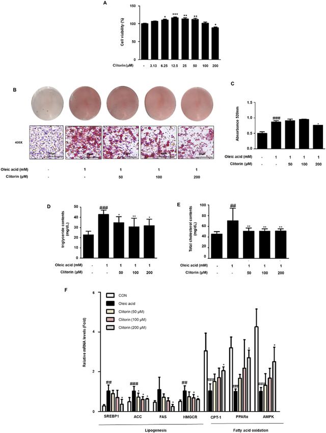
www.nature.com/scientificreports/
inflammation3. NAFLD is usually correlated with metabolic diseases, such as obesity, insulin resistance, hyper-
glycemia, and hypertension. Although considerable progress has been achieved with regard to drug development
for NAFLD, no suitable therapeutic agent has yet been a pproved2. Therefore, there is a critical need to develop
optimal therapeutic agents for NAFLD.
Under normal condition, liver processes large quantities of fatty acid, but stores only small amounts in the
form of t riglyceride4. Overnutrition directly contributes to the abundance of hepatic triglyceride accumula-
tion, which lead to NAFLD p rogression5; herein, the main causes of hepatic steatosis are increased de novo
lipogenesis and decreased fatty acid o xidation6. Many genes play important roles in lipogenesis and fatty acid
oxidation in the liver. When the high-fat diet feeding, peroxisome proliferator-activated receptor γ (PPARγ) is
the early-induced lipogenic transcription f actor7. Liver X receptor (LXR) and sterol regulatory element–bind-
ing protein 1c (SREBP1c) are key transcription factors involved in hepatic lipid s ynthesis7. Fatty acid synthase
(FAS) and 3-hydroxy-3-methylglutaryl coenzyme A reductase (HMGCR) are key enzymes in the synthesis
of fatty acid and cholesterol, respectively8. Acetyl-CoA carboxylase (ACC) catalyzes a master rate-controlling
step in de novo lipogenesis and fatty acid oxidation9,10. Peroxisome proliferator-activated receptor α (PPARα)
is closely associated with the transcription of genes related to hepatic fatty acid oxidation, including carnitine
palmitoyltransferase-1 (CPT-1)11.
Adenosine monophosphate-activated protein kinase (AMPK) is master regulator for energy homeostasis
through the inhibition of lipogenesis and the activation of fatty acid oxidation in liver12. Although multiple
factors lead to hepatic steatosis, to control lipogenesis and fatty acid oxidation can be therapeutic strategies for
management of NAFLD individuals who consume excess calories4.
Papaya (Carica papaya L.) is a fruit crop that is widely grown in tropical and sub-tropical regions. Tradition-
ally, papaya plants are used to treat various ailments such as asthma, ulcers, eczema, diabetes, helminth infections,
and fever13. Papaya plants have been reported to possess therapeutic potential for metabolic disorders, such as
diabetes mellitus type 2, causing alterations in both glycemic metabolism and lipid metabolism, oxidative stress,
and in models of arterial h ypertension13,14. Previous profiling indicates that four flavonoids, including mangha-
lants15,16. Among them, we focused on clitorin, a
slin, clitorin, rutin, and nicotiflorin, were identified in papaya p
kaempferol glycoside, because it has only been reported antioxidant effects17. Based on these findings, the present
study was designed to provide basic data to delineate the pharmacological effects of clitorin on the alleviation
of hepatic steatosis in western diet (WD)-induced hepatic steatosis mice and oleic acid-stimulated HepG2 cells.
Results
Clitorin reduced the body weight and liver weight index in the WD‑fed obese mice. We have
isolated the clitorin from papaya plants, identified by high performance liquid chromatography (HPLC) analy-
sis (Fig. 1), and investigated the effect of clitorin on the WD-fed obese mice. When mice were fed a WD for
12 weeks, we observed significant differences on total body weight and weight gain between the CON and WD
groups. However, the orlistat- or clitorin-administered groups displayed significantly lower body weight and
weight gain than the WD group (Fig. 2A,B). Herein, we did not observe any differences in food intake and
energy intake among all WD-fed groups (Fig. 2C,D), while the food efficiency ratio was significantly attenuated
after orlistat and clitorin administration in the WD-induced obese mice (Fig. 2E). In addition, we found that the
liver weight and liver index (mg/body weight) in the WD group were significantly higher than the correspond-
ing parameters in the CON group, indicating that the hepatic steatosis was induced in the WD-fed obese mice.
Notably, orlistat and clitorin administration significantly reversed these changes compared to those observed in
the WD group (Fig. 2F–H).
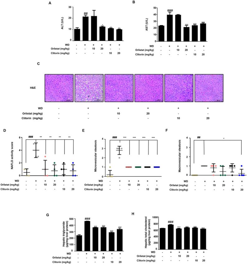
Clitorin ameliorated the features of liver steatosis in the WD‑induced hepatic steatosis
mice. Next, we checked the serum alanine aminotransferase (ALT) and aspartate aminotransferase (AST)
levels. The significantly increased serum levels of ALT and AST by HFD feeding were effectively decreased in
the 20 mg/kg orlistat-, and all clitorin-administered groups (Fig. 3A,B). To further confirm the increase in liver
weight and liver index in the WD-induced obese mice model, we conducted hematoxylin and eosin (H&E)
staining to evaluate the histological changes in the liver. We observed hepatocytic lipid vacuoles and hepatocyte
ballooning in the WD group compared to those in the CON group; however, the orlistat- or clitorin-adminis-
tered groups remarkably reversed the corresponding features in the liver of WD-fed obese mice (Fig. 3C). In
addition, the significantly increased NAFLD activity score (NAS), micro-, and macro-vesicular steatosis were
displayed in the WD group, whereas NAS and microvesicular steatosis were improved after orlistat or clitorin
administration (Fig. 3D,E); notably, the macrovesicular steatosis was significantly reduced in the only clitorin
20 mg/kg-administered group (Fig. 3F). Furthermore, the marked increases of intrahepatic triglyceride and total
cholesterol levels in the WD group were all lower in the orlistat- or clitorin-administered groups (Fig. 3G,H).
Altogether, these data indicated that WD-induced hepatic steatosis mouse model was completely established and
clitorin administration demonstrated a protective effect against hepatic steatosis in mice challenged with WD.
Clitorin regulated adipogenesis, lipogenesis, and fatty acid oxidation in the liver of WD‑induced
hepatic steatosis mice. To investigate the mechanisms of clitorin on suppressing hepatic steatosis in the
WD-induced hepatic steatosis mice, adipogenic and lipogenic transcriptional expression profiles were exam-
ined. In the liver, the protein expression levels of SREBP1, as well as PPARγ and C/EBPα, were higher in the WD
group; however, this was rescued by orlistat and clitorin administration (Fig. 4A). qRT-PCR analysis revealed
that the upregulated SREBP1, PPARγ, and C/EBPα protein expression levels in the livers of WD mice coincided
with increases in SREBP1, PPARγ, and C/EBPα mRNA levels. Notably, the marked upregulation of the mRNA
levels of SREBP1 and PPARγ was strongly suppressed in the liver of orlistat- or clitorin-administered mice com-
Scientific Reports | (2022) 12:4154 | https://doi.org/10.1038/s41598-022-07937-3 2
Vol:.(1234567890)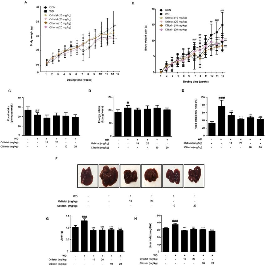
www.nature.com/scientificreports/
Figure 1. Isolation and identification of clitorin. HPLC chromatograms of (A) clitorin and (B) papaya plants.
A peak arose before clitorin was manghaslin, and two peaks arose after clitorin were rutin and nicotiflorin,
consecutively. HPLC: high performance liquid chromatography.
pared to the corresponding expression profiles in the WD group (Fig. 4B,C). The mRNA level of C/EBPα was
significantly down-regulated in the 10 mg/kg orlistat- or 20 mg/kg clitorin-administered mice; herein, 10 mg/kg
orlistat-administered group had stronger inhibitory effect than the 20 mg/kg orlistat-administered group in C/
EBPα protein expression (Fig. 4A), and this tendency was also observed in the mRNA level of C/EBPα (Fig. 4D).
We next assessed the impact of clitorin on the mRNA levels of lipogenesis (LXR, ACC, FAS, and HMGCR) and
fatty acid oxidation genes (CPT-1, PPARα, and AMPK), and it was revealed that the mRNA levels of LXR and
HMGCR were significantly inhibited in the all orlistat- or clitorin-administered groups (Fig. 4E). In addition,
the mRNA levels of ACCand FAS were significantly repressed in the 20 mg/kg orlistat- or clitorin- administered
groups, showing higher reduction in the clitorin-administered group than those in the orlistat-administered
group (Fig. 4E). In addition, CPT-1 mRNA level was significantly up-regulated in the 20 mg/kg orlistat- or
clitorin-administered mice. PPARα and AMPK mRNA levels, which was eliminated in the WD group, were sig-
nificantly augmented by 20 mg/kg orlistat- or all dose of clitorin-administered groups; additionally, these mRNA
levels were higher after the clitorin administration than those in the orlistat-administered group (Fig. 4E). These
data supported the concept that clitorin alleviates hepatic steatosis by regulating lipogenesis and fatty acid oxida-
tion genes in the liver of WD-induced hepatic steatosis mice.
Scientific Reports | (2022) 12:4154 | https://doi.org/10.1038/s41598-022-07937-3 3
Vol.:(0123456789)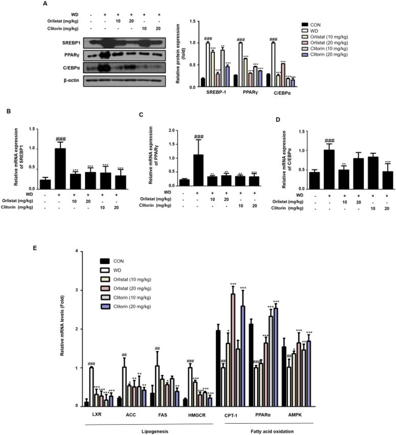
www.nature.com/scientificreports/

                                           Figure 2.  Effect of clitorin on total body weight, weight gain, and food intake in the WD-induced obese mouse
                                           model. The WD-induced mice were administered orlistat (10 or 20 mg/kg) or clitorin (10 or 20 mg/kg) for
                                           4 weeks, whereas control mice were fed a normal diet. (A) Body weight and (B) weight gain were recorded every
                                           week. (C) Food intake and (D) energy intake were calculated. (E) Food efficiency ratio (FER) was calculated by
                                           applying the equation: FER = (body weight gain (g)/food intake (g)) × 100. (F) Macroscopic images in the liver of
                                           mice in each group were taken at the end of the 13-week experimental period. (G) The weight of liver tissue and
                                           (H) relative liver weight ratio (mg/body weight) were measured. The values are represented as the mean ± SD
                                           (n = 6 per group). #P < 0.05, ##P < 0.01, and ###P < 0.001 vs. CON group; *P < 0.05, **P < 0.01, and ***P < 0.001 vs.
                                           WD group; significance was determined using two-way ANOVA followed by a Bonferroni post hoc test, and
                                           one-way ANOVA followed by Dunnett’s post hoc test. WD western diet, FER food efficiency ratio.

                                           Clitorin suppresses oleic acid‑induced lipid accumulation in HepG2 Cells. To further eliminate
                                           the effect of clitorin on lipid metabolism, relevant experiments were performed in vitro with HepG2 cells. The
                                           MTT assay showed that clitorin treatment (0 − 200 μM) did not induce cytotoxicity in HepG2 cells for 24 h
                                           (Fig. 5A). Accordingly, we designated three doses of clitorin at 50, 100, and 200 µM for further study. Oil Red O
                                           staining exhibited an obvious increase in stained lipid droplets in oleic acid-stimulated HepG2 cells compared to
                                           that in the non-treated cells, whereas it was significantly decreased after 200 µM clitorin treatment (Fig. 5B,C).
                                           Moreover, clitorin treatment significantly repressed the increased contents of triglyceride and total cholesterol

          Scientific Reports |   (2022) 12:4154 |               https://doi.org/10.1038/s41598-022-07937-3                                                     4

Vol:.(1234567890)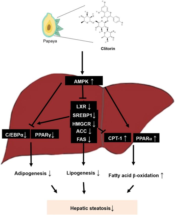
www.nature.com/scientificreports/

                                 Figure 3.  Effect of clitorin on the hepatic steatosis in the WD-induced hepatic steatosis mouse model. The
                                 levels of serum (A) ALT and (B) AST were determined using enzymatic methods. (C) The liver tissues from
                                 representative mice in each group were fixed, embedded in paraffin, and stained with H&E solution. Images
                                 are shown at an original magnification of 200 × . The scale bar is 100 µm. (D) NAS, (E) micro-, and (F)
                                 macro-vesicular steatosis were determined. The levels of hepatic (G) triglyceride and (H) total cholesterol
                                 were determined using enzymatic methods. The values are represented as the mean ± SD (n = 6 per group).
                                 ##
                                   P < 0.01 and ###P < 0.001 vs. CON group; *P < 0.05, **P < 0.01, and ***P < 0.001 vs. WD group; significance
                                 was determined using one-way ANOVA followed by Dunnett’s post hoc test. WD western diet, ALT alanine
                                 aminotransferase, AST aspartate aminotransferase, H&E hematoxylin and eosin.

Scientific Reports |   (2022) 12:4154 |               https://doi.org/10.1038/s41598-022-07937-3                                                 5

                                                                                                                                            Vol.:(0123456789)
www.nature.com/scientificreports/
Figure 4. Effect of clitorin on adipogenic, lipogenic, and fatty acid oxidation-related genes in the liver of
WD-induced hepatic steatosis mouse model. The protein levels of (A) SREBP1, PPARγ, and C/EBPα were
determined using western blot analysis. The cropped gel images are shown for clarity. Densitometric analysis
was performed using ImageJ ver. 1.50i (https://imagej.nih.gov/ij/). The mRNA levels of (B) SREBP1, (C)
PPARγ, (D) C/EBPα, and (E) LXR, ACC, FAS, HMGCR, CPT-1, PPARα, and AMPK were determined using
qRT-PCR analysis. The values are represented as the mean ± SD (n = 6 per group). #P < 0.05, ##P < 0.01, and
###
P < 0.001 vs. CON group; *P < 0.05, **P < 0.01, and ***P < 0.001 vs. WD group; significance was determined
using one-way ANOVA followed by Dunnett’s post hoc test. WD western diet, SREBP1 sterol regulatory element
binding protein 1, PPARγ peroxisome proliferator activated receptor γ, C/EBPα CCAAT/enhancer binding
protein α, LXR liver X receptor, ACCacetyl-CoA carboxylase, FAS fatty acid synthase, HMGCR3-Hydroxy-
3-Methylglutaryl-CoA Reductase, CPT-1 Carnitine palmitoyltransferase-1, PPARα peroxisome proliferator
activated receptor α, AMPK adenosine monophosphate-activated protein kinase.
Scientific Reports | (2022) 12:4154 | https://doi.org/10.1038/s41598-022-07937-3 6
Vol:.(1234567890)www.nature.com/scientificreports/
Figure 5. Effect of clitorin on oleic acid-induced lipid accumulation in HepG2 Cells. (A) Cell viability was
evaluated in HepG2 cells treated with clitorin. (B) The lipid accumulation was determined by Oil Red O
staining. Images are shown at an original magnification of 400 × . The scale bar is 50 µm. (C) The lipid content
was quantified by measuring absorbance. The levels of secreted (D) triglyceride and (E) total cholesterol
in HepG2 cells were determined using enzymatic methods. The mRNA levels of (F) SREBP1, ACC, FAS,
HMGCR, CPT-1, PPARα, and AMPK were determined using qRT-PCR analysis. The values are represented
as mean ± S.D of three independent experiments. ##P < 0.01 and ###P < 0.001 vs. non-treated cells; *P < 0.05,
**P < 0.01, and ***P < 0.001 vs. oleic acid-treated cells; significances were determined using one-way ANOVA
followed by Dunnett’s post hoc test. SREBP1 sterol regulatory element binding protein 1, ACCacetyl-CoA
carboxylase, FAS fatty acid synthase, HMGCR3-Hydroxy-3-Methylglutaryl-CoA Reductase, CPT-1 Carnitine
palmitoyltransferase-1, PPARα peroxisome proliferator activated receptor α, AMPK adenosine monophosphate-
activated protein kinase.
Scientific Reports | (2022) 12:4154 | https://doi.org/10.1038/s41598-022-07937-3 7
Vol.:(0123456789)www.nature.com/scientificreports/
in the oleic acid-stimulated HepG2 cells (Fig. 5D,E). Furthermore, the mRNA levels SREBP1, FAS, ACC, and
HMGCR were higher in the oleic acid-treated group than those in the non-treated group; however, this was
strongly reversed by 200 µM clitorin treatment (Fig. 5F). Moreover, CPT-1, PPARα, and AMPK mRNA levels,
which was significantly down-regulated in the WD group, were effectively reversed by 200 µM clitorin treatment
(Fig. 5F). These data indicated that clitorin attenuates oleic acid-induced hepatic lipid accumulation via control
of the lipogenesis and the fatty acid oxidation.
Discussion
Immoderate exposure to a high-fat diet has been determined as a key attribute in an increasing number of
NAFLD patients, which is demonstrated by the fact that the prevalence of NAFLD in obese patients is reported
to be up to 90%18; consequently, a high-fat diet is widely used to construct NAFLD animal models19. In the pre-
sent study, clitorin administration significantly reduced the body weight gain in the WD-induced obese mice
(Fig. 2A,B). To evaluate whether the hepatic steatosis was induced in this model, we first investigated the liver
weight and index (mg/body weight), and the data showed that WD feeding caused the remarkable increases in
the corresponding parameters compared to those in the CON group; on the other hand, clitorin administration
significantly reduced the liver weight and index in the WD-induced obese mice (Fig. 2G,H). It has been reported
that orlistat effectively alleviates steatosis and may serve as a viable treatment option for NAFLD20, therefore, it
was used as a positive control. Next, we confirmed that clitorin administration significantly rescued the mark-
edly increased serum levels of ALT and AST in WD-fed obese mice (Fig. 3A,B). Even though NAFLD typically
is characterized by mild increases of serum ALT and AST, the normal ALT and AST levels can be observed in up
to 50% of NAFLD p atients21. In addition, previous clinical review study suggested that relying on liver enzyme
abnormalities is unhelpful in the diagnosis of NAFLD and examination of disease severity22. For assessment of
hepatic steatosis, the NAS, which includes steatosis, lobular inflammation, and ballooning, has been used in
numerous clinical trials and cross-sectional studies23. Therefore, we conducted the H&E staining to further track
whether the hepatic histological changes were induced in this in vivo model. We found that the NAS, micro-,
and macro-vesicular steatosis were all higher in the WD-fed mice than the normal diet-fed mice; moreover, the
marked elevations in the hepatic triglyceride and total cholesterol contents were displayed in the WD group,
indicating that the WD-induced hepatic steatosis mouse model was completely established. Furthermore, clitorin
administration significantly recovered the NAS and microvesicular steatosis (Fig. 3D,E) as well as lipid profiling
(Fig. 3G,H) compared to those in the WD group. Notably, our data revealed that clitorin had stronger effect in
suppressing macrovesicular steatosis than the orlistat (Fig. 3F).
The hepatic effect of PPARγ appears to be steatogenic; hepatocyte-specific PPARγ knockout mice showed a
remarkable decrease in the number of hepatic lipid vacuoles, as well as downregulation of de novo lipogenesis
activators24. Conversely, PPARγ overexpression in the liver induced by HFD feeding leads to lipid accumula-
tion, which is the initiation step in the development of NAFLD7. CCAAT/enhancer binding proteins (C/EBPs),
including C/EBPα and SREBP1, are also considered key regulators of adipogenesis. SREBP1 plays an important
role in the regulation of de novo lipogenesis in the liver25. SREBP1c levels are enhanced in the fatty livers of
obese, insulin-resistant, and hyperinsulinemic ob/ob mice10. In addition, SREBP1c expression is also elevated in
patients with NAFLD; additionally, in concordance with its lipogenic role, hepatic triglyceride levels are higher
in SREBP1c-overexpressing transgenic mice26. Thus, SREBP1, PPARγ, and C/EBPα are crucial transcription fac-
tors that upregulate the expression of genes modulating fat accumulation in the liver. In this study, the hepatic
mRNA and protein levels of SREBP1, PPARγ, and C/EBPα were significantly reduced after clitorin administration
in a dose-dependent manner (Fig. 4A–D). However, both protein and mRNA levels of C/EBPα were effectively
abolished in the 10 mg/kg orlistat-administered mice than those in the 20 mg/kg orlistat-administered mice.
Additionally, in the orlistat-administrated groups, the mRNA levels of SREBP1 and PPARγ were not consistent
with the protein expressions; 10 mg/kg orlistat-administered group had higher inhibitory effects in the mRNA
levels of SREBP1 and PPARγ than those in the 20 mg/kg orlistat-administered group. This probably seems to
have relatively limitation in our study, with only 6 mice in each group examined in both protein and mRNA
expression; therefore, further studies (e.g. increase ‘n’ per a group) would be examined to prove these differences.
LXRs are involved in hepatic lipogenesis via direct regulation of SREBP1c27, which positively modulates ACC
expression28. ACC catalyzes a key rate-limiting step in fatty acid biosynthesis, and is also associated with the
control of fatty acid oxidation by the synthesis of malonyl-CoA, an inhibitor of CPT-16. Indeed, inhibition of the
liver-specific isoform ACC1 in mice ameliorated hepatic triglyceride levels in mice by simultaneously suppressing
fatty acid biosynthesis and augmenting fatty acid beta oxidation in the l iver6. CPT-1 leads to beta-oxidation, as
it allows fatty acids to reach the mitochondrial m atrix11. CPT-1 is also linked to PPARα expression. Among the
three PPAR isotypes, PPARα, PPARβ/δ, and PPARγ, PPARα is the most abundant isotype in hepatocytes and is
related to numerous aspects of lipid metabolism29 and high fatty acid oxidation rates30. Ineffective PPARα sens-
ing leads to diminished energy burning, resulting in hepatic steatosis and steatohepatitis31; thus, it is inferred
that these genes can potentially prevent NAFLD. AMPK, a major energy sensor of the cell, downregulates ACC
activity to suppress lipid b iosynthesis32. In addition, AMPK regulates hepatic and adipose lipid metabolism by
modulating lipogenesis, lipolysis, gluconeogenesis, and adipogenesis. AMPK inhibits de novo lipogenesis by
downregulating PPARγ, C/EBPα, and SREBP1; furthermore, it promotes fatty acid oxidation by upregulating
CPT-1a33. Our results showed that clitorin administration significantly decreased the mRNA levels of LXR, ACC,
FAS, and HMGCR, which are lipogenic genes, and it also enhanced the mRNA levels of PPARα and CTP-1, fatty
acid oxidation genes, as well as AMPK mRNA levels in the livers of WD-induced hepatic steatosis mice (Fig. 4E).
Overall, clitorin had similar or excellent inhibitory effects compared to orlistat used as a positive control in the
WD-induced hepatic steatosis mice.
Scientific Reports | (2022) 12:4154 | https://doi.org/10.1038/s41598-022-07937-3 8
Vol:.(1234567890)www.nature.com/scientificreports/
Oleic acid-stimulated HepG2 cells have been widely used to evaluate NAFLD in vitro34–39. Consistent with
in vivo experiments, our results showed that clitorin treatment significantly diminished lipid accumulation by
reducing the lipogenic genes (SREBP1, ACC, FAS, and HMGCR) and enhancing the fatty acid oxidation genes
(PPARα, CPT-1, and AMPK) in oleic acid-stimulated HepG2 cells (Fig. 5). Unexpectedly, there were significantly
increases on the cell viability in 6.25–50 µM clitorin-treated cells. This issue should be considered in additional
research to study the impact of clitorin in HepG2 cells, but we clearly demonstrated that 200 µM clitorin block
the hepatic lipid accumulation by controlling the lipogenesis and fatty acid oxidation in oleic acid-stimulated
HepG2 cells.
In this study, four flavonoids, including manghaslin, clitorin, rutin, and nicotiflorin, were identified in papaya
leaf. Among the four flavonoids, previous study reported that a recommended dose of rutin is 250–500 mg twice
per day40,41. Compare to rutin, 20 mg/kg clitorin, which equates to a 97 mg for a 60 kg person, seems not to be an
excessive dose for daily intake. However, it is necessary to evaluate clitorin content in papaya plant to determine
whether 97 mg clitorin (for human) can be taken through diet such as papaya plant or can be considered as a food
supplement. Although there is evidence on clitorin content in freeze-dried papaya leaf juice42, it is not sufficient to
figure out its quantity. Additionally, to decide the exact human dose, further study on the toxicity of clitorin must
be needed. This study contributes new knowledge to the sparse literature on clitorin, which would help specific
areas for future research including determination of clinical dose. Next, we are going to analyze the mechanisms
of clitorin by inhibiting/silencing AMPK in vivo and in vitro model, to fully understand its action on regulation
of lipolysis and lipogenesis in NAFLD. Overall, our results showed that clitorin alleviated hepatic steatosis by
reducing both adipogenesis and lipogenesis, and enhancing fatty acid oxidation (Fig. 6). The present study is the
first to report on the positive impact of clitorin on hepatic steatosis and our findings provide basic data, which
lead to deeper understanding of the pharmacological effects of clitorin on the potential improvement of NAFLD.
Materials and methods
Chemicals and reagents. Oil Red O powder, oleic acid, and methyl alcohol were purchased from Sigma-
Aldrich Co. LLC (St. Louis, MO, USA). Minimum Essential Medium (MEM), fetal bovine serum (FBS), and
penicillin were purchased from Life Technologies Inc. (Grand Island, NY, USA). Orlistat was purchased from
Tokyo Chemical Inc. (Tokyo, Japan). The Research Diets (New Brunswick, NJ, USA) provided 45% of the
WD (D-12451). Antibodies against PPARγ (cat. no. sc-7273), C/EBPα (cat. no. sc-365318), SREBP1 (cat. no.
sc-13551), and β-actin (cat. No. sc-47778) were purchased from Santa Cruz Biotechnology Inc. (Dallas, TX,
USA). Horseradish peroxidase-conjugated secondary antibodies were purchased from Jackson ImmunoRe-
search Laboratories, Inc. (West Grove, PA, USA).
Isolation and preparation of clitorin by HPLC. Clitorin, a compound derived from Carica papaya
L., was identified by Professor Agung Nugroho (Lambung Mangkurat University, Indonesia). As previously
described43, the leaves of Carica papaya were collected from a papaya farm near Pelaihari City, South Kalimantan
Province, Indonesia. Plant species was identified and authenticated at the Department of Agronomy, Lambung
Mangkurat University, and the voucher specimen (No. C-23) was deposited in the herbarium of Laboratory of
Natural Products, Department of Agro-industrial Technology, Lambung Mangkurat University. The collected
leaves were dried completely at 40 °C. The dried powder of C. papaya leaf (750 g) was extracted thrice with
methanol (6 L) under reflux at 70 °C for 5 h. The HPLC method involved two solvents for the mobile phase,
solvent A was H2O with 0.05% acetic acid, v/v) and solvent B was methanol. The linear gradient elution of the
solvents was programmed as follows: 0–20 min (20 → 65% B), 20–21 min (65 → 100% B), 21–25 min (100% B),
25–27 min (100 → 20% B), and 27–30 min (20% B). The flow rate and column temperature was set constantly at
1.0 mL/min and 40 °C, respectively. The detection wavelength was fixed at 254 nm and monitored for 30 min.
The linear calibration equation was y = 30.87 × + 17.49 with the LOD and LOQ was 5.91 µg/mL and 19.70 µg/mL,
respectively. The purity of the compound was more than 92%. For all experiments on this plants, we confirm that
all methods were carried out in accordance with relevant guidelines and regulations.
Experimental animal care protocols and treatment cycles. Six-week-old male C57BL/6J mice were
procured from Daehan Biolink (Daejeon, Republic of Korea). The mice were maintained under conditions of
controlled temperature (22 ± 2 °C) and humidity (55 ± 9%), with a 12-h light/dark cycle. After a week of adjust-
ment, the mice were fed 45% WD for 7 weeks, except for the normal diet group (CON). After 7 weeks, the mice
were randomly divided into five groups of six mice each: WD group, WD + treatment group with 10 or 20 mg/
kg orlistat as a positive control, and WD + treatment group with 10 or 20 mg/kg clitorin. Orlistat and clitorin
were dissolved in 1:1:18 ratio of ethanol, cremophor, and distilled water and orally administered to the mice
once daily for 4 weeks. Mice in the CON and WD groups were administered vehicle. The mice were allowed
free access to water and food, and their body weight and food intake were measured every week. Food efficiency
ratio (FER) was calculated by applying the equation: FER = (body weight gain (g)/food intake (g)) × 100. At
the end of the experiment, mice were anesthetized with Zoletil 50 (20 mg/kg) by i.p. injection according to the
manufacturer’s instructions, and then the mice were euthanized by cervical dislocation. The livers of the mice
were excised, cleaned with phosphate-buffered saline (PBS), weighed, and directly stored at − 80 °C. All experi-
ments were performed under the Ethical Committee for Animal Care and the Use of Laboratory Animals, Sangji
University (approval document no. 2017-22).
Serum analysis. During blood sample collection, the animals were already under the influence of termi-
nal anesthesia. Blood samples were collected via cardiac puncture. The samples were centrifuged at 1000×g
Scientific Reports | (2022) 12:4154 | https://doi.org/10.1038/s41598-022-07937-3 9
Vol.:(0123456789)www.nature.com/scientificreports/
Figure 6. Schematic diagram of clitorin on prevention of WD-induced hepatic steatosis. Clitorin alleviated
hepatic steatosis by reducing both adipogenesis and lipogenesis, and enhancing fatty acid oxidation in vivo and
in vitro model.
for 20 min to obtain the serum samples. The concentrations of ALT and AST were measured by ALT and AST
quantification kit (ASAN Pharm, Co., Ltd, Seoul, Republic of Korea).
Histological analysis. The liver tissues from the mice in each group were fixed in 10% formalin, embedded
in paraffin, and cut into 8 µm sections. Certain sections were stained with H&E for histological examination.
Stained liver sections were observed for the evaluation of hepatic steatosis using an Olympus SZX10 micro-
scope (Olympus, Tokyo, Japan). NAFLD development was examined using NAS, which includes a numerical
score for steatosis (0–3), hepatocyte ballooning (0–2), and lobular inflammation (0–3). Steatosis were deter-
mined at 200 × magnification and quantified as macrovesicular and microvesicular steatosis using ImageJ ver.
1.50i (https://imagej.nih.gov/ij/). Then, the percentage of steatotic cells was graded as follows: (1) 0: absent; (2)
1: ≤ 25%; (3) 2: > 25% and ≤ 50%; (4) 3: > 50% and ≤ 75%; or (5) 4: > 75% of the parenchyma.
Lipid profiling analysis. For liver tissues, approximately 0.1 g of liver tissue was homogenized in 2 mL of
chloroform:methanol:distilled water (2:1:1, v/v) solution, vortexed, and centrifuged at 3,000 rpm for 10 min at
Scientific Reports | (2022) 12:4154 | https://doi.org/10.1038/s41598-022-07937-3 10
Vol:.(1234567890)www.nature.com/scientificreports/
Gene Forward (5′–3′) Reverse (5′–3′)
PPARγ (m) ATCGAGTGCCGAGTCTGTGG GCAAGGCACTTCTGAAACCG
SREBP1 (m) GGCTATTCCGTGAACATCTCCTA ATCCAAGGGCAGTTCTTGTG
C/EBPα (m) GGAACTTGAAGCACAATCGATC TGGTAAAGGTTCTCA
LXRα (m) CAGGAGACCAGGGAGGCAAC GCAGGGCTGTAGGCTCTGCT
ACC(m) TTTTCGATGTCCTCCCAAACTTT GCTCATAGGCGATATAAGCTCT
FAS (m) AGGGGTCGACCTGGTCCTCA GCCATGCCCAGAGGGTGGTT
HMGCR(m) CAGGATGCAGCACAGAATGT CTTTGCATGCTCCTTGAACA
CPT-1 (m) CTCAGTGGGAGCGACTCTTCA GGCCTCTGTGGTACACGACAA
PPARα (m) CAGGAGAGCAGGGATTTGCA CCTACGCTCAGCCCTCTTCAT
AMPK (m) GGTGGATTCCCAAAAGTGCT AAGCAGTGCTGGGTCACAAG
GAPDH (m) ATGGAAATCCCATCACCATCTT CGCCCCACTTGATTTTGG
SREBP1 (h) ACCGACATCGAAGGTGAAGT CCAGCATAGGGTGGGTCAAA
ACC(h) CATGCGGTCTATCCGTAGGTG GTGTGACCATGACAACGAATCT
FAS (h) AAGGACCTGTCTAGGTTTGATGC TGGCTTCATAGGTGACTTCCA
HMGCR(h) GACCTTTCCAGAGCAAGCAC TCAACAAGAGCATCGAGGGT
CPT-1 (h) TCCAGTTGGCTTATCGTGGTG TCCAGAGTCCGATTGATTTTTGC
PPARα (h) TCCGACTCCGTCTTCTTGAT GCCTAAGGAAACCGTTCTGTG
AMPK (h) AGGATGCCTGAAAAGCTTGA GACAGCCGGAGAAGCAGAAAC
GAPDH (h) CTCCTCCACCTTTGACGCTG CTCTTGTGCTCTTGCTGGGG
Table 1. Real-time PCR primer sequences.
room temperature. After centrifugation, the bottom layer was carefully aspirated into a new test tube and dried
at 50 °C to remove chloroform. The dried lipid was weighed and dissolved in methanol prior to lipid analysis.
For HepG2 cells, the supernatant from the plates was directly used. The concentrations of triglyceride and total
cholesterol were measured by triglyceride and total cholesterol quantification kit (ASAN Pharm, Co., Ltd, Seoul,
Republic of Korea), and detected spectrophotometrically at 550 and 500 nm.
Western blot analysis. Each liver tissue (10 mg) was homogenized using 600 µL PRO-PREP® solution
(Intron Biotechnology, Gyeonggi-do, Republic of Korea), a protein extraction solution. The same amount (15–
30 µg) of protein sample was separated on an 8%–12% sodium dodecyl sulfate polyacrylamide gel and trans-
ferred onto a polyvinylidene fluoride membrane. The membranes were blocked with 2.5% skim milk solution for
30 min, incubated with PPARγ (1:1000), C/EBPα (1:1000), SREBP1 (1:1000), and β-actin (1:2500) primary anti-
bodies overnight at 4 °C, followed by incubation with anti-mouse horseradish peroxidase-conjugated secondary
antibody (1:2500) for 2 h at 25 °C. The membranes were washed thrice for 10 min with Tris-buffered saline
containing Tween 20 and visualized by enhanced chemiluminescence using X-ray film (Agfa, Belgium). The
uncropped blot images are given in Supplementary information.
Quantitative reverse‑transcription polymerase chain reaction (qRT‑PCR) analysis. qRT-PCR
escribed44. Briefly, each liver tissue (50 mg) were homogenized, and total
analysis was performed as previously d
RNA was isolated using the 1 mL Easy-Blue® reagent according to the manufacturer’s instructions (Intron Bio-
technology; Seongnam, Republic of Korea). Total RNA (1 µg) was converted to cDNA using a high-capacity
cDNA reverse transcription kit (Applied Biosystems; Foster City, CA, USA). For qPCR reactions, the 10 ng of
cDNA was amplified and measured using SYBR® Master Mix (Applied Biosystems; Foster City, CA, USA). Gene
expression was determined using the comparative threshold cycle method. GAPDH was used as an internal
control. Sequences of mouse oligonucleotide primers are presented in Table 1.
Cell culture and treatment. The human hepatoma cell line HepG2 (No. 88065) was obtained from the
Korean Cell Line Bank (KCLB, Seoul, Republic of Korea). HepG2 cells were grown in MEM containing 10% FBS
and 100 mg/L penicillin under a humidified atmosphere of 5% CO2 at 37 °C. The cells were seeded at a density of
2 × 10⁵ cells per well into 6-well plate and then treated with 1 mM oleic acid (O 7501, Sigma-Aldrich) dissolved
in culture medium containing 5% methanol with or without different concentrations of clitorin (50, 100, and
200 µM) for 48 h. The clitorin was dissolved in dimethyl sulfoxide (DMSO) and the 0.1% DMSO was treated in
the control cells as a vehicle.
Cell viability assay. HepG2 cells were seeded into a 96-well plate at a concentration of 1 × 10⁴ cells per
well for 24 h. After incubation, the cells were treated with different concentrations of clitorin (0–200 µM) for
24 h. After treatment, the cells were treated with 3-(4,5-dimethylthiazol-2-yl)-2,5-diphenyl tetrazolium bromide
(MTT) solution (5 mg/mL) and incubated again for 4 h. The supernatant from the plates was discarded, and
the purple formazan product was dissolved in DMSO. The absorbance was measured at 540 nm using an Epoch
microplate spectrometer (Biotek, Winooski, VT, USA).
Scientific Reports | (2022) 12:4154 | https://doi.org/10.1038/s41598-022-07937-3 11
Vol.:(0123456789)www.nature.com/scientificreports/
Oil red O staining of HepG2 cells. After stimulation with oleic acid, the cells were washed with PBS and
fixed with 10% formaldehyde in PBS at 25 °C for 1 h. Cells were then washed thrice with distilled water and
stained with Oil Red O working solution (3 mg/mL in 60% isopropanol) at 25 °C for 2 h. The cells were rinsed
thrice with distilled water and photographed using an Olympus SZX10 microscope. Next, the Oil Red O dye was
eluted with isopropanol to determine the intracellular lipid content and was measured using an Epoch® micro-
volume spectrophotometer at 520 nm.
Statistical analysis. Data are expressed as the mean ± standard deviation (SD) of triplicate experiments.
Statistically significant values were compared using ANOVA and Dunnett’s post hoc test, and p-values < 0.05
were considered statistically significant. Statistical analysis was performed using SPSS statistical analysis soft-
ware (version 19.0, IBM SPSS, Armonk, NY, USA).
Ethical approval. The research protocol (no. 2017-22) was approved by the Institutional animal ethics com-
mittee (IAEC) of Sangji University. Guidelines outlined in the Guide for the Care and Use of Laboratory Animals
of the National Institutes of Health and the ARRIVE (Animal Research: Reporting of In-vivo Experiments)
guidelines (http://www.nc3rs.org/ARRIVE) were followed to perform all the experiments.
Data availability
The datasets used and/or analyzed in this study are available from the corresponding authors on reasonable
request.
Received: 24 August 2021; Accepted: 28 February 2022
References
1. Drescher, H. K., Weiskirchen, S. & Weiskirchen, R. Current status in testing for nonalcoholic fatty liver disease (NAFLD) and
nonalcoholic steatohepatitis (NASH). Cells 8, 845. https://doi.org/10.3390/cells8080845 (2019).
2. Friedman, S. L., Neuschwander-Tetri, B. A., Rinella, M. & Sanyal, A. J. Mechanisms of NAFLD development and therapeutic
strategies. Nat. Med. 24, 908–922. https://doi.org/10.1038/s41591-018-0104-9 (2018).
3. Pydyn, N., Miekus, K., Jura, J. & Kotlinowski, J. New therapeutic strategies in nonalcoholic fatty liver disease: A focus on promising
drugs for nonalcoholic steatohepatitis. Pharmacol. Rep. 72, 1–12. https://doi.org/10.1007/s43440-019-00020-1 (2020).
4. Alves-Bezerra, M. & Cohen, D. E. Triglyceride metabolism in the liver. Compr. Physiol. 8, 1–8. https://doi.org/10.1002/cphy.c1700
12 (2017).
5. Lakhani, H. V. et al. Phenotypic alteration of hepatocytes in non-alcoholic fatty liver disease. Int. J. Med. Sci. 15, 1591–1599. https://
doi.org/10.7150/ijms.27953 (2018).
6. Koo, S. H. Nonalcoholic fatty liver disease: Molecular mechanisms for the hepatic steatosis. Clin. Mol. Hepatol. 19, 210–215. https://
doi.org/10.3350/cmh.2013.19.3.210 (2013).
7. Lee, Y. K., Park, J. E., Lee, M. & Hardwick, J. P. Hepatic lipid homeostasis by peroxisome proliferator-activated receptor gamma 2.
Liver Res. 2, 209–215. https://doi.org/10.1016/j.livres.2018.12.001 (2018).
8. Shin, M. R., Shin, S. H. & Roh, S. S. Diospyros kaki and Citrus unshiu mixture improves disorders of lipid metabolism in nonalco-
holic fatty liver disease. Can. J. Gastroenterol. Hepatol. 2020, 8812634. https://doi.org/10.1155/2020/8812634 (2020).
9. Wang, L. F. et al. Inhibition of NAMPT aggravates high fat diet-induced hepatic steatosis in mice through regulating Sirt1/AMP-
Kalpha/SREBP1 signaling pathway. Lipids Health Dis. 16, 82. https://doi.org/10.1186/s12944-017-0464-z (2017).
10. Musso, G., Gambino, R. & Cassader, M. Recent insights into hepatic lipid metabolism in non-alcoholic fatty liver disease (NAFLD).
Prog. Lipid Res. 48, 1–26. https://doi.org/10.1016/j.plipres.2008.08.001 (2009).
11. Souza-Mello, V. Peroxisome proliferator-activated receptors as targets to treat non-alcoholic fatty liver disease. World J. Hepatol.
7, 1012–1019. https://doi.org/10.4254/wjh.v7.i8.1012 (2015).
12. Seo, Y. J., Lee, K., Song, J. H., Chei, S. & Lee, B. Y. Ishige okamurae extract suppresses obesity and hepatic steatosis in high fat
diet-induced obese mice. Nutrients 10, 1802. https://doi.org/10.3390/nu10111802 (2018).
13. Pandey, S., Cabot, P. J., Shaw, P. N. & Hewavitharana, A. K. Anti-inflammatory and immunomodulatory properties of Carica
papaya. J. Immunotoxicol. 13, 590–602. https://doi.org/10.3109/1547691X.2016.1149528 (2016).
14. Santana, L. F. et al. Nutraceutical potential of Carica papaya in metabolic syndrome. Nutrients 11, 1608. https://doi.org/10.3390/
nu11071608 (2019).
15. Julianti, T. et al. HPLC-based activity profiling for antiplasmodial compounds in the traditional Indonesian medicinal plant Carica
papaya L. J. Ethnopharmacol. 155, 426–434. https://doi.org/10.1016/j.jep.2014.05.050 (2014).
16. Brasil, G. A. et al. Antihypertensive effect of Carica papaya via a reduction in ACE activity and improved baroreflex. Planta Med.
80, 1580–1587. https://doi.org/10.1055/s-0034-1383122 (2014).
17. Ma, H., Li, J., An, M., Gao, X. M. & Chang, Y. X. A powerful on line ABTS(+)-CE-DAD method to screen and quantify major
antioxidants for quality control of Shuxuening Injection. Sci. Rep. 8, 5441. https://doi.org/10.1038/s41598-018-23748-x (2018).
18. Hu, Y. et al. Acerola polysaccharides ameliorate high-fat diet-induced non-alcoholic fatty liver disease through reduction of lipo-
genesis and improvement of mitochondrial functions in mice. Food Funct. 11, 1037–1048. https://doi.org/10.1039/c9fo01611b
(2020).
19. Zhong, F., Zhou, X., Xu, J. & Gao, L. Rodent models of nonalcoholic fatty liver disease. Digestion 101, 522–535. https://doi.org/10.
1159/000501851 (2020).
20. Ye, J. et al. Effect of orlistat on liver fat content in patients with nonalcoholic fatty liver disease with obesity: Assessment using
magnetic resonance imaging-derived proton density fat fraction. Therap. Adv. Gastroenterol. 12, 1756284819879047. https://doi.
org/10.1177/1756284819879047 (2019).
21. Noureddin, M. & Loomba, R. Nonalcoholic fatty liver disease: Indications for liver biopsy and noninvasive biomarkers. Clin. Liver
Dis. 1, 104–107. https://doi.org/10.1002/cld.65 (2012).
22. Dyson, J. K., Anstee, Q. M. & McPherson, S. Non-alcoholic fatty liver disease: a practical approach to diagnosis and staging. Front.
Gastroenterol. 5, 211–218. https://doi.org/10.1136/flgastro-2013-100403 (2014).
23. Brown, G. T. & Kleiner, D. E. Histopathology of nonalcoholic fatty liver disease and nonalcoholic steatohepatitis. Metabolism 65,
1080–1086. https://doi.org/10.1016/j.metabol.2015.11.008 (2016).
24. Skat-Rordam, J., Hojland Ipsen, D., Lykkesfeldt, J. & Tveden-Nyborg, P. A role of peroxisome proliferator-activated receptor gamma
in non-alcoholic fatty liver disease. Basic Clin. Pharmacol. Toxicol. 124, 528–537. https://doi.org/10.1111/bcpt.13190 (2019).
Scientific Reports | (2022) 12:4154 | https://doi.org/10.1038/s41598-022-07937-3 12
Vol:.(1234567890)www.nature.com/scientificreports/
25. Jo, H. K., Kim, G. W., Jeong, K. J., Kim, D. Y. & Chung, S. H. Eugenol ameliorates hepatic steatosis and fibrosis by down-regulating
SREBP1 gene expression via AMPK-mTOR-p70S6K signaling pathway. Biol. Pharm. Bull. 37, 1341–1351. https://doi.org/10.1248/
bpb.b14-00281 (2014).
26. Shimano, H. et al. Isoform 1c of sterol regulatory element binding protein is less active than isoform 1a in livers of transgenic mice
and in cultured cells. J. Clin. Invest. 99, 846–854. https://doi.org/10.1172/JCI119248 (1997).
27. Grønning-Wang, L. M., Bindesbøll, C. & Nebb, H. I. The role of liver X receptor in hepatic de novo lipogenesis and cross-talk with
insulin and glucose signaling. Lipid Metab. 1, 61–90 (2013).
28. Kohjima, M. et al. SREBP-1c, regulated by the insulin and AMPK signaling pathways, plays a role in nonalcoholic fatty liver disease.
Int. J. Mol. Med. 21, 507–511 (2008).
29. Montagner, A. et al. Liver PPARalpha is crucial for whole-body fatty acid homeostasis and is protective against NAFLD. Gut 65,
1202–1214. https://doi.org/10.1136/gutjnl-2015-310798 (2016).
30. Pawlak, M., Lefebvre, P. & Staels, B. Molecular mechanism of PPARalpha action and its impact on lipid metabolism, inflammation
and fibrosis in non-alcoholic fatty liver disease. J. Hepatol. 62, 720–733. https://doi.org/10.1016/j.jhep.2014.10.039 (2015).
31. Reddy, J. K. & Rao, M. S. Lipid metabolism and liver inflammation. II. Fatty liver disease and fatty acid oxidation. Am. J. Physiol.
Gastrointest. Liver Physiol. 290, 852–858. https://doi.org/10.1152/ajpgi.00521.2005 (2006).
32. Liou, C. J. et al. Fisetin protects against hepatic steatosis through regulation of the Sirt1/AMPK and fatty acid beta-oxidation
signaling pathway in high-fat diet-induced obese mice. Cell Physiol. Biochem. 49, 1870–1884. https://doi.org/10.1159/000493650
(2018).
33. Inamdar, S., Joshi, A., Malik, S., Boppana, R. & Ghaskadbi, S. Vitexin alleviates non-alcoholic fatty liver disease by activating AMPK
in high fat diet fed mice. Biochem. Biophys. Res. Commun. 519, 106–112. https://doi.org/10.1016/j.bbrc.2019.08.139 (2019).
34. Kanuri, G. & Bergheim, I. In vitro and in vivo models of non-alcoholic fatty liver disease (NAFLD). Int. J. Mol. Sci. 14, 11963–11980.
https://doi.org/10.3390/ijms140611963 (2013).
35. Müller, F. A. & Sturla, S. J. Human in vitro models of nonalcoholic fatty liver disease. Curr. Opin. Toxicol. 16, 9–16 (2019).
36. Lee, M. R., Yang, H. J., Park, K. I. & Ma, J. Y. Lycopus lucidus Turcz. ex Benth Attenuates free fatty acid-induced steatosis in HepG2
cells and non-alcoholic fatty liver disease in high-fat diet-induced obese mice. Phytomedicine 55, 14–22. https://doi.org/10.1016/j.
phymed.2018.07.008 (2019).
37. Ali, O., Darwish, H. A., Eldeib, K. M. & Abdel Azim, S. A. miR-26a potentially contributes to the regulation of fatty acid and sterol
metabolism in vitro human HepG2 cell model of nonalcoholic fatty liver disease. Oxid. Med. Cell Longev. 2018, 8515343. https://
doi.org/10.1155/2018/8515343 (2018).
38. Gomaraschi, M. et al. Lipid accumulation impairs lysosomal acid lipase activity in hepatocytes: Evidence in NAFLD patients and
cell cultures. Biochim. Biophys. Acta Mol. Cell. Biol. Lipids 1864, 158523. https://doi.org/10.1016/j.bbalip.2019.158523 (2019).
39. Xia, H. et al. Alpha-naphthoflavone attenuates non-alcoholic fatty liver disease in oleic acid-treated HepG2 hepatocytes and in
high fat diet-fed mice. Biomed. Pharmacother. 118, 109287. https://doi.org/10.1016/j.biopha.2019.109287 (2019).
40. Yu, C. P. et al. Quercetin and rutin reduced the bioavailability of cyclosporine from Neoral, an immunosuppressant, through
activating P-glycoprotein and CYP 3A4. J. Agric. Food Chem. 59, 4644–4648. https://doi.org/10.1021/jf104786t (2011).
41. Volate, S. R., Davenport, D. M., Muga, S. J. & Wargovich, M. J. Modulation of aberrant crypt foci and apoptosis by dietary herbal
supplements (quercetin, curcumin, silymarin, ginseng and rutin). Carcinogenesis 26, 1450–1456. https://doi.org/10.1093/carcin/
bgi089 (2005).
42. Mohd Abd Razak, M. R. et al. Immunomodulatory activities of Carica papaya L. leaf juice in a non-lethal symptomatic dengue
mouse model. Pathogens https://doi.org/10.3390/pathogens10050501 (2021).
43. Nugroho, A., Heryani, H., Choi, J. S. & Park, H.-J. Identification and quantification of flavonoids in Carica papaya leaf and
peroxynitrite-scavenging activity. Asian Pac. J. Trop. Biomed. 7, 208–213 (2017).
44. Park, Y. J., Lee, G. S., Cheon, S. Y., Cha, Y. Y. & An, H. J. The anti-obesity effects of Tongbi-san in a high-fat diet-induced obese
mouse model. BMC Complement. Altern. Med. 19, 1. https://doi.org/10.1186/s12906-018-2420-5 (2019).
Acknowledgements
The present study was supported by a National Research Foundation of Korea (NRF) grant funded by the
Korean government (MSIP; Ministry of Science, ICT & Future Planning) (No. NRF-2020R1G1A1011494) and
the Research fund from Sangji University Graduate School.
Author contributions
D.C.C. and H.J.A. conceived and designed the experiments. D.C.C. and Y.J.P. wrote the manuscript and con-
ducted the experiments. A.N. supplied the clitorin used in experimental analyses, which is derived from the
papaya plant. Y.J.P., Y.M.K., and H.J.A. substantially contributed to the analysis and interpretation of data and
revised the manuscript. All authors read and approved the final manuscript.
Competing interests
The authors declare no competing interests.
Additional information
Supplementary Information The online version contains supplementary material available at https://doi.org/
10.1038/s41598-022-07937-3.
Correspondence and requests for materials should be addressed to H.-J.A.
Reprints and permissions information is available at www.nature.com/reprints.
Publisher’s note Springer Nature remains neutral with regard to jurisdictional claims in published maps and
institutional affiliations.
Scientific Reports | (2022) 12:4154 | https://doi.org/10.1038/s41598-022-07937-3 13
Vol.:(0123456789)www.nature.com/scientificreports/
Open Access This article is licensed under a Creative Commons Attribution 4.0 International
License, which permits use, sharing, adaptation, distribution and reproduction in any medium or
format, as long as you give appropriate credit to the original author(s) and the source, provide a link to the
Creative Commons licence, and indicate if changes were made. The images or other third party material in this
article are included in the article’s Creative Commons licence, unless indicated otherwise in a credit line to the
material. If material is not included in the article’s Creative Commons licence and your intended use is not
permitted by statutory regulation or exceeds the permitted use, you will need to obtain permission directly from
the copyright holder. To view a copy of this licence, visit http://creativecommons.org/licenses/by/4.0/.
© The Author(s) 2022
Scientific Reports | (2022) 12:4154 | https://doi.org/10.1038/s41598-022-07937-3 14
Vol:.(1234567890)You can also read