Crocodile blood supplementation protects vascular function in diabetic mice
←
→
Page content transcription
If your browser does not render page correctly, please read the page content below
Chook et al. Food Production, Processing and Nutrition               (2021) 3:19
https://doi.org/10.1186/s43014-021-00066-w
                                                                                                                  Food Production, Processing
                                                                                                                  and Nutrition

 RESEARCH                                                                                                                                           Open Access

Crocodile blood supplementation protects
vascular function in diabetic mice
Chui Yiu Bamboo Chook1, Francis M. Chen1, Gary Tse1, Fung Ping Leung1 and Wing Tak Wong1,2*

  Abstract
  Cardiovascular disease is a major cause of mortality in diabetic patients due to the heightened oxidative stress and
  pro-inflammatory state in vascular tissues. Effective approaches targeting cardiovascular health for diabetic patients
  are urgently needed. Crocodile blood, an emerging dietary supplement, was suggested to have anti-oxidative and
  anti-inflammatory effects in vitro, which have yet to be proven in animal models. This study thereby aimed to
  evaluate whether crocodile blood can protect vascular function in diabetic mice against oxidation and
  inflammation. Diabetic db/db mice and their counterparts db/m+ mice were treated daily with crocodile blood
  soluble fraction (CBSF) or vehicle via oral gavage for 4 weeks before their aortae were harvested for endothelium-
  dependent relaxation (EDR) quantification using wire myograph, which is a well-established functional study for
  vascular function indication. Organ culture experiments culturing mouse aortae from C57BL/6 J mice with or
  without IL-1β and CBSF were done to evaluate the direct effect of CBSF on endothelial function. Reactive oxygen
  species (ROS) levels in mouse aortae were assessed by dihydroethidium (DHE) staining with inflammatory markers
  in endothelial cells quantified by quantitative polymerase chain reaction (qPCR). CBSF significantly improved
  deteriorated EDR in db/db diabetic mice through both diet supplementation and direct culture, with suppression of
  ROS level in mouse aortae. CBSF also maintained EDR and reduced ROS levels in mouse aortae against the
  presence of pro-inflammatory IL-1β. Under the pro-inflammatory state induced by IL-1β, gene expressions of
  inflammatory cytokines were downregulated, while the protective transcripts UCP2 and SIRT6 were upregulated in
  endothelial cells. Our study suggests a novel beneficial effect of crocodile blood on vascular function in diabetic
  mice and that supplementation of diet with crocodile blood may act as a complementary approach to protect
  against vascular diseases through anti-oxidation and anti-inflammation in diabetic patients.
  Keywords: Crocodile blood, Diabetes mellitus, Vascular endothelial function, Anti-oxidative, Anti-inflammatory

* Correspondence: jack_wong@cuhk.edu.hk
1
 School of Life Sciences, Faculty of Science, The Chinese University of Hong
Kong, Shatin, NT, Hong Kong SAR
2
 State Key Laboratory of Agrobiotechnology, The Chinese University of Hong
Kong, Hong Kong, China

                                        © The Author(s). 2021 Open Access This article is licensed under a Creative Commons Attribution 4.0 International License,
                                        which permits use, sharing, adaptation, distribution and reproduction in any medium or format, as long as you give
                                        appropriate credit to the original author(s) and the source, provide a link to the Creative Commons licence, and indicate if
                                        changes were made. The images or other third party material in this article are included in the article's Creative Commons
                                        licence, unless indicated otherwise in a credit line to the material. If material is not included in the article's Creative Commons
                                        licence and your intended use is not permitted by statutory regulation or exceeds the permitted use, you will need to obtain
                                        permission directly from the copyright holder. To view a copy of this licence, visit http://creativecommons.org/licenses/by/4.0/.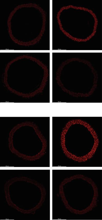
Chook et al. Food Production, Processing and Nutrition   (2021) 3:19                                                     Page 2 of 13

Highlights                                                               We thereby hypothesized that crocodile blood may
                                                                       have protective effects on vascular endothelial function
   Daily oral crocodile blood supplementation for 5                   against diabetic damage through anti-oxidation and anti-
    weeks ameliorated endothelial dysfunction in db/db                 inflammation. Using endothelium-dependent relaxation
    diabetic mice through suppressing the overproduced                 (EDR) as an indicator (Versari et al. 2009), this study ex-
    reactive oxygen species (ROS).                                     amined the effects of crocodile blood on endothelial dys-
   Direct action of improving endothelium-dependent                   function in db/db diabetic mice, aiming to evaluate the
    relaxation (EDR) and lowering ROS level on mouse                   potential of crocodile blood supplementation on improv-
    aortae were demonstrated using ex vivo organ cul-                  ing vascular health in diabetic patients.
    ture experiments on mouse aortae.
   Vasoprotective effects observed against                            Materials and methods
    inflammation-induced endothelial dysfunction pro-                  Preparation of crocodile blood soluble fraction
    vides strong evidence and a possible mechanism for                 Crocodile blood collected from farmed crocodiles in
    the beneficial effects of crocodile blood on vascular              Thailand was first sterilized at high temperature, with
    health in diabetes.                                                further confirmation on the absence of virus or patho-
                                                                       gens warranted. The blood was then freeze-dried, and
Background                                                             ground into powder. Upon receival, the crocodile blood
One in ten people worldwide suffer from diabetes melli-                powder (HK JEBN Limited, HK) was dissolved in
tus (Saeedi et al. 2019), and one in three type 2 diabetic             phosphate-buffered saline (PBS). The mixture was then
patients has cardiovascular disease (Einarson et al. 2018).            centrifuged at 3900×g for 15 mins and filtered through a
While currently used therapeutic strategies for diabetic               0.22 μm filter to obtain a clear pale-yellowish solution,
patients focus mainly on lowering blood glucose levels,                noted as crocodile blood soluble fraction (CBSF) in this
diabetes medications only modestly improve cardiovas-                  article. All the weight of CBSF in the doses (in vivo ex-
cular disease outcomes, without significantly reducing                 periments) or concentrations (in vitro experiments) refer
cardiovascular mortality in diabetic patients (Kelly 2009;             to the weight of dissolved freeze-dried crocodile blood
Turnbull et al. 2009). Therefore, there is a need for new              powder.
approaches to improve cardiovascular health in diabetes
mellitus patients. In particular, suppression of reactive              Animal models
oxygen species (ROS) and inflammation were suggested                   Animal experiments were performed on male type 2 dia-
to be a mechanism-based therapeutic approach due to                    betic db/db mice lacking the gene encoding for leptin re-
its pivotal role in the pathology of diabetic cardiovascu-             ceptor from C57BL/KSJ background, the counterpart
lar disease (Giacco & Brownlee 2010).                                  heterozygote db/m+ mice and C57BL/6 J mice, which
   Crocodile blood, marketed as a dietary supplement                   were supplied by the Laboratory Animal Service Center
since 2008 (Chaeychomsri et al. 2013), has been                        (LASEC), the Chinese University of Hong Kong
suggested to have a wide range of physiological benefits               (CUHK), with the approval from the Animal Experimen-
(Chook et al. 2021), including anti-oxidative                          tation Ethics Committee, CUHK (Ref No. 18–243-MIS).
(Jangpromma et al. 2018; Lueangsakulthai et al. 2018;                  Mice were kept in a temperature-controlled holding
Phosri et al. 2014, 2017; Theansungnoen et al. 2014),                  room (22–24 °C) with a 12-h light/dark cycle. Standard
anti-inflammatory (Jangpromma et al. 2017; Kommanee                    diet and water were provided ad libitum. All db/db mice
et al. 2014; Lueangsakulthai et al. 2018; Pata et al. 2011;            have established blood glucose level over 33.3 mmol/dL
Phosri et al. 2014, 2017), anti-microbial (Aree et al.                 before they were randomly divided into 2 groups. Db/m+
2011; Hao et al. 2012; Kommanee et al. 2012; Merchant                  mice were also divided into 2 groups without any signifi-
et al. 2011; Pata et al. 2007, 2011; Phupiewkham et al.                cant difference in body weight nor blood glucose. At 10
2018), anti-viral (Kozlowski et al. 2016), anti-tumor                  weeks of age, the db/db and db/m+ mice were treated
(Maijaroen et al. 2018; Maraming et al. 2018; Ou & Ho                  with 25 mg/kg body weight/day CBSF for 4 weeks or ve-
2016; Patathananone et al. 2015; Phosri et al. 2018),                  hicle (PBS) via oral gavage. Blood was collected into hep-
wound healing enhancing (Jangpromma et al. 2016;                       arinized tubes after anesthetizing the mice before
Pakdeesuwan et al. 2017) and anti-anemia (Chaeychomsri                 sacrifice.
2015) effects. Novel functional substances such as
Leucrocins (Pata et al. 2011) and crocosin (Preecharram                Basic parameters
et al. 2010) were identified from crocodile blood.                     Glucose, total cholesterol, and triglyceride concentra-
Crocodile blood has thus attracted growing attention and               tions of the supernatant plasma from centrifuged blood
is anticipated to bring new hope to different fields of                were determined using Glucose LiquiColor®, LiquiColor®
medications (Smith 2019).                                              Triglycerides and Cholesterol LiquiColor® tests (Stanbio,
Chook et al. Food Production, Processing and Nutrition (2021) 3:19 Page 3 of 13
Boerne, TX, USA) respectively as described in the pro- Detection of intracellular reactive oxygen species by
vided protocols. Blood pressures were measured using dihydroethidium fluorescence
the tail-cuff sphygmomanometer (CODA® High The 2-mm aortae were put into optimal cutting temperature
Throughput System, Kent Scientific Corporation, Tor- compound (O.C.T.) (Tissue-Tek® O.C.T.™ Compound,
rington, CT, USA). Sakura Finetek Europe B.V., Alphen aan den Rijn, Nether-
land), which were then snap frozen in liquid nitrogen for
embedding. The embedded aortae were cut into 5 μm-thick
Isometric force measurement slides for staining with 5 μM dihydroethidium (DHE) (Invi-
After euthanizing the mice by carbon dioxide (CO2) inhal- trogen, Waltham, MA, USA) staining solution, which is blue
ation, thoracic aortae were rapidly removed and immersed in the cytosol and turns red when oxidized and intercalated
in ice-cold oxygenated Krebs-Henseleit solution. Each aorta, with the nucleus. The emission of red light was detected by
with its perivascular adipose tissue removed, was cut into 2- confocal microscope (TCS SP8 MP, Leica, Wetzlar,
mm segments of aortic rings. Changes in isometric tension Germany). The DHE-emitted light intensity (605 nm) was
of the aortic rings were measured and recorded in the Multi normalized by the autofluorescence area (488 nm) from each
Myograph System (Danish Myo Technology, Aarhus, aortic ring. Any DHE detection was done on aortic rings
Denmark) as previously described (Huang et al. 2003). Each from the same mouse in triplicates.
aortic ring was mounted to one chamber on the Multi Myo-
graph System using two wires and stretched to an optimal Endothelial cell culture
baseline tension of 3 mN. The aortic rings were allowed to Mouse brain microvascular endothelial cells (mBMECs)
equilibrate in the chamber for 60 mins before the start of ex- (Angio-Proteomie, Boston, MA, USA) were cultured in
periment. 60 mmol/L KCl-containing Krebs solution was DMEM (Gibco) with 4.5 g/L glucose, 0.5% FBS (Gibco),
added to induce contraction of the aortic rings, which were 100 IU/mL penicillin and 100 μg/mL streptomycin (Peni-
then rinsed by Krebs solution to restore the baseline tension. cillin-Streptomycin, Gibco) at 37 °C in a CO2 incubator,
Following the contraction induced by 3 μmol/L phenyleph- divided into groups of control, 1 pg/mL IL-1β and 1 pg/
rine (Phe), endothelium-dependent relaxation (EDR) was mL IL-1β + 0.2 μg/mL CBSF.
studied along the cumulative addition of acetylcholine (ACh)
from 3×10− 9 to 1×10− 5 mol/L, which stimulates endothelial Cell proliferation assay
cells to produce the vasodilator nitric oxide (NO). The aortae Equal number of mBMECs in 100 μL culture medium
were then incubated with L-NG-nitro-L-arginine methyl were seeded into each well of 96-well plate and supple-
ester (L-NAME) to inhibit NO production by endothelial ni- mented with 0.2–51.2 μg/mL of CBSF. After overnight in-
tric oxide synthase (eNOS) in the endothelial cells. cubation, 50 μL/well Activated-XTT Solution (XTT Cell
Endothelium-independent relaxation was then studied in a Proliferation Assay Kit, Manassas, VA, USA) was added,
similar manner as EDR instead using cumulative addition of followed by reading the absorbance at 475 nm (specific ab-
sodium nitroprusside (SNP) (1×10− 9 to 1×10− 5 mol/L), sorbance) and 660 nm (non-specific absorbance).
which donates NO directly to smooth muscle cells to induce
vasodilation. Each experiment was performed on rings pre- Quantitative polymerase chain reaction (qPCR)
pared from different mice in duplicates. RNA was extracted from homogenized mBMEC samples
using TRIzol (Invitrogen) reagent. RNA was converted
into cDNA using PrimeScript™ RT Master Mix (TaKaRa,
Organ culture of mouse aortic rings Kyoto, Japan) on Veriti™ 96-Well Thermal Cycler (Ther-
Aortic rings were obtained as previously described and moFisher Scientific, Waltham, MA, U.S.). Quantitative
incubated with or without CBSF overnight in Dulbecco’s polymerase chain reaction (qPCR) experiments were
Modified Eagle’s Media (DMEM, Gibco, Gaithersberg, performed on the CFX96 Touch™ Real-time PCR Detec-
MD, USA) culture media with 1 g/L glucose, 10% fetal tion System (Bio-Rad Laboratories, Hercules, CA, USA)
bovine serum (FBS, Gibco), 100 IU/mL penicillin and with QuantiNova SYBR Green PCR kit (Qiagen, Hilden,
100 μg/mL streptomycin (Penicillin-Streptomycin, Germany). Relative expression levels of mRNAs were
Gibco). Aortic rings from db/m+ and db/db mice were calculated in relative to 36B4 as the housekeeping gene
divided into 4 groups: db/m+ alone, db/m+ + 0.4 μg/mL using the 2^(−Delta Delta C(T)) Method. Primers used
CBSF, db/db alone, db/db + 0.4 μg/mL CBSF. C57BL/6 J for qPCR analysis are available upon request.
mouse aortae were separated into groups of control, 1
pg/mL IL-1β and 1 pg/mL IL-1β + 0.2 μg/mL CBSF. All Drugs and solutions
aortic rings were incubated overnight in a CO2 incuba- IL-1β was purchased from PeproTech (Rocky Hill, NJ,
tor at 37 °C. All experiments on cultured aortic rings USA). DHE was purchased from Abcam (Cambridge,
were done in duplicates. UK). Acetylcholine (ACh), L-NG-nitro-L-arginine methylChook et al. Food Production, Processing and Nutrition (2021) 3:19 Page 4 of 13 Fig. 1 Effects of oral CBSF treatment on the basic parameters of db/m+ and db/db mice. Body weight (a), plasma glucose level (b), systolic blood pressure, diastolic blood pressure, mean arterial pressure (c), plasma cholesterol (d) and plasma triglycerides (e) of db/m+ and db/db mice treated or non-treated with CBSF (25 mg/kg/day) via oral gavage for 4 weeks. Data are presented in mean ± SEM; n = 8, *p < 0.05 and **p < 0.01. CBSF, crocodile blood soluble fraction
Chook et al. Food Production, Processing and Nutrition       (2021) 3:19                                                               Page 5 of 13

 Fig. 2 Oral CBSF treatment improved the impaired endothelial-dependent relaxation (EDR) in db/db mouse aortae. Representative tracings (a)
 with summarized data (b-c) of EDR measured by wire myograph in isolated aortae from db/m+ and db/db mice with and without in vivo CBSF
 treatment (25 mg/kg/day). Summarized data of endothelium-independent relaxation of isolated aortae from db/m+ and db/db mice treated or
 non-treated with CBSF (d-e). Data are presented in mean ± SEM; n = 8, *p < 0.05 and **p < 0.01 relative to db/db. CBSF, crocodile blood soluble
 fraction; Phe, phenylephrine; ACh, acetylcholine; SNP, sodium nitroprusside
Chook et al. Food Production, Processing and Nutrition (2021) 3:19 Page 6 of 13
ester (L-NAME), phenylephrine (Phe), and sodium nitro- A dose-dependent trial was done on C57BL/6 J
prusside (SNP) were purchased from Sigma-Aldrich mice to evaluate the toxicity of crocodile blood
Chemical (St Louis, MO, USA) and dissolved in double- (Suppl. Fig. 1). Five groups of mice were treated
distilled water. Krebs-Henseleit solution is composed of with different doses of crocodile blood: PBS
(mmol/L): 119 NaCl, 4.7 KCl, 2.5 CaCl2, 1 MgCl2, 25 (vehicle), 65, 130, 190 and 250 mg/kg. All mice
NaHCO3, 1.2 KH2PO4, and 11 D-glucose. showed no observable behavioral changes. The body
weight (Suppl. Fig. 1A) and non-fasting blood
Statistical analysis glucose (Suppl. Fig. 1B) of the mice also showed no
Results were expressed as means ± SEM for each group. significant difference between each group.
Statistical significance was determined by two-tailed Stu- Cytotoxicity on mBMECs was assessed by XTT
dent’s t-test or one-way ANOVA when appropriate assay. No significant difference in cell viability
using GraphPad Prism software (Version 8.0, San Diego, resulted from treatments of concentrations ranged
CA, USA). A Bonferroni correction was performed for from 0.2 to 51.2 μg/mL CBSF (Suppl. Fig. 1C).
multiple comparisons. P < 0.05 was regarded as statisti-
cally significant. Basic parameters
We first evaluated the effects of crocodile blood soluble frac-
Results tion (CBSF) on the general metabolic health of the mice.
Safety and toxicity Both db/m+ and db/db mice treated or non-treated with
Prior to receival as powder in our laboratory, potential CBSF showed gradual weight gain over the 4-week oral treat-
pathogens such as fungi, bacteria and virus in the ment period with no significant difference between groups
crocodile blood samples were eliminated through (Fig. 1a). Oral CBSF treatment for 4 weeks induced a subtle
high-temperature sterilization and lyophilization but significant drop in plasma glucose level in the db/db
(Barba et al. 2017; Unger et al. 2009). mice, but not in db/m+ mice (Fig. 1b). While the diastolic
Fig. 3 Ex vivo CBSF incubation ameliorated the impaired EDR in db/db mouse aortae. EDR measured by wire myograph in non-treated db/m+ and db/
db mice (a). Effect of ex vivo CBSF incubation on EDR in isolated aortae from non-treated db/db mice (b). Effect of ex vivo CBSF incubation on
endothelium-independent relaxation in isolated aortae from non-treated db/m+ and db/db mice (c-d). Data are presented in means ± SEM; n = 3 for
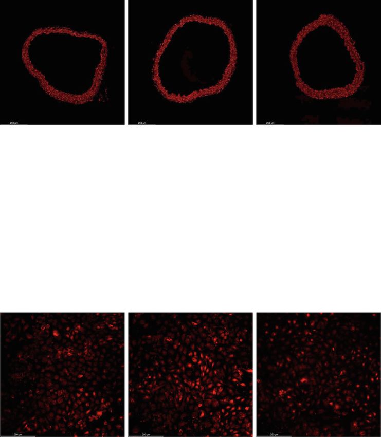
db/m+, n = 5 for db/db; *p < 0.05, **p < 0.01. CBSF, crocodile blood soluble fraction; Phe, phenylephrine; ACh, acetylcholine; SNP, sodium nitroprussideChook et al. Food Production, Processing and Nutrition (2021) 3:19 Page 7 of 13 and mean blood pressures were higher in db/db mice than arginine methyl ester (L-NAME), thus inhibiting the ability db/m+ mice, CBSF lowered the systolic, diastolic, and mean of endothelial cells to induce vasodilation, the total blood pressures significantly in the db/db mice (Fig. 1c). In endothelium-independent relaxation induced by sodium ni- addition, CBSF had no significant effects on plasma choles- troprusside (SNP) was found comparable among the groups, terol (Fig. 1d) nor triglyceride levels (Fig. 1e) in both db/m+ despite a difference in the initial relaxation between db/m+ and db/db mice. and db/db mice (Fig. 2d-e). In vivo CBSF oral treatment improved endothelium- dependent relaxation (EDR) in db/db mouse aortae CBSF directly reversed impaired EDR in ex vivo cultured We then examined whether oral CBSF treatment effects db/db mouse aortae endothelial function by wire myograph. The endothelium- Our subsequent experiments investigated whether CBSF dependent relaxation (EDR) induced by the addition of has a direct effect on endothelial function by incubating acetylcholine (ACh) was impaired in the db/db control mice the db/db and db/m+ mouse aortae with or without compared to the db/m+ counterparts (Fig. 2a-b). The 4-week CBSF overnight. While EDR was impaired in the db/db oral CBSF treatment improved the EDR in db/db mouse group in relative to the db/m+ group (Fig. 3a), CBSF in- aortae (Fig. 2a & c), yet did not show a significant effect on cubation improved the EDR performance in db/db the db/m+ mouse aortae (Fig. 2a). Following the inhibition of mouse aortae (Fig. 3b), without affecting the endothelial nitric oxide synthase (eNOS) by L-NG-nitro-L- endothelium-independent relaxation (Fig. 3c-d). Fig. 4 CBSF suppressed the ROS level in db/db mouse aortae both in vivo and ex vivo. Representative confocal images (a) with summarized data (b) of DHE stain intensity in isolated aortic rings from db/m+ and db/db mice with and without in vivo CBSF treatment (25 mg/kg/day). Representative confocal images (c) and summarized data (d) of DHE stain intensity in isolated aortic rings from non-treated db/m+ and db/db mice with and without ex vivo CBSF incubation (0.4 μg/mL). Data are presented in means ± SEM; n = 5; *p < 0.05, **p < 0.01. CBSF, crocodile blood soluble fraction; ROS, reactive oxygen species; DHE, dihydroethidium
Chook et al. Food Production, Processing and Nutrition      (2021) 3:19                                                               Page 8 of 13

CBSF reduced the ROS level in db/db mouse aortae both                      with CBSF (Fig. 5b). On the other hand, endothelium-
in vivo and ex vivo                                                        independent relaxation either incubated with or without IL-
Oxidative stress is an important factor leading to endo-                   1β was not affected by CBSF (Fig. 5c-d).
thelial dysfunction in diabetes (Giacco & Brownlee
2010). As reflected by the total dihydroethidium (DHE)
                                                                           CBSF lowered the ROS level elevated by IL-1β in both
intensity, reactive oxygen species (ROS) levels were in-
                                                                           normal mouse aortae and mouse brain microvascular
creased in db/db mouse aortic rings as compared to db/
                                                                           endothelial cells (mBMECs)
m+ control and were lowered by oral CBSF treatment
                                                                           Elevated ROS level was also observed in the IL-1β-
(Fig. 4a-b). In addition, ex vivo CBSF incubation simi-
                                                                           incubated mouse aortic rings, but was reduced by CBSF
larly reduced the heightened ROS levels in db/db mouse
                                                                           co-incubation (Fig. 6a-b). The high ROS level induced
aortic rings (Fig. 4c-d).
                                                                           by IL-1β incubation was also lowered by CBSF co-
                                                                           incubation in mBMECs (Fig. 6c-d).
CBSF directly reversed IL-1β-induced EDR impairment in
C57BL/6 J mouse aortae
It is well established that inflammation plays an important                CBSF downregulated pro-inflammatory cytokines while
role in mediating endothelial dysfunction in diabetes (Peiró               upregulating protective gene expressions
et al. 2017; Tabit et al. 2010). Using the classical pro-                  We further found that the pro-inflammatory cytokines, IL-6
inflammatory cytokine IL-1β, we investigated whether CBSF                  (Fig. 7a), TNF-α (Fig. 7b), VCAM-1 (Fig. 7c) and MCP-1
protects endothelial function against inflammation (Peiró                  (Fig. 7d), upregulated by IL-1β incubation, were reduced by
et al. 2016; Sprague & Khalil 2009). Our results showed that               CBSF. In addition, UCP2 (Fig. 7e) and SIRT6 (Fig. 7f) mRNA
EDR of mouse aortae from C57BL/6 J mice was impaired by                    expression levels downregulated by IL-1β were also restored
IL-1β incubation (Fig. 5a), but reversed by co-incubation                  by CBSF co-incubation.

 Fig. 5 Ex vivo CBSF co-incubation remedied IL-1β-impaired EDR in C57BL/6 J mouse aortae. Effect of IL-1β (1 pg/mL) incubation on EDR of
 isolated aortae from C57BL/6 J mice (a). Effect of CBSF co-incubation on EDR of IL-1β-treated aortae from C57BL/6 J mice (b). Effect of IL-1β
 incubation on endothelium-independent relaxation of isolated aortae from C57BL/6 J mice (c). Effect of CBSF co-incubation on endothelium-
 independent relaxation of IL-1β-treated aortae from C57BL/6 J mice (d). Data are presented in means ± SEM; n = 5; *p < 0.05, **p < 0.01 compared
 to IL-1β-treated group. CBSF, crocodile blood soluble fraction; Phe, phenylephrine; ACh, acetylcholine; SNP, sodium nitroprusside
Chook et al. Food Production, Processing and Nutrition (2021) 3:19 Page 9 of 13 Fig. 6 CBSF co-incubation lowered the ROS levels exaggerated by IL-1β in both aortae and endothelial cells. Representative confocal images (a) and summarized data (b) of DHE stain intensity of C57BL/6 J aortic rings incubated with and without IL-1β (1 pg/mL) and CBSF (0.2 mg/mL). Summarized data (c) and representative confocal images (d) of DHE stain intensity of mBMECs incubated with and without IL-1β (1 pg/mL) and CBSF (0.2 mg/mL). Data are presented in means ± SEM; n = 5; *p < 0.05. CBSF, crocodile blood soluble fraction; ROS, reactive oxygen species; DHE, dihydroethidium Discussion week oral CBSF treatment may exert its effects either by Endothelial dysfunction is an early manifestation and target direct or systemic influence through regulating blood for prevention of cardiovascular disease (Versari et al. 2009; glucose levels and blood pressure, the EDR improvement Widlansky et al. 2003). Endothelial dysfunction refers to by direct CBSF incubation proved a direct protective ef- the inability of endothelial cells to maintain vascular fect on the endothelial function of CBSF against diabetic homeostasis, which includes the regulation of vasoconstric- damage. tion, vasodilation, inflammation and oxidative stress (Tabit It is well established that upregulation of ROS plays a et al. 2010). Vascular endothelial cells are particularly sus- pivotal role in cardiovascular disease in diabetes (Giacco ceptible to hyperglycemic damage due to limited regulation & Brownlee 2010). Under hyperglycemia, the increased of glucose transport rate and thus exaggerated glucose in- glucose influx leads to increased ROS production. The flux in high glucose conditions (Kaiser et al. 1993), explain- high oxidative stress not only uncouples eNOS, which ing the especially high prevalence of cardiovascular disease further induces overproduction of ROS, but also reduces in diabetic patients (Einarson et al. 2018). the bioavailability of NO, which is responsible for vaso- In this study, db/db mice lacking leptin receptor were dilation and anti-inflammation (Giacco & Brownlee used to evaluate the effects of crocodile blood on endo- 2010; Kim et al. 2006). As a result, a cascade of pro- thelial functions in type 2 diabetes mellitus. While the 4- inflammatory markers including IL-1β, IL-6 and TNF-α
Chook et al. Food Production, Processing and Nutrition        (2021) 3:19                                                                Page 10 of 13

 Fig. 7 CBSF co-incubation reversed the gene expression levels of pro-inflammatory cytokines, SIRT6 and UCP2 skewed by IL-1β in endothelial
 cells. Effect of IL-1β (1 pg/mL) and CBSF (0.2 μg/mL) on the mRNA levels of IL-6 (a), TNF-α (b), VCAM-1 (c), MCP-1 (d), UCP2 (e) and SIRT6 (f) in
 mBMECs. Data are presented in means ± SEM; n = 4–6, *p < 0.05, **p < 0.01. CBSF, crocodile blood soluble fraction

were upregulated, eventually leading to endothelial dys-                     aortae. More specifically, CBSF downregulated the oxi-
function, as displayed by EDR impairment (Versari et al.                     dative stress, as well as inflammatory genes in endothe-
2009). Our results consistently showed that ROS level                        lial cells. CBSF also upregulated UCP2 and SIRT6 gene
increased in the db/db mouse aortae was lowered by                           expression in endothelial cells, which were suggested to
both in vivo and ex vivo CBSF treatment, indicating that                     have a protective role on the endothelial function (Tian
CBSF may possibly protect endothelial function against                       et al. 2012; Xu et al. 2017). Thus, our results suggest that
hyperglycemic damage through ROS suppression.                                CBSF protects endothelial functions against inflamma-
  Nevertheless, exertion of hyperglycemic damage re-                         tion, which may also be a possible mechanism under-
quires pro-inflammatory conditioning (Azcutia et al. 2010;                   lying the protective effect of CBSF on endothelial
Lafuente et al. 2008; Peiró et al. 2016). It was found that                  function in diabetic mice.
hyperglycemia alone is not sufficient to induce EDR im-                         Previous studies have shown that single-time oral
pairment, inflammation and ROS overproduction in blood                       treatment of crocodile blood lowered inflammatory cyto-
vessels (Lafuente et al. 2008; Peiró et al. 2016). In the pres-              kine production and increased the anti-oxidative enzyme
ence of pro-inflammatory IL-1β, however, EDR is im-                          levels in mouse models (Phosri et al. 2017; Phupiewk-
paired, with both ROS production and inflammatory                            ham et al. 2018). The doses used ranged from 62.5 to
signaling exacerbated by high glucose concentrations                         250 mg/kg body weight and caused no observable tox-
(Peiró et al. 2016). The upregulation of VCAM-1 by                           icity to the experimental animals. According to a Thai
hyperglycemia in endothelial cells also depends on the                       Congress Report, the protein content of freeze-dried
presence of IL-1β (Azcutia et al. 2010). Furthermore, IL-1                   crocodile blood powder is 83.1% (Chaeychomsri et al.
receptor antagonists were found to inhibit diabetic endo-                    2013). A number of short peptides were isolated from
thelial dysfunction (Vallejo et al. 2010). Thus, pro-                        the leucocyte extract and hemoglobin of crocodile blood,
inflammatory cytokines like IL-1β act as a prerequisite for                  and were suggested to possess anti-inflammatory and
hyperglycemic damage on endothelial function.                                anti-oxidative effects in vitro (Lueangsakulthai et al.
  In our study, CBSF ameliorated the IL-1β-induced                           2018; Phosri et al. 2017; Theansungnoen et al. 2014).
EDR impairment and ROS overproduction in mouse                               However, the content of these peptides in crocodile
Chook et al. Food Production, Processing and Nutrition         (2021) 3:19                                                                      Page 11 of 13

blood and whether these peptides can also exert the                          eNOS: Endothelial nitric oxide synthase; L-NAME: L-NG-nitro-L-arginine methyl
same effects in vivo have not been revealed.                                 ester; mBMECs: Mouse brain microvascular endothelial cells; NO: Nitric oxide;
                                                                             PBS: Phosphate-buffered saline; Phe: Phenylephrine; qPCR: Quantitative
   To the best of our knowledge, our study is the first                      polymerase chain reaction; ROS: Reactive oxygen species; SNP: Sodium
long-term experiment to discover the vasoprotective ef-                      nitroprusside
fects of crocodile blood in vivo. However, given that our
study primarily aimed to evaluate the effects of the avail-                  Supplementary Information
able crocodile blood supplement on vascular function in                      The online version contains supplementary material available at https://doi.
                                                                             org/10.1186/s43014-021-00066-w.
db/db mouse model, our results have two major limita-
tions. First, detailed molecular mechanisms regarding
                                                                              Additional file 1: Supplementary Figure 1. Crocodile blood does not
the effects observed were not identified. Second, the re-                     cause toxicity to C57BL/6 J mice and mBMECs. Body weight (A), and non-
sponsible active components in crocodile blood require                        fasting blood glucose level (B) of C57BL/6 J mice treated with different
                                                                              doses of crocodile blood via oral gavage for 5 weeks (n = 5). Effects of
further investigations. A number of novel peptides iden-
                                                                              CBSF on the cell viability of mBMECs assessed by XTT Assay (n = 8) (C).
tified in crocodile blood were suggested to possess anti-                     Data are represented in means ± SEM. CBSF, crocodile blood soluble frac-
oxidative properties in vitro (Pata et al. 2011; Phosri                       tion; mBMECs, mouse brain microvascular endothelial cells.
et al. 2017; Theansungnoen et al. 2014), not to mention
the many yet to be discovered. Whether these anti-                           Acknowledgements
oxidative peptides contribute to the in vivo vasoprotec-                     Not applicable.

tive effects described in this article is anticipated to be                  Authors’ contributions
verified in future studies. It is also very likely that mul-                 Conceptualization, W.T. Wong; methodology, C.Y. Chook, F.M. Chen;
tiple active components collaboratively contribute to the                    investigation, C.Y. Chook; formal analysis, C.Y. Chook; writing—original draft
                                                                             preparation, C.Y. Chook; writing—review and editing, F.M. Chen, F.P. Leung,
described effects through different molecular pathways.                      C.Y. Chook, G. Tse & W.T. Wong; visualization, C.Y. Chook; supervision, W.T.
   In addition to the previously suggested anti-                             Wong. The author(s) read and approved the final manuscript.
inflammatory, anti-oxidative, anti-microbial, anti-viral,
                                                                             Funding
anti-tumor, anti-anemia, and wound healing enhancing                         This study was supported by the Hong Kong Research Grants Council Grant
effects, our data indicated that crocodile blood may also                    ECS [24163117]; GRF [14101119]; National Natural Science Foundation of
protect vascular functions in diabetic patients. Although                    China [81970423]; and SKLA (CUHK).
how the active substances in crocodile blood are
                                                                             Availability of data and materials
digested, absorbed, and processed in the body requires                       The data used to support the findings of this study are included in the
further investigations, this study highlights the presence                   article.
of vasoactive substances in crocodile blood, and acts as a
                                                                             Declarations
first step in developing a novel vasoprotective medica-
tion or supplementation for diabetic patients. In view of                    Ethics approval and consent to participate
the recent increased incidence of zoonotic diseases in                       All animal experiments were undertaken with the approval from the Animal
                                                                             Experimentation Ethics Committee, the Chinese University of Hong Kong
humans such as SARS-CoV-2 and bird flu, further                              (Ref No. 18-243-MIS).
characterization of crocodile blood is needed to confirm
the absence of virus and any possible pathogens.                             Consent for publication
                                                                             All authors approved the final version of this manuscript.

Conclusion                                                                   Competing interests
Our results indicated that CBSF improved vascular                            The authors declare no conflict of interest. The funders had no role in the
                                                                             design of the study; in the collection, analyses, or interpretation of data; in
endothelial function both in vivo and ex vivo in db/db
                                                                             the writing of the manuscript, or in the decision to publish the results.
diabetic mice, possibly through ROS suppression. The
positive effects also applied to IL-1β-induced inflamma-                     Received: 27 August 2020 Accepted: 12 June 2021
tion model, with downregulation of pro-inflammatory
genes and upregulation of beneficial genes, which may                        References
explain how CBSF protects endothelial health in diabetic                     Aree, K., Siruntawineti, J., & Chaeychomsri, W. (2011). Crocodylus siamensis serum
condition. Although verification of the exact mechanistic                        and macrophage phagocytic activity. Journal of the Medical Association of
                                                                                 Thailand = Chotmaihet Thangphaet, 94(Suppl 7(May)), S131–S138 http://www.
pathways requires further study, our study provides in-                          ncbi.nlm.nih.gov/pubmed/22619919.
sights to the further identification of vasoprotective sub-                  Azcutia, V., Abu-Taha, M., Romacho, T., Vázquez-Bella, M., Matesanz, N., Luscinskas,
stances in crocodile blood, which can potentially be                             F. W., … Peiró, C. (2010). Inflammation determines the pro-adhesive
                                                                                 properties of high extracellular D-glucose in human endothelial cells in vitro
developed into a novel medicinal approach for diabetic                           and rat microvessels in vivo. PLoS One, 5(4), e10091. https://doi.org/10.1371/
patients targeting cardiovascular diseases.                                      journal.pone.0010091.
                                                                             Barba, F. J., Koubaa, M., do Prado-Silva, L., Orlien, V., & de Souza Sant’Ana, A.
Abbreviations                                                                    (2017). Mild processing applied to the inactivation of the main foodborne
ACh: Acetylcholine; CBSF: Crocodile blood soluble fraction;                      bacterial pathogens: A review. Trends in Food Science and Technology, 66, 20–
DHE: Dihydroethidium; EDR: Endothelium-dependent relaxation;                     35. https://doi.org/10.1016/j.tifs.2017.05.011.
Chook et al. Food Production, Processing and Nutrition (2021) 3:19 Page 12 of 13
Chaeychomsri, W. (2015). Effectiveness in the treatment of Iron deficiency anemia Lueangsakulthai, J., Phosri, S., Theansungnoen, T., Jangpromma, N., Temsiripong,
in Sprague-Dawley rats using freeze- dried crocodile blood. International T., Mckendrick, J. E., … Klaynongsruang, S. (2018). Novel antioxidant and anti-
Journal of Life Sciences Biotechnology and Pharma Research, 4(1), 42–49. inflammatory peptides from the Siamese crocodile (Crocodylus siamensis)
Chaeychomsri, W., Siruntawineti, J., Chaeychomsri, S., Hengsawadi, D., Cuptapan, hemoglobin hydrolysate. Biotechnology and Applied Biochemistry, 65(3), 455–
Y., & Rungtaweechai, W. (2013). Successful development and 466. https://doi.org/10.1002/bab.1628.
commercialization of freeze-dried crocodile blood product. In The 39th Maijaroen, S., Jangpromma, N., Daduang, J., & Klaynongsruang, S. (2018). KT2 and
Congress on Science and Technology of Thailand, (pp. 569–576). https://doi. RT2 modified antimicrobial peptides derived from Crocodylus siamensis
org/10.1017/CBO9781107415324.004. Leucrocin I show activity against human colon cancer HCT-116 cells.
Chook, C. Y. B., Chen, F. M., Leung, F. P., Chen, Z., & Wong, W. T. (2021). Potential Environmental Toxicology and Pharmacology, 62(July), 164–176. https://doi.
of crocodile blood as a medication and dietary supplement: A systemic org/10.1016/j.etap.2018.07.007.
review. In Clinical and Experimental Pharmacology and Physiology, May, 1–16. Maraming, P., Klaynongsruang, S., Boonsiri, P., Maijaroen, S., Daduang, S., Chung, J.
https://doi.org/10.1111/1440-1681.13524. G., & Daduang, J. (2018). Antitumor activity of RT2 peptide derived from
Einarson, T. R., Acs, A., Ludwig, C., & Panton, U. H. (2018). Prevalence of crocodile leukocyte peptide on human colon cancer xenografts in nude mice.
cardiovascular disease in type 2 diabetes: A systematic literature review of Environmental Toxicology, 33(9), 972–977. https://doi.org/10.1002/tox.22584.
scientific evidence from across the world in 2007-2017. Cardiovascular Merchant, M., Juneau, K., Gemillion, J., Falconi, R., Doucet, A., & Shirley, M. H.
Diabetology, 17(1), 83. https://doi.org/10.1186/s12933-018-0728-6. (2011). Characterization of serum phospholipase A2 activity in three diverse
Giacco, F., & Brownlee, M. (2010). Oxidative stress and diabetic complications. species of west African crocodiles. Biochemistry Research International, 2011,
Circulation Research, 107(9), 1058–1070. https://doi.org/10.1161/CIRCRESA 1–7. https://doi.org/10.1155/2011/925012.
HA.110.223545. Ou, Y., & Ho, W. S. (2016). Crocodile blood extract induces the apoptosis of lung
Hao, J., Li, Y. W., Xie, M. Q., & Li, A. X. (2012). Molecular cloning, recombinant cancer cells through PTEN activity. Oncology Reports, 36(3), 1457–1466.
expression and antibacterial activity analysis of hepcidin from Siamensis https://doi.org/10.3892/or.2016.4914.
crocodile (Crocodylus siamensis). Comparative Biochemistry and Physiology - B Pakdeesuwan, A., Araki, T., Daduang, S., Payoungkiattikun, W., Jangpromma, N., &
Biochemistry and Molecular Biology, 163(3–4), 309–315. https://doi.org/10.101 Klaynongsruang, S. (2017). In vivo wound healing activity of crocodile
6/j.cbpb.2012.08.002. (Crocodylus siamensis) hemoglobin and evaluation of antibacterial and
Huang, Y., Chan, F. L., Lau, C.-W. W., Tsang, S.-Y. Y., Chen, Z.-Y. Y., He, G.-W. W., & antioxidant properties of hemoglobin and hemoglobin hydrolysate. Journal
Yao, X. (2003). Roles of cyclic AMP and Ca2+−activated K+ channels in of Microbiology and Biotechnology, 27(1), 26–35. https://doi.org/10.4014/jmb.1
endothelium-independent relaxation by urocortin in the rat coronary artery. 603.03046.
Cardiovascular Research, 57(3), 824–833. https://doi.org/10.1016/S0008-6363 Pata, S., Daduang, S., Svasti, J., & Thammasirirak, S. (2007). Isolation of lysozyme
(02)00773-3. like protein from crocodile leukocyte extract (Crocodylus siamensis). KMITL
Jangpromma, N., Poolperm, N., Pornsri, K., Anwised, P., Kabbua, T., Phosri, S., … Science and Technology Journal, 7(S1), 70–85.
Klaynongsruang, S. (2017). Proteomics profiling and inflammatory factor gene Pata, S., Yaraksa, N., Daduang, S., Temsiripong, Y., Svasti, J., Araki, T., &
expression in LPS-stimulated RAW 264.7 cells treated with Crocodylus Thammasirirak, S. (2011). Characterization of the novel antibacterial peptide
siamensis hemoglobin. Chiang Mai Journal of Science, 44(3), 800–815. Leucrocin from crocodile (Crocodylus siamensis) white blood cell extracts.
Jangpromma, N., Preecharram, S., Srilert, T., Maijaroen, S., Mahakunakorn, P., Developmental and Comparative Immunology, 35(5), 545–553. https://doi.
Nualkaew, N., … Klaynongsruang, S. (2016). In vitro and in vivo wound org/10.1016/j.dci.2010.12.011.
healing properties of plasma and serum from Crocodylus siamensis blood. Patathananone, S., Thammasirirak, S., Daduang, J., Chung, J. G., Temsiripong, Y., &
Journal of Microbiology and Biotechnology, 26(6), 1140–1147. https://doi.org/1 Daduang, S. (2015). Bioactive compounds from crocodile (Crocodylus
0.4014/jmb.1601.01054. siamensis) white blood cells induced apoptotic cell death in HeLa cells.
Jangpromma, N., Suttee, K., Phosri, S., Theansungnoen, T., Lueangsakulthai, J., Environmental Toxicology, 31(8), 986–997. https://doi.org/10.1002/tox.22108.
Payoungkiattikun, W., … Klaynongsruang, S. (2018). Antioxidant properties of Peiró, C., Lorenzo, Ó., Carraro, R., & Sánchez-Ferrer, C. F. (2017). IL-1β inhibition in
Crocodylus siamensis blood components on H2O2-induced human skin cardiovascular complications associated to diabetes mellitus. Frontiers in
fibroblast cells. Chiang Mai Journal of Science, 45(3), 1359–1371. Pharmacology, 8(JUN), 1–13. https://doi.org/10.3389/fphar.2017.00363.
Kaiser, N., Sasson, S., Feener, E. P., Boukobza-Vardi, N., Higashi, S., Moller, D. E., … Peiró, C., Romacho, T., Azcutia, V., Villalobos, L., Fernández, E., Bolaños, J. P., …
King, G. L. (1993). Differential regulation of glucose transport and transporters Sánchez-Ferrer, C. F. (2016). Inflammation, glucose, and vascular cell damage:
by glucose in vascular endothelial and smooth muscle cells. Diabetes, 42(1), The role of the pentose phosphate pathway. Cardiovascular Diabetology,
80–89. https://doi.org/10.2337/diabetes.42.1.80. 15(1), 1–15. https://doi.org/10.1186/s12933-016-0397-2.
Kelly, T. N. (2009). Systematic review: Glucose control and cardiovascular disease Phosri, S., Jangpromma, N., Chang, L. C., Tan, G. T., Wongwiwatthananukit, S.,
in type 2 diabetes. Annals of Internal Medicine, 151(6), 394. https://doi.org/10. Maijaroen, S., … Klaynongsruang, S. (2018). Siamese crocodile white blood
7326/0003-4819-151-6-200909150-00137. cell extract inhibits cell proliferation and promotes autophagy in multiple
Kim, J. A., Montagnani, M., Kwang, K. K., & Quon, M. J. (2006). Reciprocal cancer cell lines. Journal of Microbiology and Biotechnology, 28(6), 1007–1021.
relationships between insulin resistance and endothelial dysfunction: https://doi.org/10.4014/jmb.1712.12002.
Molecular and pathophysiological mechanisms. Circulation, 113(15), 1888– Phosri, S., Jangpromma, N., Patramanon, R., Kongyingyoes, B., Mahakunakorn, P., &
1904. https://doi.org/10.1161/CIRCULATIONAHA.105.563213. Klaynongsruang, S. (2017). Protective effect of crocodile hemoglobin and
Kommanee, J., Phosri, S., Daduang, S., Temsiripong, Y., Dhiravisit, A., & whole blood against hydrogen peroxide-induced oxidative damage in
Thammasirirak, S. (2014). Comparisons of anti-inflammatory activity of human lung fibroblasts (MRC-5) and inflammation in mice. Inflammation,
crocodile (Crocodylus siamensis) blood extract. Chiang Mai Journal of Science, 40(1), 205–220. https://doi.org/10.1007/s10753-016-0471-7.
41(3), 627–634. Phosri, S., Mahakunakorn, P., Lueangsakulthai, J., Jangpromma, N., Swatsitang, P.,
Kommanee, J., Preecharram, S., Daduang, S., Temsiripong, Y., Dhiravisit, A., Daduang, S., … Thammasirirak, S. (2014). An investigation of antioxidant and
Yamada, Y., & Thammasirirak, S. (2012). Antibacterial activity of plasma from anti-inflammatory activities from blood components of crocodile (Crocodylus
crocodile (Crocodylus siamensis) against pathogenic bacteria. Annals of siamensis). Protein Journal, 33(5), 484–492. https://doi.org/10.1007/s10930-014-
Clinical Microbiology and Antimicrobials, 11(1), 1. https://doi.org/10.1186/1476- 9581-y.
0711-11-22. Phupiewkham, W., Lu, Q., Payoungkiattikun, W., Temsiripong, T., Jangpromma, N.,
Kozlowski, H. N., Lai, E. T. L., Havugimana, P. C., White, C., Emili, A., Sakac, D., … Lai, R., & Klaynongsruang, S. (2018). Development and characterization of an
Branch, D. R. (2016). Extracellular histones identified in crocodile blood inhibit anti-acne gel containing siamese crocodile (Crocodylus siamensis) leukocyte
in-vitro HIV-1 infection. Aids, 30(13), 2043–2052. https://doi.org/10.1097/QAD. extract. Journal of Microbiology and Biotechnology, 28(5), 707–717. https://doi.
0000000000001159. org/10.4014/jmb.1802.02027.
Lafuente, N., Matesanz, N., Azcutia, V., Romacho, T., Nevado, J., Rodríguez-Mañas, Preecharram, S., Jearranaiprepame, P., Daduang, S., Temsiripong, Y., Somdee, T.,
L., … Sánchez-Ferrer, C. F. (2008). The deleterious effect of high Fukamizo, T., … Thammasirirak, S. (2010). Isolation and characterisation of
concentrations of D-glucose requires pro-inflammatory preconditioning. crocosin, an antibacterial compound from crocodile (Crocodylus siamensis)
Journal of Hypertension, 26(3), 478–485. https://doi.org/10.1097/HJH.0b013e32 plasma. Animal Science Journal, 81(3), 393–401. https://doi.org/10.1111/j.1740-
82f331fb. 0929.2010.00752.x.Chook et al. Food Production, Processing and Nutrition (2021) 3:19 Page 13 of 13
Saeedi, P., Petersohn, I., Salpea, P., Malanda, B., Karuranga, S., Unwin, N., …
Williams, R. (2019). Global and regional diabetes prevalence estimates for
2019 and projections for 2030 and 2045: Results from the International
Diabetes Federation Diabetes Atlas, 9th edition. Diabetes Research and
Clinical Practice, 157, 107843. https://doi.org/10.1016/j.diabres.2019.107843.
Smith, E. (2019, April 13). Expert urges NT to explore use of crocodile compounds
in medical industry. ABC News. https://www.abc.net.au/news/2019-04-13/
crocodile-blood-oil-bacteria-antibiotics-antibacterial-medicine/10985546
Sprague, A. H., & Khalil, R. A. (2009). Inflammatory cytokines in vascular
dysfunction and vascular disease. Biochemical Pharmacology, 78(6), 539–552.
https://doi.org/10.1016/j.bcp.2009.04.029.
Tabit, C. E., Chung, W. B., Hamburg, N. M., & Vita, J. A. (2010). Endothelial
dysfunction in diabetes mellitus: Molecular mechanisms and clinical
implications. Reviews in Endocrine and Metabolic Disorders, 11(1), 61–74.
https://doi.org/10.1007/s11154-010-9134-4.
Theansungnoen, T., Yaraksa, N., Daduang, S., Dhiravisit, A., & Thammasirirak, S.
(2014). Purification and characterization of antioxidant peptides from
leukocyte extract of Crocodylus siamensis. Protein Journal, 33(1), 24–31.
https://doi.org/10.1007/s10930-013-9536-8.
Tian, X. Y., Wong, W. T., Xu, A., Lu, Y., Zhang, Y., Wang, L., … Huang, Y. (2012).
Uncoupling protein-2 protects endothelial function in diet-induced obese
mice. Circulation Research, 110(9), 1211–1216. https://doi.org/10.1161/
CIRCRESAHA.111.262170.
Turnbull, F. M., Abraira, C., Anderson, R. J., Byington, R. P., Chalmers, J. P.,
Duckworth, W. C., … Woodward, M. (2009). Intensive glucose control and
macrovascular outcomes in type 2 diabetes. Diabetologia, 52(11), 2288–2298.
https://doi.org/10.1007/s00125-009-1470-0.
Unger, U., Poelsler, G., Modrof, J., & Kreil, T. R. (2009). Virus inactivation during the
freeze-drying processes as used for the manufacture of plasma-derived
medicinal products. Transfusion, 49(9), 1924–1930. https://doi.org/10.1111/j.1
537-2995.2009.02218.x.
Vallejo, S., Palacios, E., Romacho, T., Villalobos, L., Peiró, C., & Sánchez-Ferrer, C. F.
(2010). The interleukin-1 receptor antagonist anakinra improves endothelial
dysfunction in streptozotocin-induced diabetic rats cardio VASCULAR
DIABETOLOGY. Chemico-Biological Interactions, 188(1), 237–245. https://doi.
org/10.1186/s12933-014-0158-z.
Versari, D., Daghini, E., Virdis, A., Ghiadoni, L., & Taddei, S. (2009). Endothelial
dysfunction as a target for prevention of cardiovascular disease. Diabetes
Care, 32(suppl_2), S314–S321. https://doi.org/10.2337/dc09-S330.
Widlansky, M. E., Gokce, N., Keaney, J. F., & Vita, J. A. (2003). The clinical
implications of endothelial dysfunction. Journal of the American College of
Cardiology, 42(7), 1149–1160. https://doi.org/10.1016/S0735-1097(03)00994-X.
Xu, S., Yin, M., Koroleva, M., Mastrangelo, M. A., Zhang, W., Bai, P., … Jin, Z. G.
(2017). SIRT6 protects against endothelial dysfunction and atherosclerosis in
mice. Aging, 8(5), 1064–1078. https://doi.org/10.18632/aging.100975.
Publisher’s Note
Springer Nature remains neutral with regard to jurisdictional claims in
published maps and institutional affiliations.You can also read