Cognitive Deficits, Apathy, and Hypersomnolence Represent the Core Brain Symptoms of Adult-Onset Myotonic Dystrophy Type
←
→
Page content transcription
If your browser does not render page correctly, please read the page content below
ORIGINAL RESEARCH
                                                                                                                                                    published: 01 July 2021
                                                                                                                                           doi: 10.3389/fneur.2021.700796

                                               Cognitive Deficits, Apathy, and
                                               Hypersomnolence Represent the
                                               Core Brain Symptoms of Adult-Onset
                                               Myotonic Dystrophy Type 1
                                               Jacob N. Miller 1*, Alison Kruger 1 , David J. Moser 1 , Laurie Gutmann 2 , Ellen van der Plas 1 ,
                                               Timothy R. Koscik 1 , Sarah A. Cumming 3 , Darren G. Monckton 3 and Peggy C. Nopoulos 1,4,5
                                               1
                                                 Department of Psychiatry, Carver College of Medicine, University of Iowa, Iowa City, IA, United States, 2 Department of
                                               Neurology, Indiana University School of Medicine, Indianapolis, IN, United States, 3 Institute of Molecular, Cell and Systems
                                               Biology, College of Medical, Veterinary and Life Sciences, University of Glasgow, Glasgow, United Kingdom, 4 Department of
                                               Neurology, Carver College of Medicine, University of Iowa, Iowa City, IA, United States, 5 Department of Pediatrics, Carver
                                               College of Medicine, University of Iowa, Iowa City, IA, United States

                          Edited by:           Myotonic dystrophy type 1 is the most common form of muscular dystrophy in adults,
                      Huifang Shang,
             Sichuan University, China
                                               and is primarily characterized by muscle weakness and myotonia, yet some of the most
                        Reviewed by:
                                               disabling symptoms of the disease are cognitive and behavioral. Here we evaluated
                     W. David Arnold,          several of these non-motor symptoms from a cross-sectional time-point in one of the
             The Ohio State University,
                                               largest longitudinal studies to date, including full-scale intelligence quotient, depression,
                         United States
                      Giovanni Meola,          anxiety, apathy, sleep, and cerebral white matter fractional anisotropy in a group of
               University of Milan, Italy      39 adult-onset myotonic dystrophy type 1 participants (27 female) compared to 79
                 *Correspondence:              unaffected control participants (46 female). We show that intelligence quotient was
                      Jacob N. Miller
            jacob-n-miller@uiowa.edu
                                               significantly associated with depression (P < 0.0001) and anxiety (P = 0.018), but
                                               not apathy (P < 0.058) or hypersomnolence (P = 0.266) in the DM1 group. When
                    Specialty section:         controlling for intelligence quotient, cerebral white matter fractional anisotropy was
          This article was submitted to
                                               significantly associated with apathy (P = 0.042) and hypersomnolence (P = 0.034),
                  Movement Disorders,
                a section of the journal       but not depression (P = 0.679) or anxiety (P = 0.731) in the myotonic dystrophy
                 Frontiers in Neurology        type 1 group. Finally, we found that disease duration was significantly associated with
             Received: 26 April 2021           apathy (P < 0.0001), hypersomnolence (P < 0.001), IQ (P = 0.038), and cerebral white
            Accepted: 03 June 2021
            Published: 01 July 2021
                                               matter fractional anisotropy (P < 0.001), but not depression (P = 0.271) or anxiety
                              Citation:
                                               (P = 0.508). Our results support the hypothesis that cognitive deficits, hypersomnolence,
        Miller JN, Kruger A, Moser DJ,         and apathy, are due to the underlying neuropathology of myotonic dystrophy type
           Gutmann L, van der Plas E,
                                               1, as measured by cerebral white matter fractional anisotropy and disease duration.
              Koscik TR, Cumming SA,
     Monckton DG and Nopoulos PC               Whereas elevated symptoms of depression and anxiety in myotonic dystrophy type 1 are
 (2021) Cognitive Deficits, Apathy, and        secondary to the physical symptoms and the emotional stress of coping with a chronic
Hypersomnolence Represent the Core
      Brain Symptoms of Adult-Onset
                                               and debilitating disease. Results from this work contribute to a better understanding of
          Myotonic Dystrophy Type 1.           disease neuropathology and represent important therapeutic targets for clinical trials.
             Front. Neurol. 12:700796.
     doi: 10.3389/fneur.2021.700796            Keywords: myotonic dystrophy, apathy, hypersomnolence, cognition, depression, fractional anisotropy

Frontiers in Neurology | www.frontiersin.org                                          1                                              July 2021 | Volume 12 | Article 700796
Miller et al. Core Brain Symptoms of DM1
INTRODUCTION the burden of having a degenerative neurologic disorder, deficits
in cognitive skills can result in sub-optimal coping strategies
Myotonic dystrophy type 1 (DM1) is a trinucleotide repeat potentially driving secondary emotional problems, including
disorder, classically characterized with motor symptoms of depression and anxiety.
prolonged muscle contractions (myotonia), progressive muscle The majority of studies evaluating depression in DM1 have
wasting, and weakness (1). As a multisystemic disease, DM1 utilized rating scales such as the Beck Depression Inventory
manifests with many additional non-motor symptoms including (BDI) (6, 9, 10, 14, 16, 22–28), the Hamilton Rating Scale for
cataracts, heart conduction abnormalities and arrythmias, Depression (HAM-D) (29–34) or a variety of other standardized,
gastrointestinal abnormalities, endocrine abnormalities, insulin self-rating scales (35–48), and comparison groups were often
resistance and diabetes, and respiratory failure. Other non- adults with no major medical illnesses. However, rating scales
motor symptoms, including cognitive deficits, sleep disturbances such as the BDI-II not only assess mood and cognitive
and affective symptoms, are thought to be due to CNS symptoms such as sadness, suicidality, and guilt, but also
pathology in DM1, a feature of the disease that has had somatic symptoms such as fatigue and sleep disturbance. When
increasing focus over the past 5–10 years. White matter using depression rating scales, it is vital to determine whether
(WM) microstructure pathology is one of the most robust the scores are increased due to higher somatic ratings or
and reproducible observations in DM1, supported by several because of depressed mood, as many of the other symptoms of
neuroimaging studies to date (2–13). Fractional Anisotropy (FA, DM1 (hypersomnolence, fatigue) will contribute to the overall
obtained via diffusion tensor MR imaging [DTI]) provides a depression score.
measure of WM microstructural integrity by quantifying local To better understand whether these symptoms in DM1 are
restrictions of the direction of diffusion of water molecules and truly related to brain pathology, a few studies have evaluated the
ranges from 0 (e.g., cerebrospinal fluid) to ∼1 (e.g., highly relationship between cerebral WM FA and symptoms. Several
myelinated WM fiber bundles). In DM1, FA has been shown to studies have shown a relationship between lower FA, lower
be globally reduced, with limited regional specificity (3–14). cognitive function (9), and hypersomnolence (4). Yet, no study
As a single gene disorder, there has been significant progress has systematically evaluated all of these brain symptom measures
toward developing gene knock-down or other therapies directed together, while accounting for relationships among measures. In
at the core site of pathology in DM1. To treat the CNS associated addition, if a symptom of a progressive, worsening disease is truly
symptoms of DM1, there is consideration and progress toward related to the pathology of that disease, that symptom would
delivery of these therapies directly to the brain (15). In the context worsen over the course of time. Therefore, the association of a
of developing endpoints for monitoring centrally administered symptom with disease duration would also support the notion
therapy, it is crucial to distinguish what features of DM1 are truly that that symptom was due to progressive brain pathology, as
related to brain pathology and which features may be secondary shown previously with cognitive performance (49, 50).
manifestations of living with a chronic neurologic disease. The Iowa DM1 study evaluates brain structure and function in
This will move the field toward more robust clinical targets, adult-onset DM1. Here, as part of this larger study, we evaluate
biomarkers, and endpoints for clinical trials. The symptoms measures of cognitive/behavioral symptoms, including FSIQ,
of DM1 considered directly related to brain pathology include depression, anxiety, apathy (obtained from both the participant
cognitive deficits, apathy, hypersomnolence/fatigue, and affective and an informant, as patients often under-rate this symptom),
symptoms of depression and anxiety (15). These symptoms and hypersomnolence. First, we evaluated the relationship
can have a significant burden on both the patient and their between cognitive impairment to ratings of depression, anxiety,
caregiver (15–17). apathy, and hypersomnolence. Then, relationships between these
It is important to consider the potential relationships between clinical measures and cerebral FA were compared. Finally, disease
the symptoms of DM1 when evaluating whether or not they duration (as a marker of disease progression) was used as a
represent core features of the disease. General cognitive deficits predictor for these measures and cerebral FA. Our results indicate
are severe in congenital and childhood forms of DM1 (18), that cognitive deficits, apathy, and hypersomnolence represent
but even in adult-onset DM1, we have shown that Full Scale that core brain symptoms of DM1.
IQ (FSIQ) is significantly lower than unaffected healthy adults
(19, 20). Although verbal skills are typically less affected
than visuospatial skills, a significantly lower FSIQ highlights a
PATIENTS AND METHODS
generalized deficit in cognitive skills. There is also significant Participants
evidence for global cognitive deficits in adult-onset DM1 present The Iowa DM1 Brain study targets individuals with adult-onset
across several different domains (21). For patients navigating DM1 (diagnosis after the age of 18 years old). Participants
were recruited from our multidisciplinary specialty clinic for
Abbreviations: AES, apathy evaluation scale; BDI-II, beck depression inventory- DM1 at the University of Iowa and through the Myotonic
II; BDA, beck anxiety inventory; DM1, myotonic dystrophy type 1; DTI, diffusion Dystrophy Foundation. Healthy adults were recruited from
tensor imaging; EEG, electroencephalography; ePAL, estimated progenitor allele spouses of DM1 participants and from the Iowa City area via
length; FA, fractional anisotropy; MRI, magnetic resonance imaging; FSIQ, full
scale intelligence quotient; SCOPA, scales for outcomes in Parkinson’s disease; SP-
advertisements. Exclusion criteria for all participants included:
PCR, small-pool polymerase chain reaction; WAIS-IV, Wechsler adult intelligence MRI contraindication, a history of serious head injury that
scale-IV; WM, white matter. resulted in a hospital stay, or a chronic neurological disorder
Frontiers in Neurology | www.frontiersin.org 2 July 2021 | Volume 12 | Article 700796Miller et al. Core Brain Symptoms of DM1
TABLE 1 | Demographics of study sample. that flanked the CTG repeats were used to amplify across the
repeat region, also adding barcoded sequencing adapters to
Healthy controls DM1
generate the sequencing library. The resulting reads were aligned
′ ′
Sample n 71 39 against reference sequences comprising the 5 and 3 -flanking
Sex n Males 25 12 sequences separated by 0–100 CTG repeats. The genotype was
Females 46 27 the CTG repeat length to which the highest number of sequence
Age at evaluation Mean (SD) 43.22 (13.07) 45.49 (9.03) reads were mapped.
Age at disease onset Mean (SD) n/a 32.35 (9.59) Research staff, clinicians, and scientists involved in this study
Disease Duration Mean (SD) n/a 12.87 (7.38) remained blind to the participant’s clinical condition (CTG
MIRS n MIRS 1 - 13 expansion length and muscular impairment). However, this was
MIRS 2 - 24
not always possible when participants exhibited moderate-to-
MIRS 3 - 9
severe symptoms of DM1 during the study. All clinical scales and
MIRS 4 - 4
measures were administered by a trained examiner experienced
MIRS 5 - 0
in DM1. All data were de-identified and all participants
consented to non-disclosure of genetic results obtained as part
CTG Range 5–43 81–501
of the study.
Median 13 146
Motor Testing
Severity of muscle weakness was measured using the Muscle
Impairment Rating Scale (MIRS) during examination by a
other than DM1. Healthy adults were additionally required to be neuromuscular specialist experienced in DM1, blinded to the
without history of substance abuse, psychiatric disease, or major participants’ genetic status (54). This scale evaluates muscular
medical disease, including heart disease, sleep disorder, vascular impairment severity according to an ordinal 5-point scale as
disease, uncontrolled hypertension, cancer, diabetes mellitus, follows: (1) no muscular impairment, (2) minimal signs, (3)
lung disease, and autoimmune conditions. A total of 8 potential distal weakness, (4) mild to moderate proximal weakness, and (5)
DM1 participants and 9 potential healthy control participants severe proximal weakness.
were either excluded or did not meet inclusion criteria for
the study and were excluded. The current sample included 39 General Cognitive Abilities
individuals with DM1 and 71 healthy adults. Demographics are Participants completed the Wechsler Adult Intelligence Scale-IV
displayed in Table 1. There was no significant difference in sex (WAIS-IV) to estimate Full Scale IQ (55).
(χ 2 = 0.068, df = 1, p = 0.794) or age [t (108) = 0.96, p =
0.337] between groups. Disease duration was defined as the time Depression and Anxiety
between onset of the first motor symptom of DM1 and time of The Beck Depression Inventory (BDI-II) is a widely used
assessment. Mean disease duration for this group was 12.9 years questionnaire measuring self-reported symptoms of depression
with a range of 2.4 years to 28.9 years. As far as the severity of on a 4-point Likert scale ranging from 0 to 3 (56). A total score is
muscular impairment, the DM1 group had 13 individuals with a summed and can be interpreted clinically as 0–13 being minimal
score of one (no impairment), 24 with a score of two (minimal depression; 14–19 being mild depression; 20–28 being moderate
signs), nine with a score of three (distal weakness), four with a depression; and 29–63 being severe depression. The Beck Anxiety
score of four (mild to moderate proximal weakness), and none Inventory (BAI) is a corollary self-report questionnaire (21
with a score of five (severe proximal weakness). questions on a 4-point Likert scale) assessing symptoms related
All participants gave written, informed consent prior to to anxiety.
enrolling in the protocol in accordance with the Declaration of
Helsinki. The study was approved by the University of Iowa Sleep Quality
Institutional Review Board. The SCOPA-Sleep (Scales for Outcomes in Parkinson’s Disease-
Genotyping of CTG repeat in DM1-affected participants was Sleep) survey was used to assess daytime sleepiness (57). The
completed by small-pool PCR (SP-PCR) (51). For each patient, self-report scale includes six items in subscale D: Sleeping
four reactions were completed, each using 300 pg genomic DNA during the day and evening, which were summed to calculate
template derived from blood leukocytes. CTG repeat lengths hypersomnolence scores. Items used to measure overall sleep
were estimated by comparison against DNA fragments of known quality from subscales A: Use of sleeping tablets, B: Sleeping at
length and molecular weight markers, using CLIQS software night, and C: Global evaluation of sleeping at night, were not
(TotalLabs UK Ltd.). The lower boundary of the expanded included in the analysis.
molecules in SP-PCR was used to estimate the progenitor
(inherited) allele length (ePAL) (52). The mean ePAL for the Apathy
DM1 group was 146 with a minimum of 81 and a maximum of The Apathy Evaluation Scale (AES) was used to determine self-
501. Repeat lengths for the non-disease-causing alleles from all reported degree of apathy (58). The AES includes 18 items that
participants were estimated using the Illumina MiSeq platform, are rated on a 4-point Likert scale. Items were summed to create
broadly as described for Huntington disease alleles (53). Primers a total score, where higher scores represent increased apathy. In
Frontiers in Neurology | www.frontiersin.org 3 July 2021 | Volume 12 | Article 700796Miller et al. Core Brain Symptoms of DM1
addition, we had a subset of 24 of the 39 participants with DM1 and clinical outcomes, FSIQ was included if the coefficient was
who had an informant fill out the informant version of the scale. statistically significant.
Finally, a set of models determined the effect of disease
Magnetic Resonance Imaging duration (predictor variable) on the clinical measures of
Individuals who participated before June 2016 (49 controls, depression, anxiety, apathy, sleep, and cerebral WM FA
25 DM1) were scanned using a 3T Siemens TrioTIM scanner (dependent variables).
(Siemens AG, Munich, Germany; 12 channel head coil, software
version: syngo B17). Those who participated after June 2016
were scanned using a 3T General Electric Discovery MR750w RESULTS
scanner (GE Medical Systems, Chicago, Il, 16 channel head and
neck coil, software versions: 25.0, 25.1, and 26.0) (21 controls, Table 2 displays the comparison of the clinical measures and
and 13 DM1). Participants completed DWI acquisitions with cerebral WM FA across groups. As expected, patients with DM1
either a single-shell (B1000, 64 directions), multi-shell (B1000 had significantly different clinical measures compared to healthy
and B2000, 29-30 directions per shell), or both. Diffusion- adults. These results included lower FSIQ [t (106) = −6.16, P <
weighted images were collected using echo planar recovery 0.0001], and higher ratings of depression [t (106) = 8.12, P <
magnitude sequences collected in the axial plane. Anatomical 0.0001] and anxiety [t (106) = 5.29, P < 0.0001). Self-reported
T1-weighted and T2-weighted images were collected and used apathy [t (106) = 5.86, P < 0.0001] and informant-reported
for co-registration, normalization, and labeling purposes using apathy scores [t (27) = 2.43, P = 0.0221] were also significantly
acquisition parameters described previously (59). higher. Additionally, DM1 patients had higher daytime sleepiness
scores [t (106) = 8.41, P < 0.0001]. Finally, cerebral WM FA was
Fractional Anisotropy significantly lower in the DM1 group compared to healthy adults
Diffusion-weighted images were processed using standard [t (98) = −12.23, P < 0.0001].
procedures of the FMRIB Diffusion toolbox from the FSL BDI-II depression scores were subdivided into ranges, where
software package (http://www.fmrib.ox.ac.uk/fsl), where phase 99% of healthy adults scored in the normal or minimal depression
encoding distortion and eddy current artifacts were removed range, and 1% in the mild depression range. For participants with
using topup and eddy tools respectively (60, 61). Following DM1, 72% were in the normal or minimal range, 18% were in the
correction, diffusion tensor models were generated using dtifit, mild range, 10% scored in the moderate range, and none scored
and from these tensors, scalar measures of anisotropy (FA) were in the severe range.
calculated. B0 maps were co-registered to T2-weighted image Table 3 shows the results of the models that determined
for each participant, which were in turn registered to their T1- associations between FSIQ and other clinical measures. FSIQ was
weighted images, which were normalized to a standard space. All significantly associated with depression scores [t (35) = −3.60, P
registrations consisted of rigid, affine, and non-linear (symmetric < 0.001] and anxiety scores [t (35) = −2.47, P = 0.018], with
normalization) components and were conducted using Advanced lower FSIQ associated with higher scores of both scales. However,
Normalization Tools (62) and were applied together in a single FSIQ was not associated with apathy (neither self-reported or
interpolation step to avoid compounding interpolation errors. informant-reported scores) or daytime sleepiness.
We focused on cerebral white matter FA for the current analysis, Table 4 shows the results of the analyses evaluating
rather than regional WM FA, given prior research indicating lack associations between cerebral WM FA and clinical measures.
of regional specificity of WM FA deficits in DM1. Cerebral FA was strongly associated with FSIQ, where higher FA
was associated with higher FSIQ [t (29) = 3.61, P = 0.001]. FSIQ
Statistical Analyses was included in the models predicting depression and anxiety
All statistical analyses were performed using R (version 3.6.2). with FA. There was no statistically significant relationship
Linear regression models were run to compare group differences between FA and depression [t (29) = −0.49, P = 0.679] or anxiety
across groups in depression, anxiety, apathy, sleep, FSIQ and [t (29) = 0.35, P = 0.731]. Cerebral FA was not associated with
cerebral WM FA. All models were controlled for age, sex apathy self-reported scores [t (29) = −1.88, P = 0.071], but
and a sex-by-group interaction. The semi-partial coefficient of was significantly associated with apathy informant-reported
determination (R2 ) for the model, and 95% confidence intervals scores with small effect size [t (29) = ]2.20, P = 0.042]. Greater
were calculated. Effect sizes were considered very small (R2 < white matter abnormality (lower FA) was associated with
0.1), small (0.1 < R2 < 0.3), moderate (0.3 < R2 < 0.5), and higher informant-reported apathy scores. Finally, FA was also
large (0.5 < R2 ) (63). The interaction term was removed from significantly associated with hypersomnolence with small effect
the model if not significant at p < 0.05 and removed from size [t (29) = −2.22, P = 0.034], with greater white matter
further analyses. abnormality being associated with higher sleepiness scores.
A second set of models determined the effect of FSIQ We found that disease duration (used as a measure of disease
(predictor variable) on the clinical measures of depression, progression) was significantly associated with cerebral FA [t (32)
anxiety, apathy, and sleep (dependent variables). = −4.28, P < 0.001] supporting the hypothesis that white matter
Additionally, clinical outcome measures were modeled with integrity decreases over time. Table 5 and Figure 1 shows the
cerebral WM FA as the predictor variable. In order to account results of the analysis evaluating associations between disease
for potential effects of FSIQ on the relationship between WM FA duration with clinical measures and cerebral WM FA. We found
Frontiers in Neurology | www.frontiersin.org 4 July 2021 | Volume 12 | Article 700796Miller et al. Core Brain Symptoms of DM1 TABLE 2 | Clinical outcome comparisons between groupsa . Variable Estimate 95% CI t-value (df) P-value R2 R2 95% CI FSIQ −14.83 (−19.60, −10.05) −6.13 (106)
Miller et al.                                                                                                                                          Core Brain Symptoms of DM1

TABLE 4 | Cerebral white matter FA as a predictor of clinical outcomes in DM1a .

Variable                                       β                     β 95% CI                    t-value (df)               P-value                   R2                   R2 95% CI

FSIQ                                         0.55                   (0.24, 0.86)                   3.61 (29)                  0.001                 0.309                (0.083, 0.566)
Beck Depression Inventory*                  −0.08                  (−0.50, 0.33)                  −0.49 (29)                  0.679                 0.006                (0.000, 0.189)
Beck Anxiety Inventory*                      0.07                  (−0.36, 0.51)                   0.35 (29)                  0.731                 0.004                (0.000, 0.183)
AES: self-report                            −0.30                  (−0.64, 0.03)                  −1.88 (29)                  0.071                 0.108                (0.001, 0.370)
AES: informant-report                       −0.41                 (−0.81, −0.02)                  −2.20 (19)                  0.042                 0.222                (0.006, 0.572)
SCOPA: hypersomnolence                      −0.37                 (−0.71, −0.03)                  −2.22 (29)                  0.034                 0.145                (0.004, 0.414)

    standardized regression coefficients (β), 95% confidence intervals, t-values, P-values, and semi-partial R2 were calculated in the linear regression model, using group, age, and sex
a All

as covariates.
AES, apathy evaluation scale; SCOPA, scales for outcomes in Parkinson’s disease; FSIQ, full scale intelligence quotient.
*FSIQ was entered into the model as a covariate.
Bold values indicate they are significant at a p-value < 0.05.

TABLE 5 | Disease duration as a predictor of clinical outcomes in DM1a .

Variable                                      β                     β 95% CI                    t-value (df)                P-value                   R2                   R2 95% CI

FSIQ                                       −0.31                  (−0.67, 0.05)                  −1.78 (32)                  0.085                  0.090                (0.001, 0.332)
Beck depression inventory                   0.19                  (−0.16, 0.55)                   1.12 (32)                  0.271                  0.038                (0.000, 0.153)
Beck anxiety inventory                      0.12                  (−0.24, 0.48)                   0.67 (32)                  0.508                  0.014                (0.000, 0.251)
AES: self-report                            0.37                   (0.05, 0.69)                   2.38 (32)                  0.023                  0.150                (0.010, 0.421)
AES: informant-report                       0.72                   (0.48, 0.96)                   6.33 (19)
Miller et al. Core Brain Symptoms of DM1
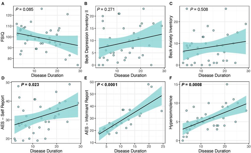
FIGURE 1 | Disease duration as a predictor of clinical measures in DM1. Disease duration was not associated with changes in (A) FSIQ (P = 0.085), (B) depression
(P = 0.271), or (C) anxiety (P = 0.508), but was significantly associated with increased scores for the core symptoms of (D) self-reported apathy (P = 0.023), (E)
informant reported-apathy (P < 0.0001), and (F) hypersomnolence (P = 0.0008). Light-blue shaded region represents 95% confidence interval.
crudely tracked by self-report in this current study, this feature progression, disease duration was significantly associated with
can also be evaluated by quantitative assessment such as EEG the core features of apathy, hypersomnolence, and cerebral FA.
from polysomnography, which could be used in future studies or Identifying features that represent the true manifestation of brain
clinical trials (82). pathology in DM1 is of particular importance given that drug
A limitation of this present study is a relatively low median companies are interested in targeting the CNS for clinical trials
CTG repeat length of 146 and mild MIRS score compared to of gene knock-down and other therapies. It is important to note
other studies, suggesting that the DM1 group is only mildly that a correlation to disease duration is only a proxy to disease
affected. However, our study was primarily focused on adult- progression. Moreover, given the wide range of disease duration
onset DM1, and by excluding congenital, childhood, and juvenile in our sample, it will be possible to evaluate disease progression
forms of DM1 the median CTG repeat length of 146 was not in the future. Whether progression in these symptoms occur
unexpected. This could lead to problems generalizing these rapidly is important to assess in the context of whether or not
results to a larger DM1 population that included congenital, they may represent appropriate end-points for clinical trials.
childhood, and juvenile DM1 patients. Like most human research Our study is designed as a prospective longitudinal study and
studies, adult-onset DM1 participants that are moderately to evaluation of change over a one- and two-year time period is
severely affected face greater barriers with research study currently underway.
participation, thus biasing the study population itself to those
less affected by disease. Another limitation to this study is the CONCLUSIONS
use of self-report questionnaires for measurement of depression,
anxiety, apathy, and hypersomnolence. This could have led to Our study supports the hypothesis that the underlying
biased results through misreporting of symptoms. neuropathology of adult-onset myotonic dystrophy type 1,
The current findings highlight the concept that core as measured by cerebral WM FA and disease duration, leads
features of the disease that are directly related to measures directly to cognitive deficits, apathy, and hypersomnolence,
of brain pathology (lower WM FA) are FSIQ, apathy, and while increased symptoms of depression and anxiety are
hypersomnolence. Additionally, as a measure of disease secondary to a combination of the physical symptoms of having
Frontiers in Neurology | www.frontiersin.org 7 July 2021 | Volume 12 | Article 700796Miller et al. Core Brain Symptoms of DM1
a neuromuscular disorder and the emotional stress of coping AK, DJM, LG, EP, TK, SC, and DGM. Resources, Supervision,
with a chronic and debilitating disorder. and Funding Acquisition: PN. Writing—Review & Editing: JM,
PN, AK, DJM, LG, EP, TK, SC, and DGM. Visualization: JM.
DATA AVAILABILITY STATEMENT All authors had full access to all the data in the study and take
responsibility for the integrity of the data and the accuracy of the
The raw data supporting the conclusions of this article will be data analysis.
made available by the authors, without undue reservation.
FUNDING
ETHICS STATEMENT
This work was supported by a grant from the National
The studies involving human participants were reviewed Institute of Neurological Disorders and Stroke (NINDS) (Ref:
and approved by University of Iowa Institutional 5R01NS094387-02) and the Wyck Foundation. The 3T scanner
Review Board. The patients/participants provided was supported by an equipment grant from the Office of the
their written informed consent to participate in Director at the National Institutes of Health (S10OD025025).
this study.
ACKNOWLEDGMENTS
AUTHOR CONTRIBUTIONS
The authors are grateful to the participants and their families for
Conceptualization and Methodology, Formal Analysis, their cooperation with the study, and also to the team of research
Writing—Original Draft: JM and PN. Investigation: JM, PN, assistants who collected the data.
REFERENCES cognitive impairment in Myotonic Dystrophy type 1. Neuroimage Clin. (2019)
24:102078. doi: 10.1016/j.nicl.2019.102078
1. Meola G, Cardani R. Myotonic dystrophies: an update on clinical aspects, 13. Caso F, Agosta F, Peric S, Rakočević-Stojanović V, Copetti M,
genetic, pathology, and molecular pathomechanisms. Biochim Biophys Acta. Kostic VS, et al. Cognitive impairment in myotonic dystrophy
(2015) 1852:594–606. doi: 10.1016/j.bbadis.2014.05.019 type 1 is associated with white matter damage. PLoS ONE. (2014)
2. Okkersen K, Monckton DG, Le N, Tuladhar AM, Raaphorst J, van Engelen 9:e104697. doi: 10.1371/journal.pone.0104697
BGM. Brain imaging in myotonic dystrophy type 1: a systematic review. 14. Serra L, Petrucci A, Spano B, Torso M, Olivito G, Lispi L, et al. How genetics
Neurology. (2017) 89:960–9. doi: 10.1212/WNL.0000000000004300 affects the brain to produce higher-level dysfunctions in myotonic dystrophy
3. van Dorst M, Okkersen K, Kessels RPC, Meijer FJA, Monckton DG, van type 1. Funct Neurol. (2015) 30:21–31.
Engelen BGM, et al. Structural white matter networks in myotonic dystrophy 15. Gourdon G, Meola G. Myotonic dystrophies: state of the art of new
type 1. Neuroimage Clin. (2019) 21:101615. doi: 10.1016/j.nicl.2018.101615 therapeutic developments for the CNS. Front Cell Neurosci. (2017)
4. Wozniak JR, Mueller BA, Lim KO, Hemmy LS, Day JW. Tractography reveals 11:101. doi: 10.3389/fncel.2017.00101
diffuse white matter abnormalities in myotonic dystrophy type 1. J Neurol Sci. 16. Phillips MF, Steer HM, Soldan JR, Wiles CM, Harper PS.
(2014) 341:73–8. doi: 10.1016/j.jns.2014.04.005 Daytime somnolence in myotonic dystrophy. J Neurol. (1999)
5. Zanigni S, Evangelisti S, Giannoccaro MP, Oppi F, Poda R, Giorgio A, et al. 246:275–82. doi: 10.1007/s004150050346
Relationship of white and gray matter abnormalities to clinical and genetic 17. Chase TN. Apathy in neuropsychiatric disease: diagnosis,
features in myotonic dystrophy type 1. Neuroimage Clin. (2016) 11:678– pathophysiology, and treatment. Neurotox Res. (2010) 19:266–
85. doi: 10.1016/j.nicl.2016.04.012 78. doi: 10.1007/978-1-4614-0785-0_5
6. Cabada T, Iridoy M, Jericó I, Lecumberri P, Seijas R, Gargallo A, et al. 18. Lindeblad G, Kroksmark AK, Ekstrom AB. Cognitive and adaptive
Brain involvement in myotonic dystrophy type 1: a morphometric and functioning in congenital and childhood forms of myotonic dystrophy
diffusion tensor imaging study with neuropsychological correlation. Arch Clin type 1: a longitudinal study. Dev Med Child Neurol. (2019) 61:1214–
Neuropsychol. (2017) 32:401–12. doi: 10.1093/arclin/acx008 20. doi: 10.1111/dmcn.14161
7. Park JS, Song H, Jang KE, Cha H, Lee SH, Hwang SK, et al. Diffusion tensor 19. Langbehn KE, van der Plas E, Moser DJ, Long JD, Gutmann L, Nopoulos
imaging and voxel-based morphometry reveal corticospinal tract involvement PC. Cognitive function and its relationship with brain structure in myotonic
in the motor dysfunction of adult-onset myotonic dystrophy type 1. Sci Rep. dystrophy type 1. J Neurosci Res. (2020) 99:190–9. doi: 10.1002/jnr.24595
(2018) 8:15592. doi: 10.1038/s41598-018-34048-9 20. Miller JN, van der Plas E, Hamilton M, Koscik TR, Gutmann L,
8. Yoo WK, Park YG, Choi YC, Kim SM. Cortical thickness and white matter Cumming SA, et al. Variant repeats within the DMPK CTG expansion
integrity are associated with CTG expansion size in myotonic dystrophy type protect function in myotonic dystrophy type 1. Neurol Genet. (2020)
I. Yonsei Med J. (2017) 58:807–15. doi: 10.3349/ymj.2017.58.4.807 6:e504. doi: 10.1212/NXG.0000000000000504
9. Baldanzi S, Cecchi P, Fabbri S, Pesaresi I, Simoncini C, Angelini C, et al. 21. Okkersen K, Buskes M, Groenewoud J, Kessels RPC, Knoop H,
Relationship between neuropsychological impairment and grey and white van Engelen B, et al. The cognitive profile of myotonic dystrophy
matter changes in adult-onset myotonic dystrophy type 1. Neuroimage Clin. type 1: a systematic review and meta-analysis. Cortex. (2017)
(2016) 12:190–7. doi: 10.1016/j.nicl.2016.06.011 95:143–55. doi: 10.1016/j.cortex.2017.08.008
10. Minnerop M, Weber B, Schoene-Bake J-C, Roeske S, Mirbach S, Anspach C, et 22. Ates S, Deistung A, Schneider R, Prehn C, Lukas C, Reichenbach JR, et
al. The brain in myotonic dystrophy 1 and 2: evidence for a predominant white al. Characterization of iron accumulation in deep gray matter in myotonic
matter disease. Brain. (2011) 134(Pt 12):3530–46. doi: 10.1093/brain/awr299 dystrophy type 1 and 2 using quantitative susceptibility mapping and R2(∗ )
11. Van der Plas E, Koscik T, Magnotta V, Monckton DG, Cumming S, Gutmann relaxometry: a magnetic resonance imaging study at 3 tesla. Front Neurol.
L, et al. Brain and functional features of individuals in premanifest myotonic (2019) 10:1320. doi: 10.3389/fneur.2019.01320
dystrophy type 1. Neurol Genet. (2020). 23. Kalkman JS, Schillings ML, Zwarts MJ, van Engelen BG, Bleijenberg
12. Labayru G, Diez I, Sepulcre J, Fernandez E, Zulaica M, Cortes JM, et G. Psychiatric disorders appear equally in patients with myotonic
al. Regional brain atrophy in gray and white matter is associated with dystrophy, facioscapulohumeral dystrophy, and hereditary motor
Frontiers in Neurology | www.frontiersin.org 8 July 2021 | Volume 12 | Article 700796Miller et al. Core Brain Symptoms of DM1
and sensory neuropathy type I. Acta Neurol Scand. (2007) 43. Geirdal AO, Lund-Petersen I, Heiberg A. Understanding the experience of
115:265–70. doi: 10.1111/j.1600-0404.2006.00737.x myotonic dystrophy. Mixed method study. J Genet Couns. (2015) 24:169–
24. Kalkman JS, Schillings ML, Zwarts MJ, van Engelen BG, Bleijenberg G. The 78. doi: 10.1007/s10897-014-9752-1
development of a model of fatigue in neuromuscular disorders: a longitudinal 44. Kobayakawa M, Tsuruya N, Kawamura M. Theory of mind impairment
study. J Psychosom Res. (2007) 62:571–9. doi: 10.1016/j.jpsychores.2006.11.014 in adult-onset myotonic dystrophy type 1. Neurosci. Res. (2012) 72:341–
25. Okkersen K, Jimenez-Moreno C, Wenninger S, Daidj F, Glennon J, Cumming 6. doi: 10.1016/j.neures.2012.01.005
S, et al. Cognitive behavioural therapy with optional graded exercise 45. Kurauchi G, Endo M, Odaira K, Ono R, Koseki A, Goto M, et
therapy in patients with severe fatigue with myotonic dystrophy type 1: a al. Caregiver burden and related factors among caregivers of patients
multicentre, single-blind, randomised trial. Lancet Neurol. (2018) 17:671– with myotonic dystrophy type 1. J Neuromuscul Dis. (2019) 6:527–
80. doi: 10.1016/S1474-4422(18)30203-5 36. doi: 10.3233/JND-190386
26. Schneider-Gold C, Bellenberg B, Prehn C, Krogias C, Schneider R, 46. Rose MR, Sadjadi R, Weinman J, Akhtar T, Pandya S, Kissel JT, et al. Role
Klein J, et al. Cortical and subcortical grey and white matter atrophy of disease severity, illness perceptions, and mood on quality of life in muscle
in myotonic dystrophies type 1 and 2 is associated with cognitive disease. Muscle Nerve. (2012) 46:351–9. doi: 10.1002/mus.23320
impairment, depression and daytime sleepiness. PLoS ONE. (2015) 47. Van Heugten C, Meuleman S, Hellebrekers D, Kruitwagen-van Reenen
10:e0130352. doi: 10.1371/journal.pone.0130352 E, Visser-Meily J. Participation and the role of neuropsychological
27. Winblad S, Jensen C, Månsson J-E, Samuelsson L, Lindberg C. Depression functioning in myotonic dystrophy type 1. J Neuromuscul Dis. (2018) 5:205–
in myotonic dystrophy type 1: clinical and neuronal correlates. Behav Brain 14. doi: 10.3233/JND-170246
Functions. (2010) 6:25. doi: 10.1186/1744-9081-6-25 48. Winblad S, Lindberg C, Hansen S. Temperament and character in patients
28. Winblad S, Lindberg C. Perceived fatigue in myotonic with classical myotonic dystrophy type 1 (DM-1). Neuromuscul Disord. (2005)
dystrophy type 1: a case-control study. BMC Neurol. (2019) 15:287–92. doi: 10.1016/j.nmd.2004.12.003
19:45. doi: 10.1186/s12883-019-1280-z 49. Winblad S, Samuelsson L, Lindberg C, Meola G. Cognition in myotonic
29. Antonini G, Soscia F, Giubilei F, De Carolis A, Gragnani F, Morino S, dystrophy type 1: a 5-year follow-up study. Eur J Neurol. (2016) 23:1471–
et al. Health-Related quality of life in myotonic dystrophy type 1 and its 6. doi: 10.1111/ene.13062
relationship with cognitive and emotional functioning. J Rehabil Med. (2006) 50. Callus E, Bertoldo EG, Beretta M, Boveri S, Cardani R, Fossati B,
38:181–5. doi: 10.1080/16501970500477967 et al. Neuropsychological and psychological functioning aspects in
30. Di Costanzo A, Mottola A, Toriello A, Di Iorio G, Tedeschi G, Bonavita V. myotonic dystrophy type 1 patients in Italy. Front Neurol. (2018)
Does abnormal neuronal excitability exist in myotonic dystrophy? II. Effects of 9:751. doi: 10.3389/fneur.2018.00751
the antiarrhythmic drug hydroquinidine on apathy and hypersomnia. Neurol 51. Gomes-Pereira M, Bidichandani SI, Monckton DG. Analysis of unstable
Sci. (2000) 21:81–6. doi: 10.1007/s100720070100 triplet repeats using small-pool polymerase chain reaction. Methods Mol Biol.
31. Peric D, Plancak D, Bulj M, Tudor V, Spalj S. Health-related quality (2004) 277:61–76. doi: 10.1385/1-59259-804-8:061
of life in soldiers in Croatia: relationship with combat readiness and 52. Monckton DG, Wong LJ, Ashizawa T, Caskey CT. Somatic mosaicism,
psychological dimensions. Cent Eur J Public Health. (2013) 21:207– germline expansions, germline reversions and intergenerational reductions in
12. doi: 10.21101/cejph.a3862 myotonic dystrophy males: small pool PCR analyses. Hum Mol Genet. (1995)
32. Peric S, Bjelica B, Bozovic I, Pesovic J, Paunic T, Banovic M, et al. Fatigue 4:1–8. doi: 10.1093/hmg/4.1.1
in myotonic dystrophy type 1: a seven-year prospective study. Acta Myol. 53. Ciosi M, Cumming SA, Alshammari AM, Symeonidi E, Herzyk P, McGuinness
(2019) 38:239–44. D, et al. Library preparation and MiSeq sequencing for the genotyping-
33. Rakocevic Stojanovic V, Peric S, Paunic T, Pesovic J, Vujnic M, Peric M, et al. by-sequencing of the Huntington disease HTT exon one trinucleotide
Quality of life in patients with myotonic dystrophy type 2. J Neurol Sci. (2016) repeat and the quantification of somatic mosaicism. Protocol Exchange.
365:158–61. doi: 10.1016/j.jns.2016.04.018 (2018). doi: 10.1038/protex.2018.089
34. Rakocevic-Stojanovic V, Peric S, Madzarevic R, Dobricic V, Ralic V, Ilic V, et 54. Mathieu J, Boivin H, Meunier D, Gaudreault M, Bégin P. Assessment of a
al. Significant impact of behavioral and cognitive impairment on quality of disease-specific muscular impairment rating scale in myotonic dystrophy.
life in patients with myotonic dystrophy type 1. Clin Neurol Neurosurg. (2014) Neurology. (2001) 56:336–40. doi: 10.1212/WNL.56.3.336
126:76–81. doi: 10.1016/j.clineuro.2014.08.021 55. Wechsler D. Wechsler Adult Intelligence Scale. 4th ed. San Antonio, TX:
35. Abe K, Fujimura H, Toyooka K, Yorifuji S, Nishikawa Y, Hazama T, et al. Pearson Assessment (2008). doi: 10.1037/t15169-000
Involvement of the central nervous system in myotonic dystrophy. J Neurol 56. Beck AT, Ward CH, Mendelson M, Mock J, Erbaugh J. An
Sci. (1994) 127:179–85. doi: 10.1016/0022-510X(94)90071-X inventory for measuring depression. Arch Gen Psychiatry. (1961)
36. Bertrand JA, Jean S, Laberge L, Gagnon C, Mathieu J, Gagnon JF, et al. 4:561–71. doi: 10.1001/archpsyc.1961.01710120031004
Psychological characteristics of patients with myotonic dystrophy type 1. Acta 57. Marinus J, Visser M, van Hilten JJ, Lammers GJ, Stiggelbout AM. Assessment
Neurol Scand. (2014) 132:49–58. doi: 10.1111/ane.12356 of sleep and sleepiness in Parkinson disease. Sleep. (2003) 26:1049–
37. Colombo G, Perini GI, Miotti MV, Armani M, Angelini C. Cognitive and 54. doi: 10.1093/sleep/26.8.1049
psychiatric evaluation of 40 patients with myotonic dystrophy. Ital J Neurol 58. Marin RS, Biedrzycki RC, Firinciogullari S. Reliability and
Sci. (1992) 13:53–8. doi: 10.1007/BF02222889 validity of the apathy evaluation scale. Psychiatry Res. (1991)
38. Duveneck MJ, Portwood MM, Wicks JJ, Lieberman JS. Depression in 38:143–62. doi: 10.1016/0165-1781(91)90040-V
myotonic muscular dystrophy. Arch Phys Med Rehabil. (1986) 67:875–7. 59. van der Plas E, Hamilton MJ, Miller JN, Koscik TR, Long JD, Cumming
39. Endo M, Odaira K, Ono R, Kurauchi G, Koseki A, Goto M, et al. S, et al. Brain structural features of myotonic dystrophy type 1 and
Health-related quality of life and its correlates in Japanese patients with their relationship with Ctg repeats. J Neuromuscul Dis. (2019) 6:321–
myotonic dystrophy type 1. Neuropsychiatr Dis Treat. (2019) 15:219– 32. doi: 10.3233/JND-190397
26. doi: 10.2147/NDT.S187607 60. Andersson JLR, Sotiropoulos SN. An integrated approach to correction
40. Franzese A, Antonini G, Iannelli M, Leardi MG, Spada S, Vichi for off-resonance effects and subject movement in diffusion MR imaging.
R, et al. Intellectual functions and personality in subjects with Neuroimage. (2016) 125:1063–78. doi: 10.1016/j.neuroimage.2015.10.019
noncongenital myotonic muscular dystrophy. Psychol. Rep. (1991) 68(3 Pt 61. Andersson JLR, Skare S, Ashburner J. How to correct susceptibility distortions
1):723–32. doi: 10.2466/pr0.1991.68.3.723 in spin-echo echo-planar images: application to diffusion tensor imaging.
41. Fujino H, Shingaki H, Suwazono S, Ueda Y, Wada C, Nakayama T, et al. Neuroimage. (2003) 20:870–88. doi: 10.1016/S1053-8119(03)00336-7
Cognitive impairment and quality of life in patients with myotonic dystrophy 62. Avants BB, Tustison NJ, Song G, Cook PA, Klein A, Gee JC. A reproducible
type 1. Muscle Nerve. (2018) 57:742–8. doi: 10.1002/mus.26022 evaluation of ANTs similarity metric performance in brain image registration.
42. Gallais B, Montreuil M, Gargiulo M, Eymard B, Gagnon C, Laberge L. Neuroimage. (2011) 54:2033–44. doi: 10.1016/j.neuroimage.2010.09.025
Prevalence and correlates of apathy in myotonic dystrophy type 1. BMC 63. Cohen J. Statistical Power Analysis for the Behavioral Sciences. 2nd ed.
Neurol. (2015) 15:148. doi: 10.1186/s12883-015-0401-6 Hillsdale, NJ: L. Erlbaum Associates (1988), p. xxi, 567.
Frontiers in Neurology | www.frontiersin.org 9 July 2021 | Volume 12 | Article 700796Miller et al. Core Brain Symptoms of DM1
64. LaDonna KA, Ghavanini AA, Venance SL. Truths and misinformation: a 76. Krogias C, Bellenberg B, Prehn C, Schneider R, Meves SH, Gold R,
qualitative exploration of myotonic dystrophy. Can J Neurol Sci. (2015) et al. Evaluation of CNS involvement in myotonic dystrophy type
42:187–94. doi: 10.1017/cjn.2015.26 1 and type 2 by transcranial sonography. J Neurol. (2015) 262:365–
65. Hagerman KA, Howe SJ, Heatwole CR, Christopher Project Reference G. 74. doi: 10.1007/s00415-014-7566-6
The myotonic dystrophy experience: a North American cross-sectional study. 77. Oyamada R, Hayashi M, Katoh Y, Tsuchiya K, Mizutani T, Tominaga
Muscle Nerve. (2019) 59:457–64. doi: 10.1002/mus.26420 I, et al. Neurofibrillary tangles and deposition of oxidative products in
66. Weber YG, Roebling R, Kassubek J, Hoffmann S, Rosenbohm A, Wolf the brain in cases of myotonic dystrophy. Neuropathology. (2006) 26:107–
M, et al. Comparative analysis of brain structure, metabolism, and 14. doi: 10.1111/j.1440-1789.2006.00662.x
cognition in myotonic dystrophy 1 and 2. Neurology. (2010) 74:1108– 78. Laberge L, Gagnon C, Dauvilliers Y. Daytime sleepiness
17. doi: 10.1212/WNL.0b013e3181d8c35f and myotonic dystrophy. Curr Neurol Neurosci Rep. (2013)
67. Cabada T, Diaz J, Iridoy M, Lopez P, Jerico I, Lecumberri P, et al. 13:340. doi: 10.1007/s11910-013-0340-9
Longitudinal study in patients with myotonic dystrophy type 1: correlation 79. Laberge L, Gallais B, Auclair J, Dauvilliers Y, Mathieu J,
of brain MRI abnormalities with cognitive performances. Neuroradiology. Gagnon C. Predicting daytime sleepiness and fatigue: a 9-year
(2020). doi: 10.1007/s00234-020-02611-9 prospective study in myotonic dystrophy type 1. J Neurol. (2020)
68. Lopez-Titla MM, Chirino A, Cruz Solis SV, Hernandez-Castillo CR, Diaz R, 267:461–8. doi: 10.1007/s00415-019-09592-7
Marquez-Quiroz LDC, et al. Cognitive decline and white matter integrity 80. Heatwole C, Bode R, Johnson N, Quinn C, Martens W, McDermott MP, et al.
degradation in myotonic dystrophy type I. J Neuroimaging. (2021) 31:192– Patient-reported impact of symptoms in myotonic dystrophy type 1 (PRISM-
8. doi: 10.1111/jon.12786 1). Neurology. (2012) 79:348–57. doi: 10.1212/WNL.0b013e318260cbe6
69. Gallais B, Gagnon C, Mathieu J, Richer L. Cognitive decline over time in adults 81. van der Werf S, Kalkman J, Bleijenberg G, van Engelen B, Schillings M,
with myotonic dystrophy type 1: a 9-year longitudinal study. Neuromuscul Zwarts M, et al. The relation between daytime sleepiness, fatigue, and
Disord. (2017) 27:61–72. doi: 10.1016/j.nmd.2016.10.003 reduced motivation in patients with adult onset myotonic dystrophy. J Neurol
70. Labayru G, Aliri J, Zulaica M, López de Munain A, Sistiaga A. Age-related Neurosurg Psychiatry. (2003) 74:138–9. doi: 10.1136/jnnp.74.1.138
cognitive decline in myotonic dystrophy type 1: an 11-year longitudinal 82. Cheung J, Ruoff C, Moore H, Hagerman KA, Perez J, Sakamuri S, et al.
follow-up study. J Neuropsychol. (2019) 14:121–34. doi: 10.1111/jnp.12192 Increased EEG Theta Spectral Power in Sleep in Myotonic Dystrophy Type
71. Hamilton MJ, McLean J, Cumming S, Ballantyne B, McGhie J, Jampana R, 1. J Clin Sleep Med. (2018) 14:229–35. doi: 10.5664/jcsm.6940
et al. Outcome measures for central nervous system evaluation in myotonic
dystrophy type 1 may be confounded by deficits in motor function or insight. Conflict of Interest: Within the last three years DM has been a scientific
Front Neurol. (2018) 9:780. doi: 10.3389/fneur.2018.00780 consultant and/or received an honoraria/stock options from AMO Pharma,
72. Serra L, Bianchi G, Bruschini M, Giulietti G, Domenico CD, Bonarota Charles River, Vertex Pharmaceuticals, Triplet Therapeutics, LoQus23 and Small
S, et al. Abnormal cortical thickness is associated with deficits in social Molecule RNA. DM also had/has research contracts with AMO Pharma and
cognition in patients with myotonic dystrophy type 1. Front Neurol. (2020) Vertex Pharmaceuticals.
11:113. doi: 10.3389/fneur.2020.00113
73. Minier L, Lignier B, Bouvet C, Gallais B, Camart N. A review of The remaining authors declare that the research was conducted in the absence of
psychopathology features, personality, and coping in myotonic dystrophy type any commercial or financial relationships that could be construed as a potential
1. J Neuromuscul Dis. (2018) 5:279–94. doi: 10.3233/JND-180310 conflict of interest.
74. van der Velden BG, Okkersen K, Kessels RP, Groenewoud J, van Engelen
B, Knoop H, et al. Affective symptoms and apathy in myotonic dystrophy Copyright © 2021 Miller, Kruger, Moser, Gutmann, van der Plas, Koscik, Cumming,
type 1 a systematic review and meta-analysis. J Affect Disord. (2019) 250:260– Monckton and Nopoulos. This is an open-access article distributed under the terms
9. doi: 10.1016/j.jad.2019.03.036 of the Creative Commons Attribution License (CC BY). The use, distribution or
75. Guercio BJ, Donovan NJ, Munro CE, Aghjayan SL, Wigman SE, Locascio JJ, reproduction in other forums is permitted, provided the original author(s) and the
et al. The apathy evaluation scale: a comparison of subject, informant, and copyright owner(s) are credited and that the original publication in this journal
clinician report in cognitively normal elderly and mild cognitive impairment. is cited, in accordance with accepted academic practice. No use, distribution or
J Alzheimers Dis. (2015) 47:421–32. doi: 10.3233/JAD-150146 reproduction is permitted which does not comply with these terms.
Frontiers in Neurology | www.frontiersin.org 10 July 2021 | Volume 12 | Article 700796You can also read