Comparison of T1 FLAIR BLADE with and without parallel imaging against T1 turbo spin echo in the MR imaging of lumbar spine in the sagittal plane
←
→
Page content transcription
If your browser does not render page correctly, please read the page content below
Journal of Radiation Research and                                                                                          Lavdas et al., J Radiat Res Imaging 2021;
                                                                                                                                                         1(1):33-40.
Imaging
                                                                                                                                         Research Article

Comparison of T1 FLAIR BLADE with and
without parallel imaging against T1 turbo spin
echo in the MR imaging of lumbar spine in the
sagittal plane
Eleftherios Lavdas1,2, Eleonora Giankou3, Panos Papanikolaou4, Aleksandra Tsikrika5, Maria Papaioannou2, Violeta Roka6, Vasiliki
Chatzigeorgiou3, Georgios Batsikas3, Spiros Kostopoulos7, Dimitrios Glotsos7, Athanasios Bakas1, Panayiotis Mavroidis8*

1
 University of West Attica, Department        Abstract
of Biomedical Sciences, Athens, Greece
                                              Purpose: Spinal cord and nerves are best visualized by MRI, which is able to show structural and functional
2
 Animus Kyanoys Stavros, Department           anomalies of the spine. The primary objective of this study is to identify advantages or disadvantages of
of Radiology, Larissa, Greece                 the T1-weighted fluid attenuated inversion recovery (FLAIR) sequence with BLADE technique (T1W-FLAIR
3                                             BLADE), with and without parallel imaging when compared with T1 Turbo Spin Echo (T1 TSE) sequence
 Department of Medical Imaging, IASO
Thessalias Hospital, Larissa, Greece          when performing MRI examination of the lumbar spine in a sagittal view.
4
 Long School of Medicine, University          Methods: L-spine examinations with T1W-FLAIR BLADE (with and without parallel imaging) and T1 TSE
of Texas Health at San Antonio, San           were acquired on 44 patients using a 1.5T scanner. These sequences were assessed by two radiologists
Antonio, TX, USA                              a) quantitatively by comparing the signal-to-noise ratio (SNR), contrast-to-noise ratio (CNR) and relative
                                              contrast (ReCon) measurements and b) qualitatively based on different features of the images such as
5
 Department of Radiology, University          cerebrospinal fluid (CSF) nulling.
Hospital of Larissa, Larissa, Greece
6
                                              Results: The quantitative analysis showed that T1W-FLAIR BLADE in most tissues had better SNR values, as
 Health Center of Farkadona, Trikala,         well as better CNR and ReCon results. SNR and CNR measurements were also better in T1W-FLAIR BLADE
Greece                                        images with parallel imaging compared to T1 TSE. In the qualitative analysis, it was found that T1W-FLAIR
7
 University of West Attica, Department        BLADE sequence had superior quality of images, better contrast among tissues, and better nulling of CSF.
of Biomedical Engineering, Athens,            Conclusion: T1W-FLAIR BLADE appeared to have less motion artifacts and improved image quality, as
Greece
                                              well as improved CSF nulling. T1W-FLAIR BLADE with parallel imaging achieved the second best results
8                                             out of the sequences compared. The clinicians showed a preference for T1W-FLAIR BLADE images which
 Department of Radiation Oncology,
University of North Carolina, Chapel          could be a useful addition to the standard imaging of the L-spine.
Hill, NC, USA
                                              Keywords: 1.5T MRI, Motion artifacts, BLADE sequences, Lumbar spine examination
*Author for correspondence:
Email: panayiotis_mavroidis@med.              Introduction
unc.edu
                                                   Magnetic Resonance Imaging (MRI) gives a very clear picture of the spine and it is very helpful in
Received date: March 17, 2021                 identifying serious spinal disorders such as cancer, infection, or nerve compression [1]. It can identify
Accepted date: April 13, 2021                 congenital abnormalities, trauma and other lesions that could be missed with other imaging tools.
                                              Currently, MRI is the gold standard for the imaging of the spinal canal and the nerves of the spine
Copyright: © 2021 Lavdas E, et al. This
                                              as well as imaging of injuries affecting the vertebrae and the discs (such as degeneration, hernias,
is an open-access article distributed
under the terms of the Creative
                                              etc.). MRI is also used as a tool for pre- and post-operative assessment, assessment of infections, and
Commons Attribution License, which            delineation of tumors [2].
permits unrestricted use, distribution,            The T1 sequence is the most important sequence for detecting bone marrow pathologies. More
and reproduction in any medium,
                                              specifically, it provides a wide understanding of anatomical and pathological variations in the lumbar
provided the original author and
source are credited.                          spine. In the imaging of the lumbar spine, T1W-Turbo Spin Echo (TSE) or Fast Spin Echo (FSE)
                                              images are acquired [3]. According to Melhem et al., who compared T1 FLAIR and T1 SE images,
                                              the images received from T1W-FLAIR sequence showed higher contrast around CSF, improved
                                              lesion delineation in the spinal cord and bone marrow, and showed less artifacts in comparison with

                             This article is originally published by ProBiologist LLC., and is freely available at probiologists.com

J Radiat Res Imaging 2021; 1(1):33-40.                                                                                                                  33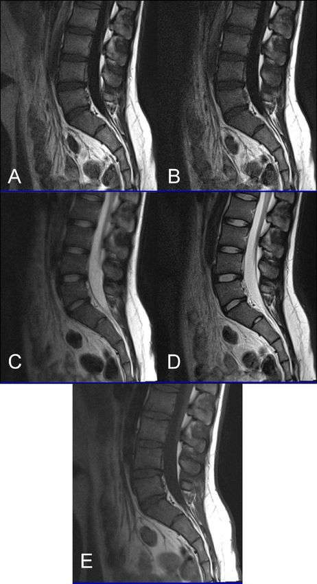
Citation: Lavdas E, Giankou E, Papanikolaou P, Tsikrika A, Papaioannou M, Roka V, et al. Comparison of T1 FLAIR BLADE with and without parallel
imaging against T1 turbo spin echo in the MR imaging of lumbar spine in the sagittal plane. J Radiat Res Imaging. 2021; 1(1):33-40.

the conventional T1W-SE sequences [4]. The study concluded that             saturation, non-contrast). The aim to show the benefits of both
T1W-FLAIR images were superior to SE sequences when analyzing               sequences and identify which one provides better lesion delineation
spinal imaging. Another study verified that T1W-FLAIR images                and reduction of artifacts.
might be an alternative approach for the imaging of L-spine instead
of T1W-TSE, as it achieved better overall image quality and contrast,       Methods and Materials
better signal nulling of CSF, as well as better definition of structures        Subjects
and lesions [5].
                                                                                This study was performed on forty-four patients (20 females and
    In most cases, patients that are to have an MRI examination of          24 males within the range of 18-82 years), who had a routine MRI
the L-spine present with severe back pain. This can be caused by            examination of the L-spine between February 2014 and July 2018.
many reasons, such as sciatica or even bone metastases. In these            All patients participating in the study signed a written consent form
cases, it is very difficult for the patient to keep still during a long     with all the information provided and the study was approved by the
MRI examination, resulting in visible patient movement in the               local institutional review board.
acquired images. Sequences that use central K-space oversampling,
a technique known as PROPELLER (Periodically Rotated                            MR Imaging techniques
Overlapping Parallel Lines with Enhanced Reconstruction) can be                 All the lumbar spine (L-spine) MRI examinations in this study
helpful in reducing image artifacts [6]. Many studies have also shown       were acquired on a 1.5T scanner (Magnetom, Avanto, Siemens
the advantages of BLADE in T2 sequences with regards to image               Healthcare Sector, Erlangen, Germany) and a Synergy body phased-
quality when compared to conventional sequences [6,7]. Advantages           array surface coil. The parameters of each sequence are presented in
included motion elimination as well as elimination of flow and slice-       Table 1.
overlap artifacts in MR lumbar spine imaging in both cooperative
and non-cooperative patients [7]. Alternatively, there have not been             Turbo Spin Echo (TSE) or Fast Spin Echo (FSE) pulse sequence
many studies that show the advantages of BLADE in T1 sequences.             is the main sequence used in almost all MR imaging nowadays [9].
Acquiring T1-weighted BLADE images requires a long echo train               TSE is basically a spin echo sequence with the advantage of a shorter
length (ETL), which may slightly increase the sequence time but a           acquisition time. While in a SE sequence a 90° excitation pulse is
long ETL would decrease motion artifacts. A few studies on brain            followed by a 180° rephasing pulse, filling only one line of K-space
MR imaging have found that T1W-FLAIR BLADE presented better                 per TR, in the TSE sequence several 180° rephasing pulses produce
image quality including CSF nulling and less artifacts [6].                 an echo train, thus producing more than one phase encoding steps
                                                                            and subsequently filling more than one line of K-space per TR [8].
     Parallel imaging is a technique in which TSE fills K-space more        This allows the TSE sequence to be acquired in much less time than
efficiently by filling multiple lines of K-space per TR. However,           the conventional spin echo (CSE) without compromising spatial
the difference is that these lines are acquired by certain coils that       resolution, image weighting or signal-to-noise ratio (SNR) [8]. To
are combined together to acquire data simultaneously [8]. Parallel          improve spatial resolution, a greater number of phase-encoding steps
imaging is an important tool that can be used to improve image              can be used. Last but not least, TSE provides a better image when
resolution and reduce scan times. With the appropriate software and         metal objects are present due to less signal loss from susceptibility
coil configurations, it can be used with most pulse sequences. Apart        artifacts [9].
from the benefits described above, parallel imaging can result in a
slight loss of SNR. In addition, chemical shift may increase due to              Generally speaking, although TSE and CSE are two of the main
different resonant frequencies being mapped across each coil. Patient       sequences that are used in most MR imaging examinations, TSE has
movement also causes misalignment between under-sampled data                largely replaced CSE in certain examinations, such as musculoskeletal
and reference scans [8].                                                    scans, especially in T2 weighting. However, an issue that can occur
                                                                            during the acquisition of TSE images is blurring at the edges of
    In this study, T1W-FLAIR BLADE without parallel imaging in              tissues with different T2 decay values. This is because each line of
the sagittal plane (sag) is compared with T1W-TSE in the sagittal           K-space filled during an echo train contains data from echoes with a
plane and T1W-FLAIR BLADE with parallel imaging (no fat                     different TE. When using long echo trains, late echoes that have low

 Sequences                                T1 TSE Sag                 T1 Sag Blade Flair                   T1 Sag Blade Flair PI
 TR (msec)                                591                        2000                                 2000
 TE (msec)                                11                         47                                   45
 TI (msec)                                                           860                                  860
 Matrix (Phase/Freq)                      384/288                    256/256                              256/256
 BW (Hz/pix)                              161                        362                                  362
 A.A (min)                                2:43                       4:06                                 2:25
 ET                                       2                          49                                   49
 Thickness (mm)                           4 mm                       4 mm                                 4 mm
 Space (mm)                               10%                        10%                                  10%
 FOV (mm)                                 280                        280                                  280
 NSA                                      2                          1                                    1
Table 1: Summary of the Sequences that were applied for the lumbar spine (L-Spine) MR examinations.

J Radiat Res Imaging 2021; 1(1):33-40.                                                                                                      34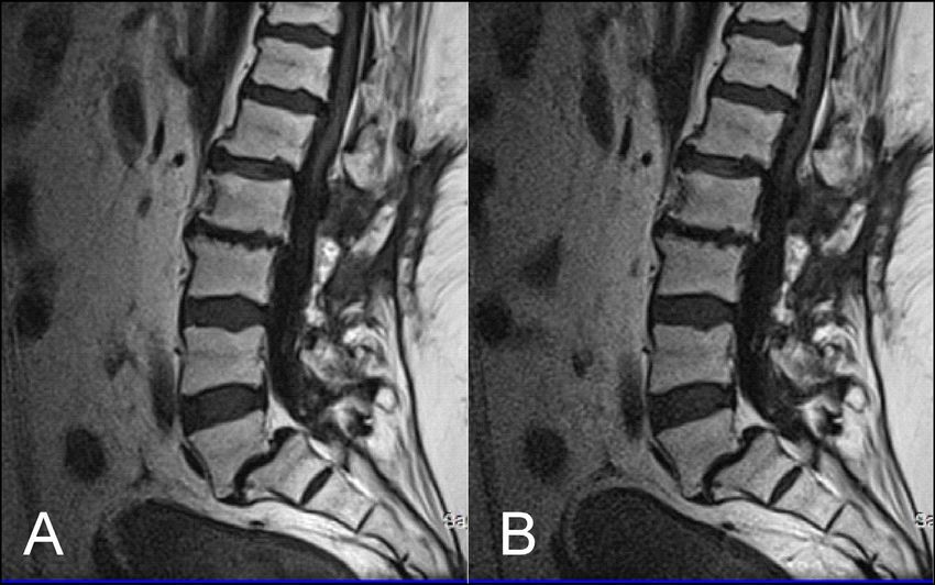
Citation: Lavdas E, Giankou E, Papanikolaou P, Tsikrika A, Papaioannou M, Roka V, et al. Comparison of T1 FLAIR BLADE with and without parallel
imaging against T1 turbo spin echo in the MR imaging of lumbar spine in the sagittal plane. J Radiat Res Imaging. 2021; 1(1):33-40.

signal amplitude, contribute to the resolution of K-space. If these        acquired in a rotating and partially overlapping manner which
echoes are negligible, then the resolution is lost from the image and      represent “blades”. Then, on each dataset acquired from the blades,
blurring occurs. This loss of resolution, however, may be reduced by       phase correction is performed to remove phase inconsistencies from
decreasing the spacing between echoes and/or the turbo factor [10].        motion artifacts. In a similar way, with the BLADE technique which
                                                                           is the equivalent of PROPELLER for SIEMENS Medical System
     Inversion recovery (IR) was initially developed to provide good
                                                                           (Erlangen, Germany), the acquired blades are rotated around the
T1 contrast but nowadays, in combination with FSE, produces
                                                                           center of K-space, achieving central K-space oversampling, which is
images in few minutes. IR is generally used to suppress the signal
                                                                           key for image artifact reduction [6].
from certain tissues in conjunction with long TE, and T2 weighting
[8]. The IR sequence begins with an 180° inverting pulse. When the              Parallel imaging is a technique that contributes to shorter
inverting pulse is removed, the magnetic vector of the tissue’s nuclei     acquisition times in MRI scans. Parallel imaging relies on the use
begins to relax back to the initial state. Then, a 90° excitation pulse    of multiple coils to receive information to fill K-space. Although,
is applied at a specific inversion time depending on the contrast          this information can be acquired more quickly when compared to
that wants to be achieved [8]. Fluid attenuated inversion recovery         conventional methods, there is a reduced amount of data, leading to
(FLAIR) is a variation of IR. In FLAIR, the signal from CSF is nulled      a slight loss of signal, without that affecting though the visualization
as described, thus making it the sequence of choice in brain and           of the lesion and the surrounding tissues. To solve this problem in
spine imaging for the diagnosis of periventricular and cord lesions.       parallel imaging, a special algorithm is used which combines all the
For spinal imaging, a STIR sequence, which is another variation of         information received from the coils to make a complete image. The
IR, can follow after FLAIR as it provides better image contrast as it      faster acquisition time that is provided with parallel imaging is very
achieves fat suppression and visualizes lesions better.                    beneficial in cases where involuntary patient motion is expected as
                                                                           well as in imaging where breath hold is needed [15,16].
     Important factors for eliminating motion artifacts are the echo
train length (ETL) and the bandwidth (BW) values which is basically        Results
the number of frequencies that can be transmitted or received during
a certain time. Although BLADE takes almost twice the time of                  Quantitative analysis
conventional sequences to acquire, it has a larger ETL and BW, thus,           The results of the quantitative analysis are presented in Table
reducing artifacts. Originally, the technique of the PROPELLER was         2. The findings show that T1W-FLAIR BLADE sequences have
introduced by Pipe and colleagues [11-14]. During this acquisition,        improved SNR in most of the tissues. In further detail, the results
in order to achieve artifact reduction, multiple ETL of a TSE are          showed that T1W-FLAIR BLADE sequence without parallel imaging

 SNR                   T1-TSE                      T1-Blade-Flair                     T1-Blade-Flair-PI                       p
 BM                    67.6 ± 35.2                 102.4 ± 38.8                       72.5 ± 33.2                             5.0E-04
 DS                    36.4 ± 22.1                 41.4 ± 17.6                        30.4 ± 14.7                             4.8E-02
 RT                    43.5 ± 23.7                 45.9 ± 34.0                        36.8 ± 29.9                             2.4E-01
 SC                    42.3 ± 14.9                 52.7 ± 17.6                        39.8 ± 15.2                             1.2E-02
 CSF                   20.0 ± 9.1                  7.8 ± 4.2                          8.3 ± 3.6                               0.0E+00
 FTS                   150.9 ± 66.1                205.0 ± 66.2                       177.9 ± 62.2                            5.2E-03
 CNR                   T1-TSE                      T1-Blade-Flair                     T1-Blade-Flair-PI                       p
 BM/DS                 31.2 ± 16.3                 61.0 ± 28.1                        42.1 ± 22.8                             0.0E+00
 CSF/SC                22.3 ± 9.1                  44.9 ± 15.3                        31.5 ± 13.8                             0.0E+00
 RT/FT                 108.0 ± 57.0                159.1 ± 62.6                       141.1 ± 56.1                            3.2E-03
 CSF/BM                47.6 ± 27.0                 94.6 ± 35.8                        64.2 ± 31.6                             0.0E+00
 CSF/DS                16.4 ± 14.8                 33.7 ± 15.8                        22.1 ± 13.7                             0.0E+00
 DS/RT                 15.7 ± 21.3                 19.1 ± 26.0                        16.1 ± 23.6                             6.5E-01
 DS/FT                 114.6 ± 47.8                163.5 ± 54.8                       147.5 ± 52.7                            1.4E-03
 ReCNR                 T1-TSE                      T1-Blade-Flair                     T1-Blade-Flair-PI                       p
 BM/DS                 31.7 ± 10.3                 42.6 ± 10.8                        40.0 ± 11.3                             1.3E-03
 CSF/SC                35.9 ± 12.6                 74.6 ± 8.5                         64.1 ± 12.1                             0.0E+00
 RT/FT                 54.9 ± 14.5                 66.2 ± 18.7                        68.1 ± 18.9                             8.0E-04
 CSF/BM                52.9 ± 7.0                  86.0 ± 4.5                         77.7 ± 9.4                              0.0E+00
 CSF/DS                24.9 ± 13.4                 68.3 ± 9.5                         55.0 ± 16.6                             0.0E+00
 DS/RT                 17.8 ± 15.2                 24.3 ± 22.8                        24.0 ± 24.7                             8.4E-01
 DS/FT                 62.4 ± 8.4                  66.6 ± 7.7                         70.8 ± 7.9                              1.5E-03
Table 2: Summary of the results of the quantitative comparison between the different sequences. The analysis of the signal to noise ratio (SNR),
contrast to noise ratio (CNR) and relative contrast (ReCon) results was performed using the Kruskal-Wallis nonparametric test. The p-values in bold
indicated statistical significance. BM: Bone Marrow; DS: Disc; RT: Root; SC: Spinal Cord; NS: Noise; CSF: Cerebrospinal Fluid; FT: Fat Tissue.

J Radiat Res Imaging 2021; 1(1):33-40.                                                                                                        35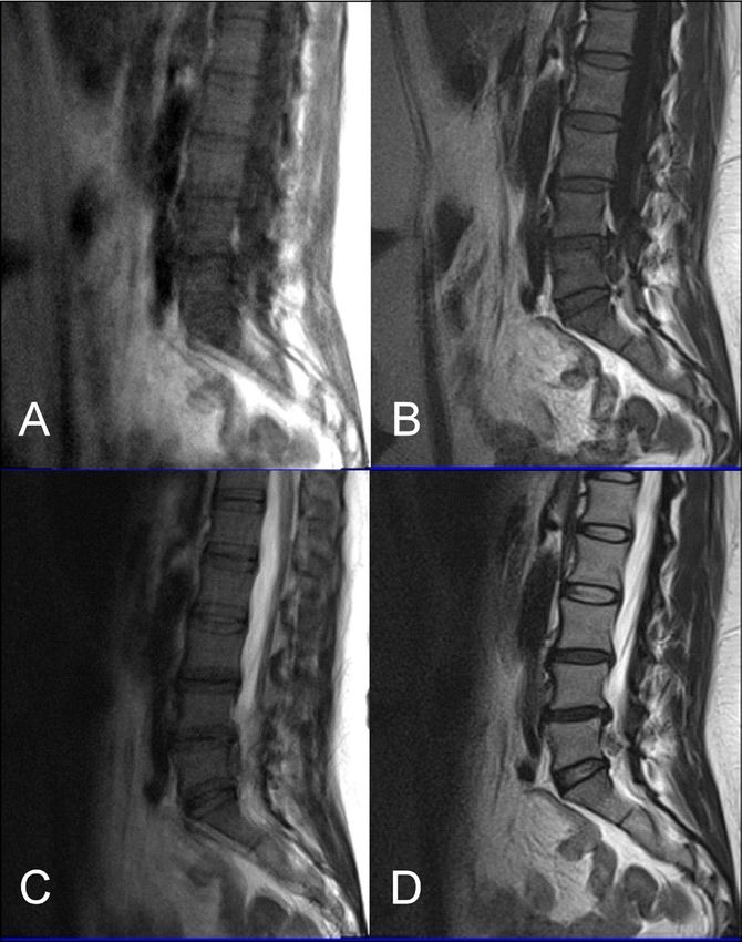
Citation: Lavdas E, Giankou E, Papanikolaou P, Tsikrika A, Papaioannou M, Roka V, et al. Comparison of T1 FLAIR BLADE with and without parallel
imaging against T1 turbo spin echo in the MR imaging of lumbar spine in the sagittal plane. J Radiat Res Imaging. 2021; 1(1):33-40.

had statistically significant difference in the imaging of bone marrow         FLAIR BLADE provided better contrast for bone marrow, spinal
(p
Citation: Lavdas E, Giankou E, Papanikolaou P, Tsikrika A, Papaioannou M, Roka V, et al. Comparison of T1 FLAIR BLADE with and without parallel
imaging against T1 turbo spin echo in the MR imaging of lumbar spine in the sagittal plane. J Radiat Res Imaging. 2021; 1(1):33-40.
Figure 2: (A) T1 FLAIR BLADE, (B) T1 TSE. The results obtained from the quantitative analysis showed that the FLAIR BLADE sequences have better
SNR in most tissues. T1 FLAIR BLADE sequence achieved better CSF nulling and better contrast between spongy bone and intervertebral disc. The
contrast was also better when comparing spongy bone and intervertebral disc with CSF.
The results obtained from the quantitative analysis of this study al., has also demonstrated the superiority in image contrast of T1W-
showed that the SNR and CNR results were significantly greater FLAIR images showing that they achieve better lesion to background
for T1W-FLAIR BLADE. More specifically, T1W-FLAIR BLADE and grey to white matter CNR [19]. The inherent restrictions of
sequence achieved better CSF nulling and better contrast between parallel imaging, i.e., reduction of SNR and increased possibility for
spongy bone and intervertebral disc as well as between spongy bone reconstruction artifacts, were confirmed in our findings where the
and intervertebral disc with CSF (Figure 2). A study by Alibek et images without parallel imaging had better SNR (Figure 3).
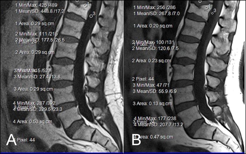
Figure 3: (A) T1 FLAIR BLADE without parallel imaging, (B) T1 FLAIR BLADE with parallel imaging. T1 FLAIR BLADE images without parallel imaging
were found to have better quality than the T1 FLAIR BLADE images with parallel imaging. Although they both eliminated motion artifacts, the images
without parallel imaging have better SNR as well as better contrast between tissues.
J Radiat Res Imaging 2021; 1(1):33-40. 37Citation: Lavdas E, Giankou E, Papanikolaou P, Tsikrika A, Papaioannou M, Roka V, et al. Comparison of T1 FLAIR BLADE with and without parallel
imaging against T1 turbo spin echo in the MR imaging of lumbar spine in the sagittal plane. J Radiat Res Imaging. 2021; 1(1):33-40.
As stated earlier, sagittal T1 weighted images play an integral or metastatic lesions. Both the quantitative and qualitative analysis
role in the MR imaging of the L-spine. One of the reasons is that showed that T1W-FLAIR BLADE achieved better results than
they provide great information about anatomical structures. In order T1W-TSE. In addition to that, T1W-FLAIR BLADE images
to assess the importance of the BLADE sequence compared to a without parallel imaging were found to have better quality than
TSE, both a qualitative and quantitative analysis were performed the T1W-FLAIR BLADE images with parallel imaging. Although
by evaluating contrast in between anatomical structures and the they both eliminated motion artifacts, the images without parallel
SNR, CNR and ReCon measurement respectively [18]. Although imaging have better SNR as well as better contrast between tissues
T1W-TSE is a faster sequence, T1W-FLAIR BLADE scored (Figure 3). Moreover, a study showed that T1W-FLAIR images can
better in quantitative and qualitative analyses. Pathologies and successfully eliminate chemical shift artifacts compared to T1W-TSE
abnormalities were better visualized in the latter one. Furthermore, due to greater receiver bandwidth [8]. This statement was confirmed
in T1W-FLAIR BLADE there was improved image contrast as well by our findings (Figure 4).
as greater depiction of anatomical structures and either degeneration
Figure 4: (A) T1 FLAIR BLADE, (B) T1 FLAIR BLADE with parallel imaging, (C) T2 TSE, (D) T2 BLADE TSE, (E) T1 TSE. T1 FLAIR BLADE achieves better CSF
nulling, better elimination of motion artifacts as well as better contrast between tissues. In T1 FLAIR BLADE there is also less chemical shift due to the
fact that a greater bandwidth (1) is used. T2 BLADE appears to have better image quality compared to T2 TSE.
J Radiat Res Imaging 2021; 1(1):33-40. 38Citation: Lavdas E, Giankou E, Papanikolaou P, Tsikrika A, Papaioannou M, Roka V, et al. Comparison of T1 FLAIR BLADE with and without parallel
imaging against T1 turbo spin echo in the MR imaging of lumbar spine in the sagittal plane. J Radiat Res Imaging. 2021; 1(1):33-40.
T1W-FLAIR BLADE also had higher values in all the relative sequence is a very useful tool to distinguish cysts within the spinal
contrast measurements. T1W-FLAIR BLADE showed better results cord, tumors and demyelinating disease. Another study showed that
when comparing CSF with spongy bone, CSF with intervertebral T1W-FLAIR images had also a higher CNR over T1W-TSE images
disc and intervertebral disc with spongy bone. These results in 16 metastatic lesions in the vertebrae, which helped diagnose the
from the quantitative analysis meant better conspicuity between extent of the lesion in the spinal cord. Moreover, the findings of
the following anatomical structures: intervertebral disc-CSF, this study are in line with the results of other studies, which show
intervertebral disc-spinal cord, vertebral body-CSF and spinal cord- that the T1W-FLAIR sequence could be more beneficial for L-spine
CSF. Both radiologists found that T1W-FLAIR images achieved imaging than T1W-TSE, due to CSF nulling, higher image contrast
better CSF nulling compared to T1W-TSE. It was also found that and definition of anatomical structures, degeneration, or metastatic
T1W-FLAIR BLADE was significantly superior to T1W-TSE lesions (Figure 5) [11]. The results of our study also support the
when reviewing the contrast at the spinal cord and cauda equina above, i.e., T1W-FLAIR BLADE being superior to T1W-TSE for
interface, demonstrating substantial inter-observer agreement. Due the aforementioned reasons.
to CSF nulling that is achieved in FLAIR sequences, T1W-FLAIR
Figure 5: (A) T1 FLAIR BLADE, (B) T1 FLAIR BLADE with parallel imaging, (C) T1 TSE. T1 FLAIR BLADE shows better image contrast. In images A and B,
a lesion is depicted behind L3-L5 which is not depicted in the TSE in image C. In T1 FLAIR BLADE we can achieve better CSF nulling, and also better
lesion delineation. The lesion is better visualized in the image with parallel imaging as parallel imaging eliminates artifacts.
J Radiat Res Imaging 2021; 1(1):33-40. 39Citation: Lavdas E, Giankou E, Papanikolaou P, Tsikrika A, Papaioannou M, Roka V, et al. Comparison of T1 FLAIR BLADE with and without parallel
imaging against T1 turbo spin echo in the MR imaging of lumbar spine in the sagittal plane. J Radiat Res Imaging. 2021; 1(1):33-40.
An interesting implementation of T1W-FLAIR BLADE images 8. Westbrook C, Roth CK, Talbot J. MRI in Practice Fourth Edition.
would be in 3T MRI. Theoretically, 3T MRI is expected to have an Wiley-Blackwell, A John Wiley & Sons, Ltd., Publication, 2011.
SNR that is twice as good as the SNR received at 1.5T. However, 9. Lavdas E, Mavroidis P, Vassiou K, Roka V, Fezoulidis IV, Vlychou
there is a slight increase in susceptibility and chemical shift artifacts M. Elimination of chemical shift artifacts of thoracic spine with
that T1W-FLAIR BLADE has been shown to eliminate successfully contrast-enhanced FLAIR imaging with fat suppression at 3.0 T.
[9,11]. Another issue that T1W-FLAIR BLADE could help Magnetic Resonance Imaging. 2010 Dec 1;28(10):1535-40.
overcome in 3T MRIs is the nulling of CSF. At 3T MRI relaxation
10. Hirokawa Y, Isoda H, Maetani YS, Arizono S, Shimada K, Togashi
times are increased so the contrast for T1 images is reduced. This K. Evaluation of motion correction effect and image quality with
may result in CSF appearing less dark which T1W-FLAIR BLADE the periodically rotated overlapping parallel lines with enhanced
could help resolve effectively as it achieves great CSF nulling [5]. reconstruction (PROPELLER)(BLADE) and parallel imaging
acquisition technique in the upper abdomen. Journal of Magnetic
To conclude, T1W-FLAIR BLADE without parallel imaging
Resonance Imaging: An Official Journal of the International
achieved the highest scores from all the sequences. T1W-FLAIR Society for Magnetic Resonance in Medicine. 2008 Oct;28(4):957-
BLADE with parallel imaging was the second-best sequence in our 62.
study with T1 TSE following. T1W-FLAIR BLADE was found to
have better CSF nulling and better SNR measurements for the spinal 11. Forbes KP, Pipe JG, Bird CR, Heiserman JE. PROPELLER MRI: clinical
cord. T1W-FLAIR BLADE also had better CNR measurements for testing of a novel technique for quantification and compensation
of head motion. Journal of Magnetic Resonance Imaging:
spinal cord-CSF with better imaging of disc-cord and cord-CSF
An Official Journal of the International Society for Magnetic
interfaces. Moreover, it showed better overall image quality than Resonance in Medicine. 2001 Sep;14(3):215-22.
T1W-TSE and greater reduction of artifacts. T1W-FLAIR BLADE
with or without parallel imaging shows great potential for 3T 12. Hennig J, Nauerth A, Friedburg HR. RARE imaging: a fast imaging
MRI as it reduces susceptibility and chemical shift errors as well as method for clinical MR. Magnetic Resonance in Medicine. 1986
improved CSF nulling. Last but not least, T1W-FLAIR BLADE was Dec;3(6):823-33.
the preferred choice of the clinicians for the imaging of the L-spine. 13. Walker MT, Partovi S, Karis JP, Fram FK. Fast, versatile, and cost-
effective FSE MR imaging: technical considerations and clinical
Ethical Approval applications. Barrow Quarterly. 2000;16:1-5.
All procedures performed in studies involving human 14. Pipe JG. Motion correction with PROPELLER MRI: application
participants were in accordance with the ethical standards of the to head motion and free‐breathing cardiac imaging. Magnetic
institutional and/or national research committee and with the 1964 Resonance in Medicine: An Official Journal of the International
Helsinki declaration and its later amendments or comparable ethical Society for Magnetic Resonance in Medicine. 1999 Nov;42(5):963-
standards. 9.
Conflict of Interest 15. Deshmane A, Gulani V, Griswold MA, Seiberlich N. Parallel
MR imaging. Journal of Magnetic Resonance Imaging. 2012
The authors declare that they have no conflict of interest. Jul;36(1):55-72.
References 16. Fellner C, Menzel C, Fellner FA, Ginthoer C, Zorger N, Schreyer A,
et al. BLADE in sagittal T2-weighted MR imaging of the cervical
1. British Association of Spine Surgeons. The Spine and MRI imaging. spine. American Journal of Neuroradiology. 2010 Apr 1;31(4):674-
[Online] 2018. 81.
2. Radiology Info. [Online] Radiological Society of North America, 17. Bushberg JT, Boone JM. The essential physics of medical imaging.
Inc. (RSNA). Lippincott Williams & Wilkins; 2011 Dec 20.
3. Wilmink JT. Lumbar spinal imaging in radicular pain and related 18. Glockner JF, Hu HH, Stanley DW, Angelos L, King K. Parallel MR
conditions: understanding diagnostic images in a clinical context. imaging: a user’s guide. Radiographics. 2005 Sep;25(5):1279-97.
Springer Science & Business Media; 2010 Feb 8.
19. Alibek S, Adamietz B, Cavallaro A, Stemmer A, Anders K, Kramer M,
4. Melhem ER, Israel DA, Eustace S, Jara H. MR of the spine with a et al. Contrast-enhanced T1-weighted fluid-attenuated inversion-
fast T1-weighted fluid-attenuated inversion recovery sequence. recovery BLADE magnetic resonance imaging of the brain: an
American Journal of Neuroradiology. 1997 Mar 1;18(3):447-54. alternative to spin-echo technique for detection of brain lesions
in the unsedated pediatric patient? Academic Radiology. 2008
5. Lavdas E, Vlychou M, Arikidis N, Kapsalaki E, Roka V, Fezoulidis IV.
Aug 1;15(8):986-95.
Comparison of T1-weighted fast spin-echo and T1-weighted fluid-
attenuated inversion recovery images of the lumbar spine at 3.0
Tesla. Acta Radiologica. 2010 Jan 1;51(3):290-5.
6. Mavroidis P, Giankou E, Tsikrika A, Kapsalaki E, Chatzigeorgiou V,
Batsikas G, et al. Brain imaging: comparison of T1W FLAIR BLADE
with conventional T1W SE. Magnetic Resonance Imaging. 2017
Apr 1;37:234-42.
7. Lavdas E, Mavroidis P, Kostopoulos S, Glotsos D, Roka V, Koutsiaris
AG, et al. Elimination of motion, pulsatile flow and cross-talk
artifacts using blade sequences in lumbar spine MR imaging.
Magnetic Resonance Imaging. 2013 Jul 1;31(6):882-90.
J Radiat Res Imaging 2021; 1(1):33-40. 40You can also read