Comparison of Two Hair Removal Methods in Sprague-Dawley Rats (Rattus norvegicus)
←
→
Page content transcription
If your browser does not render page correctly, please read the page content below
Journal of the American Association for Laboratory Animal Science                                                                           Vol 60, No 2
Copyright 2021                                                                                                                              March 2021
by the American Association for Laboratory Animal Science                                                                                 Pages 213–220

      Comparison of Two Hair Removal Methods in
        Sprague–Dawley Rats (Rattus norvegicus)
               Nicole L Rowley,1,* Elliot Ramos-Rivera,1 Sorana Raiciulescu,2 Sang H Lee,1 and Amanda C Christy1

   Rats commonly undergo surgery for research purposes. However, the effects of different methods of hair removal on
wound healing and surgical site infections (SSI) in rats has not been evaluated. The current study evaluated 2 hair removal
methods, clipping with an electric clipper and using a depilatory agent, and their effect on wound healing and SSI. Swabs
for bacterial culture were obtained on Day 0 just after hair removal, after aseptic skin preparation, and on Days 1 and 3
before conducting skin biopsies to assess bacterial load and recolonization. Full-thickness punch biopsies were taken for
histopathologic evaluation on Days 0, 1, 3, 7, and 10. The surgical incisions were assigned an ASEPSIS score on Days 1 and
3. The data revealed that the bacterial load was significantly higher with the depilatory method as compared with the clipper
method, but only on Day 1. The histopathologic evaluation found no significant difference in wound healing between the
2 methods. Although the ASEPSIS score was significantly higher for the clipping method than for the depilatory method
on Day 1, both techniques were equivalent by Day 3. We conclude that both hair removal methods are safe and efficacious
components of aseptic technique in rats.

Abbreviations: Buprenorphine-SR, Buprenorphine-Sustained Release; SSI, Surgical Site Infections

DOI: 10.30802/AALAS-JAALAS-20-000108

   Sprague–Dawley rats are often used in surgical procedures                        a sensitivity reaction.1,17,22,24 In mice, hair removal with either
for biomedical research and training. The Guide for the Care and                    clipping or depilatory agent resulted in acceptable healing.25
Use of Laboratory Animals20 requires the use of aseptic technique                   In Wistar rats, the use of a depilatory agent did not affect the
when performing survival surgery on any species. One purpose                        healing of a dorsal flap.3 Despite these findings, no studies have
of aseptic technique is to reduce or eliminate the bacterial load                   compared the effects of clipping and a depilatory agent on the
on the animal prior to the start of surgery to prevent the intro-                   prevalence of SSI and on wound healing in rats.
duction of bacteria into the sterile surface below the skin.2,30,42                    Our facility's standard practice is to remove hair with clip-
Insufficient or inappropriate skin preparation may result in sur-                   pers using a no. 40 blade. However, this approach leaves a short
gical site infections (SSI). SSI can delay or compromise wound                      stubble of approximately 1 mm.22 For surgical procedures that
healing.1,34 Aseptic technique requires the preparation of the                      require a smooth skin, using a depilatory agent appears rea-
surgical site on the animal by removing the hair such that skin                     sonable; however, little information is available on the effects
damage, abrasions or other dermal injuries are avoided, fol-                        of a depilatory agent on rat skin, SSI, and wound healing. We
lowed by cleaning the skin with topical antiseptic compound.6,30                    hypothesize that using a depilatory agent as a hair removal
   Traditionally, hair is removed from surgical sites because it                    method in rats will reduce bacterial counts, dermal trauma,
harbors bacteria and prevents thorough cleansing of the inci-                       and SSI as compared with using clippers.
sion site. Hair removal also facilitates visibility of the surgery
site and removes a potential foreign-body that may result in                                         Materials and Methods
SSI.10,22,43 The 3 most common hair removal methods are shav-                         Animals. All procedures were approved by the Uniformed
ing with a razor, clipping the hair with an electric razor, and                     Services University IACUC and were performed in accordance
using a depilatory agent. In human patients, recommendations                        with the Animal Welfare Regulations4 and the Guide for the Care
are that the hair not be removed unless visualization is needed                     and Use of Laboratory Animals.20 Uniformed Services University
or the hair would interfere with the surgical site or postsurgi-                    vivarium is an AAALAC International accredited facility. All
cal bandaging.5,43 If hair removal is necessary for humans, the                     rats were included in a routine health surveillance program and
recommendation is to use either clippers or a depilatory agent.                     were negative for all pathogens that were excluded from the rat
Using a razor has been shown to traumatize the skin, resulting in                   colony in the facility: rat parvoviruses (RPV, KRV, H-1, RMV, and
higher rates of SSI.22,28,35,36 Some research studies indicate that a               NS-1), rat theilovirus, sialodacryoadenitis virus, Pneumocystis
depilatory agent is a better method, as it is efficient, atraumatic,                carinii, Sendai virus, reovirus, lymphocytic choriomeningitis
and safe to use on or around wounds. However, it can cause a                        virus, cilia-associated respiratory bacillus, pneumonia virus
transitory lymphocytic reaction, and some individuals may have                      of mice, Mycoplasma pulmonis, adenovirus (MAV), Salmonella,
                                                                                    Helicobacter, Giardia, Pasteurella, Streptococcus, pinworms, Spiro-
Received: 17 Jul 2020. Revision requested: 14 Aug 2020. Accepted: 22 Sep 2020.
                                                                                    nucleus, and fur mites.
1Department of Laboratory Animal Resources and 2Department of Preventive Medicine     Male Sprague–Dawley rats (Rattus norvegicus) (n = 33), with
and Biostatistics, Uniformed Services University, Bethesda, Maryland                ages ranging from 4.5 mo up to 23 mo and a weight range from
   *Corresponding author. Email: Nicole.Rowley@usuhs.edu

                                                                                                                                                   213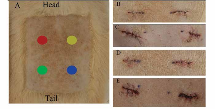
Vol 60, No 2
Journal of the American Association for Laboratory Animal Science
March 2021

488 g to 955 g. The rats were transferred from another IACUC         from the direction of growth, the depilatory agent was applied
approved protocol in which they had not participated in any          until the entire selected area was covered. The depilatory agent
experimental procedures. They were then assigned to 1 of 2           remained on the skin for 3 min, at which time a test section was
experimental groups: clipping (Arco, Wahl Clipper, Sterling, IL)     gently cleared with a gauze covered gloved finger or tongue
or depilatory agent (Nair Hair Removal Lotion Softening Baby         depressor. If the hair came off easily, the rest of the depilatory
Oil, Ewing, NJ). Half of each of these 2 experimental groups         agent was removed. If not, the depilatory agent was reapplied
were then assigned to be used in either the first or second cohort   in that section and another test was conducted 2 min later. All
of the experiment. Rats were pair-housed until the initial sur-      depilatory agent was removed no more than 10 min after the first
gery date and then individually housed for the duration of the       application. Once removed, the area was generously cleansed
study. Housing consisted of a static polycarbonate shoebox-type      with sterile water and gauze to ensure complete removal of the
cage with a filter top (Allentown, Allentown, NJ) and rodent         depilatory agent.
hardwood bedding (catalog number 7090M, Laboratory-Grade                Bacterial Sampling, Culture, and Identification. Bacterial sam-
Teklad Maple SaniChips, Harlan Teklad, Madison, WI). After           ples were taken to determine the antimicrobial activity of each
the surgery, the bedding was switched to Shepard Specialty           of the hair removal methods on the skin. On Day 0, samples
Paper (catalog no. SHE1009, Alpha-dri, Watertown, TN). While         were collected at 2 time points: once the skin had dried after
housed on the Shepard Specialty Paper, cage changes occurred         the hair removal, and after the skin was aseptically prepped.
twice weekly. Enrichment was provided daily in the form Fat Rat      On Days 1 and 3, the samples were collected prior to the skin
Huts (high-temperature polycarbonate, Bio-Serv, Flemington,          being aseptically prepped for that day’s biopsy. Samples were
NJ), nylabones (pure virgin nylon, Bio-Serv, Flemington, NJ) and     collected by defining a 1 × 1 in. area within the region from
cotton squares (iso-PAD: The Ultra Enrichment Media, Omni            which the hair had been removed. Starting in the center of the
BioResources, Cherry Hill, NJ). The rats were fed a pelleted ro-     area and rolling the swab (BD BBL Culture Swab Plus, Item
dent food (Envigo Teklad Global 18% protein T2018.15 Rodent          220118, Copan Italia, Brescia, Italy), it was moved in a spiraling
Diet) and filtered domestic water ad libitum. The room was kept      outward motion toward the outer edges, so as to not to exceed
on a 12:12-h light:dark cycle (lights on, 0600; lights off, 1800)    the defined 1 × 1 in. area.
with temperature maintained between 68 °F and 79 °F (20 °C              The samples were submitted to IDEXX BioAnalytics for
to 26 °C). The relative humidity was maintained at 30% to 70%.       aerobic culture, bacterial identification, and colony counts.
   Anesthetic Procedure. Our initial plan was to use injectable      An aliquot of approximately 1 mL of the BD Eswabs (BD Cat#
anesthetics. However, rats anesthetized with an intraperitoneal      220245) transport medium was vortexed, and a sterile calibrated
injection of ketamine hydrochloride (75 mg/kg; Ketathesia,           10 µL loop (Fisherbrand 220363 to 600) was used to inoculate 10
Henry Schein, Dublin, OH) and xylazine (7.5 mg/kg; AnaSed            µL of the liquid onto BBL Trypticase Soy Agar with 5% sheep
Injection, AKORN Animal Health, Lake Forest, IL) on Days             blood (TSA II; Becton Dickinson). Sample plates were incubated
0 and 1 of the study had prolonged recovery, with a few rats         at 35 °C (95 °F) and 7% CO2 for 48 h. Colonies of each colony
requiring supplemental oxygen and many needing reversal              type were counted up to 350 CFU/10 µL; counts greater than
with atipamezole hydrochloride (0.5 mg/kg subcutaneously;            350 were reported simply as “greater than 350 CFU/10 µL”. The
Antisedan, Zoetis, Orion Corporation, Espoo, Finland) to assist      counts were then binned into 4 categories: None = 0; Low =1 to
in the recovery. The next day these rats were still very sedated,    49; Medium = 50 to 200 and High = greater than 200. Identifica-
perhaps due to their age, and yet they required anesthesia           tion of the bacteria was conducted as previously reported.29
for experimental use on that day. We decided that switching             Surgical Technique. All rats were maintained on a surgical
to inhalational anesthesia as safer for the rats. Therefore, all     plane of anesthesia as described above. For each anesthetic
subsequent anesthetic events were conducted using inhaled            event, rats received subcutaneous fluids at 100 mL/kg/day after
isoflurane (Isothesia, Henry Schein, Dublin, OH) administered        anesthesia to maintain hydration. The experimental procedures
with a vaporizer. For the inhaled anesthesia, induction was          were performed in two equivalent cohorts. The first cohort
performed using 5% isoflurane with 100% oxygen. Once loss of         received Buprenorphine-Sustained Release (SR) Laboratory
pedal reflex occurred, the rats were maintained via a nose cone      (ZooPharm Ve terinary Compounding Pharmacy, Laramie,
at between 1% to 3% isoflurane at an appropriate anesthetic          WY) subcutaneously at 1.0 mg/kg on Day 0. Rats also received
depth, as monitored with toe pinch and respiratory rate. Full        Meloxicam-SR (ZooPharm Veterinary Compounding Pharmacy,
recovery occurred as expected. All rats received thermal support     Laramie, WY) subcutaneously at 4.0 mg/kg for 3 d alternated
during anesthesia (HotDog Patient Warming, Eden Prairie, MN).        with acetaminophen (Children’s Mapap Acetaminophen Liquid
   Skin Preparation. After confirmation of appropriate anesthetic    (160 mg/5 mL), Major Pharmaceuticals, Livonia, MI) in 2 oz
depth, hair was removed using 1 of 2 methods (clipping or            gel cups (MediGel Sucralose, Clear H2O, Portland, ME) for 24
depilatory agent) from the thoracolumbar region in an approxi-       h administered as previously described.31
mately 3 in. by 2 in. rectangular shape. For the clipping method,       The surgical technique consisted of taking biopsies. The skin
the clippers were disinfected (Oster Professional Products,          was aseptically prepped with BD ChloraPrep One-Step (2%
Spray Disinfectant, no.76300 to 102 part 110969, McMinnville,        s/v Chlorhexidine gluconate and 70% v/v Isopropyl alcohol,
TN) before and after each use. The blade was placed parallel         Becton, Dickinson and Company, El Paso, TX) according to
to the skin and advanced in the direction of the hair growth. A      product directions. The surgeon prepped aseptically, with
second pass clipping against the hair growth was performed to        hairnet, face mask, and sterile surgical gloves. A sterile drape
ensure a close clip. The area was wiped down with sterile water      was placed over the rat. On Day 0, 4 full-thickness skin punch
(Sterile 0.9% Normal Saline, USP, 100 mL, Mundelein, IL) and         biopsies, identified as sites A, B, C, and D, were taken from the
gauze to remove any loose hair. For the depilatory method, a         hairless region of each rat (clipped or depilated) with an 8 mm
gloved hand or tongue depressor was used to part the hair and        biopsy punch (Robbin Instruments, Chatham, NJ). The punch
expose the base of the hair shaft so that the product could be       biopsies were taken at the outside corners of the 1 inch × 1 in.
applied at the base of the hair shaft. Then, in a circular motion    area defined for the bacterial sampling. Hemostasis was man-
that moved the hair shaft from the direction of growth to away       aged with sterile gauze. The open wounds were closed using 5-0

214
Assessment of two hair removal methods in Sprague–Dawley rats
Monocryl RB-1 (Ethicon, Guaynabo, PR) in a simple interrupted statistics software (version 25, IBM North America, NY, NY).
pattern. After the initial 4 biopsies on each rat, another biopsy All continuous results are presented as mean ± SEM.
was performed at the same sites (sites A, B, C or D on days 1,
3, 7, and 10, respectively) using a 10 mm biopsy punch (Robbin Results
Instruments, Chatham, NJ). Prior to the biopsy, the target site Interassessor agreement regarding ASEPSIS wound score. The
was aseptically prepped with ChloraPrep Single Swabstick (2% Kendall coefficient of concordance for the assessor’s ASEPSIS
s/v Chlorhexidine gluconate and 70% v/v Isopropyl alcohol, wound scores were 0.74 for Day 1 and 0.66 for Day 3 for the clip-
Becton, Dickinson and Company, El Paso, TX) according to ping hair removal method, indicating moderate to substantial
product directions. agreement. For the depilatory methods, concordance was 0.35
Histopathologic Evaluation. To microscopically examine the on Day 1 and 0.39 on Day 3, weak to moderate agreement. In
surgical sites for bacterial contamination and wound (biopsy) addition, assessor scores showed significant concurrence (P <
healing, the punch biopsies were collected into cassettes and 0.001) for both clipping and the depilatory method Day 1 (P =
placed in 10% neutral buffered formalin, routinely processed, 0.04) and Day 3 (P = 0.02).
paraffin-embedded, sectioned at 5µm, and stained with he- ASEPSIS Score. The ASEPSIS Day 1 mean score for the clip-
matoxylin and eosin. The stained slides were evaluated by ping method was 2.1 ± 0.4, which significantly (P = 0.03) higher
a veterinary pathologist who was blind to the hair removal than the mean of the depilatory method (0.48 ± 0.54). The Day 3
method. Histopathology scoring was performed at both low- ASEPSIS mean scores between methods were not significantly
power (magnification, 100×) and high power (magnification, different, although the Day 3 mean for the depilatory method
200×) by using methods similar those previously published.26 (2.1 ± 0.5) was significantly (P = 0.01) higher than the Day 1 mean
The assessed criteria were dermal inflammation, follicular (0.48 ± 0.54). The clipping method had no significant change
changes, fibroplasia, and epidermal hyperplasia. Each criterion from Day 1 (2.1 ± 0.4) to Day 3 (2.5 ± 0.4) (Table 1).
was assigned a score ranging from 0 (absent) to 3 (robust). The Histopathology Evaluation. The clipping method score in-
scores were summed for each sample to obtain a cumulative creased at each time point from 1.5 ± 0.2 on Day 0 to reach the
histopathology lesion score, with a maximum possible score of highest score of 8.7 ± 0.3 on Day 10. With the depilatory method,
12. Images of stained slides were obtained at 200× magnification the highest score was on Day 3 (8.4 ± 0.2) and fell through Day
using a microscope (model BX41, Olympus) equipped with a 10, but none of the differences were significant. No significant
digital camera (model DP22, Olympus) by using digital imag- difference existed between the 2 methods on any day. The mean
ing software (cellSens Standard, Olympus Life Science Imaging scores for each method had a significant (P < 0.001) increase on
Software). Representative images are presented in Figure 1. Days 1, 3, 7, and 10 as compared with Day 0.
ASEPSIS Evaluation. The initial 4 biopsy sites were photo- Bacterial Assessment. The bacterial load across the 2 methods
graphed (Canon EOS Rebel T1i) on Day 0, Day 1 and Day 3. was statistically different only on Day 1, when the bacterial
On Day 0, the sites were photographed after all 4 punch biopsy growth for the depilatory method was Low (67%, or 10 of 15
sites were closed. This photograph was used as a baseline of samples) compared with the clipping method, which was None
the appearance of the incision immediately after closure. It was (83%, or 15 of 18 samples) (P = 0.002). Both methods remained
not evaluated as healing, and infection would not be evident at in either the None or Low growth category, except for the
that early time point. Day 1 and Day 3 photographs were edited depilatory method on Day 3 in which 13%, or 2 of 15 samples
to select only for sites C and D for scoring using the ASEPSIS had Medium growth (Table 2). The bacteria isolated prior to
evaluation. All photographs were evaluated by 5 assessors who the aseptic preparation step represent 8 genera: Aerococcus spp.,
were blind to the hair removal group. Assessors used a modified- Bacillus spp., Corynebacterium spp., Enterobacter spp., Enteroccocus
ASEPSIS wound chart similar to that described previously.41 spp., Klebsiella spp., Proteus spp., and Staphylococcus spp. (Table 3).
Scores were assigned for 4 criteria: serous exudate, erythema, The most frequent organisms were the Staphylococcus spp. at
purulent exudate and separation of deep tissue. The scores were 53% (30 out of 57) and Aerococcus spp. at 25% (14 out of 57). The
summed to arrive at a total wound score with a maximal possible other 6 genera ranged from 2% to 7% (1 to 4 out of 57) of the
score of 22. Total scores of 0 to 3 indicate satisfactory healing; 4 to total bacteria found. After the antiseptic step, the only genera
6 indicated disturbed healing; 7 to 12 indicate mild to moderate still present were the Staphylococcus spp. at 5% (3 of 57) and
infection; and scores greater than 13 indicated severe infection. Aerococcus spp. at 2% (1 out of 57). All 4 isolates were found in
The average ASEPSIS wound score for each treatment condition the depilatory method.
was calculated and used for the statistical analysis (Figure 2).
Statistical analysis. ASEPSIS wound scoring was evaluated
among the 5 assessors using Kendall coefficient of concordance. Discussion
Agreement was assessed for each day and each method. Using Guidance on rodent asepsis states that the hair should be
the median wound score from each animal, a linear mixed model removed, and the skin prepped with an antiseptic solution.6,30
for repeated measures assessed the main effects of day, hair re- Recently, research on skin preparations for surgical procedures
moval method, and the interaction of the 2. For histopathology, in mice investigated aseptic preparations and their impact on SSI
a similar mixed model approach was used. Model means for and healing.12,18,25 The impact of hair removal method has not
both ASEPSIS and histopathology scores are reported as age- been assessed in rats. The goal of this study was to determine
adjusted, given the significance of age in both models. Pairwise whether the hair removal method affected wound healing and
comparisons evaluating the interactions from both models were the development of SSI. The results of this study demonstrate
adjusted using the Sidak method. Bacterial load for each animal that either method of hair removal is appropriate, with no
was categorized from 0 to 3, 0 representing no growth, and 3 differential effect on the development of SSI and satisfactory
representing heavy growth. Associations between hair removal healing by Day 10.
method and bacterial load grouping were assessed on each day In humans, recommended practice is to not remove the
using a χ2 test. Unless otherwise stated, Type I error is controlled hair. However, in rodents, hair removal is necessary to ensure
at 5%, with all tests 2-sided. Analysis was conducted in SPSS visualization of the surgical site and is considered a mandatory
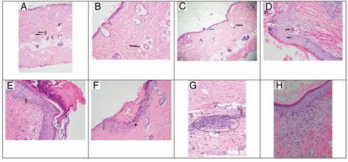
215Vol 60, No 2 Journal of the American Association for Laboratory Animal Science March 2021 Figure 1. Dermal pathology on Days 0, 3, and 10. (A) Normal full thickness intact skin with the black arrows indicating normal hair follicles (40× magnification). (B) Day 0 Depilatory method with the arrow indicating a normal hair follicle with central hair shaft. The asterisks (*) indicate hair follicles that are moderately dilated with hair shaft fragments, flattened follicular epithelium, and compressed sebaceous glands (10× magnifica- tion). (C) Day 0 Clipper method with the arrow indicating a markedly dilated hair follicle void of the hair shaft and a compressed sebaceous gland. The blue arrow indicates a moderately dilated hair follicle with no hair shaft (40× magnification). (D) Day 3 Clipper method shows mildly transmural thickened epidermis (blue line) and the arrow shows single cell necrosis (20× magnification). (E) Day 3 Depilatory method shows a moderate transmural thickened epidermis (blue line) with the arrow showing cellular bridging (edema). A thick mat of serocellular debris is seen covering the epidermis (20× magnification). (F) Day 3 Clipper method with the white arrows indicating a demarcation for a discontinuous epidermis; black astrick indicating inflammatory cellular infiltration covered by a mat of serocellular material (10× magnification). (G) Day 3 Depilatory method with a large aggregate of inflammatory cells within the deep dermis marked by the oval; the black arrow indicates fibrosis (20× magnification). Image H: Day 10 Clipper method showing an area of organized fibrosis with fibroblasts stacked in a linear fashion subjacent to the epidermis (20× magnification). component of asepsis.10,35 The 3 common methods of remov- 4 gauze cloth. Despite the attention to gentleness, this may still ing hair are a razor blade, an electric clipper and a depilatory have been too harsh a removal method given the skin's reaction agent. This study originally planned on assessing all 3 methods. to the depilatory agent. Using a soft gauze with a higher weave However, 2 different single-blade razors, BIC Sensitive Shaver may protect the skin better. disposable and Gallant Disposable Prep Razor (Process-Con- One reason for hair removal is to reduce bacterial load.30 Both struction AB, Sweden), could not cut the rat hair such that a clear hair removal methods had no or low bacterial growth after the patch of skin was visible. We therefore only assessed clippers antiseptic step. Because the depilatory agent is a chemical, it and depilatory agent. potentially could have antimicrobial properties. In one study,25 Both clippers and the depilatory agent are effective methods mice treated with a depilatory agent had less bacterial growth as of removing hair in rats. In contrast to a previous study17 in compared with clipping. However, in our study, the depilatory- which the clipper caused widespread nicking and the depilatory treated rats had a bacterial load of 2 [Low] of 15, while the agent appeared to cause no damage, our study found no appar- clipper-treated rats had a load of 0 [None] of 18; this difference ent damage due to the clippers, while 10 of the 15 rats given the was not significant. As time progressed and bacteria began to depilatory agent developed a mild to moderate sensitivity reac- recolonize the skin, the bacterial load of the depilatory-treated tion (small nonerythemic bumps and petechiae of the skin). All rats remained higher than the clipper treated rats. Because all sensitivity reactions were healed by Day 10 without requiring wounds healed well, with no signs of infection, the method treatment. While 9 of the rats developed sensitivity reactions of hair removal is likely not clinically relevant. However, our in less than 24 h, one rat did not develop a reaction until Day data indicate that a depilatory agent should not be considered 3 after the original application. The total contact time did not to reduce bacterial load. exceed the product directions of 10 min, making the number Staphylococcus spp. was the most commonly isolated bacte- of sensitivity reactions unexpected. The hair was not trimmed ria. This is not an unexpected finding, as Staphylococcus spp. prior to the application of the depilatory agent, which may have is ubiquitous in the environment and is known as a common, contributed to the length of time the depilatory agent was in commensal bacteria in both humans and mice.25,27,37 Character- contact with the skin. Clipping the hair before using the depila- izing the microflora of the rat skin was not a goal of this study; tory agent may decrease contact time and decrease sensitivity samples were taken prior to the aseptic step only to ensure that reactions. Another possibility for the unexpected sensitivity known pathogens were not present. A potential limitation of reaction may be the way the depilatory agent was removed from our bacterial characterization is the possibility of not capturing the rat's skin. A standard 4 × 4 gauze cloth or tongue depressor all bacteria and the potential of one species to outgrow another was used to gently remove the chemically treated hair, and then while plated. We may have missed bacteria by using a gentle the area was generously rinsed and wiped with a standard 4 × rolling technique rather than a swabbing technique. Dilution of 216
Assessment of two hair removal methods in Sprague–Dawley rats

Figure 2. Photographs of surgical sites on Days 0, 1 and 3. (A) Representative of hair removal site (clipper method) with rat orientation noted.
Each colored circle represents a punch biopsy site: A (red circle), B (yellow circle), C (green circle), D (blue circle). After Day 0, in which all 4 sites
were initially created, the healing punch biopsy site was removed on Days 1, 3, 7 and 10 respectively. The following images are of only Sites C
and D which were used for the ASEPSIS scoring. (B) Day 1 Clipper method ASEPSIS scoring photo. The wound total average score is a 1 (sat-
isfactory healing). (C) Day 1 Depilatory method ASEPSIS scoring photo. The wound total average score is a 1 (satisfactory healing). Sensitivity
reaction can be seen in the red circular lesions between the punch biopsy sites. (D): Day 3 Clipper method ASEPSIS scoring photo. The wound
total average score is a 3 (satisfactory healing). (E) Day 3 Depilatory method ASEPSIS scoring photo sensitivity resolving sensitivity reaction can
be seen between the incision sites. The wound total average score is a 2 (satisfactory healing).

Table 1. Comparisons of ASEPSIS means between hair removal methods             rat and guinea pig models, surgical wounds were considered
on each day.                                                                   infected based on the presence of pus or an abscess.13,23,44 In
                               Method                                          human medicine, more than 80 methods and 6 grading systems
Day               Clippers               Depilatory                P value     have been described for assessing surgical wounds, with the
                                                                               ASEPSIS grading system being the most frequently used.7 We
1                2.10 ± 0.45              0.48 ± 0.54               0.03
                                                                               used the ASEPSIS grading system for this study. To support
3                2.51 ± 0.45              2.07 ± 0.54               0.53       the ASEPSIS determination, we collected samples for histology.
Results reported as age-adjusted means with standard error. P values           The rationale is that the histology would show indications of
are adjusted via the Sidak method.                                             delayed wound healing or infection, and it is often the ’gold
                                                                               standard‘ for identifying infected wounds and for describing
                                                                               delayed wound healing.25,32,33
the transport medium may also have limited growth, although                       The mean ASEPSIS scores of both hair removal methods
IDEXX has found their current standard balances the number                     were low, indicating satisfactory healing. The Day 1 score for
of plates with too many colonies to count with those display-                  the depilatory method was significantly lower than the clip-
ing no or limited growth. More fastidious organisms could                      per method. However, by Day 3 their scores were statistically
be outcompeted by less fastidious organisms, resulting in not                  equivalent, and both scores were within the satisfactory heal-
identifying some bacteria that may be present on the skin. As                  ing category. The histopathology assessment revealed normal
we found no published articles describing the microflora of                    healing with no indication of deep dermal bacterial infection
rat skin, our work may provide an initial examination of rat                   for either method. Mean scores in the Dermal Inflammation
skin microflora. We used a single step aseptic preparation,                    and Epidermal Hyperplasia categories were highest for both
which is not a standard preparation method for our facility, as                methods on Day 3, which indicates the incisions were in the
compared with the standard triplicate method. The bacterial                    acute healing stage. Scores then fell through Day 10. By Day 10,
counts after the aseptic step were reduced by 93%, and only 4                  the highest scores were in the Fibroplasia category, indicating
isolates were found. Because a standard is not available for the               that the incisions had started forming scars, as characterized by
level of reduction that should be achieved with the antiseptic                 organizing and remodeling fibroblasts. In the Follicular Change
step, we considered this percentage of reduction to be effective               category on Day 0, we expected that the score would be zero,
as an aseptic process.                                                         as others had previously reported no follicular change for their
   In the human health care setting, providers make a determi-                 depilatory method group on Day 0.25 In our study, however,
nation of a SSI based on gross evaluation, and they may have                   both hair removal methods had scores in the Follicular Change
formal guidance on what constitutes an SSI.8 In laboratory                     category. For the clipping method, the scores ranged from 1 to
animal medicines, such formal guidance is not available. How-                  3 (highest possible score of 3 possible) in 15 of 18 rats, whereas
ever, several animal models of wound infection do exist.11 In                  in the depilatory method group, scores ranged from 1 to 3 in
                                                                                                                                                      217
Vol 60, No 2
Journal of the American Association for Laboratory Animal Science
March 2021
Table 2. Percent bacterial load of each hair removal method across Days 0, 1, and 3.
Day 0 Day 1* Day 3
Bacterial Load Clippers Depilatory Clippers Depilatory Clippers Depilatory
None 100 86.7 83.3 33.3 61.1 33.3
Low 0 13.3 11.1 66.7 33.3 53.3
Medium 0 0 5.6 0 5.6 13.3
*P = 0.002
Data represents bacterial load from Clipper group (n = 18) and Depilatory (n = 15) across 3 d. Statistical significance on Day 1 in which majority
of Clipper group is “None” while Depilatory is “Low”. High group is not listed as bacterial load never elevated above Medium. Note: Day 0
bacterial samples used in the table were collected after the aseptic preparation step.
Table 3. Bacteria cultured from samples collected on Day 1 posthair removal and postaseptic preparation
Bacteria Total count (n = 57) Post-hair removal proportion Post-aseptic proportion
Aerococcus sp. 14 0.25 0.02
Bacillus cereus 1 0.02 0
Corynebacterium stationis 3 0.05 0
Enterobacter cloacae 1 0.02 0
Enterococcus casselitavus 1 0.02 0
Klebsiella sp. 4 0.07 0
Proteus mirabilis 3 0.05 0
Staphylococcus sp. 30 0.53 0.05
Data represents the number of times the bacteria were cultured across the rat population. Proportions are relative to the original count.
14 of 15 rats. Depilatory agents act by dissolving the hair, while mo of age. Gross necropsy and histologic evaluations did not
clippers only cut the hair.36 Chemical components of the de- indicate an obvious cause of death, and no reasons for the
pilatory agent could potentially seep into the intradermal hair deteriorating condition were found in the 2 euthanized rats. A
follicular lumen, damaging the hair shafts. However, clippers possible contributor was that 2 injections of anesthetics at 24
should have no influence on the intradermal follicular changes h apart, in addition to the injection of buprenorphine-SR, was
noted in the histopathology evaluation. Because the mean age too stressful for the aged rats. Bradycardia and hypothermia
of the rats on this study is 10 mo, this could be a nonspecific, are common problems in rats given injectable anesthetics
age-related change. However, this is speculative, as no studies like ketamine and xylazine, while the inhaled anesthetic,
report skin changes in aging rats. isoflurane, has less cardiorespiratory influence.40 Although an
Several unexpected events occurred during the study. The external heat source was provided, and subcutaneous fluids
initial event was the response of the first cohort to buprenor- were administered to support the rats, the stress on their car-
phine-SR. Pica behavior, compulsively bringing bedding into diovascular system may have exceeded their tolerance. While
the mouth, was seen in 11 of 17 rats. Once anesthetized and ex- research is available on the effects of anesthetics in neonates,
amined, 2 of 11 rats had mouths full of bedding. Buprenorphine research on the effects of anesthesia in aged rats is lacking.39
is an accepted analgesic in rats, with the buprenorphine-SR ver- In conclusion, both hair removal methods used in this study
sion commonly used to ensure dosing compliance and reduce resulted in satisfactory healing of a biopsy site without dermal
stress due to handling.9,14,15 Pica is a known potential side effect surgical site infections. We believe both methods are safe and
of buprenorphine in rats.38 Some evidence indicates that pica effective hair removal methods. As sensitivity reactions oc-
development may be strain-dependent, with Sprague–Dawley curred with the depilatory agent, a prudent strategy might be to
rats being susceptible.16,38 In our facility, Sprague–Dawley shorten the hair length prior to application, thus shortening the
is the most commonly used strain in surgical models, and contact time needed to achieve appropriate hair removal. Future
buprenorphine-SR the preferred analgesic. While pica has only research on the effects of using both clippers and a depilatory
occasionally been seen, the rate of pica-like behaviors experi- agent over the same area and its joint effects on dermal changes
enced in this study is unprecedented in our facility. While strain or SSI would be beneficial.
has been implicated as a potential factor, the possible influence
of age has not. The most common age of rats in research ranges Acknowledgments
from 2 to 3 mo.21 However, the mean age of the rats in this study The opinions, interpretations, conclusions, and recommendations
was 10 mo, and ranged from 4.5 to up to 23 mo. The advanced are those of the authors and are not necessarily endorsed by the Uni-
age of the rats in this group may have predisposed them to the formed Services University of the Health Sciences, the Department of
behavioral side effects of buprenorphine-SR that were not seen Defense, or the United States Federal Government. The authors want
in studies using younger rats. to especially thank Alli Oliver, Justin Brown, Andreanna Atkins, Kasie
The other unexpected event was 3 deaths, which we at- Carriveau, Constance Nichols, Amory Koch, Jennifer LeFors, Anna
Mullins, Kerrie Farrar, Philip Bowling, Michael Bencivenga, and War-
tributed to the age of the rats. One rat died within 24 h of
ren W McNeil for their technical assistance in completing this study.
the initial biopsy, and the other 2 rats required euthanasia
based on veterinary guidance, one on Day 3 and the other
on Day 7 after the initial biopsy. All 3 rats were the oldest
animals used, with 2 of them 23 mo of age and the other 13
218Assessment of two hair removal methods in Sprague–Dawley rats
References 21. Jackson SJ, Andrews N, Ball D, Bellantuono I, Gray J, Hachoumi
1. American Academy of Orthopaedic Surgeons Patient Safety L, Holmes A, Latcham J, Petrie A, Potter P, Rice A, Ritchie A,
Committee, Evans RP. 2009. Surgical site infection prevention and Stewart M, Strepka C, Yeoman M, Chapman K. 2016. Does age
control: an emerging paradigm. J Bone Joint Surg Am 91 Suppl matter? The impact of rodent age on study outcomes. Lab Anim
6:2–9. https://doi.org/10.2106/JBJS.I.00549. 51:160–169. https://doi.org/10.1177/0023677216653984.
2. American College of Laboratory Animal Medicine. 2016. ACLAM 22. Jose B, Dignon A. 2013. Is there a relationship between pre-
position statement on rodent surgery. J Am Assoc Lab Anim Sci operative shaving (hair removal) and surgical site infection? J
55:822–823. Perioper Pract 23:22–25. https://doi.org/10.1177/1750458913023001-
3. Angel MF, Jorysz M, Schieren G, Knight KR, O’Brien BM. 1992. 203.
Hair removal by a depilatory does not affect survival in rodent 23. Kaiser AB, Kernodle DS, Parker RA. 1992. Low-inoculum model
experimental flaps. Ann Plast Surg 29:297–298. https://doi. of surgical wound infection. J Infect Dis 166:393–399. https://doi.
org/10.1097/00000637-199210000-00003. org/10.1093/infdis/166.2.393.
4. Animal Welfare Regulations. 2013. 9 CFR § 3.129. 24. Karegoudar JS, Prabhakar PJ, Vijayanath V, Anitha MR, Surpur
5. Association of Surgical Technologist. [Internet]. 2008. AST RR, Patil VM. 2011. Shaving versus depilation cream for pre-
standards of practice for skin prep of the surgical patient. [Cited operative skin preparation. Indian J Surg 74:294–297. https://doi.
17 April 2020]. Available at: http://www.ast.org/uploaded- org/10.1007/s12262-011-0368-5.
files/main_site/content/about_us/standard_skin_prep.pdf 25. Kick BL, Gumber S, Wang H, Moore RH, Taylor DK. 2019.
6. Bernal J, Baldwin M, Gleason T, Kuhlman S, Moore G, Talcott Evaluation of 4 presurgical skin preparation methods in mice.
M. 2009. Guidelines for rodent survival surgery. J Invest Surg J Am Assoc Lab Anim Sci 58:71–77. https://doi.org/10.30802/
22:445–451. https://doi.org/10.3109/08941930903396412. AALAS-JAALAS-18-000047.
7. Bruce J, Russell EM, Mollison J, Krukowski ZH. 2001. The 26. Klopfleisch R. 2013. Multiparametric and semiquantitative scor-
measurement and monitoring of surgical adverse events. ing systems for the evaluation of mouse model histopathology—a
Health Technol Assess 5:1–194. https://doi.org/10.3310/ systematic review. BMC Vet Res 9:1–15.
hta5220. 27. Nagase N, Sasaki A, Yamashita K, Shimizu A, Wakita Y, Kitai S,
8. Centers for Disease Control and Prevention. [Internet]. 2020. Na- Kawano J. 2002. Isolation and species distribution of Staphylococci
tional Healthcare Safety Network (NHSN). Surgical Site Infections from animal and human skin. J Vet Med Sci 64:245–250. https://
(SSI) Events. [Cited 17 April 2020]. Available at: https://www.cdc. doi.org/10.1292/jvms.64.245.
gov/nhsn/faqs/faq-ssi.html. 28. Orsi GB, Ferraro F, Franchi C. 2005. [The preoperative trichotomy:
9. Chum HH, Jampachairsri K, McKeon GP, Yeomans DC, Pacha- role in the prevention of surgical site infections]. [Article in Italian].
rinsak C, Felt SA. 2014. Antinociceptive effects of sustained-release Ann Ig 17:401–412.
buprenorphine in a model of incisional pain in rats (Rattus nor- 29. Philips BH, Crim MJ, Hankenson FC, Steffen EK, Klein PS, Brice
vegicus). J Am Assoc Lab Anim Sci 53:193–197. AK, Carty AJ. 2015. Evaluation of presurgical skin preparation
10. Cooper DM, Mciver R, Bianco R. 2000. The thin blue line: a review agents in African clawed frogs (Xenopus laevis). J Am Assoc Lab
and discussion of aseptic Technique and postprocedural infections Anim Sci 54:788–798.
in rodents. Contemp Top Lab Anim Sci 39:27–32. Erratum.2001. 30. Pritchett-Corning KR, Mulder GB, Luo Y, White WJ. 2011. Prin-
Contemp Top Lab Anim Sci 40:49. ciples of rodent surgery for the new surgeon. J Vis Exp (47). 1–4.
11. Dai T, Kharkwal GB, Tanaka M, Huang YY, Bil de Arce VJ, https://doi.org/10.3791/2586.
Hamblin MR. 2011. Animal models of external traumatic wound 31. Riddle LE, Raiciulescu S, Mullins AB, Foster CD. 2019. Evalua-
infections. Virulence 2:296–315. https://doi.org/10.4161/ tion of medicated gel as a supplement to providing acetaminophen
viru.2.4.16840. in the drinking water of Sprague Dawley rats after surgery. The
12. Del Valle JM, Fisk EA, Noland EL, Pak D, Zhang J, Crim MJ, Internet Journal of Veterinary Medicine 15:1–8. DOI: 10.5580/
Lawrence FR, Hankenson FC. 2018. Comparison of aqueous IJVM.53539.
and alcohol-based agents for presurgical skin preparation meth- 32. Schaudinn C, Dittmann C, Jurisch J, Laue M, Gunday-Tureli
ods in mice. J Am Assoc Lab Anim Sci 57:401–414. https://doi. N, Blume-Peytavi U, Vogt A, Rancan F. 2017. Development,
org/10.30802/AALAS-JAALAS-17-000128. standardization and testing of a bacterial wound infection model
13. Fallon MT, Shafer W, Jacob E. 1996. Use of cefazoline micro- based on ex vivo human skin. PLoS One 12:1–13. https://doi.
spheres to treat localized methicillin-resistant staphylococcus org/10.1371/journal.pone.0186946.
aureus infections in rats. J Surg Res 86:97–102. https://doi. 33. Schultz G, Bjarnsholt T, James GA, Leaper DJ, McBain AJ,
org/10.1006/jsre.1999.5686. Malone M, Stoodley P, Swanson T, Tachi M, Wolcott RD, Global
14. Foley PL, Liang H, Crichlow AR. 2011. Evaluation of a sustained- Wound Biofilm Expert P. 2017. Consensus guidelines for the iden-
release formulation of buprenorphine for analgesia in rats. J Am tification and treatment of biofilms in chronic nonhealing wounds.
Assoc Lab Anim Sci 50:198–204. Wound Repair Regen 25:744–757. https://doi.org/10.1111/
15. Food and Drug Administration. [Internet]. 2013. Freedom of wrr.12590.
Information Summary: BUPRELAB-RAT (Buprenorphine Extend- 34. Smith MA, Dahlen NR. 2013. Clinical practice guideline surgical
ed-release Injection). [Cited 17 April 2020]. Available at: https:// site infection prevention. Orthop Nurs 32:242–248, quiz 249–250.
www.fda.gov/media/87135/download https://doi.org/10.1097/NOR.0b013e3182a39c6b.
16. Guillory AN, Clayton RP, Prasai A, Jay JW, Wetzel M, El Ayadi 35. Spruce L. 2016. Back to basics: surgical skin antisepsis. AORN J
A, Herndon DN, Finnerty CC. 2018. Buprenorphine-sustained 103:96–100, quiz 101–102.
release alters hemodynamic parameters in a rat burn model. 36. Tanner J, Moncaster K, Woodings D. 2007. Preoperative hair re-
J Surg Res 232:154–159. https://doi.org/10.1016/j.jss.2018. moval: a systematic review. J Perioper Pract 17:118–121. https://
03.016. doi.org/10.1177/175045890701700304.
17. Hamilton HW, Hamilton KR, Lone FJ. 1977. Preoperative hair 37. Tavakkol Z, Samuelson D, deLancey Pulcini E, Underwood RA,
removal. Can J Surg 20:269–274. Usui ML, Costerton JW, James GA, Olerud JE, Fleckman P. 2010.
18. Huss MK, Casey KM, Hu J, Moorhead RC, Chum HH. 2020. Resident bacterial flora in the skin of C57BL/6 mice housed under
Evaluation of 3 alcohol-based agents for presurgical skin prepa- SPF conditions. J Am Assoc Lab Anim Sci 49:588–591.
ration in mice. J Am Assoc Lab Anim Sci 59:67–73. https://doi. 38. Thompson AC, Kristal MB, Sallaj A, Acheson A, Martin LBE,
org/10.30802/AALAS-JAALAS-19-000053. Martin T. 2004. Analgesic efficacy of orally administered bu-
19. IBM Corporation. 2017. Released 2017. IBM SPSS Statistics for prenorphine in rats: methodologic considerations. Comp Med
Windows, Version 25.0. Armonk (NY): IBM Corps. 54:293–300.
20. Institute for Laboratory Animal Research. 2011. Guide for the care 39. Tsukamoto A, Konishi Y, Kawakami T, Koibuchi C, Sato R,
and use of laboratory animals, 8th ed. Washington (DC): National Kanai E, Inomata T. 2017. Pharmacological properties of vari-
Academies Press.
219Vol 60, No 2
Journal of the American Association for Laboratory Animal Science
March 2021
ous anesthetics protocols in 10-day-old neonatal rats. Exp Anim April 2020]. Available at: https://www.who.int/gpsc/appendix8.
66:397–404. https://doi.org/10.1538/expanim.17-0037. pdf?ua=1.
40. Tsukamoto A, Niino N, Sakamoto M, Ohtani R, Inomata T. 43. World Health Organization. [Internet]. 2016. WHO surgical site
2018. Validity of anesthetic protocols for the surgical procedure of infection prevention guidelines. Appendix 7: Summary of a sys-
castration in rats. Exp Anim 67:329–336. https://doi.org/10.1538/ tematic review on the effectiveness and optimal method of hair
expanim.18-0003. removal. [Cited 18 April 2020]. Available at: https://www.who.
41. Wilson AP, Sturridge MF, Treasure T, Grüneberg RN. 1986. A int/gpsc/appendix7.pdf.
scoring method (ASEPSIS) for postoperative wound infections for 44. Yarboro SR, Baum EJ, Dahners LE. 2007. Locally administered
use in clinical trials of antibiotic prophylaxis. Lancet 327:311–312. antibiotics for prophylaxis against surgical wound infection. J Bone
https://doi.org/10.1016/S0140-6736(86)90838-X. Joint Surg Am 89:929–933. https://doi.org/10.2106/00004623-
42. World Health Organization. [Internet]. 2016. Global guidelines 200705000-00002.
on the prevention of surgical site infection. Appendix 8: Summary
of the systematic review on surgical site preparation. [Cited 18
220You can also read