Connexin43 gap junction drives fascia matrix mobilization and repair of deep skin wounds
←
→
Page content transcription
If your browser does not render page correctly, please read the page content below

Aus der Medizinische Klinik und Poliklinik V

       Klinik der Ludwigs-Maximilians-Universität München

              Director: Prof. Dr. med. Jürgen Behr

Connexin43 gap junction drives fascia matrix

mobilization and repair of deep skin wounds

                          Dissertation

       Zum Erwerb des Doktorgrades der Humanbiologie

               an der Medizinischen Fakultät der

          Ludwig-Maximilians-Universität zu München

                         Vorgelegt von

                             Li WAN

                               aus

                         Sichuan, China

                              Jahr

                              2021

Mit Genehmigung der Medizinischen Fakultät

                                der Universität München

Berichterstatter:                      Prof. Dr. med. Jürgen Behr

Mitberichterstatter:                   PD. Dr. med. Gerd Gauglitz

                                       Prof. Dr. med. Thilo Schenck
Mitbetreuung durch den                 Dr. Yuval Rinkevich
promovierten Mitarbeiter:

Dekan:                                 Prof. Dr. med. dent. Reinhard Hickel

Tag der mündlichen Prüfung:            08.07.2021
1
Table of content

Zusammenfassung .......................................................................................................... 5

Abstract ............................................................................................................................. 7

1            Introduction ........................................................................................................ 9

    1.1      Skin ...................................................................................................................... 9

    1.1.1 Epidermis ............................................................................................................. 9

    1.1.2 Dermis .................................................................................................................. 9

    1.1.3 Fascia ................................................................................................................. 10

    1.1.4 Murine skin ......................................................................................................... 11

    1.2      Cutaneous wound healing .............................................................................. 15

    1.2.1 Scarring .............................................................................................................. 15

    1.2.2 En1 lineage-positive skin fibroblasts ................................................................. 16

    1.2.3 EPF in superficial fascia .................................................................................... 19

    1.2.4 ECM on scar formation ...................................................................................... 21

    1.3      Gap junction ..................................................................................................... 22

    1.3.1 Gap junction in wound healing .......................................................................... 25

    1.3.2 Gap junction targeting therapy .......................................................................... 26

    1.3.3 Calcium signalling .............................................................................................. 27

2            Hypothesis of the study .................................................................................. 29

3            Materials and methods .................................................................................... 31

    3.1      Human samples ............................................................................................... 31

    3.2      Mice strains ...................................................................................................... 31

    3.3      Genotyping ....................................................................................................... 31

    3.4      Skin-fascia explants assay ............................................................................. 32

                                                                    2
3.5    Excisional wounds on mice ............................................................................ 33

    3.6    Splinted wounds on mice ............................................................................... 33

    3.7    Masson’s trichrome staining .......................................................................... 34

    3.8    Immunostaining ............................................................................................... 35

    3.9    3D imaging for murine skin ............................................................................ 36

    3.10 Dermal EPFs purification ................................................................................ 36

    3.11 Live imaging of cells migration ...................................................................... 37

    3.12 Cell tracking analysis ...................................................................................... 38

    3.13 Dye transfer assay in fibroblasts ................................................................... 38

    3.14 Time-lapse Ca2+ imaging in fibroblasts ......................................................... 38

    3.15 Chimeric skin transplantations ...................................................................... 39

    3.16 In situ matrix tracing experiment ................................................................... 40

    3.17 Image analysis and statistic ........................................................................... 40

4          Results .............................................................................................................. 43

    4.1    Injury induces gap junction formation in fascia fibroblasts....................... 43

    4.1.1 Expression pattern of Cx43 on human normal skin and keloid......................... 43

    4.1.2 Compact collagen deposition in skin-fascia explants ........................................ 44

    4.1.3 Cx43 expression in skin-fascia explants ........................................................... 45

    4.1.4 Multiphoton microscopic analyses of Cx43 expression on skin-fascia explants

                  47

    4.1.5 Cx43 expression in in vivo wounds ................................................................... 47

    4.2    Inhibition of Cx43 reduces scar formation. .................................................. 50

    4.2.1 Gap junction inhibitors reduce scar formation on skin-fascia explants ............. 50

    4.2.2 Gap junction inhibitors reduce Cx43 expression on EPFs ................................ 52

                                                                3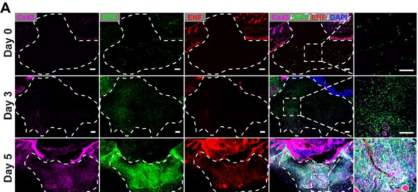
4.2.3 Gap junction inhibitors reduce scar formation in splinted full-thickness wounds

             in vivo ................................................................................................................. 54

    4.3      Disrupted calcium oscillation by gap junction inhibition underlies the

             reduced scarring .............................................................................................. 56

    4.3.1 Gap junction inhibitors disturb the functional communication on fibroblasts. ... 56

    4.3.2 Gap junction inhibitors interrupt calcium oscillation on fibroblasts. .................. 58

    4.3.3 Calcium inhibitor reduces scar formation on skin-fascia fascia. ....................... 60

    4.3.4 Calcium inhibitor reduces scar formation in vivo ............................................... 61

    4.4      Inhibition of Cx43 abrogates EPF swarming and fascial matrix migration

             62

    4.4.1 EPF swarming were abrogated by Cx43 inhibition on skin-fascia explants ..... 62

    4.4.2 Absent collective migration by gap junction inhibition in vivo chimeric

             transplantation model......................................................................................... 63

    4.4.3 Matrix movement was inhibited in labelled matrix tracing in situ ...................... 66

5            Discussion ........................................................................................................ 69

    5.1      Cx43 on EPF involved in scarring.................................................................. 69

    5.2      Gap junction inhibition decrease scar formation by N-cadherin and

             calcium oscillations ......................................................................................... 70

    5.3      Cx43 coordinates collective migrations into open wounds. ...................... 71

Abbreviations ................................................................................................................. 75

Reference ........................................................................................................................ 79

Publications .................................................................................................................... 87

Affidavit ........................................................................................................................... 89

Acknowledgment ........................................................................................................... 90

                                                                   4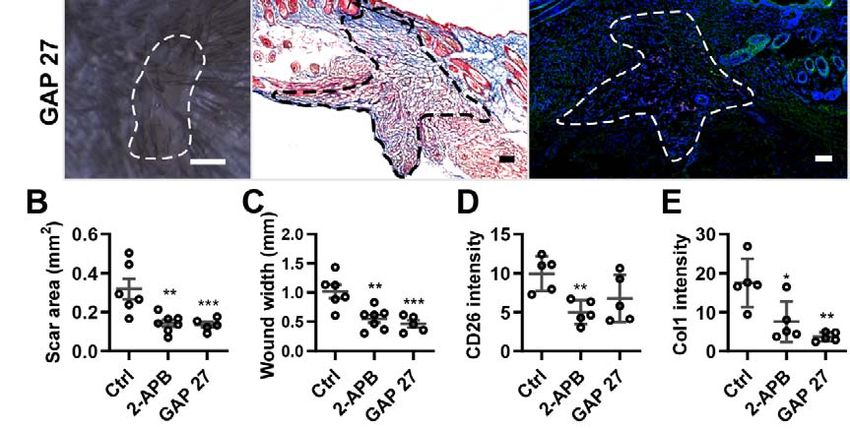
Zusammenfassung

Tiefe und voluminöse Hautwunden werden repariert, indem Fibroblasten und

extrazelluläre Matrix aus der Faszie tief unter der Haut austreten und die verletzten

Oberflächen mit Narben verschließen. Der molekulare Auslöser dieses neuartigen

Reparaturmechanismus ist nur unvollständig verstanden. Hier zeigen wir, dass Cx43

der Schlüssel zur Patch-Reparatur von tiefen Wunden ist. Durch die Kombination von

Full-Thickness-Wundmodellen mit Fibroblastenlinien-spezifischen transgenen Linien

zeigen wir, dass Cx43 ausschließlich in tiefen Wunden hochreguliert wird, und zwar in

spezialisierten Fibroblasten der Faszie tief unter der Haut, die für die Narbenbildung

verantwortlich sind. Mit Hilfe von Live-Imaging von Faszienfibroblasten und Fate Tracing

der extrazellulären Matrix der Faszien zeigen wir, dass die Hemmung von Cx43 die

Oszillationen des Kalziumspiegels in tiefen Faszienfibroblasten unterbricht und dass

dies ihre kollektive Wanderung hemmt, die notwendig sind, um die Faszienmatrix in

offene Wunden auszuschütten. Cx43 und die Zell-Zell-Kommunikation sind somit

Vehikel der Matrixausschüttung und Narbenbildung und notwendig für die Patch-

Reparatur von voluminösen Wunden. Diese Erkenntnisse haben weitreichende klinische

Implikationen für die Behandlung von Fibrose, überschießender Narbenbildung und

gestörter Wundheilung.

                                             5
6

Abstract

Deep and voluminous skin wounds are repaired, by outpouring of fibroblasts and

extracellular matrix from fascia, deep below the skin, plugging breached surfaces with

scars. The molecular trigger of this novel repair mechanism is incompletely understood.

Here we reveal that Cx43 is the key to patch repair of deep wounds. By combining full-

thickness wound models with fibroblast lineage specific transgenic lines, we show Cx43

is upregulated exclusively in deep wounds, in specialised fibroblasts of the fascia deep

beneath the skin that are responsible for scar formation. Using live imaging of fascia

fibroblasts and fate tracing of the fascia extracellular matrix, we show that Cx43 inhibition

disrupts calcium level oscillations in deep fascia fibroblasts and that this inhibits their

collective migrations necessary to outpour fascia matrix into open wounds. Cx43 and cell-

cell communication are thus vehicles of matrix outpouring and scar formation, and

necessary for patch repair of voluminous wounds. These findings have broad clinical

implications for treating fibrosis, aggravated scarring and impaired wound healing.

                                              7
8
2 Introduction

2.1 Skin

Skin, the largest and outermost organ in mammals, is highly organized into multiple

strata layers, from outer to inner, that include the epidermis, dermis and hypodermis

(Kanitakis, 2002). It serves as a protective barrier from the external environment, as well

as provides a primary defence against bacterial and viral infections, physical protection

from external environmental insults such as dehydration, chemical, mechanical, and UV

exposure (Lee et al., 2006). In addition, the skin has further versatile roles in sensation,

thermoregulation, metabolic regulation and immune surveillance (Kanitakis, 2002).

2.1.1 Epidermis

The epidermis is the outermost strata layer of the skin, and it is mainly composed of four

different cellular lineages. The predominant one are the stratified keratinocytes, which

synthesize and secrete the major structural proteins of the stratum corneum (SC).

Melanocytes, the second cell type, generate pigment granules, termed melanosomes,

containing melanin (Wickett and Visscher, 2006). The third cell type is Langerhans cells,

a type of dendritic cells presenting epitope once activated by antigen. Merkel cells is last

one, involved in sensation (Moll et al., 2005).

2.1.2 Dermis

The dermis originates from the embryonic mesoderm, and it represents the most

abundant connective tissue of the skin. It is composed by two main layers: upper

papillary and lower reticular dermis. Both dermal compartments are fundamentally made

up of the same fibrillary and connective tissue components namely collagens, which

                                              9
contributes to the rigidity of the dermis and that supports skin appendages.
Approximately 80% of dermal collagen is collagen type I and the remaining belongs to
collagen type III and elastin (Epstein and Munderloh, 1978). In addition to connective
tissue framework, dermis houses specialised cell types termed fibroblasts, which are the
primary cellular source of collagen and elastin (Lynch and Watt, 2018). In addition to
fibroblasts, dermis also contains a range of other supportive cells including mast cells,
macrophages, lymphocytes and melanocytes, as well as blood vessels and peripheral
nerves. The dermis further contains various epidermal appendages that invaginate and
reside within the dermis, including sweat glands, apocrine glands, sebaceous glands,
hair follicles and nails, which support the functions of the upper epidermal layer. Below
the dermis directly is the hypodermis layer, also called subcutaneous layer, which is
actually not part of integument system. The hypodermis, consisting areolar and adipose
connective tissue, mainly protects the body from damage, provides thermal insulation,
and functions as energy reservoir. A layer of striated muscle, below hypodermis known
as Panniculus carnornosus (PC) muscle, provides skin twitching and contraction
functions.
2.1.3 Fascia
Fascia is a gelatinous viscoelastic layer in subcutaneous locations, which implements a
frictionless interface between the upper skin and inner rigid structures. The thickness of
fascia varies considerable regionally and sexually. For example, murine back skin houses
a single layer of fascia underneath muscle and skin. In contrast, several fascia layers,
incorporated with adipose tissue, neurovascular and lymphatic tissue, are continuous with
skin directly in human without any PC muscle. The fascia underneath the dermis also
serves as fat storage, insulator and protection, providing a buffer effect for the underlying
muscles, tendons, bones and joints.
102.1.4 Murine skin
Human and murine skin are highly similar in structure and composition. Murine skin is
composed of the same layers as human skin, except a far thinner epidermis and hair
follicle-populated dermis (Lee et al., 2018; Sougrat et al., 2002).
In mouse skin, hair follicles cycle and transition much more frequently than in human
skin (Muller-Rover et al., 2001). In other hand, subcutaneous layers in mouse skin lies
directly above a striated muscle layer, the panniculus carnosus muscle, as compared to
human skin. Whereas human skin lacks muscle layer, hypodermis is adjacent to fascia
layers directly (Dorsett-Martin, 2004). Resident immune cells are apparent similarly
present, as both mouse and human skin houses macrophages, mast cells, conventional
αβ T cells and a small population of innate lymphoid cells (ILCs). However, the most
prevalent immune cells in human skin are Langerhans cells and CD8+ T cells, in
contrast with prominent Vγ5+ dendritic and T cells (DETCs) distributing in mouse skin
(Pasparakis et al., 2014).
Below is a schematic that summaries the major differences in cell composition and
structure between mouse and human skin (Fig. 1.1, 1.2, Table 1.1).
11Figure 1.1 Structure of the skin in murine and human. Mouse skin (a) shows
denser hair follicle but thinner epidermis with less cell layers compared with human skin
(b). Several immune cells presented in murine and human skin (Pasparakis et al.,
2014).
12Figure 1.2 Comparative structure of human and murine skin. Trichrome staining
of human skin and murine dorsal skin section. ep stands epidermis, de represents
dermis (10x).
Table 1.1 Major differences between mouse and human skin (Wong et al.,
2011)
Mouse Human
Hair cycle ~ 3 weeks Regionally, sexual dependent
Epithelial structure Without rete ridges Rete ridges
Extend from mammary Appeared in axilla, inguinal, and
Apocrine sweat glands
glands perianal skin regions
Thick, relatively stiff, adherent to
Physical properties Thin, compliant, loose
underlying tissues
13Hypodermal thickness Depends on hair cycle Fixed
Knowns as panniculus
Subcutaneous muscle In neck as platysma
carnosus muscle
Granulation tissue formation, re-
Wound healing Contraction, fascia patching
epithelization
142.2 Cutaneous wound healing
Intact healthy skin provides protection from outside irritation or injury. Therefore, proper
wound healing is an essential physiological process for tissue homeostasis (Karppinen et
al., 2019). Generally, skin wound healing is a highly hierarchically orchestrated process
involving three overlapping phases. Wound healing initiates with haemostasis, an immune
reaction that forms a temporary fibre clot at sites of injury. Haemostasis rapidly stops
bleeding and covers the injury site with a provisional fibrin barrier, which deters pathogens
and microorganisms within open wounds, amplifies the pro-inflammatory cytokines and
immediately consecutive recruiting activated neutrophils into the wound from damaged
blood vessels for the next 1 or 3 days, and establishes a first provisional matrix bedding
for subsequent stages (Kim et al., 2008). Subsequently within days, the healing process
progresses into a proliferative phase, involving fibroblast proliferations in dermis,
keratinocyte proliferation in epidermis, and endothelial proliferation that end with
angiogenesis. At the same time, wound contraction initiates from the edges of the wound,
gradually closing the open skin barrier (Shaw and Martin, 2009). Lastly, fresh matrix is
synthesized, deposited within wound, and further remodels over time until maturation,
finally, as scar tissue (Dovi et al., 2004; Shaw and Martin, 2009).
2.2.1 Scarring
Over 100 million patients experience skin scarring from burns, surgery, and recreational
injury per year, which are a huge burden for both patients and global healthcare
systems (Marshall et al., 2018). Scarring is characterized by excess collagen deposition
which initiates by specialized fibroblast immigration into wounds (Marshall et al., 2018).
Human skin is primarily repaired by scarring. Whereas, superficial skin wounds
regenerate as minimal scars, deep and voluminous wounds are physically covered with
15dense plugs of connective tissue matrix, called scar tissue (Bayat et al., 2003; Driskell et
al., 2013; Gurtner et al., 2008; Rinkevich et al., 2015; Stappenbeck and Miyoshi, 2009;
Watts, 1960). However, wound healing in the oral mucosa proceeds rapidly with minimal
scar formation compared to back skin injuries (Jiang et al., 2020). Similarly, foetal skin
wounds also exhibit highly regeneration ability. Fortunately, stromal cell-derived-factor-
1(SDF1) contribute elder to acquire this ability again as wound healing with less scarring
(Larson et al., 2010; Nishiguchi et al., 2018).
However, keloids and hypertrophic scars, two fibrotic scars in humans, are usually
different from normal mature scars in composition and size, appearing as shiny,
hairless, rising above surrounding skin, reaching the reticular dermis. Fibrotic scars are
pathological scars that are populated with inflammatory cells and exuberant numbers of
fibroblasts, enriched neo-vessels and collagen deposition. So far, Researcher cannot
reduce systemic and genetic risk factors though treatments, such as laser treatment,
cryotherapy, were developed for keloids and hypertrophic scars (Gauglitz et al., 2011;
Ogawa, 2017).
2.2.2 En1 lineage-positive skin fibroblasts
Fibroblasts are the major producers of ECM in both embryonic and adult organs, as well
as in tissue fibrosis, and cutaneous scarring (Hinz et al., 2007; Wynn, 2008). Though
multiple embryonic lineages of fibroblasts were discovered in the dorsal skin, all scars
primarily come from a single fibroblastic lineage, termed as Engrailed-1 (En1) lineage
positive fibroblasts (EPFs). EPFs appear as a small subset at early embryogenesis, and
expand within the developing skin during embryogenesis. In contrast, En1 lineage
negative fibroblasts (ENFs), characterised in the back skin, do not contribute to scar
formation. Both fibroblast subsets co-exist in the back skin, where the balance of their
16abundance drives the emergence of scar formation during skin development (Jiang et
al., 2018; Rinkevich et al., 2015).
Figure 1.3 Schematic showing differential labeling of connective tissue depending
on EPFs and ENFs in mTmG system (Rinkevich et al., 2015).
17Figure 1.4 EPFs contributing to connective tissue deposition, whereas ablation/
inhibition of En1 fibroblasts reduced scarring (Rinkevich et al., 2015).
182.2.3 EPF in superficial fascia
Superficial fascia, in murine back skin, is a connective tissue that is separated by
panniculus carnosus muscle, whereas, fascia in humans is much thicker and
incorporates fibroblasts, vascular and matrix. Studies discovered that subcutaneous
fascia largely took part in provisional scar tissue and reduced wound size. En1 lineage
fibroblasts appear to be the major fascia fibroblasts contributing to scar. Fascia
fibroblasts expand in skin surface after wounding, and trigger surrounding extracellular
jelly-like matrix moving into wound (Correa-Gallegos et al., 2019). However, it is still
unclear how fascia EPFs, but not dermal or oral mucosa fibroblasts, cause scarring after
wound.
19Figure 1.5 Fascia in human and mouse skin. Line separates fascia and dermis.
Figure 1.6 Process of deep skin wounds healing. Fascia, containing EPFs, ECM,
blood vessels, macrophages and nerve, penetrates into skin and seals the wound.
(Coles and Buckley, 2019).
202.2.4 ECM on scar formation
ECM, is a dynamic scaffold composed of water, proteins and polysaccharides. It
undergoes active remodeling and rearranging during development as well as during
organ morphogenesis. Overproduction of ECM, especially in scar formation, causes
pathological scarring and load a larger burden for internal organs (Coentro et al., 2018;
Karppinen et al., 2019). Interestingly, cultured dermal fibroblasts could steer ECM
movement or remove individual fibronectin fibers and collagen in vitro. In addition,
matrix movements occurring during injury quickly seal large open wounds, and
inversely, blocking EPFs migrations inhibits scar formation (Correa-Gallegos et al.,
2019; Kurpios et al., 2008; Tseng Q, 2012).
Figure 1.7 Extracellular matrix (ECM) functions and cross talking at the cell–
cell and cell–matrix interfaces (Chen and Liu, 2016).
212.3 Gap junction
Mammalian gap junctions are communication channels that stitch adjacent cells
(Tarzemany et al., 2017). Six connexin (Cx) subunits of gap junction, merge together
coupling cytoplasmic and permitting intercellular passage of ions and small molecules
such as Ca2+, ATP and cAMP (Chen et al., 2016; Wong et al., 2016). Cx43, one of the
major Connexin in mammalian skin, is a ubiquitous gap junction targeting wound healing
responses (Wong et al., 2016). For example, Cx43 deficiency and downregulation
accelerated wound closure in mice, and attenuated diabetic wounds in rat model (Becker
et al., 2012; Cogliati et al., 2015; Qiu et al., 2003). The oligo peptide inhibiting Cx43 is
also currently under Phase III clinical trial for treating diabetic foot ulcers (Montgomery et
al., 2018). However, the direct functions of Cx43 in vivo, especially in fascia and deep
skin wounds remain unknown. As connexin-based therapeutics are being further explored
pre-clinically in injury of organ, identifying repair mechanism allow the development of
improved, targeted therapies and elucidation of universal repair mechanisms.
Figure 1.8 3D map of recombinant gap junction. (a and b) showed the side view
and density of gap junction. (c) Transverse view of gap junction at position indicated by
22white arrows. Letter C represents cytoplasmic space, M is membrane bilayers, E means
extracellular gap (Unger et al., 1999).
Gap junction, encoded by connexin gene in both vertebrates and invertebrates,
classifies three types, known as α, β and γ, responding to conserved protein motifs
(Harris, 2001). All connexins present highly specialized transmembrane structures,
linking double tandem extracellular loops with triple cysteine residues forming disulfide
bonds such as CX6CX3C in one extracellular terminus and CX4CX5C in the other
extracellular terminus, and similar topology according to their structure, especially in
humans and mice (Krutovskikh and Yamasaki, 2000). However, the structure of C-
terminus, for example, in connexin 43 (Cx43), is randomly coiled with two helical
structures. It interacts with various protein partners, including ZO-1, cadherin, catenin,
tubulin and microtubules (Sorgen et al., 2004). Residues 228~260 of Cx43 directly
interact with tubulin and microtubules, and regulate permeability of Cx43 by
23phosphorylation and signal transduction regulated by TGF-β pathway (Saidi Brikci-
Nigassa et al., 2012).
Figure 1.9 The C-terminus Cx43 binding with tubulin. The binding negatively
regulated by phosphorylation Y247, a target for Src kinase (Saidi Brikci-Nigassa et al.,
2012).
Genomic study revealed that connexin ubiquitously expressed on all vertebrates, most
of them clustering together within chromosomes 1 and 13 in human. However, more
than 20 connexins are named into three subfamilies α, β and γ in humans (Nielsen et
al., 2012). Almost all cells express at least one kind of connexins due to gene diversity,
for example Cx26, Cx30, Cx43 has been found in keratinocytes, while in Schwann cells
is Cx30.2 and Cx32 (Lin et al., 2003; Zhang and Cui, 2017). Those mostly similarity
imply that the connexin are likely derived from one gene duplication events.
24Figure 1.10 All connexins family from human alignment. More than 20 connexins
are classified into three subfamilies α, β and γ in humans.
2.3.1 Gap junction in wound healing
Gap junction play important roles in cell-cell communication, cell morphology and
polarity, and influence the adhesiveness of cells and directionality of cell migration
(Wright et al., 2009). However, abnormal connexin expression was associated with
dysregulated cell proliferation, migration and wound healing rates. Cx43, stabilizing a
series of proteins, including CDH2 and ZO-1, are required for cell-to-cell adhesion and
cell migration. Consecutive high levels of Cx43 in diabetic wounds significantly
ameliorating the process of healing was observed in diabetic skin (Wright et al., 2012).
25Alternatively, as the C-terminus of Cx43 is known to interact with cytoskeletal
components or with P120ctn/ Rho GTPase, downregulation of Cx43 could redirect the
motility of keratinocytes at the wound edge, spurring them to migrate and close the
wound, which underlie Cx43 has potential tremendous therapeutic value (Derangeon et
al., 2008). Blocking Cx43 by antisense peptide accelerated the wound closure and
granulation tissue formation and reduced scar size after burn injury, which was
beneficial effect in rat model diabetic ulcer (Nakano et al., 2008; Wong et al., 2016).
Hence, competing inhibitory peptide of Cx43 in intracellular signalling, currently, is under
clinical trial of Phase III for curing diabetic foot ulcers (Montgomery et al., 2018; Zhang
and Cui, 2017). On the other side, increased Cx43 expression was induced by TGF-β1,
which promotes scar formation via Erk/ MMP-1/ Collagen III pathway (Li et al., 2019).
Further reports illustrated that the sustained inhibition of Cx43 accelerated efficient
wound healing, and altered mesenchymal cell movement patterns and feather bud
elongation in chicken dorsal skin explants (Li et al., 2018).
2.3.2 Gap junction targeting therapy
Mimetic peptides, identical to defining sequences of connexin lying in the N-terminus,
extracellular loops and transmembrane domains, are more specific targeting connexin.
The popular peptide, Gap 27, possessing sequence homology of various connexin
subtypes, has been frequently employed to inhibit intercellular communication in various
inflammatory diseases (Elbadawy et al., 2016). Indirectly inferring from arterial
contractility studies, the IC50 of Gap27 is 20-30 μM whereas higher concentration, at
200-300 μM, may block other channels, like Pannexin-1 (Glass et al., 2015). In parallel
with a reduction of Cx43 phosphorylation, gap junction also mediates apoptotic cell
death or increases sensitivity to pro-apoptotic agents (Lin et al., 2003). Although the
26therapeutic potential of connexin is undeniable, more effort should be taken into
studying the regulation and functions of these proteins.
2.3.3 Calcium signalling
Organ development and tissue regeneration require extensive coordination among
heterogeneous cell populations to generate complex organ morphologies (Brodskiy and
Zartman, 2018). Ca2+, a ubiquitous intracellular signalling molecule responds to diverse
stimuli and participates in various physiological processes, such as cell proliferation,
differentiation, apoptosis and migration (Brodskiy and Zartman, 2018). When
concentrating on wound healing in multi-layered skin, a rapid influx of calcium, firstly,
comes around the wound; then a short-lived ranged wave spreads through healthy
neighbouring cells. Moreover, the larger the wound injured, the faster the wave
spreading, in which calcium waves pass through adjacent cells suggesting that gap
junctions and calcium ions make up signalling bridges. Furthermore, X-ray crystal
structures of the human Cx26 gap junction channel with and without bound Ca2+ has
been described.(Bennett et al., 2016). Due to T- and L- type Ca2+ channels involving
cellular and organ functions, Ca2+ channel blockers, e.g., efonidipine which disrupts the
Ca2+ oscillations by selective inhibition of both T- and L-type channels, are pivotal to the
synthetic function of human pulmonary fibroblasts, which have been used in clinical
practice (Mukherjee et al., 2013). Furthermore, rapid, transient increases in intracellular
calcium have been reported in scratch wounds and single-cell wound assays (McNeil
and Steinhardt, 2003; Nakano et al., 2008). Induced Ca2+ oscillations also enhanced
feather bud elongation in chicken (Li et al., 2018). In Summary, calcium has long been a
candidate for discovering the earliest signal during wound repair. Understanding
mechanisms under the wound healing process with personalized condition would pave a
27road for developing pharmacological tools to slow down or even to speed up normal
wound healing.
Figure 1.11 Gap junction bound with calcium. (a) The calcium-bound (orange) and
calcium-free (cyan) gap junction are almost identical. (b, c) depicted gap junction
electrostatic surfaces, in which calcium binding (b) establishes a positive surface
potential in the pore (blue) that limits molecular permeation (Bennett et al., 2016)
283 Hypothesis of the study
Deep and voluminous skin wounds are repaired by plugging breached surfaces with
scars. The molecular mechanism steering fibroblast and ECM from fascia into scars is
incompletely uncovered. The hypothesis was that Cx43, cell-cell communication
junction, are vehicles of matrix outpouring and scar formation, and necessary for patch
voluminous wounds.
To reveal the Cx43 role in deep wounds healing, full-thickness wound models in specific
fibroblast lineage transgenic mouse lines, and fate tracing of the fascia extracellular
matrix were applied.
2930
5 Materials and methods

5.1 Human samples

Fresh human skin and keloid biopsies were collected by the department of Dermatology

and Allergology, Klinikum rechts der Isar, Technical University Munich (reference

number 85/ 18S). Informed consent was obtained from all subjects prior to skin biopsies.

Upon collection, these samples were directly processed with PFA fixation and then OCT

embedding followed by histological or immunofluorescent analyses.

5.2 Mice strains

All Mouse strains (C57BL/6J, En1Cre, R26mTmG, R26LSL-H2B-mCherry, R26VT2/ GK3, Rag2 -/-) were

obtained from Jackson laboratories or generated at the Stanford University Research

Animal Facility as described previously (Rinkevich et al., 2015). En1cre transgenic mice

were crossed with R26mTmG two-color membrane reporter mice or R26LSL-H2B-mCherry nuclear

reporter mice. All mice were bred and maintained at the animal facility of Helmholtz Centre

Munich. All animal experiments were approved by the Government of Upper Bavaria and

registered under the project 55.2-1-54-2532-61-2016 and conducted under strict

governmental and international guidelines. This study is compliant with all relevant ethical

regulations regarding animal research.

5.3 Genotyping

Cre-positive (Cre+) was identified by detecting relevant fluorescence of the dorsal

dermis under microscope. Genotype of Cre+ inserted with a 200-base pair fragment

(Cre+/-) was performed by PCR to distinguish from the wildtypes (Cre-/-). Briefly, genomic

                                             31
DNA from the ear clips was extracted using Quick Extract DNA extraction solution
(Epicenter) following the manufacturer’s guidelines. Then 1 μL DNA extract was added
to 24 μL Qiagen PCR reaction mixture containing forward primer “Cre_genotype_4F”- 5´
ATT GCT GTC ACT TGG TCG TGG C- 3´ (Sigma) and reverse primer
“Cre_genotype_4R”- 5´ GGA AAA TGC TTC TGT CCG TTT GC- 3´ (Sigma). PCR
reactions were initiated into denaturation for 10 min at 94 °C, amplification for 30 cycles:
denaturation for 30 s at 94 °C, hybridization for 30 s at 56 °C, elongation for 30 s at
72 °C and final elongation for 8 min at 72 °C then cooled to 4 °C. Reactions were
analysed by gel electrophoresis.
The 2% agarose gel was prepared by mixing 2 mL of 50x TAE buffer, 2 g agarose in
100 mL ddH2O and heating with microwave to melt completely. To stain DNA fragments
in gel, 5 μL SYBR Green Master Mix was added once the solution was cooled to rough
60 °C. The gel was filled into a mould with the fitted comb and allowed to cool down at
room temperature. Once the gel solidified, it was placed in a tank with the TAE buffer. A
DNA ladder was loaded in the first lane and 15 μL sample reaction solution in the rest
wells. Then electrophoresis was run at constant voltage 120 for 12 min. Finally, gel was
visualized on a UV trans-illuminator and photographs were taken on a digitall camera
(UVP Bioimaging Systems).
5.4 Skin-fascia explants assay
Dorsal skin was harvested from new-born (postnatal day 0-1) C57BL/6J or
En1cre;R26mTmG mice or nuclear reporter En1Cre;R26LSL-H2B-mCherry mice and washed with
HBSS. Full-thickness round skin pieces were taken with 2 mm biopsy punches (Stiefel)
and cultured in 200 μL DMEM/F-12 (Thermo Fisher) medium containing 10% FBS (Life
technologies), 1% GlutaMax (Thermo Fisher), 1% Penicillin/streptomycin (Thermo
32Fisher) and 1% MEM non-essential amino acids (Thermo Fisher) in 96-well plates in a
humidified 37 °C, 5% CO2 incubator. In addition, the skin pieces were cultured
submerged in medium with dermal side face up.
Medium was routinely replaced every other day stimulated with 10 ng/mL TGF-β 1, 2
μM SB 431542, 1 μg/mL 2-APB, 8 μM GAP 27 separately. On day 3 or day 5 of culture,
samples were washed by PBS and fixed with 2% PFA overnight at 4 °C. After fixation,
the tissue were processed for whole-mount bright field imaging or whole-mount
fluorescent staining directly or embedded in OCT following with 6 μm cryosections for
trichrome staining or immunofluorescence staining.
5.5 Excisional wounds on mice
The 8-10 weeks old En1cre;R26mTmG mice were anesthetized with medetomidine at 500
μg/kg, midazolam at 5mg/kg and fentanyl at 50 μg/kg (MMF) body weight. Two full-
thickness excisional wounds were created with a 5 mm diameter biology punch (Stiefel)
under supply with analgesia. Wounds were harvested at day 0, 3, day 5, day 7 post-
surgery by cutting out and washing with PBS, following fixation with 2% PFA
immediately at 4 °C overnight. After washing 3 times with PBS, wounds were sliced into
two half parts in the middle and then embedded in OCT and processed for histology
staining.
5.6 Splinted wounds on mice
Splinting rings with outer diameter 12 mm and inner diameter 6 mm were made of 0.5
mm silicone sheet (Grace Bio-Labs, CWs-0.5), and sterilized with 70% ethanol for 30
min followed by washing with PBS. Air dried rings were kept in a sterile bottle until
surgery. Before operation, mice were anesthetized with MMF. Dorsal hair was removed
33by incubating skin with hair removal cream for 5 min. Two full-thickness excisional
wounds on back skin were created with a 5 mm diameter biology punch (Stiefe). To
prevent skin contraction around wounds and allow the wound to close by re-
epithelialization and granulation tissue formation, silicone rings with one side applied
with silicone elastomer super glue (KwiK-Sil Adhesive) were sutured onto the excisional
wounds. Mice were supplied with analgesia after surgery. Mice were subcutaneously
injected with 60 μL 2-APB at 4 μg/mL or 60 μL GAP27 at 80 μM or 60 μL Nifedipine at
10 μM, 60 μL 0.1% DMSO or saline on every second day for each group. After 21 days,
the hair follicle of the back skin was shaved followed by hair removal cream. Then scars
were dissected and washed with PBS, following fixation with 2% PFA immediately in
4 °C overnight. After washing 3 times with PBS, wounds were sliced into two parts in the
middle and then embedded in OCT.
5.7 Masson’s trichrome staining
Skin-fascia explants tissues were fixed with 2% PFA at 4 °C overnight, and washed
three times with PBS. Whole mount bright-field images were taken with a Leica M50
stereo microscope equipped with a Leica DFC310 FX camera (Leica). Subsequently,
tissues were embedded in OCT (Sakura Finetek) and subjected to slice in 6 μm
cryosections with Hyrax C50 Cryostat (Zeiss).
Masson’s trichrome staining was performed using a Masson’s trichrome staining kit
(Sigma-Aldrich HT15) according to manufacturer instructions. Briefly, cryosections fixed
in cold acetone in minus 20 °C for 5 min, and then air dry for 5 min. After wetting slides
in deionized water for 2 min, sections were incubated in preheated Bouin’s solution
(Sigma-Aldrich HT10132) at 56 °C for 15 min. Then wash in cold tap water to remove
yellow color from sections, the sections were stained with Weigert’s Iron Hematoxylin
34(Sigma-Aldrich HT1079) at working concentration for 5 min. Thereafter, the sections
were treated with Massion’s trichrome stain kit (Sigma-Aldrich HT15) by sequentially
incubating at room temperature in Biebrich scarlet-acid fuchsin solution for 5 min,
working concentration of Phosphotungstic / Phosphomolybdic acid (HT152-250ML/
HT153-250ML) for 5 min, aniline blue solution for 10 min, and 1% acetic acid for 2 min.
After dehydration in 80% ethanol, 100% ethanol I, 100% ethanol II consecutively for 5
min separately, the sections were cleared with Roti-Histol (Roth 6640) and mounted with
a Roti-HistoKit (Roth 6638). In white field, collagen was stained blue, cells in red and
nuclear in black. For the quantification of scar areas, trichrome images were converted
to CMYK and the cyan channel was assigned to quantify blue-stained collagens.
5.8 Immunostaining
For immunostaining, sections were rinsed with PBS for three times and then incubated
with 5% BSA in PBS for one hour at room temperature to block the non-specific binding
of antibodies. Then sections were incubated with the primary antibodies rabbit anti-Cx43
(Sigma-Aldrich C6219, 1:200), goat anti-CD 26 (Sigma-Aldrich, 1:200) or rabbit anti-
collagen I (Rockland 600-401-103-0.5, 1:150) or goat anti-CD 26 (Sigma-Aldrich, 1:200),
Goat anti α-SMA (Abcam, ab21027, 1:200) or Rat anti F4/80 (Abcam, ab90247, 1:200)
in 2% BSA at 4 °C overnight. Sections were washed with PBS for three times and
incubated with the AlexaFluor 594-, or AlexaFluor 647-conjugated secondary antibodies
(Life technologies, 1:500) against the relevant species for 1 hour at room temperature.
Next, sections were washed three times in PBS and finally mounted with fluorescent
mounting media with DAPI (Fluoromount-G, Thermo Fisher scientific 00-4958-02).
Photomicrographs were taken with a Zeiss AxioImager microscope with ZEN blue
software (Carl Zeiss).
355.9 3D imaging for murine skin
Fixed skin-fascia explants from En1cre;R26mTmG mice were pre-incubated with PBS,
containing 0.01% Thimerosal (Sigma T8784), 0.5% Triton-X100 (Sigma X100) and 0.2%
gelatin (Sigma G1393), termed as PBS-GT, at room temperature for 18 h. Then samples
were incubated with the primary antibodies anti-43 (Sigma-Aldrich, C6219, 1:100) in PBS-
GT at room temperature for 24 h. After washing in PBS-GT, samples were incubated in
Alexa Fluor 647-conjugated goat anti-rabbit IgG (Thermo Fisher Scientific, 1:250) in PBS-
GT at RT for 24 h. After incubation, samples were washed with PBS-GT. Finally, samples
were embedded into 2% NuSieve GTG agarose (Lonza 859081) in a 35 mm dish (Falcon
351008). Whole-mount fluorescence 3D imaging was performed under a Leica SP8 MP
(Leica, Germany). Tiles were merged by using the Leica Application Suite X (v4.8, Leica)
with smoothing overlay blending. Then data were visualized with Imaris software (v9.1.0,
Bitplane, UK) under contrast and brightness optimisation.
5.10 Dermal EPFs purification
Back skin from En1cre; R26mTmG adult mice were harvested and minced with surgical
scissors. The tiny pieces were digested with an enzymatic cocktail containing 1 mg/mL of
collagenase IV, 0.5 mg/mL of Hyaluronidase, and 25 U/mL of DNase I in HBSS (Thermo
Fisher Scientific), and incubated in 37 °C water bath for 30 min. DMEM containing 10%
FBS was added to stop the enzymatic reaction. The suspension was filtered through a 40
μm cell strainer. After washing, the pellets were incubated with APC-conjugated lineage
maker anti-mouse CD31 (PECAM-1), CD45, Ter119, Tie2 (CD202b) or EpCam (CD326)
(BioLegend) and eFluor660- conjugated anti-mouse Lyve-1 (Thermo Fisher) on ice in dark
for 30 min. Subsequently, cells were resuspended in FACs buffer with sytox blue dye
(Thermo Fisher Scientific) for dead cell exclusion. Finally, cells were sorted on a BD
36FACSAria III with 100 μm nozzle. The viable (Sytox blue), lineage-negative cells (Lin-:
CD31-, CD45-, Ter119-, Tie2-, EpCam-, Lyve-1) were sorted into ENFs (Lin-RFP+GFP-)
and EPFs (Lin-RFP-GFP+) based on RFP and GFP fluorescence.
Collected cells were seeded on the 8 well chamber (ibidi) with DMEM/F-12 medium
containing 10% FBS, 1% GlutaMax (Thermo Fisher), 1% Penicillin/streptomycin (Thermo
Fisher) and 1% MEM non-essential amino acids (Thermo Fisher), and incubated in
humidified 37 °C, 5% CO2 incubator. Every other day, the media in the chamber was
replaced by gently pipetting out the old media and replacing it with pre-warmed fresh
media. Once the cells became 60% - 70% confluent on the bottom of the chamber, EPFs
were stimulated with 10 ng/mL TGF-β 1, or 2 μM SB 431542, 1 μg/mL 2-APB, 8 μM GAP
27 separately for 24 h.
5.11 Live imaging of cells migration
Full-thickness back skin biopsy collected from En1Cre; R26LSL-H2B-mCherry newborn embryos
were cultured for 4 days. Then sample were embedded in 4% agarose (Lonza, 859081)
in a 35 mm dish (Corning, 351008). Then samples were submerged in imaging medium
phenol-red free DMEM/F-12 (Thermo Fisher Scientific, 21041025) containing 10%
KnockOut Serum Replacement (Thermo Fisher Scientific, A3181501), 1% GlutaMAX
(Thermo Fisher Scientific, 35050038), 1% Penicillin/streptomycin (Thermo Fisher
Scientific, 15140122), and 1% MEM non-essential amino acids (Thermo Fisher Scientific,
11140035). Time-lapse imaging was performed with Leica SP8 MP (Leica, Germany)
under a modified heating and gas control incubation system (ibidi 10915 & 1192) with
3737 °C and 5% CO2 during imaging. Z-stacks images were recorded every 15 minutes for
12 h.
5.12 Cell tracking analysis
Cell tracking of Skin-fascia explants from En1Cre;R26LSL-H2B-mCherry mice was performed
under Imaris software package (v9.1.0, Bitplane, UK). Tracks were generated using the
fluorescence intensity-based detection tool. Particles represents the nuclei of Cre-positive
cells were filtered out. Tracks were visualized as a line indicated in the image.
5.13 Dye transfer assay in fibroblasts
NIH 3T3 fibroblasts (ATCC) were cultured in DMEM/F-12 medium containing 10% FBS
(Life technologies), 1% GlutaMax (Thermo Fisher), 1% Penicillin/streptomycin (Thermo
Fisher) and 1% MEM non-essential amino acids (Thermo Fisher). Culture medium was
replaced every other day. Monolayer of 3T3 cells at 70% confluence was labelled with 2.5
ug/mL calcein-AM (Invitrogen) at 37 °C for 30 min. After dissociation with 0.25% trypsin-
EDTA (Gibco), labelled cells were mixed with unlabelled cells at a ratio of 1:10. One hour
time-lapse imaging was performed every 5 min using Zeiss Observer Z1 (Zeiss, Germany).
During capturing, ambient temperature was set to 37 °C with 5% CO2-supplemented air.
5.14 Time-lapse Ca2+ imaging in fibroblasts
NIH 3T3 cells were cultured in an 8-well glass chamber (ibidi). The mixture of 4 μM AM-
Fluo-4 Ca2+ dyes (Invitrogen) and 0.04% pluronic F-127 (Sigma) were loaded on cells for
25 min in a 37 °C with 5% CO2-supplemented incubator. After washing with PBS, 20 min
time-lapse imaging was performed every 5 s using Zeiss Observer Z1 (Zeiss, Germany)
38at 37 °C with 5% CO2-supplemented air. Labelled calcium indicators are shiny once Ca2+
binding. Cell signal tracking was processed with ImageJ (version 1.52 e).
5.15 Chimeric skin transplantations
Full-thickness with 6 mm-diameter biopsies were collected from the back skin from
R26mTmG, R26VT2/GK3 adult mice. At the bench, the fascia together with muscle layer was
carefully separated from the dermis and epidermis using forceps and a 26G needle
under a fluorescent stereomicroscope (Leica). Chimeras were stacked by placing the
epidermis+dermis portion of a mouse strain on top of the muscle+fascia of another
strain and leave it in a 35 mm culture dish with 2 mL of DMEM/F12 at 4 °C for 20 min.
Then, a 2 mm “deep” full-thickness was excised from the chimeric graft using a biopsy
punch in the middle of the biopsy. To create “superficial” wounds, the 2 mm excision
was operated only in the epidermis+dermis, prior to reconstitution with the bottom part.
“Wounded” chimeric grafts were then transplanted into freshly-made 5 mm-diameter full-
thickness excisional wounds on the back of either RAG2-/- or Fox Chase SCID
immunodeficient mice. To prevent graft moving, a transparent dressing (Tegaderm, 3M)
was placed on the top after the graft drying for 20 minutes. Mice received subcutaneous
injection of saline or 80 uM GAP 27 every other day after post-wounding. Wounds
samples were collected by 8 mm punch at 14 days post-wounding and processed for
cryosection and imaging by fluorescence microscopy. To increase the success rate of
transplantation, emptying host blood from the fresh wound before the transplant and
leaving the graft dry at least 20 min before ending the anaesthesia should be executed
carefully.
395.16 In situ matrix tracing experiment
To label matrix subcutaneously, 8-10 weeks C57BL6/J mice received 20 µL FITC NHS
ester at 10 mg/mL in physiological saline with 0.1 M sodium bicarbonate pH 9 (Life
technologies, 46409) on four and two days before wounding. Full thickness excisional
wounds in 5 mm were created along the labelled area. Subsequently 60 µL saline or GAP
27 at 80 µM was subcutaneously injected around the wounds on every other day. Wounds
were harvested on day 7 post-wounding, and fixed in 2% PFA at 4 °C overnight. After
washing with PBS, tissue was embedded in OCT and subjected to cryosections.
5.17 Image analysis and statistic
Images were processed and analysed with ImageJ (version 1. 52e). For measuring scar
area, a brush tool was used to mask scar area. Calcein-AM and calcium Fluo-4 AM signal
in time-lapse images were tracked post subtracting background.
Statistical analyses were performed with GraphPad Prism (v8.0). Statistical significance
was assessed by Student’s t-test to compare two groups or one-way ANOVA with Tukey’s
multiple comparisons. All results are represented as mean ± SEM. The exact numbers N
and p values are depicted in the figure legends.
Table 3.1 Components of the quantitative qPCR master mix
Mastermix component Volume per Reaction
Coral Buffer 2.5 µL
dNTPs 0.5µL
Taq polymerase 0.125 µL
40Water 19.875 µL
DNA 1µL
F-Primer 0.5µL
R-Primer 0.5 µL
Total 25 µL
Table 3.2 Main chemicals and reagents
Chemicals and reagents Source Identifier
2-Aminoethyldiphenylborinate (2-APB) Sigma-Aldrich D9754-5G
GAP 27 Sigma-Aldrich G1794-2MG
Recombinant murine TGF-β1 R and D Systems 7666-MB-005
TGF-β RI Kinase Inhibitor VI (SB 431542) Merck Millipore 616461-5MG
Nifedipine Sigma N7634-1G
Calcein-AM LIFE Technologies C1430
Fluo-4, AM Life technologies F14201
4142
7 Results

7.1 Injury induces gap junction formation in fascia fibroblasts

7.1.1 Expression pattern of Cx43 on human normal skin and

       keloid

To investigate whether fibrosis involved with Cx43 in skin wounds, human normal

skin and keloid scars were collected and performed histological staining. When

looking into Cx43 expression in human keloid scars compared to normal skin, the

immunostaining showed that Cx43 was scarcely expressed in the dermis of normal

skin, but overwhelmingly abundant in keloid scars, with near uniform expression

(Fig. 4.1A), which indicates that high expression of Cx43 is associated with scar

formation. Quantification of relative Cx43 intensity showed that relative Cx43

intensity was significantly increased in keloid compared to normal skin (Fig. 4.1B).

Trichrome staining showed compact and fibrotic collagen patterns compared with

normal skin (Fig. 4.1C). Therefore, the expression pattern of Cx43 implied that Cx43

played a pivotal role in scar formation.

                                           43
Figure 7.1 Cx43 on human normal skin and keloid. (A) Immunofluorescence
staining of Cx43 (red) in human normal skin and keloid. DAPI (blue) was
counterstained to profile nuclei. (B) Quantification of fluorescence intensity of Cx43
in figure (A. C). Masson’s trichrome staining of the cryosections from human normal
skin and keloid. Mean ± SEM, n = 5, unpaired two-tailed student’s t-test, ***, p <
0.001. Scale bars = 100 μm.
7.1.2 Compact collagen deposition in skin-fascia explants
A whole skin-fascia explant undergoes skin contraction with scar formation outside
the animal is one novel model which describes scarring very well in ex vivo. Briefly,
skin-fascia explants were created from the back-skin from En1Cre;R26mTmG mice,
double transgenic reporter mice in which scar-prone fibroblast, EPFs in contrast with
remaining fibroblasts, ENFs, contributing to scar formation, which allowed us to track
both two functionally distinct fibroblastic lineages in genuine whole skin conditions in
high detail and throughout the wound repair process. Skin-fascia explants cultured
44for 5 days developed typical scars with denser plug of collagen fibres, as compared
to fresh day 0 skin-fascia explants (Fig. 4.2A,A’, 4.2B). Based on quantification of
area of collagen fibres, cultured explants in day 5 was around 4 times higher than
fresh tissue (Fig. 4.2B).
Figure 7.2 Collagen deposition in murine skin-fascia explants. (A) Masson’s
Trichrome staining of cryosections of a Day 0 and a Day 5 skin-fascia explants. (A’)
Magnification of scar area in Day 5 skin-fascia explants. (B) Quantitative collagen
fibres in scar areas of Day 0 and Day 5 skin-fascia explants. Mean ± SEM, n = 5,
unpaired two-tailed student’s t-test, ***p < 0.001. Scale bars = 100 μm.
7.1.3 Cx43 expression in skin-fascia explants
Regarding our well-established skin-fascia explants model, Cx43 distribution in
tissue was observed subsequently. In the immunostaining section, Cx43 was
specifically upregulated in EPFs along within the scar area; Cx43 was completely
45absent from EPFs in healthy skin (Fig. 4.3A, A’). Quantification of relative Cx43
intensity showed significant increased intensity on skin-fascia explants compared to
day 5 and day 0 (Fig. 4.3B).
Figure 7.3 Cx43 expression in murine skin-fascia explants.
(A) Immunofluorescence staining of Cx43 (magenta) in a day 0 and a day 5 skin-
fascia explant from the back-skin of En1Cre;R26mTmG mice. EPFs are green, other
cells are red, nuclei are blue. (B) Quantification of fluorescence intensity of Cx43 in
explants at days 0 and 5. Mean ± SEM, n = 5, unpaired two-tailed Student’s t-test,
***p < 0.001. Scale bars = 100 μm.
467.1.4 Multiphoton microscopic analyses of Cx43 expression
on skin-fascia explants
To visualize furtherly EPF lineage-specific expression of Cx43 in more detail, we
performed whole-mount 3D immunolabelling of the skin-fascia explants at Day 5.
Cx43 expressed on the skin-fascia explants in 3D imaging, in which explants
containing EPFs and ENFs were punched off from En1cre;R26mTmG double transgenic
mice dorsal skin (Fig. 4.4A). In addition, whole skin was undergoing contraction with
scar formation, which revealed EPFs collectively expressed Cx43, as a whole
population. Taken last Cx43 immunostaining together, Cx43 mainly distributed on
the centre area of skin-fascia explants tended to co-localizing with EPFs which are
primary fibroblast lineage contributing to scar formation (Rinkevich et al., 2015).
Figure 7.4 Multiphoton microscopy analyses Cx43 expression on skin-
fascia explants. (A) 3D immunolabelling of Cx43 in skin-fascia explants from the
back-skin of En1cre;R26mTmG mice were imaged by multiphoton microscopy, in which
Cx43 was stained as magenta, EPFs indicated as green, ENFs as red. Scale bars =
100 μm.
7.1.5 Cx43 expression in in vivo wounds
To analyse the physiologic relevance of expression patterns of Cx43 during wound
healing in vivo with our findings, full-thickness excisional wounds on the back skin of
En1cre;R26mTmG double transgenic mice were operated and the protein expression of
47Cx43 on histologic slides of these wounds were analysed. Cx43 protein expression in
live mice completely mirrored and overlapped with the patterns of expression we
initially found using ex vivo skin-fascia explants. Wounds samples from adult mouse
dorsal skin were collected post wounding at day 0, 3, 5 and 7. Protein expression was
absent from healthy skin, and was unregulated upon wounding in both epidermis and
wound bed. In wound bed, Cx43 was exclusively expressed in fascia EPFs, peaking
at 5 days post-wounding, and co-localizing with fascia EPFs during the later steps of
the wound healing process (Fig. 4.5A, B). Collectively, these results demonstrate that
Cx43 is exclusively expressed in scar-progenitor EPFs, during wound healing and may
function in these cells to control connective tissue matrix outpouring and scar severity.
Figure 7.5 Cx43 expression on wounds in vivo. Wounds on En1cre;R26mTmG
adult mouse dorsal skin were collected post wounding at days 0, 3, 5, and 7. (A)
48You can also read