Critical Reviews and Perspectives The structural and functional workings of KEOPS - Oxford Academic Journals
←
→
Page content transcription
If your browser does not render page correctly, please read the page content below
10818–10834 Nucleic Acids Research, 2021, Vol. 49, No. 19                                                            Published online 6 October 2021
https://doi.org/10.1093/nar/gkab865

Critical Reviews and Perspectives
The structural and functional workings of KEOPS
Jonah Beenstock1,* and Frank Sicheri                                1,2,3,*

1
 The Lunenfeld-Tanenbaum Research Institute, Mount Sinai Hospital, Toronto, Ontario, M5G 1X5, Canada,
2
 Department of Molecular Genetics, University of Toronto, Ontario, M5S 1A8, Canada and 3 Department of
Biochemistry, University of Toronto, Ontario, M5S 1A8, Canada

                                                                                                                                                                  Downloaded from https://academic.oup.com/nar/article/49/19/10818/6382379 by guest on 08 November 2021
Received July 30, 2021; Revised September 09, 2021; Editorial Decision September 12, 2021; Accepted October 04, 2021

ABSTRACT                                                                          Chromatin-associated) (2). For simplicity, we will use
                                                                                  KEOPS naming throughout this review. Three key findings
KEOPS (Kinase, Endopeptidase and Other Proteins                                   illuminated by these landmark studies include:
of Small size) is a five-subunit protein complex that
is highly conserved in eukaryotes and archaea and is                             (i) All KEOPS subunits are conserved across archaeal and
essential for the fitness of cells and for animal devel-                             eukaryotic organisms apart from Gon7 which appeared
opment. In humans, mutations in KEOPS genes un-                                      specific to eukaryotes.
derlie Galloway–Mowat syndrome, which manifests                                 (ii) The Kae1 subunit is even more well conserved represent-
in severe microcephaly and renal dysfunction that                                    ing one of only ∼60 genes that are universally found in
lead to childhood death. The Kae1 subunit of KEOPS                                   the genomes of all living cells (3).
                                                                               (iii) The presence of all subunits in KEOPS are required for
catalyzes the universal and essential tRNA modifica-
                                                                                     its ability to support the well-being of cells as measured
tion N6 -threonylcarbamoyl adenosine (t6 A), while the
                                                                                     by growth.
auxiliary subunits Cgi121, the kinase/ATPase Bud32,
Pcc1 and Gon7 play a supporting role. Kae1 or-                                       Over the past 15 years, a flurry of studies resolved many
thologs are also present in bacteria and mitochondria                             mysteries of how KEOPS functions, including its core bio-
but function in distinct complexes with proteins that                             chemical role, together with the Sua5 protein, in catalyzing
are not related in structure or function to the auxil-                            the universal N6 -threonylcarbamoyl adenosine (t6 A) mod-
iary subunits of KEOPS. Over the past 15 years since                              ification of transfer RNAs (tRNAs), its role in human dis-
its discovery, extensive study in the KEOPS field has                             ease and its structure. However, many critical questions re-
provided many answers towards understanding the                                   main unanswered. In this review, we will provide a compre-
                                                                                  hensive overview into how the KEOPS complex was dis-
roles that KEOPS plays in cells and in human disease
                                                                                  covered, the role of KEOPS in cells and in human disease,
and how KEOPS carries out these functions. In this                                the atomic structure of KEOPS and how this informs on
review, we provide an overview into recent advances                               function and lastly, the pressing frontier questions in the
in the study of KEOPS and illuminate exciting future                              KEOPS field that remain to be addressed.
directions.
                                                                                  HOW KEOPS WAS DISCOVERED
INTRODUCTION
                                                                                  The pre-KEOPS period
In 2006, a previously unknown five-subunit protein com-
plex was discovered in two independent studies. This com-                         Bud32 was first identified in Saccharomyces cerevisiae as a
plex contained the putative protein kinase Bud32 and the                          gene required for normal cell growth and polarized cell di-
putative endopeptidase Kae1 as well as the three unchar-                          visions, commonly known as ‘budding’ (hence its name) (4).
acterized proteins Gon7, Pcc1 and Cgi121. The presence                            Sequence analysis predicted that Bud32 is a member of the
of Kae1 and Bud32 led to the naming of this complex                               eukaryotic protein kinase superfamily (5). Protein kinases
as KEOPS (Kinase, Endopeptidase and Other Proteins of                             adopt a bi-lobal architecture with a smaller N-terminal lobe
Small size) (1) or EKC (Endopeptidase-like and Kinase                             (denoted N-lobe) and a larger C-terminal lobe (denoted

* To
   whom correspondence should be addressed. Tel: +1 416 586 8471; Fax: +1 416 586 8869; Email: sicheri@lunenfeld.ca
Correspondence may also be addressed to Jonah Beenstock. Email: beenstock@lunenfeld.ca

C The Author(s) 2021. Published by Oxford University Press on behalf of Nucleic Acids Research.
This is an Open Access article distributed under the terms of the Creative Commons Attribution-NonCommercial License
(http://creativecommons.org/licenses/by-nc/4.0/), which permits non-commercial re-use, distribution, and reproduction in any medium, provided the original work
is properly cited. For commercial re-use, please contact journals.permissions@oup.com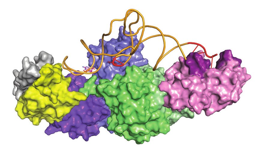
Nucleic Acids Research, 2021, Vol. 49, No. 19 10819

C-lobe). An inter-lobe cleft harbors elements projecting          Converging roads to the discovery of the KEOPS complex
from the two lobes that are required for ATP binding
                                                                  Two unrelated genetic screens conducted in S. cerevisiae us-
and for the catalytic transfer of the gamma phosphate of
                                                                  ing different strategies led to the discovery of the KEOPS
ATP to a hydroxyl bearing sidechain in proteinaceous sub-
                                                                  complex. The first study conducted a genome-wide screen
strates (for a review on the structure of kinases, see (6)).
                                                                  for suppressors of the growth defect of cells lacking a func-
Although the N-lobe of Bud32 harbors all the canoni-
                                                                  tional form of Cdc13, an essential protein that protects
cal elements characteristic of a functional protein kinase
                                                                  telomere ends from erosion (1). The analysis revealed that
enzyme, the C-lobe appears abnormally minimalistic (see
                                                                  deletion of the CGI121 gene enabled growth in the ab-
below). Despite the unusually abbreviated C-lobe, Bud32
                                                                  sence of Cdc13. Tandem affinity purification experiments
demonstrated autophosphorylation activity in vitro (5,7–9),
                                                                  of Cgi121 from yeast cells coupled to mass spectrometry re-
a property of most protein kinases (10). As expression of
                                                                  vealed that Cgi121 resides in a protein complex with Bud32,
catalytically dead Bud32 mutants did not fully rescue the
                                                                  Kae1 and Gon7. The physical interaction between KEOPS
slow growth phenotype of the bud32 yeast strain (11),

                                                                                                                                   Downloaded from https://academic.oup.com/nar/article/49/19/10818/6382379 by guest on 08 November 2021
                                                                  proteins was further supported by a large-scale analysis of
Bud32’s phospho-transfer function was inferred to be crit-
                                                                  the yeast interactome (25). Supporting the notion that the
ical for its cellular function. Within the human kinome,
                                                                  newly identified subunits of KEOPS form a functional com-
Bud32 bears closest resemblance to the Rio kinases (12).
                                                                  plex, knockout of any of these genes led to highly similar
Since the Rio kinases and Bud32 are the only two protein
                                                                  slow growth and shortened telomeres phenotypes (1), which
kinases conserved across eukaryotes and archaea (12,13),
                                                                  are most likely an indirect result of the role KEOPS plays
the two may represent primordial members of the eukary-
                                                                  in translation fidelity (elaborated on later in this review).
otic protein kinase superfamily from which the numerous
                                                                     The second study (2) utilized a different starting point,
other family members in eukaryotes have evolved.
                                                                  searching for suppressors of splicing defects caused by a
   Subsequent studies identified a Bud32 ortholog in hu-
                                                                  U to A mutation at the fifth position of U1snRNP, which
mans named Tp53 reactive kinase or p53-related protein
                                                                  yields a cold-sensitive phenotype (26). The analysis re-
kinase (TPRK or PRPK respectively) based on a report
                                                                  vealed that the cold-sensitive phenotype could be over-
assigning p53 as a PRPK substrate (14), which was most
                                                                  come by expression of an intron-less version product of
likely an in vitro artifact. A yeast two-hybrid analysis using
                                                                  the S. cerevisiae YKR095-A gene. This gene encoded a
PRPK as bait identified TPRK-binding protein (TPRKB),
                                                                  small uncharacterized protein that was required for the
the human ortholog of yeast Cgi121, as a binding partner
                                                                  transcriptional activation of pheromone and galactose re-
(15). Cgi121 was a protein of unknown function described
                                                                  sponsive genes and for polarized cell divisions. Chromatin-
only to be conserved between Caenorhabditis elegans and
                                                                  immunoprecipitation experiments showed the YKR095-A
humans in a Comparative Gene Identification study (hence
                                                                  protein associated with chromatin, leading to its naming as
its name) (16).
                                                                  polarized growth chromatin-associated controller 1 (Pcc1)
   A separate yeast two-hybrid study with S. cerevisiae
                                                                  (2). Tandem affinity purification coupled to mass spectrom-
Bud32 as bait identified OSGEP, a poorly character-
                                                                  etry revealed that Pcc1 resides within a complex with Gon7,
ized protein that was erroneously reported to have O-
                                                                  Kae1, Bud32 and Cgi121.
sialo-glycoprotease activity, as a physical interactor (17).
This led to the renaming of OSGEP to kinase-associated
endopeptidase 1 (Kae1) (11). In some archaeal species such        THE ROLE OF KEOPS IN CELLS AND IN HUMAN DIS-
as Methanocaldococcus jannaschii, Kae1 and Bud32 are              EASE
fused into a single polypeptide, hinting that their functions
                                                                  KEOPS catalyzes the t6 A modification of tRNA
are intertwined.
   As noted above, Kae1-family enzymes are part of a small        The requirement for KEOPS for cell growth suggested that
group of ∼60 genes present in the genomes of all au-              it fulfills an essential biochemical function. However, the
tonomous cells (3). Family members in archaea and eukary-         precise role of KEOPS remained elusive until 2011. In this
otes are called Kae1 while the bacterial orthologs are called     section, we provide an overview on how KEOPS activity
YgjD/TsaD (18). Similar to Kae1, TsaD is essential for            is essential for the fidelity of translation through the role
bacterial growth (19). Eukaryote genomes also encode the          KEOPS plays in generating the universal tRNA modifica-
Kae1 paralogue Qri7 (18). The open reading frame (ORF)            tion N6 -threonylcarbamoyl adenosine (t6 A).
of Qri7 contains an N-terminal mitochondrial targeting sig-          tRNAs undergo a complex and multi-staged biogenesis
nal peptide. Accordingly, although Qri7 is encoded in the         processes that includes the instalment of multiple posttran-
nucleus, it resides exclusively in the mitochondria (20–22).      scriptional modifications, catalyzed by tRNA-modifying
Analogous to the essential role of Kae1 and TsaD in cellu-        enzymes (27). To date, over 100 tRNA posttranscriptional
lar fitness, Qri7 is essential for the function of mitochondria   modifications have been identified (28). Each tRNA is esti-
(20,21).                                                          mated to carry 5 to 15 modifications distributed through-
   Very little was known about Pcc1 and Gon7. Deletion of         out its structure. These modifications regulate a range of
the GON7 gene (as well as BUD32) perturbed the transfer           tRNA functions from degradation to roles in translation
of mannosyl-phosphate groups to cell surface oligosaccha-         and modification dysregulation is implicated in human dis-
rides in S. cerevisiae (23). Pcc1 was completely uncharac-        ease (29–32). In particular, the decoding activities of tRNAs
terized, in part because of a lack of annotation resulting        are dependent on base modifications at positions 34 and 37
from its coding by an ORF containing an intron with a non-        within the tRNA anticodon loop (29), which critically in-
canonical splice site in S. cerevisiae (24).                      fluence the rate and fidelity of translation and the integrity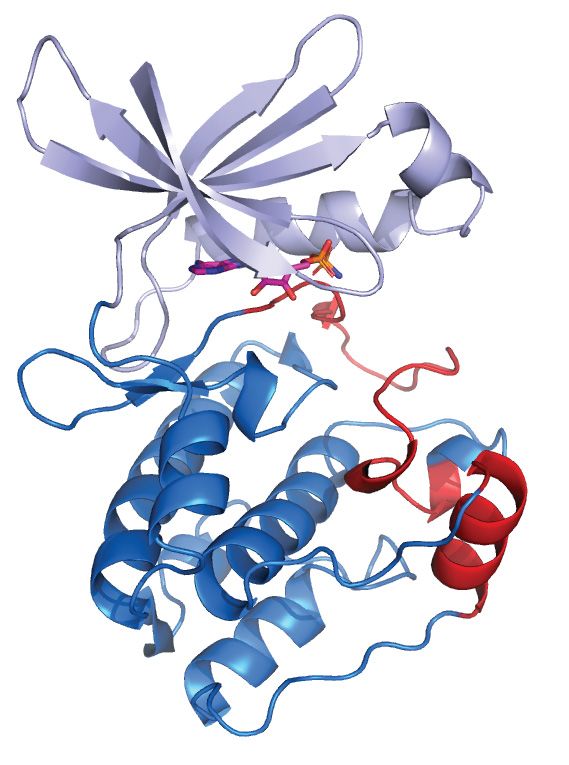
10820 Nucleic Acids Research, 2021, Vol. 49, No. 19

of the proteome (33). Of all tRNA modifications character-         DNA and RNA levels were only mildly affected in S. cere-
ized, approximately 17% are conserved in all domains of life       visiae cells in the absence of Kae1 activity, protein synthe-
(34). Not unexpectedly, conserved modifications are gener-         sis was dramatically reduced, implicating Kae1 in transla-
ated by similarly conserved enzymes.                               tion. This finding led to the hypothesis that Kae1 works with
   t6 A is a universal tRNA modification discovered in 1969        Sua5 to t6 A modify tRNAs. The second study (46) used a
(35) that is specifically found in tRNAs that decode ANN           comparative genomics approach, reasoning that since t6 A
codons, where N denotes any nucleotide (Figure 1A) (28).           is universally conserved the candidate enzymes involved in
t6 A occurs exclusively on A37, one position 3 to the an-         its biosynthesis would have a universal phylogenetic dis-
ticodon triplet (encompassing positions 34 to 36) and re-          tribution and an unknown biochemical function. Remark-
models and stabilizes the structure of the tRNA anticodon          ably, one of the few enzymes that met both criteria were the
loop by preventing an inter-loop hydrogen bond between             Kae1/TsaD/Qri7 family of enzymes (57) hinting that Kae1
A37 and U33 (Figure 1B) (36–40). This remodeling facil-            would collaborate with Sua5 in the t6 A biosynthesis path-
itates enhanced binding to the ribosome (41). In the ribo-         way. Both the first and second studies proved their working

                                                                                                                                      Downloaded from https://academic.oup.com/nar/article/49/19/10818/6382379 by guest on 08 November 2021
some, t6 A reinforces A1 :U36 codon-anticodon binding by           hypotheses by showing that mutations in Kae1 in S. cere-
inducing base stacking interactions between A37 and A38            visiae resulted in a loss of t6 A. The third study (55) revealed
of the tRNA and the A at the first position of the codon           that the knockout of KEOPS genes leads to transcriptional
triplet in the mRNA (Figure 1B) (40,42,43). As a result, t6 A      activation of target genes of the transcription factor Gcn4
is essential for the fidelity of translation and for the func-     in a Gcn2-independent manner. Gcn4 protein expression is
tionality of ANN-decoding tRNAs (44–47). t6 A can be fur-          normally suppressed by upstream ORFs that contain AUG
ther modified to five different derivative forms (28). In Es-      start codons. The Gcn4 protein is synthesized when the up-
cherichia coli and possibly in other species, almost all t6 A      stream kinase Gcn2 phosphorylates eIF2␣, suppressing the
is cyclized (denoted ct6 A) by the CsdL/TsdA enzyme (48).          inhibitory effect of the upstream ORFs (for an extensive
Crystal structures show that ct6 A adopts a hydantoin struc-       review of Gcn4 translational regulation see (58)). There-
tural form. Like t6 A, ct6 A stabilizes the structure of the an-   fore, increased Gcn4 expression in the absence of KEOPS
ticodon loop. However, it is not clear if ct6 A helps codon–       activity hinted that KEOPS would influence the selection
anticodon binding in the ribosome (49). t6 A can also be           of AUG start sites. Because Sua5 (45) and its downstream
N6 -methylated by TRMO (tRNA methytransferase O) to                product t6 A (47) are important for translation initiation,
form m6 t6 A (50) or N2 -methyl-thiolated by MtaB/Cdkla1l          this led the authors to hypothesize that KEOPS is involved
to form ms2 t6 A (51). These two specific t6 A derivative mod-     in generating the t6 A modification.
ifications provide further tuning to the decoding activities of       Together, these breakthrough studies unequivocally
t6 A modified tRNAs.                                               proved that Sua5 and Kae1/TsaD/Qri7 are needed for the
   In 2009, 40 years after the discovery of t6 A, the first        t6 A modification of tRNA in vivo (46,47,55,56). Subsequent
enzyme in the t6 A biosynthesis pathway was discovered             studies with purified proteins (together with their respective
through a comparative genomics approach that surveyed              binding partners) demonstrated the sufficiency of these en-
universally conserved proteins of unknown function (47)            zymes to reconstitute the t6 A modification of tRNA in vitro
(for a detailed review of the conservation and diversity of        (59–61). The specific role each enzyme plays in this process
t6 A biosynthesis enzymes, see (18)). This analysis revealed       and how these roles were assigned is described later in this
that Sua5 (Suppressor of upstream AUG 5) (52) is directly          review.
involved in t6 A biosynthesis. However, since Sua5 in isola-
tion was not sufficient to t6 A modify tRNA in vitro (47),
the t6 A biosynthesis pathway was predicted to involve ad-         KEOPS activity is essential for diverse aspects of cell bi-
ditional gene products. Interestingly, some bacterial and ar-      ology and mutations in KEOPS underlie Galloway–Mowat
chaeal genomes encode proteins that contain a Sua5-like            syndrome
domain fused to a Kae1-like domain (53) hinting at a linked        Genetic studies conducted in the eukaryotic species yeast,
functional role between these distinct classes of enzymes.         zebra fish, fruit flies and mice as well as the archaeon
Rhizobium fredii NolO, a Kae1-Sua5 like fusion protein,            Haloferax volcanaii have established an essential role for
functions as a carbamoyl-transferase (54), which raised the        KEOPS in normal cell growth and in animal development.
possibility that Sua5 and Kae1 could participate in catalyz-          KEOPS function has been most extensively character-
ing reactions with similar chemistry (53), which ultimately        ized in vivo in budding yeast. Knockout of any individ-
proved to be the case.                                             ual KEOPS gene leads to pleiotropic phenotypes, ranging
   In 2011, three breakthrough studies identified the              from slow growth to shortened telomeres and to defects in
Kae1/TsaD/Qri7 family of enzymes as the missing com-               transcription (1,2,44,55,62,63). These phenotypes are most
ponent in t6 A biosynthesis (46,55,56). All three studies          likely an indirect result of the role KEOPS plays in tRNA
noted the similarity between Kae1 to the Kae1-Sua5 like fu-        modification and translation and of the far-reaching down-
sion proteins as an indication that both enzymes might co-         stream effects that the perturbation of translation has on
operate to generate t6 A. The first study (56) noted that the      virtually all cellular processes. The impact of KEOPS activ-
extraordinary conservation of the Kae1 protein indicated           ity on translation has been studied using reporter based as-
that it would be involved in a universal cellular process. This    says, which showed that the absence of the t6 A modification
led to a search for a potential role Kae1 might play in the        leads to an increase in translation mis-initiation and in ± 1
synthesis of the major macromolecules in cells. While global       frame-shift events (46,47,55) as well as defects in stop codon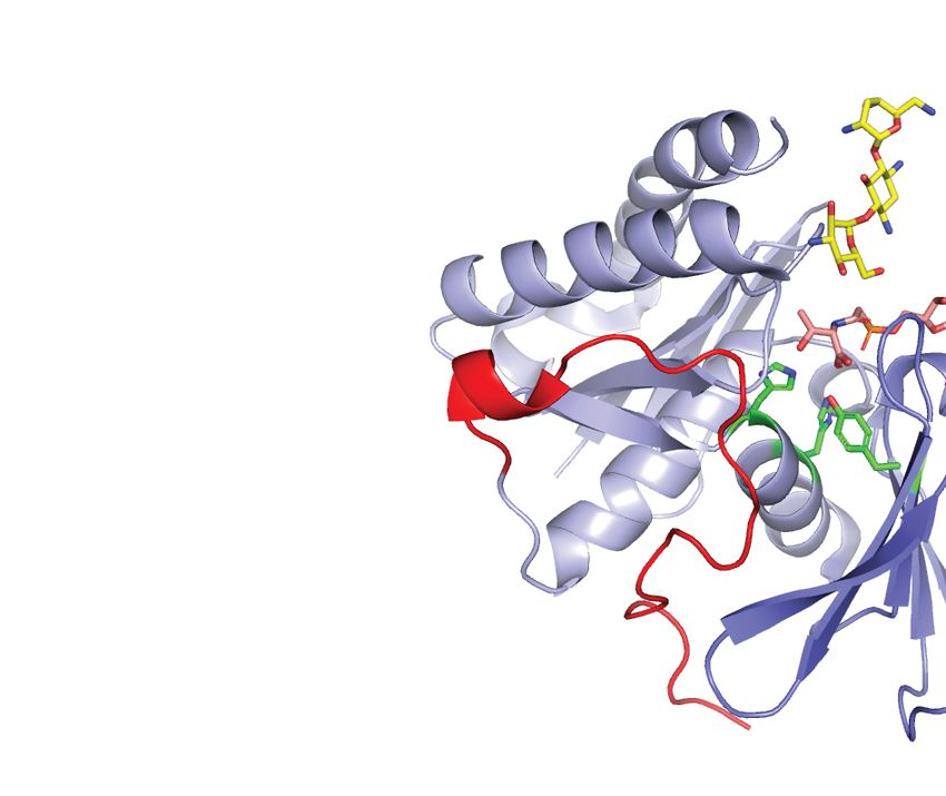
Nucleic Acids Research, 2021, Vol. 49, No. 19 10821

                                                                                                        3’                               5’
            A                                      3’ CCA tail                           B                         tRNA backbone
                                                   (positions      3’
                        t6A                                        A
                                                     74-76)        C                                                            2. Hyrdogen bond
                 threonyl-                                         C 5’                      tRNA                                   prevented
                                                                                                       1. Base stacking
                carbamoyl                                                                    Lys
                                                                          tRNA                  UUU       interaction               (t6A-U33)
                  moeity                                                                                 (A38-t6A-A1)
                                                         T-arm
                                                                          D-arm                                                    U33
              position N6                                                                             A38
                                                 UAA motif
                                              (positions 36-38)                                threonyl-
                                                                    A        Anticodon        carbamoyl
                                                                 t6A           loop             moiety       t6A          U36
                                                                    UNN
                              Adenosine
                                              5’                    ANN            3’

                                                                                                                                                          Downloaded from https://academic.oup.com/nar/article/49/19/10818/6382379 by guest on 08 November 2021
                                                                                                                                     Anticodon
                                                                             mRNA               5’      A1
                                              A1 position                                                                            (U34-U36)
                                                                  Codon-
                                                                 anticodon
                                                                  paring                               mRNA
                                                                                                       codon                                  3’
                                                                                                      (A1-A3)

Figure 1. t6 A is a posttranscriptional modification of A37 in the anticodon loop of ANN-decoding tRNAs. (A) (left) Chemical structure of N6 -
threonycarbamoyladenosine (t6 A). The threonylcarbamoyl moiety is shown in red and the adenosine is shown in black. (right) A schematic structure
of an ANN-decoding tRNA engaged with an ANN codon within mRNA. Codon–anticodon pairing is shown in blue. The 34-UAA-36 motif and the
position of t6 A as well as the CCA tail are shown in red. (B) Structure of the anticodon loop of t6 A modified Thermus thermophilus tRNALys UUU (PDB
1XM). The UUU anticodon triplet is shown in grey engaged with an AAA codon in yellow. For simplicity, the ribosome is not shown. A38 (green) and
t6 A (purple) of the tRNA form base stacking interactions with A1 of the mRNA (yellow). This stabilizes the binding of U36 (grey) with A1. The presence
of t6 A prevents base pairing with U33 (green). This stabilizes anticodon loop structure.

recognition (45,64). A higher resolution study using ribo-                         tprkb knockout (Kae1 and Cgi121 orthologs respectively)
some profiling revealed that the role t6 A has in translation                      leads to microcephaly and to a shortened life span (74,75).
varies considerably from one ANN codon to another. The                             Similarly, knockout of either of the osgep, lage3, prpk or
translation rates of ANN codons that are decoded by rare                           tprkb (Kae1, Pcc1, Bud32 and Cgi121 orthologs respec-
tRNAs or tRNAs with weak G34 :U3 pairing at the wob-                               tively) genes in Mus musculus leads to severe microcephaly
ble position decreased in the absence of t6 A. In contrast,                        and early lethality (74). Why the developmental defects re-
the translation rates of ANN codons that are dependent on                          sulting from KEOPS dysregulation localize with great fre-
high abundance tRNAs or tRNA with strong I34 :C3 pairing                           quency to the brain remains an open question.
at the wobble position were in fact increased by the absence                          In line with studies in model organisms, genetic analysis
of t6 A (44). Both types of changes in translation rate likely                     in humans have identified pathogenic mutations that under-
have a major role in the perturbed translation observed in                         lie Galloway–Mowat syndrome (GAMOS) in each of the
the absence of t6 A.                                                               five KEOPS genes and in Sua5 (74,76,77). GAMOS is a re-
   The importance of KEOPS activity for the well-being of                          cessive genetic disease that manifests in renal and neuro-
cells has been confirmed in a variety of other model or-                           logical dysfunctions with a considerable variety of sever-
ganisms. Briefly, in the archaeon H. volcanaii, the cgi121                         ity (74,76–78). As a result, GAMOS patients die at very
and kae1-bud32 fusion genes are essential for cell growth,                         early stages of life. Since GAMOS inducing mutations are
while knockout of pcc1 yields a less severe growth de-                             common to KEOPS and Sua5 encoding genes, it is highly
fect (65). In Drosophila melanogaster, only Pcc1, Kae1 and                         likely that reduced t6 A levels underpins the GAMOS dis-
Bud32 (termed Prpk) orthologs have been identified thus                            ease phenotype. Consistent with this notion, introduction
far (18,66). Knockout or knockdown of Kae1, Bud32, Pcc1                            of GAMOS disease mutations into yeast Kae1 inhibits the
or Sua5 in Drosophila leads to reduced larval size and trans-                      modification of tRNA in vivo (74,76). KEOPS pathogenic
formation to pupa (66–69). The sensitivity to Kae1 knock-                          mutations elicit complex cellular phenotypes that likely
down varies between different tissues, with mitotic tissues                        stem from perturbed translation including activation of the
especially sensitive while non-proliferating tissues much less                     DNA-damage and unfolded protein response, ER stress
sensitive (68). Interestingly, the rare larvae that manage to                      and defects in actin regulation (74,76). Some pathogenic
transform to pupa despite knockdown of Prpk develop into                           mutations in KEOPS exert their effect by disrupting the
flies with reduced cell and organ size but without extreme                         formation of KEOPS, while others are pathogenic for un-
defects in body patterning (70). This result is reminiscent                        known reasons (74,77). Further studies are required for a
of mutants in the TOR pathway (71) and hints at a poten-                           complete understanding of the molecular mechanisms con-
tial role for KEOPS in regulating TOR signaling (67,72).                           necting KEOPS mutations to GAMOS.
In agreement with this possibility, the growth defects in-                            t6 A derivative modifications are also important for cel-
duced by Prpk knockdown are reversed by expression of an                           lular processes and their dysregulation is implicated in hu-
activated mutant of the S6 kinase (70), a downstream ef-                           man disease. Mammalian Cdkal1 for example catalyzes the
fector of the TOR pathway (73). In Danio rerio, osgep and                          methythiolation of t6 A to ms2 t6 A (51). In the absence of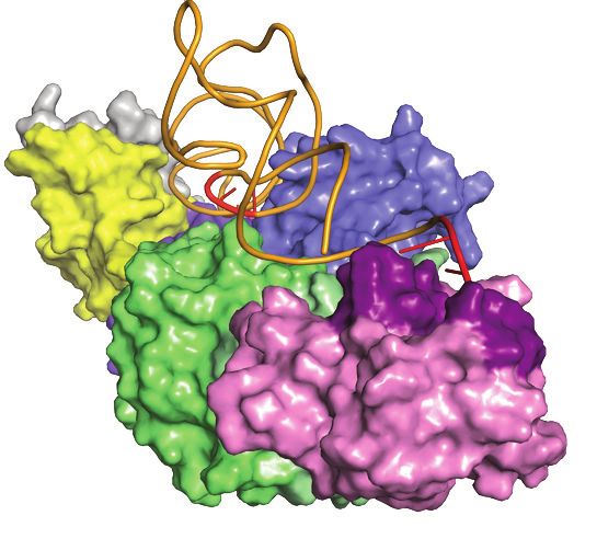
10822 Nucleic Acids Research, 2021, Vol. 49, No. 19
Cdkal1 activity, insulin synthesis is inhibited (51) due to in chambers separated by a 2 kDa size cut off membrane
misreading of lysine codons in pro-insulin (79) leading to but only when tRNA was in the same chamber as Qri7 (61).
development of type 2 diabetes. In vivo, it is currently unclear how the unstable TC-AMP is
efficiently shuttled to the active site of the Kae1/TsaD/Qri7
HOW KEOPS FUNCTIONS TO MODIFY TRNA enzymes before its breakdown.
Remarkably, although all three Kae1 family orthologs
Sua5 makes the t6 A modification reaction intermediate thre- perform the same biochemical reaction, each ortholog func-
onlycarbamoyl adenylate, which Kae1 uses to modify tRNA tions in a completely different protein complex (Figure 2A).
The mechanism by which Sua5 and Kae1 collaborate to The mitochondrial Qri7 is the most simplified member of
modify tRNA with t6 A was delineated through a series of the family and catalyzes tRNA modification without any
elegant biochemical and structural studies described below. auxiliary proteins (61,89). The bacterial TsaD protein de-
Briefly, the t6 A modification of tRNA occurs in a reaction pends on its binding to YaeZ/TsaB, which is a structural
mimic of Kae1 enzymes (90–92). TsaD bound to TsaB can
Downloaded from https://academic.oup.com/nar/article/49/19/10818/6382379 by guest on 08 November 2021
with two major steps. In the first step, Sua5 utilizes thre-
onine, CO2 /HCO3 − and ATP precursors to generate the bind tRNA but has limited catalytic capacity and likely cat-
diffusible intermediate molecule threonylcarbamoyl adeny- alyzes only a single round of tRNA modification (93). For
late (TC-AMP) while releasing PPi from ATP. In the second multiple rounds of tRNA modification, TsaD-TsaB depend
step, TC-AMP and tRNA are bound by Kae1/TsaD/Qri7, on the activity of the ATPase subunit YjeE/TsaE (59,92–95)
which then transfer the threonylcarbamoyl moiety from for reasons that are not entirely understood. Notably, TsaB
TC-AMP to the N6 acceptor site of A37 in tRNA in a reac- and TsaE are not similar in structure and apparent function
tion that generates AMP (Figure 2A). to any KEOPS subunits. The significance of the different
complex compositions is one of the most exciting mysteries
Sua5 generates TC-AMP. As early as 1974, t6 A biosyn- in the field that remain unresolved (see ‘CONCLUDING
thesis was known to require the precursors threonine, ATP REMARKS’ section).
and HCO3 − (80). Knowing that Sua5 was involved in the The tRNA modification activity of Sua5 and
t6 A modification process and that Sua5 has ATP binding Kae1/TsaD/Qri7 enzymes extends to tRNA modifi-
properties (81,82), Deutsch et al. and Perrochia et al. ex- cations other than t6 A. Sua5 can generate adenylated
amined the effect of threonine and tRNA on its ATP hy- variants of TC-AMP using serine or hydroxy norvaline
drolysis activity. They found that ATP consumption by Es- (hn) in place of threonine. In turn, the bacterial TsaD
cherichia coli, Pyrococcus abyysi and S. cerevisiae Sua5 pro- protein can utilize hn-carbamoyl adenylate to generate
teins was dependent on threonine (but not tRNA). This an hn6 A modification on substrate tRNAs (96). Since the
hinted that Sua5 produces an adenylated threonine con- hn6 A modification is also observed in archaeal species
taining molecule for use in the t6 A modification reaction (97,98), this would suggest that KEOPS in both archaea
(59,60). Subsequent studies using Bacillus subtilis and S. and eukaryotes may similarly utilize hn-carbamoyl adeny-
cerevisiae Sua5 proteins showed this molecule to be TC- late to modify tRNA. However, the functional relevance of
AMP (Figure 2B) (83). TC-AMP is a relatively short-lived this modification remains in question.
molecule that breaks down to threonylcarbamoyl and AMP
moieties. Its stability is strongly affected by pH and tem- Structure of the individual subunits
perature, with a half-life of ∼20 seconds at pH 7.5 and
at 37◦ C (83). TC-AMP can be stabilized by its binding to Bud32 is an atypical protein kinase that lacks the infrastruc-
Sua5, but not by its binding to Kae1/TsaD/Qri7 enzymes ture for canonical protein substrate recognition. Eight crys-
(84). The structural basis by which Sua5 generates TC-AMP tal structures of complexes containing Bud32 have been de-
was delineated by X-ray co-crystal and NMR structures of posited to the PDB (for a list of the structures of proteins
Sua5 with threonine, ATP and HCO3 − precursors (81,85– that take part in t6 A modifying complexes, see Table 1).
87). After the discovery that Sua5 produces TC-AMP, re- These include structures of Bud32 orthologs from archaea,
analysis of a previously determined crystal structure of Sul- yeast and human in complex with either Kae1, Cgi121 or
folobus tokodaii Sua5 bound to ATP, revealed serendipi- both. These structures reveal a bi-lobal domain organiza-
tously that the ATP moiety bound in the Sua5 active site tion characteristic of the eukaryotic protein kinase family
was actually TC-AMP (81,88). as exemplified by PKA, a well-studied prototypical kinase
(Figure 3A) (for review on the composition of the kinase
Kae1/TsaD/Qri7 uses TC-AMP to make t6 A. The discov- domain, see (6)). The kinase N-lobe of Bud32 possesses the
ery that Sua5 generates the precursor TC-AMP suggested canonical antiparallel −strands, buttressed against a he-
that the Kae1/TsaD/Qri7 enzymes would employ TC-AMP lix ␣C (Figure 3A) (8,9,99). The C-lobe, which is connected
and tRNA to catalyze the remainder of the t6 A modifica- to the N-lobe through a flexible hinge region, is predomi-
tion reaction. In support, P. abyssi KEOPS binds tRNA in nantly ␣-helical and greatly abbreviated (i.e. of smaller size)
vitro and co-purifies with tRNA when expressed in E. coli relative to the C-lobes of other eukaryotic protein kinases.
cells, suggesting that this complex directly acts on tRNA One peculiar feature of the abbreviated kinase C-lobe is the
(60). Evidence that the Kae1/TsaD/Qri7 enzymes carry out absence of all infrastructure typically required for protein
the final step of the t6 A modification reaction came from substrate recognition, including helix-␣G and the activation
the analysis of Qri7, which together with Sua5 are sufficient segment (100) (Figure 3A). A second unique feature of the
to carry out the t6 A modification of tRNA in vitro (61). kinase C-lobe is the presence of a basic C-terminal tail with
tRNA modification could be achieved with Sua5 and Qri7 no apparent homology in other members of the kinome (5).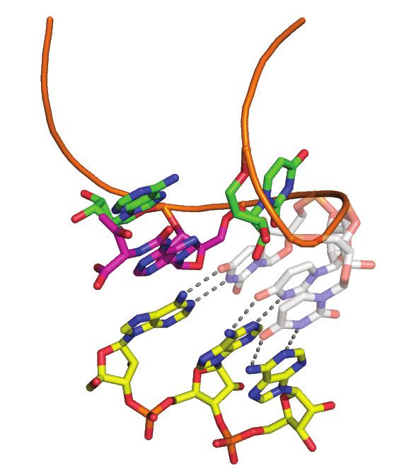
Nucleic Acids Research, 2021, Vol. 49, No. 19 10823
A
Downloaded from https://academic.oup.com/nar/article/49/19/10818/6382379 by guest on 08 November 2021
B
Figure 2. t6 A is universally biosynthesized in a two-step reaction by Sua5 and Kae1/TsaD/Qri7 enzymes. (A) Schematic of the universal t6 A biosynthesis
pathway. Step 1- Sua5 utilizes ATP, threonine and HCO3 − /CO2 to catalyze the formation of threonylcarbamoyl adenylate (TC-AMP). Step 2- TC-AMP
is used by Kae1/TsaD/Qri7 enzymes to t6 A modify ANN-decoding tRNA substrates. (top) TsaD functions with the TsaB and TsaE subunits in bacteria.
(middle) Kae1 functions with Cgi121, Bud32, Pcc1 and Gon7 subunits in the cytoplasm in eukaryotes and in archaea. (Bottom) Qri7 functions either as
a homo-dimer as shown in figure or as a monomer. (B) Chemical structure of threonylcarbamoyl adenylate (TC-AMP). The threonylcarbamoyl moiety is
shown in red and the adenylate is shown in black.
ATP binds to the catalytic cleft of Bud32 situated be- periments showing that the autophosphorylation acceptor-
tween the N and C- lobes. The ATP binding mechanism sites are not required for physiological activity (7,102) sug-
in Bud32 is conserved with that of other protein kinases gests an alternate role for its phospho-transfer function. In
(6,99,101). Specifically, the adenine base of ATP is wedged line with this prediction, the chief function of Bud32 ap-
towards the kinase hinge. The ribose and phosphate groups pears directed at ATP hydrolysis (i.e. transfer of phosphate
are coordinated by the conserved −3 Lys, ␣C-helix Glu, to water) (102).
Gly rich loop, and Asp side chain of the conserved Asp-
Phe-Gly (DFG) motif through a bridging Mg/Mn ion. Kae1 has a bi-lobal ASKHA fold. Nine crystal structures
Phospho-transfer function of protein kinases is mediated of Kae1 alone or in complex with different KEOPS pro-
in part by a catalytic Asp residue within a conserved His- teins have been deposited to the PDB (Table 1). These in-
Arg-Asp (HRD) motif in a region termed the catalytic loop clude structures of Kae1 orthologs from archaea, yeast and
(6). This Asp residue functions as a catalytic base to ex- humans either alone, in complex with Pcc1, in complex
tract a proton from the target hydroxyl group and is con- with Pcc1 and Gon7, in complex with Bud32 or in com-
served in Bud32, suggesting that Bud32 also shares the abil- plex with Bud32 and Cgi121. Likewise, multiple structures
ity to catalyze phosphate transfer. Accordingly, Bud32 pro- of the Kae1 orthologs Qri7 and TsaD have been solved
teins manifest autophosphorylation activity (5,8,9) and its in their respective complexes. These structures reveal that
phospho-transfer function is essential for biological func- Kae1 adopts a bi-lobal domain organization characteris-
tion (9,11). However, the absence of structural elements nor- tic of the ASKHA (acetate and sugar kinases/Hsc70/actin)
mally required for protein phosphorylation as well as ex- family of enzymes (Figure 3B) (103,104). The two subdo-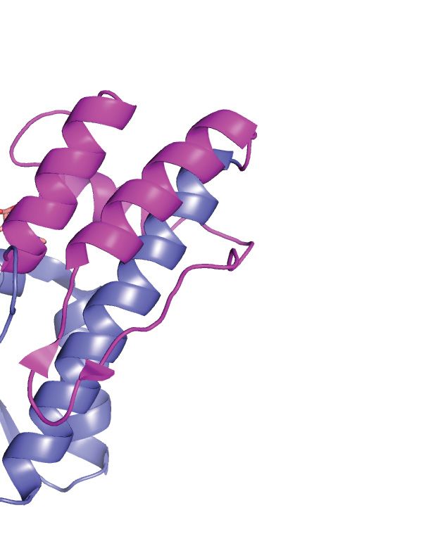
10824 Nucleic Acids Research, 2021, Vol. 49, No. 19
Table 1. Structures of KEOPS proteins and sub-complexes. All structures reported are crystal structures unless stated otherwise. f = fusion protein.
car-AMP = carboxy-adenylate. BK951 = TC-AMP mimetic.
t6 A modifying complex Proteins Species Ligand PDB code
Eukaryotes (cytoplasm) and archaea Bud32-Cgi121 S. cerevisiae - 4WWA
Bud32-Cgi121 S. cerevisiae AMP 4WW7
Bud32-Cgi121 S. cerevisiae ADP 4WW9
Bud32-Cgi121 S. cerevisiae AMP-PnP 4WW9
Bud32-Cgi121 H. sapiens AMP-PnP 6WQX
Kae1-Bud32(f) M. jannaschii AMP-PnP 2VWB
Kae1-Bud32(f) M. jannaschii - 3EN9
Kae1 P. abyssi - 2IVO
Kae1 P. abyssi ATP 2IVP
Kae1 P. abyssi AMP-PnP 2IVN
Downloaded from https://academic.oup.com/nar/article/49/19/10818/6382379 by guest on 08 November 2021
Kae1-Pcc1 T. acidophilum, P. furiosus - 3ENO
Kae1-Pcc1 M. jannaschii, P. furiosus AMP 5JMV
Kae1-Bud32(f) -Cgi121 M. jannaschii - 3ENH
Pcc1-Pcc1 dimer P. furiosus - 3ENC
Pcc1-Gon7 S. cerevisiae - 4WXA
Pcc1-Gon7 S. cerevisiae - 4WX8
Gon7-Pcc1-Kae1 H. sapiens - 6GWJ
Cgi121 S. cerevisiae - 4XAH
Cgi121 H. sapiens - 3ENP
Cgi121 (NMR) M. jannaschii - 2K8Y
Cgi121 M. jannaschii tRNA 7KJT
Mitochondria Qri7-Qri7 dimer S. cerevisiae 4K25
Qri7 S. cerevisiae AMP 4WUH
Bacteria TsaB dimer E. coli - 1OKJ
TsaB dimer S. enterica - 2GEM, 2GEL
TsaB dimer V. parahaemolyticus - 3R6M
TsaB dimer P. aeruginosa - 4Y0W, 5BR9
TsaB dimer T. maritima - 2A6A
TsaE H. influenzae - 1FL9
TsaE H. influenzae ADP 1HTW
TsaE B. subtilus ADP 5MVR, 5NP9
TsaD-TsaB E. coli BK951, AMP 6Z81
TsaD-TsaB E. coli ADP 4YDU
TsaD(E12A mutant) -TsaB E. coli ATP 4WQ4
TsaD(V85E mutant) -TsaB E. coli ATP 4WQ5
TsaD-TsaB S. typhimurium AMP 3ZET
TsaD-TsaB S. enterica ATP-␥ -S 3ZEU
TsaD-TsaB-TsaE T. maritima ATP, car-AMP 6N9A
TsaD-TsaB-TsaE T. maritima AMPcPP 6S84
mains of Kae1 bear considerable similarity to each other cation bound by the metal binding triad serves to coor-
and contain a five stranded -sheet core flanked by ␣- dinate the carboxylate group of the threonine moiety of
helices. In the cavity between the two subdomains, lies a TC-AMP (Figure 3B). This binding mode directs the thre-
conserved metal binding triad, which co-ordinates a Zn, Mg onylcarbamoyl group towards a cavity in the active site
or Fe ion. Kae1 enzymes have two unique inserts relative cleft, presumably guiding it to the binding site for tRNA.
to other ASKHA fold enzymes, termed Kae1 specific in- How the threonylcarbamoyl group is activated by Kae1
sert I and II, which are tightly associated with sub-domain for the transfer reaction to N6 of A37 remains unclear. To
2 and 1 respectively (Figure 3B) (9,104). Unique inserts in date, there is no structure of a Kae1 family enzyme with a
ASKHA-fold enzymes commonly contribute to the unique tRNA substrate. The expected position of A37 can at best
sub-family functions (103), which also holds true for Kae1 be inferred from the co-structure of TobZ, a NodU family
proteins (see below). carbamoyl transferase, with its substrate the antibiotic to-
How does the active site of Kae1 bind its substrates TC- bramycin (53,88). Since A37 and tobramycin should occupy
AMP and tRNA? The unstable nature of TC-AMP hin- a similar position in their respective enzyme’s active site, su-
dered the ability to obtain atomic level insight into how perimposing the structure of the TobZ-tobramycin complex
it is bound by Kae1-family enzymes. However, recently, onto Kae1 places A37 in close proximity to TC-AMP and
Kopina et al. developed a non-hydrolysable TC-AMP ana- the metal binding triad (Figure 3B).
logue (termed BK951) and reported its structure bound to What features of substrate tRNAs dictates the substrate
the E. coli TsaD-TsaB heterodimer (84). The structure re- selectivity of Kae1 enzymes? ANN-decoding tRNAs uni-
vealed that the adenine base portion of TC-AMP is ‘sand- versally harbor a conserved 36-UAA-38 motif within the
wiched’ between subdomain 2 and Kae1 specific insert I, anticodon loop with the A37 site at its center (Figure 3C).
similar to the nucleotide binding mechanism of Kae1 en- This motif partially overlaps with the anticodon at posi-
zymes reported in previous structures (8,84,91,95,105). A tions 34 to 36. The presence of the 36-UAA-38 motif can be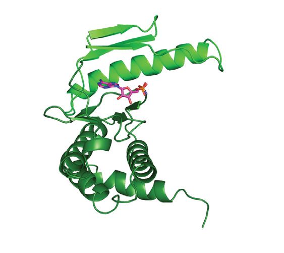
Nucleic Acids Research, 2021, Vol. 49, No. 19 10825

              A                                                                  B                            Active
                                                                                                             site cleft
                        Bud32                              PKA                             Pcc1                                  Kae1-specific
                                                                                          binding                                  insert I
                          Cgi121 binding                                                  surface                   Tob
                             surface

                                             N-lobe
             ATP                                                                                                                             Bud32
                                                                    ATP                                                                      binding
                                Kae1                                                     Kae1-                                               surface
                               binding                                                  specific
                               surface                                                                                              TC-AMP
                                                                                        insert II
                                              C-lobe

                                                                                                           metal binding

                                                                                                                                                              Downloaded from https://academic.oup.com/nar/article/49/19/10818/6382379 by guest on 08 November 2021
                  Unique C-terminal tail                 Canonical activation              Kae1-               triad                 Kae1-
                                                        segment and helix G             Subdomain 1                               Subdomain 2

             C                                 Anticodon    UAA                 D                Hydrophobic dimerization
                                                 triplet    motif                                       interface
                                                                                      Pcc1 protomer 2             Pcc1 protomer 1
                           Lys-UUU:        5’-...CUGGCUUUUAACCA...-3’
                           Ile-GAU:        5’-...UCGGCUGAUAACCG...-3’
                                                                                                                                  Kae1
                    ANN-   Met-CAU:        5’-...CCGGCUCAUAACCG...-3’
                                                                                         Kae1                                    binding
                  decoding Thr-GGU:        5’-...CUGCUUGGUAAGCA...-3’
                                                                                        binding                                  surface
                   tRNAs Asn-GUU:          5’-...CGGACUGUUAAUCC...-3’
                                                                                        surface
                           Arg-UCU:        5’-...CGGCCUUCUAAGCC...-3’
                           Ser-GCU:        5’-...GGGACUGCUAAUCC...-3’
                           Ala-GGC:        5’-...CGCAUUGGCUGUGC...-3’
                    Other  Arg-GCG:        5’-...UGGCCUGCGGAGCC...-3’                                                     Pcc1
                   tRNAs Asp-GUC:          5’-...GGGACUGUCACUCC...-3’
                           Cys-GCA:        5’-...CGGACUGCAGAUCC...-3’
                                                                                                                                  Kae1
                   Nucleotide position :
                                                 30

                                                        34
                                                        36
                                                        38
                                                              40

                                                                                                                                 binding
                                                                                                                                 surface
                                                                                                    Gon7

              E
                                         tRNA binding
                                           surface

                             Bud32
                             binding                    Cgi121
                             surface

Figure 3. Structural characteristics of the individual KEOPS subunits. (A) Bud32 is an atypical protein kinase. (left) The structure of M. jannaschii Bud32
(PDB 2VWB) is shown with AMP-PnP in the active site (depicted as ATP for simplicity). Highlighted is the unique C-terminal tail and the location of
the binding surfaces for Cgi121 and Kae1. (Right) Structure of the kinase domain of M. musculus PKA (PDB 1ATP) is shown with ATP in the active site.
Highlighted are the canonical activation segment and helix-␣G (red) that are distinctively absent from the structure of Bud32. (B) Kae1 is an ASKHA fold
enzyme with two unique insertions. The structure of M. Jannaschii Kae1 (PDB 2VWB) is shown with the threonylcarbamoyl adenylate (TC-AMP) mimetic
BK951 (PDB 6Z81) and substrate analogue tobramycin (tob, PDB 3VET) superimposed on its active site. Highlighted are the Kae1-specific inserts I and
II (purple and red, respectively) and the location of the binding surfaces for Bud32 and Pcc1. (C) Kae1/TsaD/Qri7 enzymes substrate tRNAs harbor a
universal UAA motif in the anticodon loop. A sequence alignment of representative ANN-decoding and non-ANN-decoding tRNAs from M. jannaschii
is shown highlighting that ANN-decoding tRNAs harbor a 36-UAA-38 motif (red) within the anticodon loop that partially overlaps with the anticodon
triplet (positions 34–36). Note that none of the non-ANN-decoding tRNAs have this motif. (D) Pcc1 can homo-dimerize or hetero-dimerize with Gon7.
(top) The structure of P. furiosus Pcc1 homo-dimer is shown (PDB 3ENC). The dimerization interface and the location of the Kae1 binding surface are
highlighted. (bottom) The structure of human Pcc1-Gon7 hetero-dimer is shown (PDB 6GWJ). Note the similarity between the Pcc1 homo-dimer and
Pcc1-Gon7 hetero-dimer configuration. (E) Cgi121 is a single domain protein. The structure of M. jannaschii Cgi121 (PDB 3ENH) is shown. The location
of the binding surfaces for Bud32 and tRNA are highlighted.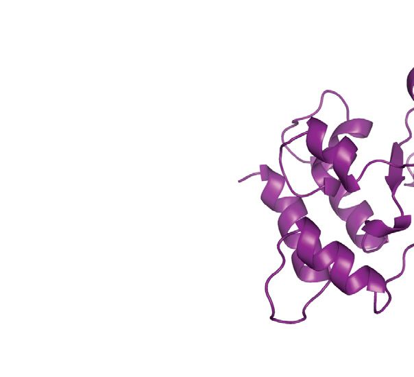
10826 Nucleic Acids Research, 2021, Vol. 49, No. 19
explained in part through the roles these nucleotides play in two anti-parallel -sheets and a single ␣-helix (Figure 3D).
translation (elaborated on above and shown in the structure The C-terminus of Gon7 (residues 51–100) is enriched with
presented in Figure 1B). However, the UAA motif also plays acidic residues, a feature that is conserved among Gon7 or-
a role in the ability of the tRNA to be modified by Kae1. thologs (77,99).
Mutation of U36 or A38 disrupts t6 A modification, demon-
strating the necessity of the UAA motif for tRNA recog- Cgi121 structure. Nine crystal structures and 1 solution
nition by Kae1. Insertion of a 36-UAA-38 motif into the structure solved by NMR of Cgi121 alone or in complex
anticodon loop of non-substrate tRNAs can lead to their with other KEOPS proteins or substrate tRNA have been
modification (106–108) demonstrating sufficiency in some deposited to the PDB (Table 1). These include the structures
instances. For example, mutations of either U36 or A38 of Cgi121 orthologs from archaea, yeast, and human either
in tRNAIle disrupts its t6 A modification in Xenopus laevis alone, in complex with Bud32, in complex with Bud32-Kae1
oocytes (107). In the same system, converting the 36-CAC- or in complex with tRNALys UUU . These structures revealed
38 motif of tRNAVal , which is normally not t6 A modified, to that Cgi121 is a single domain protein containing a core an-
Downloaded from https://academic.oup.com/nar/article/49/19/10818/6382379 by guest on 08 November 2021
a 36-UAA-38 motif was sufficient for inducing its t6 A mod- tiparallel -sheet of 3 to 5 strands sandwiched between ␣-
ification (107). Highlighting biological significance, muta- helices on either side (9) (Figure 3E).
tion of the UAA motif in substrate tRNAs can contribute
to human disease. For example, pathogenic mutations that Basis for KEOPS subunit interaction with each other and
cause myoclonus epilepsy associated with ragged red fibers with substrate tRNA
(MERRF) disease alters the 36-UAA-38 motif of mito- Overview: KEOPS subunits adopt a linear binding topology.
chondrial (m) tRNAThr to UAG. This mutation inhibits its The architecture and tRNA binding mechanism of KEOPS
t6 A modification by the human Qri7 orthologue OSGEPL1 have been elucidated by solving a series of binary and tri-
(106,108). In contrast, mtRNAIle naturally harbors a non- nary structures of its subunits (Table 1). In brief, KEOPS
canonical 36-UAG-38 motif and is therefore normally not a adopts a linear binding architecture (Figure 4A). The two
substrate for t6 A modification. However, a mtRNAIle G38A enzymes Kae1 and Bud32 bind directly to each other at
pathogenic mutation that leads to Leigh syndrome recon- the center of KEOPS. On opposing ends, Bud32 binds to
stitutes a canonical 36-UAA-38 motif, inducing the unnat- Cgi121 and Pcc1 binds to Kae1 (Figure 4A). Bound to
ural t6 A modification of this tRNA (106). As noted below, Kae1, Pcc1 exists as a homo-dimer or as a hetero-dimer with
there are other substrate determinants that are distal to an- Gon7 (Figure 3D).
ticodon region (see below).
The kinase domain of Bud32 ‘wraps’ around subdomain 2 of
Pcc1 has homo-dimerization capabilities. Six structures of Kae1. Consistent with the initial yeast two hybrid analysis
Pcc1 from archaeal, yeast and human species have been (11), Bud32 binds directly to Kae1. The Kae1 binding sur-
deposited to the PDB (Table 1). These structures include face in Bud32 is composed of regions from both N and C
those of the Pcc1 homo-dimer and of the Pcc1-Kae1, Pcc1- lobes (Figure 4A). The reciprocal binding surface on Kae1
Gon7 and Pcc1-Kae1-Gon7 complexes. These structures re- is composed of subdomain 2, including the Kae1-specific in-
veal that Pcc1 is a small protein (∼8 kDa in Pyrococcus fu- sert I. The highly conserved C-terminal tail of Bud32 inter-
riosus for example) composed of a three-strand antiparallel acts with the active site of Kae1 (Figure 4A) (8,9). While not
-sheet flanked on one side by two ␣-helices (Figure 3D). essential for binding to Kae1, the C-terminal tail of Bud32 is
Pcc1 alone crystalizes as a homo-dimer with an antiparal- required the ability of KEOPS to modify tRNA in vitro and
lel configuration of the two monomers. The dimerization of for KEOPS functionality in vivo as assessed in yeast (9,102).
Pcc1 is mediated through a hydrophobic interface, which is
generated mainly by the longer of the two helices in each Pcc1 binds to subdomain 1 of Kae1 and can control KEOPS
monomer. Mutations in this interface in P. furiosus Pcc1 dimerization. The Pcc1 binding surface on Kae1 is com-
yields insoluble proteins, suggesting that archaeal Pcc1 is posed primarily by helices ␣1 and ␣2, which pack against
an obligatory dimer (9). The Pcc1 dimer generates a con- helices ␣1 and ␣2 of Pcc1 (Figure 4A). The Kae1-Pcc1 bind-
tinuous six anti-parallel -sheet on one face of the dimer ing mechanism is similar to that of TsaD-TsaB and to the
packed against four continuous helices of the other face. dimerization mechanism of Qri7. The significance of this
Since there are no structures of a eukaryotic Pcc1 protein convergent binding mode is currently unclear (see CON-
in its apo form, it is unclear if all Pcc1 proteins are obliga- CLUDING REMARKS section). The Pcc1 homo-dimer
tory dimers, but the ability to dimerize has been reported presents two non-overlapping Kae1-binding surfaces, rais-
for the human Pcc1 ortholog Lage3 (109). ing the enticing possibility that KEOPS might dimerize
through Pcc1. In support, archaeal KEOPS with wild-type
Gon7 is an intrinsically disordered protein that becomes par- Pcc1 forms dimers in solution (60,110). In contrast, KEOPS
tially structured in binding to Pcc1. Gon7 alone is an in- reconstituted with a synthetic asymmetric Pcc1-Pcc1 fu-
trinsically disordered protein (109). However, a portion of sion protein with disruptive mutations on one of two Kae1
Gon7 assumes a stable structure upon binding to Pcc1 binding surfaces is monomeric (110). Since monomeric and
(77,99). Three structures of Gon7 from yeast and human dimeric KEOPS complexes have comparable tRNA modi-
species have been deposited to the PDB (Table 1). These fication activity in vitro, the role of this dimerization is un-
structures include those of Gon7-Pcc1 and Gon7-Pcc1- clear (110) (for a model of a KEOPS dimer see (9)). Gon7
Kae1 complexes. The structured region of Gon7 (residues 1 binds to Pcc1 through the surface that mediates the dimer-
to 20 and residues 25–50, human nomenclature used) form ization of archaeal Pcc1 (Figure 3D), Gon7 was therefore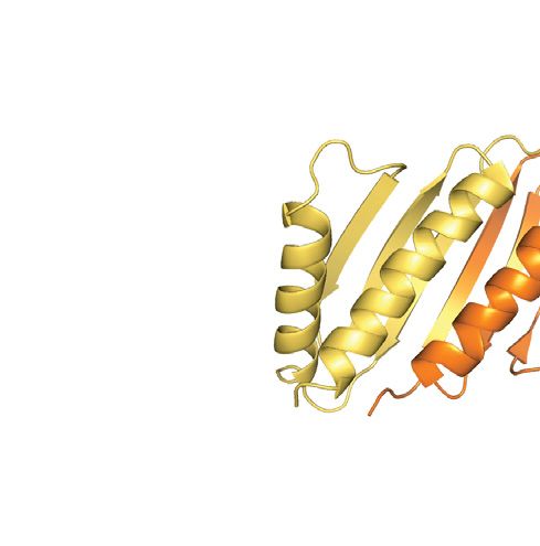
Nucleic Acids Research, 2021, Vol. 49, No. 19 10827
Kae1 sub-
A domain 2 Bud32-Cgi121 interaction
Pcc1-Kae1 interaction helix-DC and E-strand 4 of Bud32 bind
helices-D1 and D2 of Kae1 pack against helix-D8 of Cgi121
helices D1 and D2 of Pcc1
Bud32
D2 D1
Gon7
D1
Pcc1 D2
Cgi121
Downloaded from https://academic.oup.com/nar/article/49/19/10818/6382379 by guest on 08 November 2021
sub- Kae1-Bud32 interaction
domain 1
C-lobe and N-lobe of Bud32 wrap around subdomain 2 of Kae1
C-tail of Bud32 interacts with Kae1 active site
B CU motif
tRNA CCA tail
A37 binding suface
CCA tail
(helices D1, D3
Gon7 Kae1 and D4)
Cgi121
Pcc1
Bud32
Anchor 2:
Kae1 and Pcc1
bind the anticodon Anchor 3: Anchor 1:
90° Cgi121 binds the
loop of the tRNA Bud32 and Kae1 rotation
bind the D-arm of CCA tail of the
the tRNA tRNA tRNA
CU motif
Kae1
Gon7
CCA tail
Pcc1 CCA tail
C
bindi suface
binding
(helices D1, D3
(helic
a D4)
and
Bud32
Cgi121
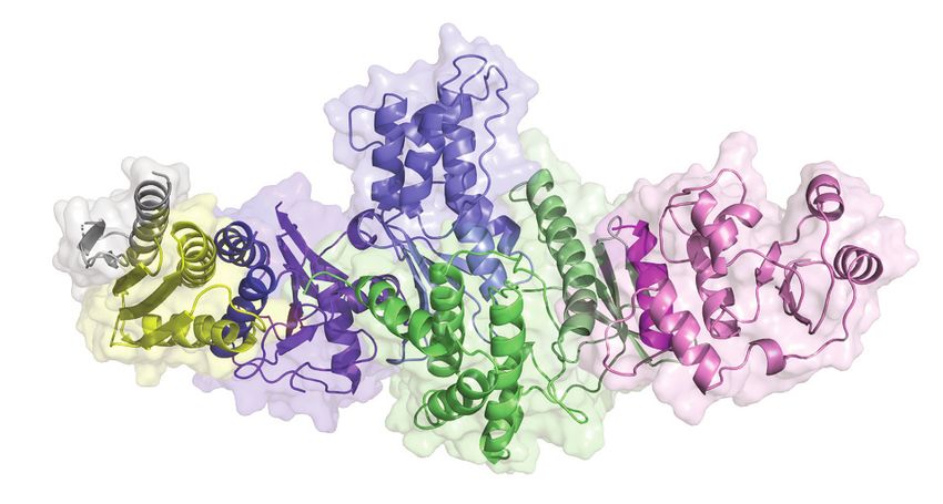
Figure 4. A model for the KEOPS-tRNA holo-enzyme-substrate complex. (A) Model of KEOPS reveals the basis for the interactions between its subunits
and shows that they adopt a linear binding. A cartoon representation with semi-transparent surface of a model of the KEOPS complex. The model was
generated using the structures of the Cgi121-Bud32-Kae1, Kae1-Pcc1 and Gon7-Pcc1-Kae1 sub-complexes (PDB 3ENH, 3ENO and 6GWJ respectively,
see Table 1). Gon7 (gray) binds to Pcc1 (yellow), which binds to Kae1 (blue). Kae1 binds also to Bud32 (green), which binds Cgi121 (purple). Bud32 (green)
binds to subdomain 2 of Kae1 (light blue) through an extensive surface that is composed of the N and C lobes. The C-tail of Bud32 interacts with the active
site cleft of Kae1. Pcc1 (yellow) binds to helices ␣1 and ␣2 of subdomain 1 of Kae1 (dark blue) in vicinity of the active site cleft of Kae1. Cgi121 (purple)
binds to helix-␣C and -strand 4 of the N-lobe of Bud32 (dark green) through helix ␣8. (B) A model of KEOPS-tRNA holoenzyme substrate complex
shows the basis for substrate interaction by KEOPS. For clarity, proteins are shown in surface representation and tRNA in cartoon representation and
complex is shown in two different orientations. According to the model, Cgi121 (pink) binds the tRNA CCA tail (red) via helices ␣1, ␣3 and ␣4 (purple).
Kae1 (blue) and Pcc1 (yellow) interact with the tRNA anticodon loop and Bud32 (green) and Kae1 interact with the tRNA D-arm. Gon7 (gray) does not
participate in tRNA binding. This binding mode directs A37 of tRNA (magenta) into the active site of Kae1 and positions the CU motif of tRNA (red)
to the interface between Bud32 and Kae1.10828 Nucleic Acids Research, 2021, Vol. 49, No. 19
proposed to regulate the dimerization function of Pcc1 and CCA tail of tRNA substrates. The biological role of this de-
thus KEOPS dimerization (99). In support, yeast KEOPS pendency is still not understood (see CONCLUDING RE-
that includes Gon7 is monomeric in solution with a 1:1 MARKS section).
binding stoichiometry between its all five subunits (99).
Furthermore, 4-subunit human KEOPS (without hGon7) A model for the KEOPS-tRNA holo-enzyme-substrate com-
forms dimers in solution while 5-subunit human KEOPS is plex. A model for the KEOPS-tRNA holo-enzyme sub-
monomeric (109,111). Lastly hGon7 appears to enhance or strate complex was generated using the Cgi121-tRNA struc-
stabilize the expression levels of KEOPS in vivo since in the ture and structures of KEOPS sub-complexes (Figure 4B)
absence of hGon7 overall expression levels of KEOPS sub- (102). According to this model, all four core KEOPS sub-
units are repressed (77). These findings might hint indirectly units (i.e. Cgi121, Bud32, Kae1 and Pcc1) form an extended
that KEOPS dimerization could regulate its degradation in tRNA binding surface that is highly complementary to the
eukaryotes, however this possibility requires further explo- structure of the tRNA. The tRNA is anchored on both ends
ration.
Downloaded from https://academic.oup.com/nar/article/49/19/10818/6382379 by guest on 08 November 2021
to the KEOPS complex with the CCA end bound by Cgi121
and the anticodon loop interacting with Kae1 and Pcc1. At
Cgi121 binds and regulates Bud32 and recruits tRNA to its center, the tRNA D-arm is predicted to contact Bud32
KEOPS. Consistent with the initial yeast two-hybrid and Kae1 (Figure 4B). Therefore, the tRNA binding mode
analysis (15), Cgi121 binds directly to Bud32. In archaeal of KEOPS seems to interrogate the entire ‘inner side’ of the
protein structures, the Cgi121 binding surface on Bud32 L shape of tRNA. Although all subunits are predicted to
is composed primarily of N-lobe helix-␣C and -strand 4 take part in contacting tRNA, only mutations on Bud32
(Figure 4A). The reciprocal binding surface on archaeal and Cgi121 adversely affect tRNA binding affinity. Bud32
Cgi121 is composed primarily of helix ␣8. The Cgi121 bind- does not bind tRNA independently but serves to potenti-
ing mode in the eukaryotic proteins is essentially the same ate the tRNA binding activity of Cgi121. Bud32 achieves
with slight elaborations. The N-lobe of eukaryotic Bud32 is this likely by forming secondary contacts with tRNA and
slightly larger than its archaeal ortholog with an extra he- by allosterically inducing a high affinity state in the CCA
lix (termed ␣A-helix) that participates in binding eukary- tail binding surface of Cgi121 (102). Despite not contribut-
otic Cgi121. The binding mode between Bud32 and Cgi121 ing to binding affinity, the predicted tRNA binding surface
is reminiscent of cyclin-dependent kinases binding to cy- in Kae1 and Pcc1 are none-the-less vital for tRNA modifi-
clins (112). Thus, not unexpectedly, Cgi121 potentiates both cation activity (102).
the ATPase and low level autophosphorylation activity of
Bud32 (note that the autophosphorylation activity is un-
The ATPase activity of Bud32 is regulated by tRNA bind-
likely to be physiologically relevant) (9,101,102). Structures
ing to KEOPS. The ATPase activity of Bud32 is essen-
of Bud32 in complex with Cgi121 reveal an active-like con-
tial for the ability of KEOPS to modify tRNA substrates
formation with productively positioned catalytic elements
in vitro and is required for Bud32’s essential function in vivo
(99,101). To date, no structures of Bud32 alone have been
(9,102). Interestingly, activation of Bud32 ATPase activity
reported for comparison.
is strongly potentiated by tRNA, but only in the context of
Apart from of its ability to bind Bud32, Cgi121 has in-
the Cgi121-Bud32-Kae1 sub-complex or the full KEOPS
trinsic ability to bind tRNA. This ability is essential for
complex (102). Mutations in any of the KEOPS subunits
the efficient recruitment of tRNA to KEOPS. A co-crystal
on the predicted tRNA binding surfaces inhibit the abil-
structure of a Cgi121-tRNA complex revealed that tRNA
ity of tRNA to activate Bud32 ATPase. How tRNA sub-
binding by Cgi121 is directed almost exclusively towards
strates regulate Bud32 ATPase activity remains an unre-
the 3’ CCA tail (positions 74–76, 5’-C74 -C75 -A76 -3’) (Fig-
solved question. However, in archaea this ability is not cor-
ure 4B) (102). The binding surface on Cgi121 for tRNA
related with the anticodon loop UAA motif but rather the
is composed primarily of ␣−helices 1, 3 and 4. The CCA
presence of a di-nucleotide CU motif located at positions
tail is a universal element present in all mature tRNAs that
10–11 within the tRNA D-arm (102). This motif is almost
plays multiple essential roles in the functionality and reg-
completely conserved in ANN-decoding tRNAs and absent
ulation of tRNAs. Perhaps most importantly, the termi-
from almost all non-ANN-decoding tRNAs with the sole
nal nucleotide of the CCA tail serves for the attachment
exception of tRNAAla . In the KEOPS-tRNA complex, the
of amino acids (aminoacylation) by aminoacyl tRNA syn-
CU motif is predicted to localize at the interface between
thetases (113). The CCA tail also plays an important role
Bud32 and Kae1 in proximity to the active site of Bud32
in quality control of tRNAs and translation. For example,
(Figure 4B) (102). The role this element has in the activa-
the CCA tail is excised by ANKZF1 to help the recycling of
tion of Bud32 and in the ability of KEOPS to modify tRNA
stalled ribosomes (114) and a second CCA tail is added by
awaits further investigation.
the CCA-adding enzyme to structurally unstable tRNAs as
a signal for their destruction by the tRNA degradation ma-
chinery (115). Cgi121 tRNA binding activity is dependent
CONCLUDING REMARKS AND QUESTIONS RE-
on the presence of an intact and non-aminoacylated CCA
MAINING TO BE ANSWERED
tail. Specifically, deletion of the CCA tail, addition of a sec-
ond CCA or attachment of bulky groups to the 3 of A76 Although many questions about how KEOPS functions
inhibits tRNA binding by Cgi121 (102). This dependency have been answered over the past 15 years, many more re-
renders KEOPS activity highly sensitive to the status of the main to be addressed, as outlined below.Nucleic Acids Research, 2021, Vol. 49, No. 19 10829
Why has KEOPS evolved added complexity? the added complexity of KEOPS actually provides relative
to Qri7? Possible advantages include the ability to read-
Fifteen years of studying the structure and function of
out tRNA features beyond the anticodon loop (102). This
KEOPS have revealed an intricate mechanism of action.
enhancement of substrate binding mechanism could serve
Unlike Qri7, which functions without binding partners, the
as a substrate quality control mechanism that would help
activity of Kae1 is dependent on at least three binding part-
discriminate between functional and non-functional tRNA
ners. This raises the question of what function does this
substrates, which might perturb the translation machinery.
added complexity serve when in principle Kae1 could func-
A second advantage may be the ability to coordinate t6 A
tion like Qri7?
modification with other tRNA modification and processing
We consider two tantalizing possibilities. First, the ma-
events. tRNAs undergo many processing steps from their
chinery built around Kae1 could enable the regulation of
initial transcription to their degradation, and many of these
its tRNA modifying activity. What benefit could there be
steps occur in a hierarchic manner (124,125). In support
from KEOPS downregulation? Although the total absence
of this possibility, since a subset of tRNAs contain an in-
Downloaded from https://academic.oup.com/nar/article/49/19/10818/6382379 by guest on 08 November 2021
of t6 A is harmful for cells, temporal control of t6 A levels
tron that is located between U36 and A37, the t6 A modifi-
could be beneficial for cells as a mean to control translation
cation by KEOPS can only occur after splicing. Likewise,
under specific cellular conditions. For example, Ser and Arg
the tRNA binding mechanism entails that t6 A modifica-
can be coded by t6 A-dependent (AGT/C and AGG/A re-
tion will occur after CCA tailing and before aminoacyla-
spectively) and t6 A-independent codons (TCT/C/A/G and
tion (102). How the activity of KEOPS is affected by other
CG/T/C/A/G respectively). As such, KEOPS downreg-
tRNA modifications and processing events and what role
ulation would favor the translation of mRNA transcripts
this might play awaits further study.
enriched with Ser and Arg codons that are not dependent
on t6 A modification. Thaiville et al. reported that in yeast
mRNAs encoding ∼300 proteins exclusively contain Arg How does the ATPase activity of Bud32 promote the t6 A
codons that are t6 A-dependent (44). These would be prime modification of tRNA by Kae1?
candidates for downregulation in response to KEOPS inhi- The ATPase activity of Bud32 is required for KEOPS activ-
bition. ity in vitro (102) and in vivo (9). However, it is still unclear
Translation is regulated by modulation of other types of how this activity exerts a specific effect on the tRNA t6 A
tRNA modifications at positions 34 and 37 (for examples, modification reaction. Since the ATPase activity of Bud32
see references (116–119)) and mRNAs that are differentially precedes the tRNA modification activity of Kae1 (102) it is
translated due to the presence or absence of tRNA modifi- reasonable to infer that it serves to activate Kae1. A can-
cations have been termed modification tunable transcripts didate region that could mediate an activating effect on
(MoTTs) (120). If the temporal regulation of t6 A is ex- Kae1 is the conserved C-terminal tail of Bud32. This re-
ploited for regulatory means, the added complexity of gion is dispensable for the activation of ATPase activity by
KEOPS provides multiple opportunities to achieve this end. tRNA (and thus exerts its essential effect downstream of
For example, the tRNA binding mechanism of KEOPS ren- the ATPase activating event) but is required for the tRNA
ders its function sensitive to Cgi121 expression levels. The modification activity of Kae1. How could this region of
short half-life of Cgi121 in yeast (∼30 min) (9) and its Bud32 mediate the activation of Kae1? Structures of Kae1-
sub-stoichiometric level relative to the other subunits (121), Bud32 complexes reveal that the C-terminus of Bud32 re-
make it an excellent restriction point for regulation. sides near the Kae1 active site and the Pcc1 binding surface
A second possibility is that the complexity of KEOPS ad- on Kae1 (Figure 4A). From this perch, the C-terminal tail
dresses a specific challenge or need of modifying cytoplas- of Bud32 is well placed to help remodel the active site of
mic tRNAs that is not required for mitochondrial tRNAs. Kae1 into a productive conformation. Alternatively, its po-
The cytoplasm contains a much larger number of tRNAs, sition could serve in some capacity to collaborate with Pcc1.
both ANN and non-ANN-decoding, which synthesize a far Either way, since ATP binding and hydrolysis by protein ki-
larger number of proteins relative to mitochondria. The hu- nases is known to influence inter-lobe closure (126), Bud32’s
man mitochondrial genome transcribes only 22 different catalytic function could generate movements of the C-tail of
tRNAs (122), of which 5 are substrates for t6 A modifica- Bud32 to enable its supporting role in Kae1 catalytic func-
tion (28,123). These tRNAs are needed for the synthesis tion.
of only 13 mitochondrial proteins (122). The human nu-
clear genome in contrast contains approximately 500 tRNA
genes, including hundreds that are not KEOPS substrates, What are the precise roles of Pcc1 and Gon7 in the activity
and these participate in the synthesis of 20 000–30 000 dif- of KEOPS?
ferent proteins. Perhaps Kae1 requires its auxiliary sub- One of the main unanswered issues in the KEOPS field is
units to deal with the greater diversity of tRNAs and the the precise role of Pcc1. Pcc1 binds close to the active site
greater demand for modified tRNAs in the cytoplasm rela- of Kae1. Interestingly, the absence of Pcc1 has the same
tive to those observed in mitochondria. In support of this effect on KEOPS as the deletion of the C-tail of Bud32,
possibility, while Qri7 and KEOPS display comparable cat- namely the absence of both has minimal effect on the tRNA
alytic rates in purified systems, massive overexpression of binding activity of KEOPS or the activation of Bud32 AT-
cytoplasmic Qri7 is required for its ability to rescue the Pase activity in response to binding tRNA (102). However,
growth defects resulting from the knockout of KEOPS (61). both are necessary for the tRNA modification activity of
This in turn raises the question of what specific advantage KEOPS in vitro and in vivo (9,102). The proximity of Pcc1You can also read