RPA2 winged-helix domain facilitates UNG-mediated removal of uracil from ssDNA; implications for repair of mutagenic uracil at the replication fork
←
→
Page content transcription
If your browser does not render page correctly, please read the page content below
3948–3966 Nucleic Acids Research, 2021, Vol. 49, No. 7                                                             Published online 30 March 2021
doi: 10.1093/nar/gkab195

RPA2 winged-helix domain facilitates UNG-mediated
removal of uracil from ssDNA; implications for repair
of mutagenic uracil at the replication fork
Bodil Kavli 1,2,* , Tobias S. Iveland1,3 , Edith Buchinger4 , Lars Hagen1,2,5 , Nina B. Liabakk1,2 ,
Per A. Aas1,2 , Tobias S. Obermann1,2 , Finn L. Aachmann4 and Geir Slupphaug 1,2,5,*
1
  Department of Clinical and Molecular Medicine, NTNU Norwegian University of Science and Technology, NO-7491
Trondheim, Norway, 2 Clinic of Laboratory Medicine, St. Olavs Hospital, Trondheim University Hospital, NO-7006

                                                                                                                                                              Downloaded from https://academic.oup.com/nar/article/49/7/3948/6204653 by guest on 24 December 2021
Trondheim, Norway, 3 Cancer Clinic, St. Olavs Hospital, Trondheim University Hospital, NO-7006 Trondheim, Norway,
4
  NOBIPOL, Department of Biotechnology and Food Science, NTNU Norwegian University of Science and
Technology, N-7034 Trondheim, Norway and 5 PROMEC Proteomics and Modomics Experimental Core at NTNU and
the Central Norway Regional Health Authority, NO-7491 Trondheim, Norway

Received December 23, 2020; Revised March 04, 2021; Editorial Decision March 09, 2021; Accepted March 10, 2021

ABSTRACT                                                                        ficient excision of AID-induced uracils in transcribed
                                                                                immunoglobulin loci.
Uracil occurs at replication forks via misincorpora-
tion of deoxyuridine monophosphate (dUMP) or via
deamination of existing cytosines, which occurs 2–3                             GRAPHICAL ABSTRACT
orders of magnitude faster in ssDNA than in dsDNA
and is 100% miscoding. Tethering of UNG2 to pro-
liferating cell nuclear antigen (PCNA) allows rapid
post-replicative removal of misincorporated uracil,
but potential ‘pre-replicative’ removal of deaminated
cytosines in ssDNA has been questioned since this
could mediate mutagenic translesion synthesis and
induction of double-strand breaks. Here, we demon-                              INTRODUCTION
strate that uracil-DNA glycosylase (UNG), but not                               Recent research indicates that DNA replication and deam-
SMUG1 efficiently excises uracil from replication pro-                          ination of cytosine and 5-methylcytosine (5-mC) are the
tein A (RPA)-coated ssDNA and that this depends on                              major sources of cancer-associated mutations (1,2). This
functional interaction between the flexible winged-                             is supported by analysis of mutational signatures associ-
helix (WH) domain of RPA2 and the N-terminal RPA-                               ated with C>T transition across a wide spectrum of hu-
                                                                                man cancers (3). Two of the C>T signatures are corre-
binding helix in UNG. This functional interaction is
                                                                                lated with age (‘clock-like’), further supporting a link to
promoted by mono-ubiquitination and diminished by                               replication (4) (SBS1 and SBS5, https://cancer.sanger.ac.uk/
cell-cycle regulated phosphorylations on UNG. Six                               cosmic/signatures/SBS/index.tt). Whereas C>T mutations
other human proteins bind the RPA2-WH domain, all                               in CpG contexts (SBS1) apparently originate from deam-
of which are involved in DNA repair and replication                             ination of 5-mC, the biological processes underlying C>T
fork remodelling. Based on this and the recent dis-                             transitions outside CpG sites (SBS5) remain obscure. One
covery of the AP site crosslinking protein HMCES,                               potential source of these C>T transitions is deamination
we propose an integrated model in which templated                               of cytosine to uracil within single-stranded DNA (ssDNA)
repair of uracil and potentially other mutagenic base                           regions at the replication fork. Spontaneous and enzymatic
lesions in ssDNA at the replication fork, is orches-                            cytosine deamination occurs 2–3 orders of magnitude faster
trated by RPA. The UNG:RPA2-WH interaction may                                  in ssDNA than in dsDNA (5–7) and unless corrected be-
                                                                                fore encounter of replicative polymerases, these would lead
also play a role in adaptive immunity by promoting ef-
                                                                                to C>T mutations after two replicative cycles. ssDNA re-
                                                                                gions continuously form at the lagging strand and could be

* To
   whom correspondence should be addressed. Tel: +47 91 82 54 55; Fax: +47 72 57 64 00; Email: geir.slupphaug@ntnu.no
Correspondence may also be addressed to Bodil Kavli. Tel: +47 97 66 14 42; Fax: +47 72 57 64 00; Email: bodil.kavli@ntnu.no


C The Author(s) 2021. Published by Oxford University Press on behalf of Nucleic Acids Research.
This is an Open Access article distributed under the terms of the Creative Commons Attribution License (http://creativecommons.org/licenses/by/4.0/), which
permits unrestricted reuse, distribution, and reproduction in any medium, provided the original work is properly cited.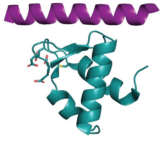
Nucleic Acids Research, 2021, Vol. 49, No. 7 3949

extensive at either strand when the replicative polymerases       the RPA2 domain D (Figure 1A), would position UNG2
pause or stall and uncoupled fork progression occurs (8).         in an optimal position to attack uracil sites in dsDNA near
   Once formed, ssDNA is rapidly coated with replication          the junction (
3950 Nucleic Acids Research, 2021, Vol. 49, No. 7

                A
                  ssDNA

                 5’                                                             3’
                               A           B        C             D                    ssDNA-binding: RPA1-A,B,C and RPA2-D
                                                                              C
                 N
                      F                                      E               WH        Protein-binding: RPA1-F and RPA2-WH
                                   RPA1 (70 kDa)    RPA3 (14 kDa)        RPA2 (32kDa)

                B                     N-terminal regulatory domain of human UNG2                        UNG catlytic domain

                                                                                                                                                 Downloaded from https://academic.oup.com/nar/article/49/7/3948/6204653 by guest on 24 December 2021
                                       P                                    P   P                             CD
                     PIP-box                                                            RPA-binding helix
                 MIGQKTLYSFFSPSPARKRHAPSPEPAVQGTGVAGVPEESGDAAAIPAKKAPAGQEEPGTPPSSPLSAEQLDRIQRNKAAALLRLAARNV
                 1        10          20       30       40       50        60         70       80        90          313

                                                                      Identical sequence in UNG1 and UNG2

Figure 1. Protein domain architecture and important motifs in human RPA and UNG. (A) Domain structure and orientation of the RPA trimer (RPA1,
RPA2, RPA3) bound to ssDNA. DNA-binding (RPA1-A,B,C; RPA2-D) and protein-binding (RPA1-F; RPA2-WH) domains are indicated. (B) Sequence
and important motifs in the ∼90 aa N-terminal regulatory domain of UNG2. Binding motifs for PCNA (PIP-box), RPA and cell-cycle regulated phos-
phorylation sites are indicated. The UNG1 isoforms also contain residues 45–313, including the RPA-binding helix.

   UNG has a high preference for excision of uracil from                        UNG N-terminal domain regulate the RPA2-WH interac-
ssDNA compared to dsDNA (36). However, in vivo, ss-                             tion and uracil excision from RPA-coated ssDNA.
DNA is bound by RPA and other ssDNA-binding pro-                                   We propose a model in which the RPA2-WH domain pro-
teins that protect the DNA strand against attack by nucle-                      motes uracil excision of deaminated cytosines in ssDNA at
ases and other DNA-modifying enzymes. We previously re-                         the replication fork and coordinates fork remodelling to re-
ported that uracil excision by mitochondrial UNG1 from                          store dsDNA and allow downstream error-free BER. RPA
ssDNA is strongly inhibited by the human mitochondrial                          is also detected together with RNA polymerase II in tran-
ssDNA-binding protein mtSSB (44). A potential function                          scribed regions of active genes (47), where it can function as
of this could be to delay uracil removal from replicative                       a sensor of R-loops (48). In activated B-cells, this mediates
single-stranded mtDNA until the dsDNA conformation is                           recruitment of AID to immunoglobulin genes to facilitate
restored. In the same study, we also found that RPA medi-                       SHM and CSR (49,50). It is thus tempting to speculate that
ated virtually no inhibition of UNG1 activity. To what de-                      RPA located at actively transcribed Ig loci recruits UNG
gree RPA hinders access of UNG2 to uracil in RPA-coated                         to ssDNA in R-loops to promote mutagenic processing of
ssDNA, has previously not been investigated. Earlier stud-                      AID-generated uracil during adaptive immunity in B cells.
ies in our group demonstrated that RPA mediated a mod-
erate reduction of UNG2-mediated uracil excision from ds-
DNA substrates, whereas excision from ssDNA was moder-                          MATERIALS AND METHODS
ately enhanced (12,45). However, these studies were under-
taken with large molar excess of long DNA substrates (nick-                     Recombinant proteins and mutagenesis
translated calf thymus DNA) and without pre-incubation of                       Plasmid encoding human trimeric RPA (p11d-tRPA) was a
RPA/DNA.                                                                        gift from Prof. Marc S. Wold (University of Iowa). Recom-
   Here, we explored the functional relevance of the in-                        binant RPA was expressed in Escherichia coli BL21(DE3)
teraction between RPA and UNG by employing uracil-                              RIPL and purified with Affi-Gel Blue (BioRad), hydroxya-
containing oligonucleotides preincubated with large molar                       patite (BioRad), and Mono Q (GE Healthcare) chromatog-
excess RPA to ensure pre-formation of RPA/DNA com-                              raphy as described (51). A plasmid expressing RPA lack-
plexes. This would be biologically relevant, since the number                   ing the WH domain (RPA-WH, codon 190 of RPA2 mu-
of RPA molecules within human cells is two orders of mag-                       tated to a TGA stop codon) was generated by the Q5 site-
nitude higher than UNG as well as the single-strand selec-                      directed mutagenesis kit (New England Biolabs) accord-
tive uracil DNA glycosylase SMUG1 (46). We demonstrate                          ing to manufacturer’s instructions. Cloning of the RPA2-
that UNG2 mediates highly efficient uracil excision from                        WH domain (RPA2 residues 172–270) into the pTYB12 ex-
RPA-bound ssDNA, while the same substrate is protected                          pression vector was performed by a sequence- and ligation-
against attack from the uracil-DNA glycosylase SMUG1.                           independent strategy as described (52). RPA2-WH-intein
We further show that this ability of UNG depends on the                         fusion protein was expressed in ZYP-5052 autoinduction
specific interaction between the RPA2-WH domain and the                         medium at 16◦ C overnight and the WH domain purified
N-terminal RPA-binding helix motif in UNG. Moreover,                            according to standard protocol (52). Recombinant human
we show that phosphorylation and ubiquitination of the                          UNG2 and SMUG1 were prepared as described previously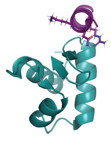
Nucleic Acids Research, 2021, Vol. 49, No. 7 3951
(36,45,53). Constructs expressing N-terminally deleted and 50% LED power in optimised MST buffer (50 mM Tris–
mutated UNG proteins were generated by the Q5 site- HCl pH 8, 150 mM NaCl, 10 mM MgCl2 , 0.05% Tween-20,
directed mutagenesis kit and Quick-change site-directed 0.5 mg/ml BSA). A constant amount of RPA (330 nM) and
mutagenesis kit (Stratagene), respectively. Mutations were a concentration gradient (∼10 nM–200 M) of each pep-
verified by Sanger-sequencing (GATC Biotech AG, Ger- tide were used in all experiments. Kd values were calculated
many) and confirmed by mass spectrometry (MS) analysis from four runs for each experiment using the MO-Affinity
of purified proteins. Cloning, expression, and purification Analysis software (NanoTemper Technologies).
of the 15 N-labelled UNG2 N-terminal (residues 1–93) was
as described previously (52). RPA affinity capture
Peptides (EV-34, pEV-34 and ppEV-34) were covalently
Uracil excision assays with RPA-coated and naked DNA sub- coupled to epoxy beads (Dynabeads M-270 Epoxy, Thermo
strates Fisher) as described by the producer. Coupled beads (15 l)
Downloaded from https://academic.oup.com/nar/article/49/7/3948/6204653 by guest on 24 December 2021
3 -FAM-labelled, PAGE-purified oligonucleotide sub- were added to HeLa whole cell extract (WCE, 1 mg pro-
strates were from Sigma-Aldrich. Unless otherwise tein) and incubated for 30 min before washing in PBS and
indicated, the substrate (25 nt) harboured uracil at position elution in LDS loading buffer. Input and affinity captured
10 in a polyC sequence to avoid secondary structures RPA were quantified by western analysis using monoclonal
(U10-25*: CCACCCCCCUCCCCCCCCCCCCCCC- rabbit anti-RPA2 [EPR2877Y] (ab76420) primary antibody
FAM). Double-stranded substrate was generated by (1:1000, Abcam) and swine anti-rabbit HRP (1:5000, Dako)
annealing (heating followed by slow cooling) U10- as secondary antibody.
25* to a non-labelled complementary oligo (A16-25:
GGGGGGGGGGTGGGGAGGGGGGTGG). All as- Circular dichroism
says were performed at 22◦ C in 10 mM Tris–HCl pH 7.5,
All CD-experiments were performed on Chirascan (Applied
50 mM NaCl, 1 mM DTT, 0.1 mM EDTA, 0.5 mg/ml
Photophysics) using a 1 mm cuvette at 25◦ C. Samples were
BSA.
measured within the range of 180–260 nm in 20 mM phos-
By monitoring the activity of the catalytic UNG domain
phate buffer pH 7.0, 10 mM NaCl. The measured millide-
(0.2 nM UNG-CD), lacking the N-terminal RPA-binding
gree from all spectra were transformed to mean residue el-
helix), we found that >400 nM RPA fully abolished uracil
lipticity . The 222/208 nm ratios were used to compare the
excision when pre-incubated with 100 nM U10-25* ssDNA
helicity of the peptides.
substrate (data not shown), indicating that at such condi-
tions all ssDNA was bound to RPA and not accessible for
processing by UNG-CD. Based on this, 10-fold molar ex- NMR and paramagnetic relaxation enhancement analysis
cess of RPA over substrate was employed in subsequent The purified WH domain was MTSL-labelled at its sin-
experiments unless otherwise stated. DNA substrate (100 gle cysteine residue (C219) by adding 10-fold molar ex-
nM final) and RPA (1 M final) were mixed and incubated cess of MTSL (S-[(1-oxyl-2,2,5,5-tetramethyl-2,5-dihydro-
on ice for 15 min to form RPA/ssDNA complexes. Vary- 1H-pyrrol-3-yl)methyl] methanesulfonothioate) dissolved
ing amounts of UNG were then added, and the mixtures in DMSO to a sample of RPA2-WH in 20 mM phos-
incubated for 10 min in a water bath. To avoid unspecific phate buffer pH 7.0, 10 mM NaCl. The mixture was in-
interactions and binding of enzyme and DNA to the assay cubated in darkness overnight to complete the reaction.
tube surface, low-DNA binding tubes and excess of BSA MTSL labelled RPA2-WH was extensively washed with 20
was used in all reactions. Reactions were quenched and AP mM phosphate buffer pH 7.0, 10 mM NaCl. NMR spectra
sites cleaved in 10% piperidine at 90◦ C for 20 min. Sam- of 15 N-labelled N-terminal UNG2 in presence of 1.5 mo-
ples were dried by vacuum centrifugation and suspended lar excess of MTSL-RPA2-WH before and after addition of
in formamide-containing loading buffer. Product and sub- ascorbic acid were recorded at 25◦ C in NMR buffer (20 mM
strate were separated in 12% PAGE/7M urea–0.5× TBE phosphate buffer pH 7.0, 10 mM NaCl, H2 O/D2 O 9:1) on a
gels, bands visualised in ChemiDoc™ Imager (Bio-Rad) and Bruker Ascend 800 MHz Avance III HD NMR spectrome-
quantified by Image Lab software (Bio-Rad). Importantly, ter equipped with 5 mm z-gradient TXI (H/C/N) cryogenic
all assays in which two parameters were compared (e.g. probe and processed with Bruker TopSpin version 3.2/3.5.
RPA-coated versus naked substrate or WT versus mutated Spectral analysis and peak intensities were determined by
protein) were performed in parallel with the exact same using CARA version 1.9.1.7.
DNA/protein dilutions.
In vitro ubiquitination
MicroScale Thermophoresis (MST)
In vitro ubiquitination for E2 ligase determination was per-
Recombinant human RPA trimer was labelled and pu- formed on purified recombinant human UNG2 using ubiq-
rified using the Monolith NT™ Protein Labelling RED- uitination kit from Enzo Life Sciences (BML-UW9920-
MALEIMIDE kit (NanoTemper Technologies, Germany) 0001) according to the manufacturer’s instructions, using
according to the manufacturer’s protocol. UNG peptides 0.036 g/l His-UNG2 and HeLa protein extract (0.6
were from Proteogenix (Schiltigheim, France). MST was g/ml final) as E3 ligase donor. Ubiquitination of UNG2
performed on Monolith NT.115 (NanoTemper Technolo- Lys to Arg mutants were performed by the same protocol
gies) using standard capillaries with settings 60% MST and using UBCH2 as E2 ligase. Ubiquitinated UNG for uracil-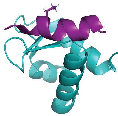
3952 Nucleic Acids Research, 2021, Vol. 49, No. 7
excision assays was generated with UBCH2 in absence of strates. By contrast, the highly efficient UNG-CD displayed
E3 ligase by using 0.068 g/l N-terminally deleted UNG2 1000-fold reduced activity with RPA-coated ssDNA com-
and 0.05 g/l BSA instead of HeLa protein extract. Mock pared to naked DNA substrate.
samples were treated identically, except that ATP was not To investigate whether positioning of uracil or length
included in the reaction. Ubiquitination was verified by of ssDNA substrate have impact on the results, we pre-
western blot analysis using polyclonal rabbit anti-UNG pared a 25 nt oligonucleotide identical to U10-25*, but with
(PU59, made in-house) primary antibody and swine anti- uracil shifted three nucleotides in the 3 direction (U10U13-
rabbit HRP (Dako) as secondary antibody and single ubiq- 25*). Based on the RPA:ssDNA complex structure (Figure
uitination at K78 was identified by LC–MS/MS analysis. 2A), this position is predicted to be less accessible. We also
made extended versions (43 nt) of both DNA substrates,
UNG affinity capture by Ugi harbouring 18 additional 3 -terminal nucleotides (U10–43*
and U13–43*) (Supplementary Figure S2A). Uracil exci-
The UNG-specific inhibitor protein Ugi (54) was covalently
sion from the three modified substrates (100 nM) by UNG-
Downloaded from https://academic.oup.com/nar/article/49/7/3948/6204653 by guest on 24 December 2021
coupled to epoxy beads (Dynabeads M-270 Epoxy, Thermo
CD was completely abolished when coated with 1 M
Fisher) as described by the producer. Coupled beads (15
RPA (Supplementary Figure S2B). Conversely, full-length
ul) were added to 1 mg HeLa WCE and incubated for 30
UNG2 was able to excise uracil from all substrates even
min before washing in PBS. Affinity captured proteins were
at 4 M RPA, but with varying efficiency. The substrate-
trypsinised directly on the beads for MS analysis.
dependent variation observed and the ability of UNG2 to
excise uracil with only slightly reduced efficiency (U10 sub-
Mass spectrometry analysis strates) in presence of 10 000-fold molar excess of RPA com-
Proteins were digested and desalted as described (55,56) pared to UNG (40-fold compared to DNA) demonstrates
evaporated to dryness and resuspended in 0.1% formic acid that UNG is not titrated out by free RPA molecules that
prior to analysis on a LC–MS/MS platform consisting of an are not bound to DNA.
Easy-nLC 1000 UHPLC interfaced with an LTQ-Orbitrap In the 25 nt substrates, shifting uracil in the 3 direc-
Elite hybrid mass spectrometer via a nanospray ESI ion tion mediated markedly reduction in excision (Supplemen-
source (Thermo Scientific/Proxeon). Peptides were injected tary Figure S2C, left panel), suggesting increased steric hin-
onto a C-18 trap column (Acclaim PepMap100 (75 m i. drance by DBD-C, as predicted (Figure 2A). U10 in the 43
d. × 2 cm, C18, 5 m, 100 Å, Thermo Scientific) and sepa- nt substrate was excised as efficiently as in the 25 nt sub-
rated on a C-18 analytical column (Acclaim PepMap100 (75 strate. Notably, in the longer substrate, much less reduced
m i.d. × 50 cm, C18, 3 m, 100 Å, Thermo Scientific) us- excision was observed when uracil was shifted to the 13 po-
ing a 84 min gradient from 10 to 40% CH3 CN, 0.1% formic sition, compared to the 25 nt substrate. This may be con-
acid at a flow rate of 250 nl/min. Peptides were analysed in tributed by a more ‘relaxed’ positioning of RPA along the
positive ion- and data dependent acquisition (DDA) mode length of the substrate. Disregarding the number of RPA
using the following parameters: Electrospray voltage 1.9 kV, complexes bound to the 43 nt substrate, U13 would then be
CID fragmentation with normalised collision energy 35, au- closer to DBD-A/B than in the 25 nt substrate since the 5
tomatic gain control (AGC) target value of 1E6 for Orbi- half is identical in both substrates and alternative binding
trap MS and 1E3 for MS/MS scans. Each MS scan (m/z must occur towards the 3 half. This would be in agreement
400–1600) was acquired at a resolution of 120 000 FWHM, with the findings that DBD-A and B are more dynamic than
followed by 20 MS/MS scans triggered for intensities above the trimerization core (C, D and E) (16–18), thereby allow-
500, at a maximum ion injection time of 200 ms for MS and ing increased access to U13 in the longer substrate.
50 ms for MS/MS scans. In separate experiments we compared UNG2 and the
single-strand selective monofunctional uracil DNA gly-
RESULTS cosylase SMUG1 using the U13–43* ssDNA substrate.
SMUG1 is catalytically slow compared to UNG (58),
UNG2 efficiently excises uracil from RPA-coated ssDNA
prefers double-stranded substrates (59) and is expressed at
UNG2-mediated excision of uracil from dsDNA was re- 3–10-fold lower levels across human cell lines than UNG
cently shown to be stimulated by the presence of an RPA- (46,53). Whereas UNG2 peaks during S-phase (32,45,60),
coated ssDNA junction (31). However, whether UNG2 can SMUG1 is constitutively expressed through the cell cy-
access uracil embedded within RPA-coated ssDNA itself cle and, in contrast to UNG2, is not localized to repli-
has not been investigated. To address this, we employed a 25 cation foci (58,60). Although this suggests that SMUG1
nt oligonucleotide substrate harbouring uracil at position does not have an important function in uracil sanitation
10 and 6FAM-label at the 3 -end. This construct was based at replication forks, a potential function in ssDNA outside
on the crystal structure of Ustilago maydis RPA trimer com- of replication forks has not been investigated. As shown
plexed to a 25 nt ssDNA (57) (Figure 2A). The substrate in Supplementary Figure S3, SMUG1 activity was virtu-
was pre-incubated with RPA to form a stable complex in ally blocked by RPA-coating of the ssDNA substrate. These
which the RPA trimer would cover the entire DNA strand. experiments demonstrate that (i) access to uracil in ss-
After pre-incubation in the presence/absence of RPA, the DNA is restricted by binding of RPA to the substrate, (ii)
substrates were incubated with UNG2 or UNG-CD as con- UNG2, but not SMUG1, can excise uracils from RPA-
trol, and uracil excision quantified as described (Figure 2B). bound ssDNA and (iii) the N-terminal regulatory domain
As illustrated in Figure 2C, UNG2 was almost equally capa- of UNG2 promotes access to catalytic removal of these
ble of excising uracil from the RPA-coated and naked sub- uracils.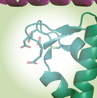
Nucleic Acids Research, 2021, Vol. 49, No. 7 3953
Downloaded from https://academic.oup.com/nar/article/49/7/3948/6204653 by guest on 24 December 2021
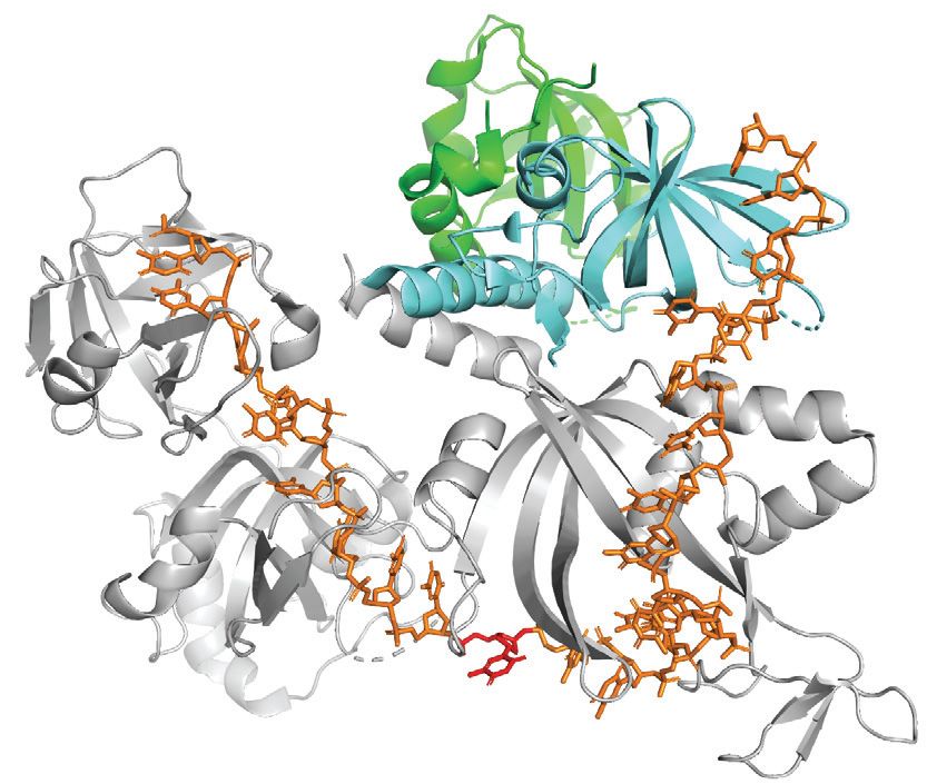
Figure 2. UNG2 promotes highly efficient uracil excision from RPA-coated ssDNA. (A) 3D structure of the RPA heterotrimer bound to ssDNA (25 nt).
The DNA binding domains (A, B and C) in RPA1 (grey) and D in RPA2 (cyan) are indicated. RPA3 is green and the direction of the DNA strand and
nt position 10 are indicated. The structure is visualised using PyMOL2 software with PDB accession code 4GNX (Ustilago maydis) (57). (B) Illustration
of the uracil-excision assay used to analyse UDG activity. The yellow asterisk indicates 6FAM labelling at the 3 end of the uracil-containing ssDNA
substrate (25 nt) and product (15 nt). (C) Uracil-excision activity with RPA (1 M)-coated and naked ssDNA substrates (100 nM U10-25*) for full-length
UNG2 and N-terminally truncated UNG (UNG-CD). Upper panels show representative PAGE gels for one experiment. Each curve represents mean
activity calculated from three independent experiments, (I15nt /I15nt+25nt ) × 100%. Standard deviations are indicated with error bars. Note that the UNG
concentrations (x-axes) are represented on a logarithmic scale.
Access to uracil in RPA-coated ssDNA depends on specific in- tions of either RPA-WT or RPA-WH. Notably, deletion
teraction between the WH domain and the UNG N-terminal of the WH domain resulted in a markedly reduced capabil-
helix ity of UNG2 to excise uracil from the RPA-coated substrate
(Figure 3B). Whereas uracil excision was essentially elimi-
To investigate whether the ability of UNG2 to excise uracil
nated in the presence of 400 nM RPA-WH, there was no
from the RPA-coated substrate was dependent on the WH
decrease in uracil excision by RPA-WT. Thus, the ability of
domain of RPA2, we deleted the domain and purified
UNG2 to target uracil in RPA-coated ssDNA is facilitated
the corresponding RPA trimer (RPA-WH) (Figure 3A).
by its interaction with the RPA2-WH domain.
Next, we compared UNG2 uracil-excision activity with ss-
To further identify which parts of the ∼90 aa N-terminal
DNA substrate preincubated with increasing concentra-
domain of UNG2 that contribute to substrate recognition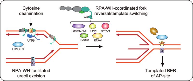
3954 Nucleic Acids Research, 2021, Vol. 49, No. 7
Downloaded from https://academic.oup.com/nar/article/49/7/3948/6204653 by guest on 24 December 2021
Figure 3. Uracil excision from RPA-coated ssDNA depends on the C-terminal RPA2-WH domain and the UNG2 N-terminal RPA-binding helix. (A)
Coomassie blue-stained SDS-PAGE gel of purified RPA trimer (1 g) containing either RPA2-WT or RPA2-WH. (B) RPA-WH, but not RPA-WT,
inhibits uracil-excision from ssDNA by UNG2. Upper panels show representative PAGE gels for one experiment. Substrate (25 nt) and product (15 nt)
bands are indicated. 100 nM ssDNA substrate and 0–400 nM RPA (WT or WH) were used in each reaction. Curves represent mean activity calculated
from three independent experiments. Standard deviations are indicated as error bars. (C) UNG mutants with partial N-terminal truncation, but that still
contain the RPA-binding helix, retain activity on RPA(WT)-coated ssDNA. RPA (500 nM, WT or WH) and 100 nM ssDNA were used in the assays.
Note logarithmic scale on the x-axes. Mutations of UNG2 residues involved in RPA binding disrupt the ability to target uracil in RPA-coated ssDNA
(D–F). (D) PAGE gels showing uracil-excision experiments with RPA-coated ssDNA (1 M RPA and 100 nM ssDNA) and naked ssDNA (100 nM)
together with UNG2 single mutant (R84D) and double mutant (N77/R84D). (E) NMR structure of a peptide segment (UNG2 residues 73–88) bound to
the RPA2-WH domain. Original side chains of the mutated UNG residues are indicated. The figure was generated using PyMOL2 and PDB coordinates
1DPU. (F) Calculated uracil excision activity ratios from several experiments as shown in panel D, representing activity with RPA-coated ssDNA (1 M
RPA and 100 nM ssDNA) divided by activity with 100 nM naked ssDNA (ssDNA+RPA/ssDNA). Each curve represents the mean of three independent
experiments and standard deviations are indicated as error bars.Nucleic Acids Research, 2021, Vol. 49, No. 7 3955
in RPA-coated ssDNA, we generated partial UNG2 N- To investigate whether phosphorylation at T60 and S64
terminal deletion mutants starting at either aa 57 (U2– regulates RPA-binding, we first measured the RPA bind-
57) or 66 (U2–66). In contrast to UNG-CD (starting at ing affinity for the EV-34 peptides by MST, using increas-
UNG2 aa 93) (Figure 1B), U2–57 and U2–66 that both ing concentrations of non-labelled peptides and a constant
contain the helix motif known to bind RPA2-WH, excised amount of RPA trimer labelled with NT-67 RED dye. The
uracil from RPA-WT-coated substrate as effectively as full- identified dissociation constants (Kd ) show that phosphory-
length UNG2 (Figure 3C). Conversely, when the ssDNA lation at T60 mediated two-fold reduction in RPA binding
substrate was bound to RPA lacking the WH domain, the whereas double phosphorylation at T60 and S64 resulted in
uracil excision activities were low for all four UNG forms a further decrease (Figure 4A). The analyses also revealed
(Figure 3C). This demonstrates that the UNG2 N-terminal that the EV-34 peptides displayed 10–26-fold stronger bind-
helix (starting at residue 66) and the RPA2-WH domain ing to RPA than the short RV-15 peptide. This demonstrates
are both necessary for uracil excision from RPA-coated that UNG2 residues outside the ‘core’ (RV-15) contribute to
ssDNA. the interaction surface with RPA.
Downloaded from https://academic.oup.com/nar/article/49/7/3948/6204653 by guest on 24 December 2021
Finally, we mutated UNG2 N-terminal helix residues Next, we covalently attached the EV-34 peptides to mag-
N77 and R84, which are directly involved in RPA2-WH netic epoxy beads to investigate the effect of T60 and S64
binding and abolish interaction with RPA when mutated to phosphorylations on RPA pull-down from HeLa whole cell
aspartate (12) (Figure 3D,E). Whereas full-length UNG2- extracts. These experiments confirmed that the single- and
WT excised uracil from naked and RPA-coated substrates double phosphorylations have an increasingly negative im-
with comparable efficiency (Figure 2C), both mutants dis- pact on RPA binding (Figure 4B). In another approach,
played compromised excision from the RPA-coated sub- we tested whether the peptides could outcompete binding
strate (Figure 3D). This effect was highly significant when of UNG2 to RPA and thereby inhibit uracil excision from
activity ratios between the two substrates where calcu- RPA-coated ssDNA. In the presence of increasing concen-
lated from several independent experiments in which RPA- trations of EV-34, UNG2 activity was reduced (Figure 4C).
coated and naked substrates were analysed in parallel with In accordance with the increased Kd in MST assay and re-
the same enzyme dilutions (Figure 3F). In summary, this duced pull-down efficiency of RPA, phosphorylated ppEV-
demonstrates that UNG has a unique capability among 34 peptides showed less inhibition of the uracil excision
the human UDGs to excise uracil from RPA-bound ss- activity than unphosphorylated EV-34 (Figure 4D, upper
DNA and that this depends on the specific interaction panel). The inhibitory effect of the EV-34 peptide was even
between the UNG N-terminal helix and the RPA2-WH more pronounced for a UNG2 phospho-mimicking mutant
domain. with decreased affinity to RPA compared to WT-UNG2
(45) (Figure 4D, lower panel).
We previously reported chemical shift assignment of full-
Cell-cycle regulated phosphorylations adjacent to the UNG2 length UNG2 (52) (BRMB entry 27133). For a structural
N-terminal helix regulate binding to RPA assessment of how phosphorylation of T60 and S64 reg-
Protein-protein interactions are commonly regulated by ulates RPA binding, we used paramagnetic relaxation en-
post-translational modifications (PTMs). We previously hancement (PRE) NMR measurements to probe inter-
identified two stepwise and cell-cycle regulated phospho- molecular interactions. The RPA2-WH domain contains a
rylations in the UNG2 N-terminal domain just upstream single solvent-exposed cysteine (RPA2 C219), making it an
of the RPA-binding helix (Figure 1B), and phospho- excellent candidate for attaching a PRE label like MTSL to
mimicking mutants suggested that these regulate affinity to- it. To measure PREs, NMR spectra of the 15 N-labelled N-
wards RPA (45). To investigate this further, we synthesised terminal region of UNG2 were recorded in the absence and
a panel of peptides, including the short RPA2-WH-binding presence of the MTSL-labelled RPA2-WH domain. Using
core peptide (RV15, UNG2 residues R76-V90) (43) and this technique, NMR signals of residues near (within 20 Å)
three N-terminally extended versions thereof containing 3, to the PRE label will experience a reduction in signal inten-
10 and 19 additional residues, respectively (Supplementary sity, as a function of residence time and distance to the la-
Figure S4). The longest, EV-34 (UNG2 residues E57-V90), bel (62). While none of the residues in the helical part of the
harbours both phosphorylation sites and was synthesised UNG2 N-terminal region were affected, signals of residues
as non-phosphorylated (EV-34), mono-phosphorylated on in the regions Q55-S63 were markedly reduced (Supplemen-
T60 (pEV-34) and di-phosphorylated on T60 and S64 tary Figure S5). This suggests that the region in UNG2 en-
(ppEV-34) peptides (Figure 4A). Secondary structures were compassing the two phosphorylation sites (T60 and S64)
determined by CD spectroscopy and secondary chemical is located close (3956 Nucleic Acids Research, 2021, Vol. 49, No. 7
Downloaded from https://academic.oup.com/nar/article/49/7/3948/6204653 by guest on 24 December 2021
Figure 4. UNG phosphorylation regulates RPA binding affinity. (A) RPA binding affinity to various UNG peptides. Dissociation constants (Kd ) were
measured by MicroScale Thermophoresis (MST). Residues forming the UNG RPA-binding helix, N77 and R84 essential for RPA binding, and adjacent
phosphorylated sites are highlighted. (B) Western blot showing pull-down of endogenously expressed RPA from HeLa whole cell extract (WCE) using the
non-, mono- (pT60), and di-phosphorylated (pT60, pS64) UNG peptide-coated beads as bait. (C) Peptide competition assay showing reduced access of
UNG2 specifically to uracil in RPA-coated ssDNA in the presence of EV-34 peptide. Curves represent the mean activity measured in two independent
experiments using 0.4 nM UNG2, 1 M RPA and 100 nM ssDNA (U10–25*) substrate. (D) Peptide competition experiments on UNG2-WT and UNG2 P-
mimicking mutant, with the same conditions as in panel C, comparing non-phosphorylated (EV-34) and di-phosphorylated (ppEV-34) peptides. The curves
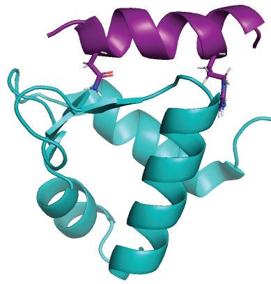
represent normalised uracil-excision activity ratios (RPA+ssDNA/ssDNA). (E) Structural interpretation of the results, including paramagnetic relaxation
enhancement analysis of RPA2-WH domain (MTSL-labelled at C219) and N-terminal UNG2 residues 1-93 (15 N-labelled). The structural model illustrates
that the UNG2 Q55-S63 region is within 20 Å from RPA2 C219 residue. The negatively charged patch on the surface of the RPA2-WH domain consists
of side chains E223, D260, D261 and D262. The structural model is visualised using PyMOL2 software based on the PDB coordinates 4MQV.Nucleic Acids Research, 2021, Vol. 49, No. 7 3957
Ubiquitination at K78 in the UNG2 N-terminal helix stimu- zymes (Figure 5G and H). The UNG enzymes were anal-
lates uracil-excision from RPA-coated ssDNA ysed with both RPA-coated and naked DNA substrates in
parallel and significantly increased activity against RPA-
The UNG2 protein level and phosphorylation status are
coated substrates by K78 ubiquitination was demonstrated
tightly regulated through cell cycle. T60 and S64 phospho-
when comparing the activity ratios (ssDNA+RPA/ssDNA)
forms gradually accumulate through S-phase, preceding a
calculated from several independent experiments (Figure 5I
mono-ubiquitinated isoform that accumulates in G2 (45).
and J). This demonstrates that ubiquitination of the UNG
To identify the ubiquitination site, we synchronised HeLa
RPA-binding helix does not block RPA binding but rather
cells with double thymidine block and harvested cells in
modestly stimulates the capability of UNG2 to excise uracil
G2. UNG isoforms were then enriched from the G2 cell ex-
from RPA-coated ssDNA.
tract, using magnetic beads coupled to the UNG-inhibitor
Finally, we investigated to what degree pre-binding of
protein Ugi. Mass spectrometry analysis identified a sin-
UNG to RPA affected UBCH2-mediated ubiquitination of
gle ubiquitination site harbouring Gly-Gly at K78 (Supple-
K78. Here, we found that RPA did not reduce ubiquitina-
mentary Figure S6A). This Ub site has also been reported
Downloaded from https://academic.oup.com/nar/article/49/7/3948/6204653 by guest on 24 December 2021
tion of UNG2 (or U2–66), and as expected, no ubiquitina-
in several high-throughput screens (www.phosphosite.org),
tion occurred within the catalytic domain (Supplementary
but the ubiquitin ligases involved and the functional conse-
Figure S8). Thus, ubiquitination of K78 occurs on both un-
quences of K78 ubiquitination remain unknown.
bound and RPA-bound UNG2 and may be a means to both
To identify potential ubiquitin ligases that target UNG2,
promote recruitment and to increase the binding strength of
we first subjected recombinant UNG2 to in vitro ubiquitina-
UNG2 already bound to RPA2-WH.
tion, using a panel of 11 E2 ubiquitin ligases and HeLa nu-
clear extract as E3 donor. UNG2 was readily and uniquely
mono-ubiquitinated by the UBCH2 E2 ligase (Figure 5A). RPA stimulates uracil excision from dsDNA by substrate
Moreover, in accordance with the endogenous Ub site iden- binding and WH-mediated UNG recruitment
tified in G2-enriched cells, a screen of UNG2 single mu- It has been shown that RPA can bind and transiently un-
tants (all K sites individually mutated to R) confirmed that wind double-stranded DNA (64,65) and stimulate uracil ex-
UBCH2 in presence of the E3 ligase source uniquely ubiq- cision, likely by creating single-stranded substrate (66). To
uitinates K78 in vitro (Figure 5B). UBCH2 has previously address the role of the WH domain in this context, we gen-
been shown to work as an E3-independent E2 ligase for erated a dsDNA substrate (A:U10-25*) with an A:U base
histone H2A (63). To test if UBCH2 could perform E3- pair in position 10 and high GC content to stabilise the
independent ubiquitination of UNG2, we replaced HeLa double-helix structure (Tm = 88◦ C). We first investigated
nuclear extract with BSA. Surprisingly, this increased the whether binding of the WH domain to the N-terminal he-
ubiquitination efficiency to almost 100% (Figure 5C), com- lix allosterically activated UNG, by analysing activity of
pared to the partial ubiquitination obtained in presence of U2–66 in the presence of excess purified WH domain. As
E3 ligase donor (Figure 5A and B). However, the increased shown in Supplementary Figure S9A, addition of the free
ubiquitination efficiency came with reduced specificity, as WH domain did not affect uracil excision from dsDNA nei-
MS analysis also revealed partial ubiquitination at K5 and ther in presence nor absence of RPA, demonstrating that
K50 in the N-terminal domain in the absence of E3 ligase the WH domain does not stimulate uracil excision by al-
(data not shown). losteric activation of UNG. Next, we monitored activity of
K78 is strongly conserved and is positioned within the a fixed amount of UNG2 or N-terminally truncated ver-
RPA-binding helix (Supplementary Figure S6B). Structural sions, in the presence of increasing amounts of purified WH
inspection shows that the side chain extrudes from the helix domain. Whereas the free WH domain had little effect on
on the opposite side of the WH-binding UNG residues N77 uracil excision from naked ss- and dsDNA substrates, uracil
and R84 (Figure 5D), suggesting that ubiquitinated UNG excision was markedly reduced from both RPA-bound sub-
may still interact with RPA. However, the size of ubiqui- strates (Supplementary Figure S9B). This indicates that the
tin (76 aa) is comparable to the UNG2 N-terminal domain WH domain must be present as part of the RPA complex to
and when situated in the N-terminal helix it may influence promote uracil excision from RPA-bound ss- and dsDNA
RPA binding, as suggested (32). To address this, we sub- and that outcompeting this interaction by free WH domain
jected the purified (Figure 5E) UNG2 N-terminal deletion markedly decreases excision.
mutants U2-57 and U2–66 (to avoid K5/K50 ubiquitina- To further investigate the mechanism whereby RPA stim-
tion) to E3-independent in vitro ubiquitination as above. We ulates uracil excision from dsDNA, we pre-incubated the
obtained near 100% ubiquitination of K78 in both dele- dsDNA substrate with/without RPA-WT or RPA-WH
tion mutants (Figure 5F), and MS analysis revealed no prior to addition of UNG2 or N-terminally truncated ver-
additional ubiquitination sites (data not shown). We first sions thereof (Figure 6A). UNG2, U2-57 and U2-66 were
compared the capability of fully K78-ubiquitinated versus all stimulated by RPA-WT but not by RPA-WH, whereas
mock-ubiquitinated (reactions lacking ATP) forms of both UNG-CD was strongly inhibited by both RPA variants
mutants to excise uracil from naked ssDNA. This revealed and required ∼50- and 200-fold increased enzyme concen-
no (U2–57) or modestly decreased (U2–66) uracil excision trations to convert similar amounts of substrate in pres-
by the ubiquitinated enzymes (Supplementary Figure S7). ence of RPA-WH and RPA-WT, respectively (Figure 6A,
Conversely, both ubiquitinated enzymes displayed modestly right panel). This supports that RPA stimulates excision of
increased activity with RPA-coated ssDNA substrates com- uracil from dsDNA by a mechanism dependent on both the
pared to the corresponding mock treated non-modified en- RPA2-WH domain and the UNG N-terminal helix, as pre-3958 Nucleic Acids Research, 2021, Vol. 49, No. 7
Downloaded from https://academic.oup.com/nar/article/49/7/3948/6204653 by guest on 24 December 2021
Figure 5. Ubiquitination at K78 in the UNG RPA-binding helix stimulates uracil-excision from RPA-coated ssDNA. (A) Western blot demonstrating in
vitro ubiquitination of purified recombinant human UNG2 by a panel of different E2 ligases with HeLa nuclear extract as E3 ligase donor. (B) UBCH2-
mediated in vitro ubiquitination of purified recombinant UNG2 Lys to Arg (K to R) mutants verified by western analysis. HeLa nuclear extract was added
to the reactions. (C) Western blot showing near complete UBCH2-mediated in vitro ubiquitination of purified recombinant UNG2-WT in absence of E3
ligase. (D) Position of K78 within the helix and position/direction of the side chain in the complex viewed from two angles (UNG peptide:RPA2-WH,
PDB:1DPU). (E) Coomassie blue-stained SDS-PAGE gels of purified recombinant UNG2 N-terminal deletion mutants starting at residue 57 (U2-57)
and residue 66 (U2-66), respectively. (F) Western blot demonstrating in vitro ubiquitination of UNG2 deletion mutants U2-57 and U2-66. (G) Uracil
excision assay employing either ubiquitinated or mock-treated UNG2 deletion mutant U2-57 with RPA-coated ssDNA substrate (500 nM RPA and 100
nM U10–25* ssDNA). The curves represent mean values from three independent experiments. (H) Similar experiment as in G using the mock-treated and
ubiquitinated forms of UNG2 deletion mutant U2-66. Curves represent mean values from four independent experiments. Results from experiments with
naked ssDNA (run in parallel) are illustrated in Supplementary Figure S7. (I) Uracil excision ratio (activity with RPA-coated ssDNA substrate divided
by activity with naked ssDNA) for ubiquitinated (U2-57-Ub) and mock-treated (U2–57) UNG deletion mutant. Curves represent mean values of three
experiments performed in parallel with RPA-coated and naked U10-25* ssDNA. (J) Similar experiments as in Figure I performed with the ubiquitinated
(U2–66-Ub) and mock-treated (U2-66) UNG deletion mutant (four experiments). Standard deviations are indicated as error bars (I, J).Nucleic Acids Research, 2021, Vol. 49, No. 7 3959
Downloaded from https://academic.oup.com/nar/article/49/7/3948/6204653 by guest on 24 December 2021
Figure 6. RPA stimulates uracil excision from dsDNA by DNA binding and RPA2-WH domain mediated UNG2 recruitment. Uracil excision assay with
UNG2-WT, N-terminally truncated UNG variants U2-57 and U2-66, and the UNG catalytic domain (UNG-CD). 100 nM U10–25* DNA substrate (ss
or ds) were used in all experiments. 500 nM RPA (WT or WH) was preincubated with the substrate where indicated. (A) Uracil excision experiments
analysing the effect of RPA-WT and RPA-WH together with dsDNA substrate. (B) Experiments comparing naked ssDNA and dsDNA uracil-excision
activity. (C) Experiments comparing ssDNA and dsDNA, both preincubated with RPA.3960 Nucleic Acids Research, 2021, Vol. 49, No. 7
viously suggested (31). In the above experiments we also in- dependent degradation, potentially mediated by K78 acety-
cluded the corresponding ssDNA substrates in parallel as lation that would block ubiquitination (68). In agreement
controls. In accordance with previous analyses, UNG2 and with this, Bao et al. (69) recently demonstrated that acety-
N-terminally truncated variants displayed higher uracil- lation of K78 was a prerequisite for binding of the E3 lig-
excision with ssDNA than with dsDNA (Figure 6B). By ase UHRF1. This mediated polyubiquitination of a yet
contrast, the activity profiles with ssDNA and dsDNA sub- unidentified lysine in the UNG2 N-terminal and proteaso-
strates in presence of RPA were almost overlapping (Figure mal degradation of UNG2. Upon ROS exposure, UNG2
6C). These results conform with a model in which RPA tar- is deacetylated at K78 and this could be a means to in-
gets UNG2 to uracil in both ssDNA and dsDNA, thereby crease the UNG2 protein level to sanitize oxidative base le-
promoting uracil excision from both substrates with sim- sions (69). Since mono-ubiquitination would block acety-
ilar efficiency in vivo. Finally, we analysed to what extent lation of K78, it is reasonable to anticipate that this
K78 ubiquitination (U2-66) affected uracil excision from would also promote UNG2 stability. A small fraction of
dsDNA substrates in the presence or absence of RPA. For UNG2 persists through G2/M, among which a mono-
Downloaded from https://academic.oup.com/nar/article/49/7/3948/6204653 by guest on 24 December 2021
direct comparison, these experiments were run in paral- ubiquitinated species dominates (45). It is possible that
lel with ssDNA substrates. A weak reduction of uracil ex- cells maintain a small amount of K78-ubiquitinated UNG2
cision was observed from naked ssDNA for the ubiquiti- through G2/M-phase to conduct specific tasks, potentially
nated form, whereas a modest increase was observed from associated with CENP-A assembly (70,71) or processing
the corresponding dsDNA substrate (Supplementary Fig- of uracil in RPA-coated ssDNA arising from DNA cate-
ure S10A). These effects were essentially abolished when the nates at centromeres/rDNA loci or late replication interme-
substrates were preincubated with either WT RPA or RPA diates (72). Notably, the dsDNA-specific uracil–DNA gly-
lacking the WH domain, and the activity curve profiles be- cosylase TDG is oppositely cell-cycle regulated compared
came virtually identical for ssDNA and dsDNA (Supple- to UNG2, and peaks in G2/M (73). K78-ubiquitination of
mentary Figure S10 B and C, respectively), supporting that UNG2 could thus be a means of functionally segregating
RPA converts the dsDNA substrates into ssDNA (64–66). these two glycosylases in G2/M by increasing association
with RPA-coated ssDNA.
Our demonstration that the UNG:RPA2-WH interac-
DISCUSSION
tion mediates a 1000-fold increased ability to excise uracil
By interacting with the N-terminal helix of UNG, the from RPA-coated ssDNA (Figure 2C) conforms with a
RPA2-WH domain promotes efficient uracil-excision from model where PCNA and RPA target UNG to excise ge-
RPA-coated ssDNA. At replication forks, this would be bi- nomic uracil in dsDNA and ssDNA, respectively, including
ologically relevant to avoid mutations due to cytosine deam- RPA-dependent targeting of UNG to deaminated cytosines
ination in the ssDNA regions preceding the replicative poly- in the lagging strand ss template (Figure 7A). This may also
merases (Figure 7A). Binding of RAD52 to the RPA2-WH hold true in the leading strand when DNA polymerase ε is
domain was recently proposed to induce loading of RAD52 blocked. Many lesions on the leading strand template do
towards the 3 -end of a 30 nt oligonucleotide with con- not block the replicative CMG helicase, but pause the poly-
comitant reduced binding of DBD-D and other RPA ele- merase, potentially mediating uncoupling and formation of
ments towards the 3 -end (19). It is less likely that UNG ssDNA in the leading strand template (74).
can displace the RPA trimerization core to the same ex-
tent as RAD52, given its smaller binding interface with ss-
A model for downstream processing of uracil in replicative ss-
DNA (67) than the oligomeric RAD52 (PDB 5XRZ). Our
DNA
initial results employing oligonucleotides of varying length
and uracil positioning rather conform to a model in which AP sites generated from uracil excision cannot be further
the UNG:RPA2-WH interaction promotes internal rear- processed by AP endonuclease 1 (APE1, APEX1) when
rangement of the DBDs and increased accessibility to the present in RPA-coated ssDNA (75), probably to safeguard
region bound by DBD-A/B. However, the exact mechanism against formation of double-strand breaks. Thus, to al-
whereby the UNG:RPA2-WH interaction allows access to low safe backbone cleavage and faithful BER, the dsDNA
uracil must await structural studies involving UNG2 and conformation must be restored prior to further process-
the intact RPA trimer bound to ssDNA. ing of the AP site. This may be facilitated by fork re-
The increased accessibility to uracil in RPA-coated ss- versal, which recently has emerged as a global response
DNA observed after mono-ubiquitination of UNG2 K78 to replication arrest (76,77). AP sites are potent blocks
in the WH-binding helix was unexpected. Potentially, this of replicative polymerases but may be bypassed by error-
modification fine-tunes binding to RPA2-WH by counter- prone translesion synthesis (TLS) (78,79). However, TLS
acting the weakened binding mediated by T60 and S64 may be counteracted by the newly discovered suicide en-
phosphorylation. These phosphorylations occur in late S- zyme 5-hydroxymethylcytosine (5hmC) binding, ES-cell-
G2 phase (45) and may facilitate release of UNG2 from specific (HMCES). HMCES forms covalent crosslinks to
RPA2 during replication fork disassembly. This would AP sites in ssDNA (80–82) and was suggested to travel
expose the largely unstructured N-terminal domain of with replication forks bound to PCNA via a C-terminal
UNG2, thereby inducing proteasomal degradation in late PIP-box (80). Whereas the PIP-box is believed to recruit
S-G2 in the absence of polyubiquitination. We recently housekeeping proteins to the replication forks, the alter-
demonstrated that histone deacetylase inhibitors medi- native APIM motif apparently mediate stress-induced re-
ated hyperacetylation of UNG2 and robust proteasome- cruitment of proteins to PCNA (83). Closer inspection ofNucleic Acids Research, 2021, Vol. 49, No. 7 3961
Downloaded from https://academic.oup.com/nar/article/49/7/3948/6204653 by guest on 24 December 2021
Figure 7. Model showing targeting of UNG to ssDNA regions in replication forks and transcription loops. (A) Recruitment of UNG2 to post-replicative
U:A repair is facilitated by binding of the N-terminal PIP-box to PCNA (nascent strands in red). Correspondingly, recruitment of UNG2 to mutagenic,
deaminated cytosines in ssDNA template in front of the replicative polymerases (illustrated in lagging strand only) is mediated by binding of the N-terminal
helix to the flexible WH domain of RPA2. Targeting to RPA-bound ssDNA in locally melted dsDNA outside of replication forks is also indicated. CMG
complex; replicative helicase complex (Cdc45/Mcm2–7/GINS). (B) Hypothetical model illustrating repair of uracil generated by cytosine deamination
in replicative ssDNA. White box illustrates known and suspect (HMCES) RPA2-WH -binding proteins. During unperturbed replication, the majority
of replicative ssDNA is formed at the lagging strand, which would face the highest risk of cytosine deamination. If not removed prior to encounter by
POLD, this would be 100% mutagenic. Similarly, uracil excision from the ssDNA template and fill-in by TLS polymerases would be highly error-prone
(red box). These mutagenic events are counteracted by UNG2, which excises the uracil, and by HMCES, which crosslinks to the AP site and blocks TLS.
Blocked replication induces RPA2-WH -dependent recruitment of SMARCAL1, which promotes fork reversal and migration of the AP site into dsDNA
ahead of the fork (light green box, right). Prior to further processing, crosslinked HMCES is degraded by the DNA-structure specific protease SPRTN or
by proteasomal degradation, thereby facilitating error-free pre-replicative BER. Alternatively, RPA2-WH recruits RAD52 to induce template switching,
allowing dGMP insertion across the AP site by employing the nascent leading strand as template (light green box, left). HJ resolution would then allow
post-replicative BER. (C) RPA-mediated targeting of AID and UNG to ssDNA regions at transcription sites (e.g. variable and switch regions of Ig loci in
B cells).3962 Nucleic Acids Research, 2021, Vol. 49, No. 7
the proposed PCNA-binding motif in HMCES actually re- bination (29). Subsequent resolution of the recombination
veals that it conforms better with APIM (consensus: R/K- intermediate would then allow error-free post-replicative
F/W/Y- L/I/V/A- L/I/V/A- K/R (84)) (Supplementary BER.
Figure S11) than with the PIP-box (consensus: Qxxxx, There are several details that remain to be elucidated to
where is an aliphatic hydrophobic residue (L, M, I,V), is validate such a model. For example, it is not clear to what
aromatic (most often Y or F), and x can be any amino acid). degree different WH-binding factors can be dynamically ex-
In support of this, HMCES-deficient cells are hypersensitive changed on a single RPA molecule during repair. Since mul-
to DNA-damaging agents that induce AP sites (80). Very re- tiple copies of RPA are bound to replicative ssDNA, dam-
cently, HMCES was directly linked to processing of deam- age processing may involve coordinated action of the WH-
inated cytosines in ssDNA at replication forks. By fusing binding proteins at different RPA molecules. It is also pos-
the ssDNA-specific cytidine deaminase APOBEC3A to a sible that the RPA2-WH domain simply promotes repair by
mutant estrogen receptor, Mehta et al. (85) induced nuclear mediating elevated concentrations of the interacting pro-
localisation of APOBEC3A. This mediated reduced cell vi- teins at the replication fork. However, our demonstration
Downloaded from https://academic.oup.com/nar/article/49/7/3948/6204653 by guest on 24 December 2021
ability and slowed replication fork progression due to TLS that the WH interaction directly facilitates access to uracil
polymerase engagement, both of which were exacerbated by in RPA-bound ssDNA, suggests that downstream steps
inactivation of HMCES. Collectively, these studies strongly may also be coordinated by WH-binding. Various binding
suggest that HMCES plays an important role in protect- affinities (Kd ) of UNG2-derived peptides to the WH do-
ing cells from mutagenic and cytotoxic effects of uracil- main of RPA2 have been reported. Xie et al. (61) found Kd
mediated AP sites formed in ssDNA, but do not explain = 6.6 M for a 24 aa peptide by isothermal calorimetry
how the AP sites are further processed. We hypothesise and Mer et al. (43) reported Kd < 1 M for a 16 aa peptide
that RPA contributes to orchestrate this through its RPA2- by NMR titration. Values within this range have also been
WH domain. Among the seven proteins known to bind the reported for binding of full-length UNG2 to RPA (39,66).
WH domain, UNG, HMCES, SMARCAL1, RFWD3 and Although the Kd values vary depending on the methods em-
TIPIN travel with replication forks, as demonstrated by ployed, values for the other WH-binding factors are also
iPOND coupled with mass spectrometry (80,86). Despite in the low micromolar to nanomolar range (43,61,94,95),
numerous efforts, we have not been able to delete the WH suggesting transient and interchangeable binding. Further-
domain from RPA2 by CRISPR/Cas9-mediated genome- more, we find that binding of UNG to the WH domain can
editing, supporting that this domain may be essential even be decreased by phosphorylation and modestly increased
in unperturbed cells. Based on our results and other stud- by ubiquitination, indicating that binding is highly regu-
ies, we propose a replication-dependent model (Figure 7B) lated. Several PTMs have been reported in the RPA2-WH
in which the RPA2-WH domain coordinates a process in- domain as well as in regions flanking the RPA2-WH bind-
volving uracil-induced replication fork arrest by recruit- ing motifs (www.phosphosite.org) of its binding partners.
ment of UNG to excise uracil in the ssDNA template and SMARCAL1, which contains a binding motif highly ho-
generate replication-blocking AP sites. The AP site is then mologous to UNG and TIPIN is phosphorylated at the
crosslinked to HMCES, which may arrive bound to PCNA upstream S2. This corresponds to a position between T60
(80). Potentially, HMCES may also be recruited via RPA2 and S64 in UNG2 and could thus contribute to lowering
since it (annotated as C3orf37) was found to bind RPA2 the affinity to RPA (Supplementary Figure S1A). More-
with high confidence in three BioPlex human interactome over, TIPIN is ubiquitinated at K207, which is situated at
studies (87–89). In support of this, the C-terminal of HM- the same position as K78 in UNG and could increase affin-
CES that contains the proposed PCNA-binding motif also ity towards RPA (Supplementary Figure S11). RFWD3 is
contains an overlapping motif that is highly homologous to subject to either acetylation or ubiquitination of two lysins
the RPA2-WH-binding motif of RFWD3 (Supplementary in the RPA2-WH binding motif (K364 and K370) and that
Figure S11), rendering an RPA-mediated ‘passing the ba- could constitute affinity switches. The ETAA1 RPA2-WH
ton’ mechanism (90) of the lesion from UNG to HMCES binding motif contains a serine (S894) that has been re-
possible. Downstream processing of free and crosslinked ported to be phosphorylated in stressed and unstressed
AP sites may follow different paths mediated by RPA2-WH. cells (www.phosphosite.org) and that conforms to phospho-
Recruitment of SMARCAL1 would promote fork reversal rylation both by cyclin-dependent kinases (SP) and Akt
to translocate the AP site into dsDNA and allow error- (RxRxxS/T). RAD52 is phosphorylated at S251 and acety-
free pre-replicative BER. Here, RPA-mediated recruitment lated at K262 and K274 in the binding motif. The potential
of RAD52 would hinder uncontrolled fork reversal and roles of these modifications in orchestrating DNA repair re-
unscheduled degradation (91,92). Potentially, concomitant main, however, to be investigated.
nascent strand synthesis by template switching may occur It is also not known what fraction of AP sites in repli-
within the chicken-foot structure, aided by RAD52, which cation fork ssDNA that become crosslinked to HMCES
is involved in most aspects of HDR (25,93). After repair prior to induction of fork regression or recombination,
is complete, RPA2-WH co-ordinates the action of TIPIN and to what degree crosslinked HMCES is completely de-
and ETAA1 to facilitate replication restart. Alternatively, graded prior to further processing of the AP site. HMCES
initial recruitment of RAD52 promotes correct insertion of degradation was originally suggested to occur via ubiquitin-
dGMP across the AP site by employing the nascent leading mediated proteasomal degradation (80). Very recently, a
strand as template. Here, RFWD3 could play an important novel DNA-structure specific protease named SPRTN was
role by ubiquitinating and removing both RPA and RAD51 reported (96). SPRTN contains two DNA-binding inter-
from DNA damage sites to promote homologous recom- faces able to read out structural features and DNA context,You can also read