Cultivable autochthonous bacteria of the intestinal mucosa of Arapaima gigas (Pisces: Arapaimidae) with probiotic potential
←
→
Page content transcription
If your browser does not render page correctly, please read the page content below
Received: 24 August 2020 | Revised: 19 October 2020 | Accepted: 11 November 2020
DOI: 10.1111/are.15025
S H O R T C O M M U N I C AT I O N
Cultivable autochthonous bacteria of the intestinal mucosa of
Arapaima gigas (Pisces: Arapaimidae) with probiotic potential
Aldo Aparecido Proietti-Junior1 | Luciana Sampaio Lima2 | Verônica Duarte Gonçalves3 |
Bruno Rocha Pribul3 | Dália Prazeres Rodrigues3 | Marcos Tavares-Dias4
1
Programa de Pós-Graduação em Biodiversidade e Biotecnologia da Rede Bionorte, Universidade Federal do Amapá (UNIFAP), Macapá, Brasil
2
Programa de Pós-Graduação em Biologia Parasitária na Amazônia, Universidade Estadual do Pará (UEPA), Belém, Brasil
3
Laboratório de Enterobactérias, Instituto Oswaldo Cruz, Fundação Oswaldo Cruz, Rio de Janeiro, Brasil
4
Embrapa Amapá, Macapá, Brasil
Correspondence
Marcos Tavares-Dias, Embrapa Amapá, Rodovia Juscelino Kubitschek, km 5, 2600, 68903-419, Macapá, Brasil.
Email: marcos.tavares@embrapa.br
Funding information
Conselho Nacional de Desenvolvimento Científico e Tecnológico, Grant/Award Number: 303013/2015-0
Keywords: diversity, enterobacteriaceae, microbiota, pirarucu, virulence
The autochthonous microbiota is closely related to the mucosa of compared to those in culture conditions. No study has investigated
the gastrointestinal tract, while the allochthonous microbiota is the probiotic potential of autochthonous Enterobacteriaceae spe-
considered as only the transient bacteria (He et al., 2016; Sedláček cies in Arapaima gigas intestine for use in dietary supplementation.
et al., 2016). Autochthonous bacteria play an important role in fish Thus, the present study analysed the diversity, characterized and
nutrition and other biological processes (Brenner & Farmer, 2015; identified species of autochthonous enterobacteria of the intesti-
La Patra et al., 2014; Merrifield & Rodiles, 2015; Ramirez & nal mucosa of wild and farmed A. gigas, aiming to isolate species of
Romero, 2017a; Solovyev et al., 2019). Enterobacteriaceae with probiotic potential for supplementation in
Bacterial communities of fish are complex, and the most fre- the diet of this giant fish species from Amazon.
quent taxa in the gastrointestinal tract are species of the phyla Twenty specimens of A. gigas that showed good health and no
Proteobacteria, Firmicutes, Actinobacteria, Bacteroidetes, signs of disease were collected to obtain samples of intestinal tissue,
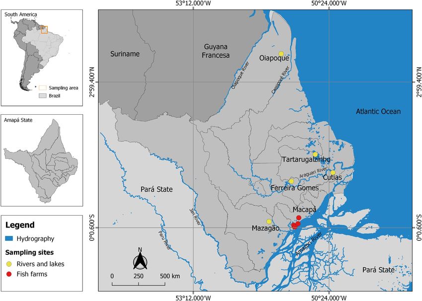
Fusobacteria and Tenericutes (Merrifield & Rodiles, 2015; Pereira of which 10 fish specimens (54.0 ± 18.4 cm and 1187.0 ± 750.1 g)
et al., 2017; Ramirez & Romero, 2017b; Salas-Leiva et al., 2017; were from two commercial fish farms located in the city of Macapá
Solovyev et al., 2019). Proteobacteria is a dominant taxon (Ramirez and 10 fish (71.1 ± 9.1 cm and 1854.3 ± 209.9 g) were obtained
& Romero, 2017b; Salas-Leiva et al., 2017; Solovyev et al., 2019), from rivers or lakes in the state of Amapá, Brazil (Figure 1). Farmed
and Enterobacteriaceae species have shown good performance in fish were fed with commercial feed for carnivorous fish, but with
the metabolism of cofactors, vitamins, amino acids, carbohydrates no additional additives such as probiotics, prebiotics and immuno-
and proteins, thus improving fish nutrition and helping to prevent stimulants. All fish were cryo-euthanized immediately after capture,
the colonization of pathogens. Enterobacteriaceae inhibit pathogens individually packed in sterile plastic bags and transported on ice in
through competition for nutrients and adhesion sites in the mucosa, isothermal coolers to the Laboratory of Applied Microbiology at the
which aids the fish immune system (Brenner & Farmer, 2015; La Patra Federal University of Amapá (UNIFAP), Macapá, state of Amapá.
et al., 2014; Ramírez & Romero, 2017a, 2017b). Therefore, such ben- The fish were initially washed in sterile sodium chloride solu-
efits indicate that species of autochthonous Enterobacteriaceae in tion (0.85%) and, subsequently, were sprayed with asepsis with
fish intestinal mucosa may have probiotic potential for supplemen- ethyl alcohol (70%) and subjected to three washes with sterile dis-
tation in their diet. tilled water. After laparotomy, a ventral incision was made from the
Little is known about the diversity and structure of autoch- urogenital pore to the operculum to remove the viscera and collect
thonous bacterial communities in the intestine of wild fish when the intestine. The abdominal cavity was rinsing with sterile distilled
1788 | © 2020 John Wiley & Sons Ltd wileyonlinelibrary.com/journal/are Aquaculture Research. 2021;52:1788–1796.APARECIDO PROIETTI-JUNIOR et al. | 1789
F I G U R E 1 Sampling sites of Arapaima gigas in fish farms, and rivers and lakes from the state of Amapá, in eastern Amazon (Brazil) [Colour
figure can be viewed at wileyonlinelibrary.com]
water. Then, an excision was made on the distal portion and ligation 300 colony-forming units (CFU) were quantified in a colony counter
of the intestinal loops was performed with sterile sutures, washing (Phoenix Luferco CP600-Plus, Brazil). The isolates were grouped
with sterile sodium chloride solution (0.85%) and spraying with asep- by behaviour in a chromogenic medium, and by the morphological
sis with 70% alcohol, followed by three washes with sterile distilled (Gram stain) and metabolic characteristics and were subsequently
water. The following steps were carried out inside a Class II B2 bio- re-isolated to verify purity.
logical safety cabinet (Trox®Technik, Brazil). Identification of bacterial isolates and susceptibility tests to
After removing the sutures from the intestinal ligations, all in- antimicrobials (Ceftazidime, Ceftriaxone, Cefepime, Ertapenem,
testinal contents were removed, and the lumen was washed 3 times Imipenem, Meropenem, Amikacin, Gentamicin, Ciprofloxacin,
using a disposable syringe containing 20 ml of sterile sodium chlo- Tigecycline and Colistine) were performed in the VITEK 2 system
ride solution (0.85%). A longitudinal incision was made in the intes- (BioM 'Etoile, France), and the isolates were then preserved in a trip-
2
tine wall to collect a 10 cm area sample to analyse the microbiota tych soy broth medium with glycerol (TSB 15% glycerol) and stored
attached to the intestinal surface of the fish. Each intestine sample in skim milk in a freezer at −70°C. Autochthonous bacterial isolates
was then transferred to a sterile Falcon tube and was completed to from the intestine of A. gigas were inoculated using streaking on sur-
10 ml with tryptic soy broth/TSB (BD Bacto™, USA) and homoge- face and depth on blood agar medium (Sigma-Aldrich, Germany) with
nized in an orbital shaker (Phoenix Luferco-AP59, Brazil) for 60 s, and 5% sheep blood, and incubated at 35°C for 24 and 48 hr.
serial dilutions were made in TSB to a concentration of 10−4. The preserved isolates, after reactivation in TSB at 37°C for
Each dilution was transplanted by surface streaking in tripli- 24 hr, were individually inoculated in tryptic soy agar/ TSA (BD
cates with 100 µl of suspensions in concentrations of 10−3 and 10−4, Bacto™, USA) and incubated again at 37°C. After 24 hr, two colonies
®
using 90 mm Petri dishes containing Chromocult Coliform Agar/ of each culture were transferred to a microplate (96 MSP, Bruker
CCA (Merck, Germany), incubated in a bacteriological incubator - Billerica, USA). The bacterial biomass was covered by a cell lysis
(REVCO ELITE II®-Kendro Laboratory Products™, USA) and main- solution (70% formic acid, Sigma-Aldrich). Then, an aliquot of 1 μL
tained at 35°C for 24 hr. Plates showing growth between 30 and of matrix solution (alpha-cyano-4-hydroxy-cinnamic acid diluted in1790 | APARECIDO PROIETTI-JUNIOR et al.
50% acetonitrile and 2.5% trifluoroacetic acid, Sigma-Aldrich) was were adjusted in a densitometer (DensichekTM, BioMerieux,
added to each bacterial biomass. The spectra of each sample were France) according to the standard 3 of the McFarland scale, for
generated in Matrix-Assisted Laser Desorption Ionization Time use of 100 µl to be inoculated in the wells. For this test, a standard
of Flight Mass Spectrometry (MALDI-TOF LT Microflex, Bruker) strain of Aeromonas hydrophila (American Type Culture Collection/
equipped with a 337 nm nitrogen laser in a linear path, controlled by ATCC-7966) provided by Fundação Oswaldo Cruz (CENT/LABENT,
the FlexControl 3.3 program (Bruker). The spectra were collected in Rio Janeiro, RJ, Brazil) was used, in addition to A. hydrophila and
a mass range between 2000–20,000 m/s, and then analysed using Aeromonas jandaei previously isolated from A. gigas with bacteriosis
the MALDI Biotyper 3.1 (Bruker) program, using the standard con- (Proietti-Junior et al., 2017). The excavated well technique was used,
figuration for bacteria identification, whereby the sample spectrum in which cylinders of 8 mm in diameter were cut using sterilized can-
is compared with the references in the database. For each plate, a nulas and removed from Petri dishes with AST medium previously
bacterial test standard was included to calibrate the instrument and inoculated with suspension of each Aeromonas, in the 0.5 standard
validate the run. The identification scoring criteria were performed of the McFarland scale, not exceeding 30 min. The plates were in-
as recommended by Bruker Daltonics who assessed the following: cubated for 24 hr at 35ºC and were subsequently analysed using an
a score of 2000 indicated identification at the species level; a score analog pachymeter (Etalon, Switzerland) to determine the inhibition
of 1700–1999 indicated gender-level identification; and a score rings around the wells containing suspensions of candidate bacteria
of 1700 was interpreted as an absence of identification (Alatoom for probiotics and their secondary metabolites (Balouiri et al., 2016).
et al., 2011). The virulence genes eaeA, lt, st, stx1, stx2, ial and eagg were in-
Tolerance test of the bacteria to low levels of pH was adapted vestigated using the Multiplex Polymerase Chain Reaction (PCR mul-
from Vinderola and Reinheimer (2003) as described below. The tiplex) technique. The purified bacterial DNA was extracted using
stored bacterial isolates were reactivated in TSB medium, re-iso- a commercial PureLin® kit (Invitrogen Life Technologies, Canada).
lated on CCA agar after 24 hr of incubation at 35°C. Colonies were Each PCR reaction was prepared in a total volume of 20 and 2.5 µl of
suspended and washed twice in buffer solution (0.05 M dibasic 10x PCR buffer (New England BioLabs, Ipswich, MA, USA), 5 pmol
potassium phosphate and potassium chloride, pH 6.5) and cen- of each primer (MWG-Biotech AG, Germany), ‘deoxynucleoside tri-
trifuged at 75 g/5ºC/20 min. The pellets were resuspended in the phosphates’ (dNTP) in a concentration of 200 µM (Promega), 1U Taq
same buffer and the concentrations were adjusted in a densitometer DNA polymerase (New England BioLabs) and 50 ng of genomic DNA.
(DensichekTM, BioMerieux, France) according to the 0.5 standard The activation temperature was 95°C in 15 min, followed by 30 cy-
of the McFarland scale. One mL of this suspension was added in a cles of denaturation at 94°C for 45 s, annealing at 55°C for 45 s, ex-
solution of pepsin (0.3% w/v) and sodium chloride (0.5% w/v) and tension at 68°C for 2 min and final extension step at 72°C for 5 min.
adjusted to pH 3.5, 4.0 and 4.5, and the mixtures were in triplicates The amplicons were subjected to electrophoresis in a 2% agarose
and were incubated for 3 hr. For each assay, 100 µl of suspension gel (Sigma-Aldrich, USA) and stained with ethidium bromide (Omar
was plated by streaking, incubated for 24 hr at 35°C, and colonies & Barnard, 2014) and visualized on a transilluminator (ImageQuant™
were counted after incubation. 300, GE, USA). The detection of the qnrA, qnrB, qnrS and rrs genes
The bile of each A. gigas specimen (farmed and wild) was collected was also performed using multiplex PCR. The reactions were per-
aseptically with the aid of disposable syringes and frozen at −20°C, formed using the previously extracted DNA in a Verit2 thermocycler
after sterilization using a 0.22 μm Filtermax vacuum filtration system (Applied Biossystems, USA), with a total volume of 20 and with 2.5 µl
(Techno Plastic Products AG, Switzerland). Subsequently, the bile of 10× PCR buffer (New England BioLabs, USA), 5 pmol of each
samples were thawed for 12 hr at 4°C and then at room temperature primer (MWG- Biotech AG, Germany), dNTP at a concentration of
in a Biological Security Cabinet. From the bacterial isolates activated 200 µM (Promega), 1U Taq DNA polymerase (New England BioLabs)
in TSB medium and re-isolated in CCA medium, bacterial suspen- and 50 ng of genomic DNA.
sions were prepared in sterile PBS pH 7.2 buffer and centrifuged at The isolates were evaluated for genetic polymorphism using the
90 g/4ºC/10 min. After two washes, the pellets were resuspended Pulsed Field Electrophoresis (PFGE) technique according to the pro-
in sterile PBS pH 7.2 buffer and the concentrations were adjusted in tocol of the Disease Control and Prevention Center (https://www.
a densitometer (DensichekTM, BioMerieux, France) according to the cdc.gov/ pulsenet/index.html). The fragments generated by the en-
0.5 standard of the McFarland scale. Then, serial dilutions of fish bile zymatic restriction were visualized in UV light using a transillumina-
were prepared in sterile PBS buffer for final concentrations of 0.5% tor (ImageQuantTM 300, GE, USA), and photo documentation was
and 1.0% (v/v). Each tube (9 ml final volume) was inoculated with performed. PFGE standards were analysed using the BioNumerics®
1 ml of the bacterial suspension and incubated for 90 min at 35°C. software, version 7.6 (Applied Maths, SintMartens-Lantem, Belgium).
The samples were then diluted in a series of sterile PBS buffer and The ecological terms prevalence, intensity and abundance were
100 µl aliquots were seeded by streaking in CCA medium and incu- used as recommended by Bush et al. (1997). The Brillouin index (HB),
bated for 24 hr at 35°C (Mukherjee & Ghosh, 2014; Nikoskelainen evenness (E) associated with HB, Berger–Parker dominance index
et al., 2001), and colonies were counted after incubation. (d), species richness (Magurran, 2004) and frequency of dominance,
The probiotic candidate bacteria were reactivated in TSB me- which is the percentage of infracommunities for each species of bac-
dium 24 hr in advance at 35°C. After propagation, the concentrations teria is numerically dominant (Rohde et al., 1995), were calculatedAPARECIDO PROIETTI-JUNIOR et al. | 1791
to evaluate the component community of enterobacteria using the The growth in chromogenic medium allowed the isolation of
software Diversity (Pisces Conservation Ltda, UK). All data were 17 species (84 strains) and the results of the identification of the
tested for normality and homoscedasticity using the Shapiro–Wilk bacterial isolates were identical when comparing those obtained by
and Barlett test respectively. The t test was used to compare data on the MALDI-TOF MS system to those of the VITEK 2 system. The
diversity parameters (HB, E, d and species richness) between farmed prevalence, mean intensity, mean abundance and frequency of dom-
and wild fish. One-way ANOVA was used to compare the size of the inance are shown in Table 1. High prevalence and mean intensity
inhibition rings of the isolates of the candidate bacteria to probiotics were shown for Enterobacter cloacae and Proteus mirabilis in wild
in the in vitro assays of antagonism against the three pathogens, fol- fish when compared to the farmed fish, whereas for Escherichia
lowed by the Tukey test (Zar, 2010). coli, there was a high prevalence in wild fish and greater intensity in
captive fish. Edwardsiella tarda, Citrobacter braakii and Plesiomonas
TA B L E 1 Prevalence (P), mean
FD
intensity (MI), mean abundance (MA)
Phenospecies P (%) MI MA (%)
and frequency of dominance (FD)
of autochthonous enterobacteria Environments Enterobacter cloacae 20.0 17,375 3475 0.8
(CFU/10 cm2) of the intestinal mucus of Citrobacter freundii 30.0 127,778 38,333 8.6
Arapaima gigas sampled in two different
Citrobacter werkmanii 20.0 198,333 39,667 8.9
environments in eastern Amazon (Brazil)
Photorhabdus 20.0 34,167 6833 1.5
luminescens
Morganella morganii 30.0 174,444 52,333 11.7
Kluyvera intermedia 30.0 160,926 48,278 10.8
Proteus vulgaris 50.0 110,533 55,267 12.3
Proteus mirabilis 10.0 73,333 7333 1.6
Farmed Cedecea neteri 10.0 86,667 8667 1.9
Cedecea davisae 30.0 61,667 18,500 4.1
Edwardsiella tarda 0 0 0 -
Escherichia coli 30.0 197,556 59,267 13.2
Aeromonas veronii 30.0 158,889 47,667 10.6
Aeromonas sobria 20.0 96,667 19,333 4.3
Citrobacter braakii 0.0 0 0 -
Klebsiella pneumoniae 40.0 107,292 42,917 9.6
Plesiomonas shigelloides 0 0 0 -
Enterobacter cloacae 90.0 161,926 145,733 20.8
Citrobacter freundii 40.0 131,667 52,667 7.5
Citrobacter werkmanii 10.0 216,667 21,667 3.1
Photorhabdus 0 0 0 -
luminescens
Morganella morganii 20.0 95,000 19,000 2.7
Kluyvera intermedia 0 0 0 -
Proteus mirabilis 80.0 145,000 116,000 16.6
Proteus vulgaris 0 0 0 -
Cedecea neteri 0.0 0 0 -
Wild Cedecea davisae 0.0 0 0 -
Edwardsiella tarda 60.0 242,889 145,733 20.8
Escherichia coli 60.0 122,222 73,333 10.5
Aeromonas veronii 0 0 0 -
Aeromonas sobria 0 0 0 -
Citrobacter braakii 20.0 153,333 30,667 4.4
Klebsiella pneumoniae 50.0 112,667 56,333 8.1
Plesiomonas shigelloides 40.0 95,000 38,000 5.41792 | APARECIDO PROIETTI-JUNIOR et al.
shigelloides occurred only in the intestinal mucosa of wild fish, Isolates of A. veronii, E. tarda, A. sobria and Plesiomonas shigel-
while Photorhabdus luminescens, Kluyvera intermedia, Cedecea neteri, loides of A. gigas (Table 2) were excluded as bacteria with probiotic
Cedecea davisae, Aeromonas veronii, Aeromonas sobria and Proteus potential due to their pathogenicity for fish (Castañeda-Monsalve
vulgaris occurred only in farmed fish. There was a frequency of dom- et al., 2019; Shama et al., 2000) and humans. In the present study,
inance for several species in farmed fish that were absent in wild 47% of the species of autochthonous enterobacteria showed
fish, which showed dominance of E. cloacae, P. mirabilis and E. tarda. the ability to colonize the intestinal mucosa surface of A. gigas.
In addition, 11 isolates of E. cloacae showed probiotic potential. Autochthonous Klebsiella pneumoniae of A. gigas showed resistance
In wild A. gigas, the Brillouin diversity, evenness and richness to the antimicrobial drugs tested here and has also shown the pres-
of the bacteria species were greater (p < 0.05) than in farmed ence of resistance genes in isolates obtained from some marine fish
A. gigas, but the Berger–Parker dominance was greater in farmed species (Singh et al., 2017).
fish (Figure 2). On the other hand, farmed A. gigas which showed a The isolates of Citrobacter freundii, Photorhabdus luminescens,
greater value for Berger–Parker dominance index due the predomi- Morganella morganii, Kluyvera intermedia and P. mirabilis of A. gigas
nance of several species. In Danio rerio, no difference was reported were excluded as bacteria with probiotic potential due to the hae-
in the structure of the composition and diversity of allochthonous molytic activity presented, while the other species were negative
bacteria between wild and captive fish (Roeselers et al., 2011). (Table 2). Citrobacter freundii also showed haemolytic activity and
Studies of the bacterial microbiota of the mucosa and intestinal con- pathogenicity to A. gigas (Pereira et al., 2017) and Pseudoplatystoma
tent of fingerlings and adults of A. gigas showed that most of the reticulatum, and resistance to antimicrobial drugs (Pádua et al., 2014).
identified isolates were members of Proteobacteria, Fusobacteria Isolates of P. luminescens, M. morganii and K. intermedia from the in-
and Firmicutes, and that this microbiota had more richness in finger- testinal mucosa of A. gigas in the present study also showed hae-
lings (Pereira et al., 2017). Although the structure of the intestinal molytic activity. Thus, these isolates were discarded as a probiotic
bacterial communities of fish can be influenced by biotic (e.g. devel- potential for A. gigas, due to its potential pathogenic in case of a
opmental stage, intestinal structure, diet, species, age and trophic disequilibrium in pathogen–host relationship. Isolates of E. coli in
level of the host) and abiotic (e.g. habitat and characteristics of the A. gigas were more prevalent in the intestinal mucosa of wild fish
surrounding environment) factors, including the cultivation condi- than in farmed fish, which had a greater abundance. In addition, the
tions (Ramirez & Romero, 2017a; Roeselers et al., 2011; Salas-Leiva isolates showed multidrug resistance to antimicrobials. Bollache
et al., 2017), the differences observed in the present study may be et al. (2018) reported that E. coli are producers of broad-spectrum
due to the differences in the environment and feeding of the fish. beta-lactamases in several fish species. Therefore, E. coli isolates
F I G U R E 2 Diversity parameters of
autochthonous enterobacteria in the
intestinal mucosa of Arapaima gigas from
fish farm, and rivers and lakes of eastern
Amazon (Brazil). Similar letters in the same
column indicate no significant differences
(p > 0.05) according to the t testAPARECIDO PROIETTI-JUNIOR et al. | 1793
TA B L E 2 Results of the analyses used in the selection of results of antimicrobial susceptibility tests for E. cloacae isolates are
enterobacteria species with probiotic potential for Arapaima gigas compatible with characteristics of commensal bacterial microbiota
Species P H R pH B since such autochthonous bacteria were sensitive to all tested anti-
microbial drugs (Table 2). Proteus vulgaris isolates of A. gigas showed
Edwardsiella tarda + NA NA NA NA
a reduction in viable cell count after 3 hr of exposure to simulated
Aeromonas veronii + NA NA NA NA
gastric fluid in different pH ranges. However, all isolates of autoch-
Aeromonas sobria + NA NA NA NA
thonous E. cloacae showed resistance to simulated gastric fluid at
Plesiomonas shigelloides + NA NA NA NA
the three pH levels (Table 3). The autochthonous E. cloacae isolates
Citrobacter freundii ND + NA NA NA
of A. gigas with probiotic potential showed tolerance to A. gigas bile
Photorhabdus luminescens ND + NA NA NA fluid (Table 4). A reduced change in the growth of these autochtho-
Morganella morganii ND + NA NA NA nous isolates was observed after 24 hr of exposure to bile fluid (data
Kluyvera intermedia ND + NA NA NA not shown). Isolates 11 and 16 of A. gigas showed greater inhibi-
Cedecea davisae ND ND + NA NA tion ring against Aeromonas hydrophila and A. jandaei from A. gigas.
Citrobacter werkmanii ND ND + NA NA In addition, the inhibition rings of all isolates against A. hydrophila
Cedecea neteri ND ND + NA NA and Aeromonas jandaei were greater when compared to A. hydroph-
Escherichia coli ND ND + NA NA ila ATCC-7966 (Table 5). None of the E. cloacae isolates of A. gigas
showed the virulence genes eaeA, lt, st, stx1, stx, eaeA, ial and eagg. In
Klebsiella pneumoniae ND ND + NA NA
addition, none of the E. cloacae isolates showed the resistance genes
Proteus mirabilis ND ND + NA NA
qnrA, qnrB, qnrS and rrs. The isolates used in the genotyping allowed
Citrobacter braakii ND ND ND NT NA
the detection of two distinct species of E. cloacae, with a gene simi-
Proteus vulgaris ND ND ND ND NT
larity of approximately 85% (Figure 3).
Enterobacter cloacae ND ND ND ND ND
Studies have shown the contribution of autochthonous in-
Abbreviations: +, Positive; B, Absence of growth in any concentration testinal bacterial microbiota to fish health (Pereira et al., 2017;
of bile fluid; H, Haemolysis activity; NA, not analysed; ND, no Sedláček et al., 2016), of which efficacy is due to the specificity
detection; NT, no tolerance; P, Pathogenicity; pH, Absence of growth
in the strain–host interaction (Salas-Leiva et al., 2017). Studies
in the analysed ranges of pH; R, Resistance to at least three classes of
antimicrobial drugs. on autochthonous bacteria in the intestine of A. gigas with pro-
biotic potential have focused only on lactic acid species (Fujimoto
TA B L E 3 Reduction of the counting of viable cells (UFC/mL) of et al., 2014; Pereira et al., 2017). However, the use of the probiotic
Enterobacter cloacae exposed to simulated gastric fluid, after 3 hr in Enterobacter spp. from the gastrointestinal tract of Oncorhynchus
three levels of pH acid mykiss demonstrated the ability to inhibit the in vitro growth of the
Fish/isolates pH 3.5 pH 4.0 pH 4.5 pathogen Flavobacterium psychrophilum and reduced fish mortality
after being challenged, due to the protection of the fish immune
1 3.3 ± 0.5 2.4 ± 0.8 2.3 ± 0.5
system (La Patra et al., 2014). In vitro and in vivo studies with the
4 4.2 ± 0.2 3.3 ± 0.5 3.8 ± 0.5
11 2.4 ± 0.4 1.9 ± 0.3 1.6 ± 0.6
TA B L E 4 Growth (%) of autochthonous Enterobacter cloacae
12 2.8 ± 0.8 2.1 ± 1.2 1.8 ± 0.5
after 90 min of exposure to concentrations of bile fluid of Arapaima
13 2.9 ± 0.6 2.4 ± 0.8 1.9 ± 0.3 gigas
14 2.6 ± 0.6 2.2 ± 1.0 1.8 ± 1.2
Concentrations of bile fluid
16 3.7 ± 0.8 2.8 ± 0.5 1.7 ± 0.8
17 2.9 ± 1.0 2.6 ± 1.3 1.9 ± 1.5 Fish/isolates 0.5% 1.0%
18 2.7 ± 0.4 2.3 ± 0.5 1.8 ± 0.8 1 63.8 ± 2.7 59.9 ± 1.8
19 2.5 ± 0.3 2.2 ± 0.9 1.9 ± 0.6 4 67.6 ± 3.2 41.0 ± 2.7
20 2.8 ± 1.2 2.3 ± 0.8 1.8 ± 1.2 11 83.4 ± 4.0 77.5 ± 2.9
12 73.8 ± 2.7 69.9 ± 1.5
13 69.5 ± 1.9 63.8 ± 2.3
were excluded as a probiotic potential for A. gigas, although this spe-
14 71.6 ± 4.1 65.5 ± 3.4
cies of Gammaproteobacteria has been used as a probiotic for other
16 74.5 ± 1.3 68.6 ± 1.9
animal species by one century (Wassenaar, 2016).
17 62.8 ± 3.7 57.9 ± 3.2
The isolates of Cedecea davisae, Citrobacter werkmanii, Cedecea
18 66.4 ± 2.5 62.8 ± 2.1
neteri, Escherichia coli, Klebsiella pneumoniae and P. mirabilis of A. gigas
19 68.8 ± 3.3 65.9 ± 2.2
were excluded as bacteria with probiotic potential because they are
resistant to three or more classes of antimicrobial. However, further 20 71.7 ± 3.5 68.4 ± 2.6
studies on these isolates are needed carried out. In contrast, the Note: Values expressed as mean ± standard deviation.1794 | APARECIDO PROIETTI-JUNIOR et al.
TA B L E 5 Measures of the ring
Aeromonas hydrophila Aeromonas Aeromonas
of inhibition (mm) of the isolates of
Bacteria (ATCC−7966) hydrophila jandaei
Enterobacter clocae by in vitro inhibition of
Fish/isolates Mean ± SE Mean ± SE Mean ± SE p-value Aeromonas species of Arapima gigas
a b b
1 11.3 ± 0.1 12.5 ± 0.1 12.5 ± 0.3APARECIDO PROIETTI-JUNIOR et al. | 1795
in A. gigas, since aquaculture of this fish requires supplementation of Brenner, D. J., & Farmer, J. (2015). Enterobacteriaceae. In M. E. Trujillo,
S. Dedysh, P. DeVos, B. Hedlund, P. Kämpfer, F. A. Rainey, & W. B.
the diet to increase its production and reduce mortality of fry and
Whitman (Eds.), Bergey's manual of systematics of archaea and bacteria
fingerlings. (pp. 1–24). Willey.
In conclusion, this is the first study regarding the diversity of Bush, A. O., Lafferty, K. D., Lotz, J. M., & Shostak, A. W. (1997).
cultivable autochthonous enterobacteria of the intestinal mucosa of Parasitology meets ecology on its own terms: Margolis
et al Revisited. The Journal of Parasitology, 83(4), 575–583. https://
A. gigas, and 84 isolates of the bacterial taxon were characterized,
doi.org/10.2307/3284227.
of which 17 species were identified. Only E. cloacae isolates demon- Castañeda-Monsalve, V. A., Junca, H., García-Bonilla, E., Montoya-
strated probiotic potential. However, further research is needed to Campuzano, O. I., & Moreno-Herrera, C. X. (2019). Characterization
validate the probiotic effects of these isolates, especially isolates 11 of the gastrointestinal bacterial microbiome of farmed juvenile and
and 16, on the growth performance and immunity of A. gigas, and to adult white cachama (Piaractus brachypomus). Aquaculture, 512,
734325. https://doi.org/10.1016/j.aquaculture.2019.734325
determine the best concentration for supplementation in the diet.
Duarte, S., Silva, F. C. P., Zauli, D. A. G., Nicoli, J. R., & Araújo, F. G. 2014).
Studies with the same isolates are also needed for the development Gram-negative intestinal indigenous microbiota from two Siluriform
of biotechnological products for use in the farming of this fish. fishes in a tropical reservoir. Brazilian Journal of Microbiology, 45(4),
1283–1292. https://doi.org/10.1590/S1517-83822014000 400019
Fujimoto, R. Y., Gabbay, M. I., Martins, M. L., Bezerra, A. M., Silva, B.
AC K N OW L E D G E M E N T S
C., & Mouriño, J. L. P. (2014). Isolamento e seleção de bactérias áci-
The authors thank the Brazilian National Council for Scientific and do-láticas com potencial probiótico para pirarucu (pp. 1–4). Aracaju,
Technological Development (CNPq, Brasil) for the scientific research Brazil: Embrapa Tabuleiros Costeiros, (Embrapa Tabuleiros Costeiros:
grant awarded to Tavares-Dias, M. (Grant 303013/2015-0). Comunicado Técnico 148).
Girijakumari, N. R., Ethiraja, K., & Marimuthu, P. N. (2018). In vitro and
in vivo evaluation of probiotic properties of Enterobacter cloacae in
C O N FL I C T O F I N T E R E S T Kenyi cichlid, Maylandia lombardoi. Aquaculture International, 26,
The authors declare no conflict of interests. 959–980. https://doi.org/10.1007/s10499-018-0262-2
He, S., Zhou, Z., Banerjee, G., Huang, L., Ray, A. K., & Ringø, E. (2016).
Bacterial diversity in the digestive tracts of four Indian air-breath-
E T H I C A L A P P R OVA L
ing fish species investigated by PCR based denaturing gradient gel
The present study was carried in accordance with the princi- electrophoresis. Brazilian Archives of Biology and Technology, 59,
ples adopted by the Brazilian College of Animal Experimentation e16160332. https://doi.org/10.1590/1678-4324-2016160332
(COBEA) and was approved by the Ethics Committee on the Use of La Patra, S. E., Fehringer, T. R., & Cain, K. D. (2014). A probiotic Enterobacter
sp. provides significant protection against Flavobacterium psychrophi-
Animals (CEUA/UFAC: No 23107.009564/2014-29) of the Federal
lum in rainbow trout (Oncorhynchus mykiss) after injection by two dif-
University of Acre and National System for the Management of ferent routes. Aquaculture, 433, 361–366. https://doi.org/10.1016/j.
Genetic Heritage and Associated Traditional Knowledge (SISGEN aquaculture.2014.06.022
Nº A7EC29D) and of the Biodiversity Authorization and Information Magurran, A. E. (2004). Measuring biological diversity (pp. 1–256). New
System (SISBIO No 62153-2), according to the regulations of York, NY: Blackwell Science.
Merrifield, D. L., Dimitroglou, A., Foey, A., Davies, S. J., Baker, R. T.
research of the Ministry of the Environment (MMA).
M., Bøgwald, J., Castex, M., & Ringø, E. (2010). The current status
and future focus of probiotic and prebiotic applications for salmo-
DATA AVA I L A B I L I T Y S TAT E M E N T nids. Aquaculture, 302(1–2), 1–18. https://doi.org/10.1016/j.aquac
The data that support the findings of this study are available from ulture.2010.02.007
Merrifield, D. L., & Rodiles, A. (2015). The fish microbiome and its in-
the corresponding author upon reasonable request.
teractions with mucosal tissues. In B. H. Beck, & E. Peatman (Eds.),
Mucosal health in aquaculture (pp. 273–295). Academic Press-Elsevier.
ORCID Mukherjee, A., & Ghosh, K. (2014). Antagonism against fish pathogens
Marcos Tavares-Dias https://orcid.org/0000-0002-8376-1846 by cellular components and verification of probiotic properties in
autochthonous bacteria isolated from the gut of an Indian major
carp, Catla catla (Hamilton). Aquaculture Research, 47(7), 2243–2255.
REFERENCES https://doi.org/10.1111/are.12676
Alatoom, A. A., Cunningham, S. A., Ihde, S. M., Mandrekar, J., & Patel, Nikoskelainen, S., Salminen, S., Bylund, G., & Ouwehand, A. C. (2001).
R. (2011). Comparison of direct colony method versus extraction Characterization of the properties of human and dairy-derived
method for identification of gram-positive cocci by use of Bruker probiotics for prevention of infectious diseases in fish. Applied
Biotyper matrix-assisted laser desorption ionization-time of flight and Environmental Microbiology, 67(6), 2430–2435. https://doi.
mass spectrometry. Journal of Clinical Microbiology, 49(8), 2868– org/10.1128/AEM.67.6.2430-2435.2001
2873. https://doi.org/10.1128/JCM.00506-11 Omar, K. B., & Barnard, T. G. (2014). Detection of diarrhoeagenic Escherichia
Balouiri, M., Sadiki, M., & Ibnsouda, S. K. (2016). Methods for in vitro coli in clinical and environmental water sources in South Africa using sin-
evaluating antimicrobial activity: A review. Journal of Pharmaceutical gle-step 11-gene m-PCR. World Journal of Microbiology and Biotechnology,
Analysis, 6(2), 71–79. https://doi.org/10.1016/j.jpha.2015.11.005 30, 2663–2671. https://doi.org/10.1007/s11274-014-1690-4
Bollache, L., Bardet, E., Depret, G., Motreuil, S., Neuwirth, C., Pádua, S. B., Marques, D. P., Sebastião, F. A., Pilarski, F., Martins, M.
Moreau, J., & Hartmann, A. (2018). Dissemination of CTX-M- L., & Ishikawa, M. M. (2014). Isolation, Characterization and pa-
producing Escherichia coli in freshwater fishes from a French wa- thology of Citrobacter freundii infection in native Brazilian catfish
tershed (Burgundy). Frontiers in Microbiology, 9, 3239. https://doi. Pseudoplatystoma. Brazilian Journal of Veterinary Pathology, 7(3),
org/10.3389/fmicb.2018.03239 151–157.1796 | APARECIDO PROIETTI-JUNIOR et al.
Pereira, G. V., Cunha, D., Mouriño, J. L., Rodiles, A., Jaramillo, A., & Singh, A. S., Lekshmi, M., Prakasan, S., Nayak, B., & Kumar, S. (2017).
Merrifield, D. (2017). Characterization of microbiota in Arapaima gigas Multiple antibiotic-resistant, extended spectrum-β-Lactamase
intestine and isolation of potential probiotic bacteria. Journal of Applied (ESBL) - producing Enterobacteria in fresh seafood. Microorganisms,
Microbiology, 123, 1298–1311. https://doi.org/10.1111/jam.13572 5(3), https://doi.org/10.3390/microorganisms5030053
Proietti-Junior, A. A., Lima, L. S., Cardoso, F. M. N., Rodrigues, D. P., & Solovyev, M. M., Kashinskaya, E. N., Bochkarev, N. A., Andree, K. B., &
Tavares-Dias, M. (2017). Bacterioses em alevinos de pirarucu de cultivo, Simonov, E. (2019). The effect of diet on the structure of gut bac-
com ênfase em edwardsielose e aeromonose (pp. 1–9). Macapa, Brazil: terial community of sympatric pair of whitefishes (Coregonus lava-
Embrapa Amapá, Comunicado Técnico 149. retus): one story more. PeerJ, 7, e8005. https://doi.org/10.7717/
Ramírez, C., & Romero, J. (2017a). The Microbiome of Seriola lalandi of peerj.8005
wild and aquaculture origin reveals differences in composition and Vinderola, C. G., & Reinheimer, J. (2003). Lactic acid starter and probiotic
potential function. Frontiers in Microbiology, 8, 1844. https://doi. bacteria: Comparative “in vitro” study of probiotic characteristics
org/10.3389/fmicb.2017.01844 and biological barrier resistance. Food Research International, 36(9–
Ramírez, C., & Romero, J. (2017b). Fine flounder (Paralichthys adspersus) 10), 895–904. https://doi.org/10.1016/S0963-9969(03)00098-X
microbiome showed important differences between wild and reared Wanka, K. M., Damerau, T., Costas, B., Krueger, A., Schulz, C., & Wuertz,
specimens. Frontiers in Microbiology, 8, 271. https://doi.org/10.3389/ S. (2018). Isolation and characterization of native probiotics for fish
fmicb.2017.000271 farming. BMC Microbiology, 18(1), 119. https://doi.org/10.1186/
Roeselers, G., Mittge, E. K., Stephens, W. Z., Parichy, D. M., Cavanaugh, s12866-018-1260-2
C. M., Guillemin, K., & Rawls, J. F. (2011). Evidence for a core gut Wassenaar, T. M. (2016). Insights from 100 years of research with pro-
microbiota in the zebrafish. ISME Journal, 5(10), 1595–1608. https:// biotic E. coli. European Journal of Microbiology and Immunology, 6(3),
doi.org/10.1038/ismej.2011.38 147–161. https://doi.org/10.1556/1886.2016.00029
Rohde, K., Hayward, C., & Heap, M. (1995). Aspects of the ecology Yamashita, M. M., Ferrarezi, J. V., Pereira, G. V., Bandeira, G., Silva, B. C.,
of metazoan ectoparasites of marine fishes. International Journal Pereira, S., Martins, M. L., & Mouriño, J. L. (2020). Autochthonous
for Parasitology, 25(8), 945–970. https://doi.org/10.1016/0020- vs allochthonous probiotic strains to Rhamdia quelen. Microbial
7519(95)00015-t Pathogenesis, 139, 103897. https://doi.org/10.1016/j.micpa
Salas-Leiva, J., Opazo, R., Remond, C., Uribe, E., Velez, A., & Romero, J. th.2019.103897
(2017). Characterization of the intestinal microbiota of wild-caught Zar, J. H. (2010). Biostatistical analysis (pp. 1–944). New York, NY: Prentice
and farmed fine flounder (Paralichthys adspersus). Latin American Hall.
Journal of Aquatic Research, 45(2), 370–378. https://doi.org/10.3856/
vol45-issue2-fulltext-12
Sedláček, I., Staňková, E., & Švec, P. (2016). Composition of cultiva-
How to cite this article: Aparecido Proietti-Junior A, Sampaio
ble enteric bacteria from the intestine of Antarctic fish (family
Lima L, Duarte Gonçalves V, Rocha Pribul B, Prazeres
Nototheniidae). Czech Journal of Animal Science, 61(3), 127–132.
https://doi.org/10.17221/8785-CJAS Rodrigues D, Tavares-Dias M. Cultivable autochthonous
Shama, S., Brandão, D. A., Vargas, A. C., Costa, M. M., & Pedrozo, A. F. bacteria of the intestinal mucosa of Arapaima gigas (Pisces:
(2000). Bactérias com potencial patogênico nos rins e lesões exter- Arapaimidae) with probiotic potential. Aquaculture Research.
nas de jundiás (Rhamdia quelen) cultivados em sistema semi-inten-
2021;52:1788–1796. https://doi.org/10.1111/are.15025
sivo. Ciência Rural, 30(2), 293–298. https://doi.org/10.1590/S0103
-847820 00000200016You can also read