Detection and assessment of electrocution in endangered raptors by infrared thermography
←
→
Page content transcription
If your browser does not render page correctly, please read the page content below
Melero et al. BMC Veterinary Research 2013, 9:149
http://www.biomedcentral.com/1746-6148/9/149

 CASE REPORT                                                                                                                                       Open Access

Detection and assessment of electrocution in
endangered raptors by infrared thermography
Mar Melero1*, Fernando González2, Olga Nicolás3, Irene López2, María de los Ángeles Jiménez4,
Susana Jato-Sánchez2 and José Manuel Sánchez-Vizcaíno1

  Abstract
  Background: Most European birds of prey find themselves in a poor state of conservation, with electrocution as
  one of the most frequent causes of unnatural death. Since early detection of electrocution is difficult, treatment is
  usually implemented late, which reduces its effectiveness. By considering that electrocution reduces tissue
  temperature, it may be detectable by thermography, which would allow a more rapid identification. Three
  individuals from three endangered raptor species [Spanish imperial eagle (Aquila adalberti), Lammergeier (Gypaetus
  barbatus) and Osprey (Pandion haliaetus)] were studied thermographically from the time they were admitted to a
  rehabilitation centre to the time their clinical cases were resolved.
  Cases presentation: The three raptors presented lesions lacking thermal bilateral symmetry and were consistent
  with electrocution of feet, wings and eyes, visible by thermography before than clinically; lesions were well-defined
  and showed a lower temperature than the surrounding tissue. Some lesions evolved thermally and clinically until
  the appearance of normal tissue recovered, while others evolved and became necrotic. A histopathological analysis
  of a damaged finger amputated off a Lammergeier, and the necropsy and histopathology examination of an
  osprey, confirmed the electrocution diagnosis.
  Conclusions: These results suggest that thermography is effective and useful for the objective and early detection
  and monitoring of electrocuted birds, and that it may prove especially useful for examining live animals that require
  no amputation or cannot be subjected to invasive histopathology.
  Keywords: Thermography, Electrocution, Raptor, Bird of prey, Spanish imperial eagle, Lammergeier, Osprey

Background                                                                              survival problem for one of the most endangered raptors
Birds of prey have been proposed as good sentinels of                                   in the world, the Spanish imperial eagle [6].
environmental changes as they are placed at the top of                                     The accuracy of electrocution mortality estimates is
the food chain and are widespread worldwide [1]. How-                                   limited given the difficulty in determining cause of death
ever, 36 (64%) of the 56 species of raptors inhabiting                                  [7]. Since determination of electrocution is usually based
Europe find themselves in an unfavourable state of con-                                 on anamnesis and clinical signs, i.e., electrical burns [1],
servation [2]. Most raptor deaths are caused by direct                                  detecting and evaluating an electrocuted bird can prove
and indirect actions of humans [1]. One of the most                                     most difficult since anamnesis is often incomplete, and
common causes of unnatural death is electrocution as a                                  evidence for electrical trauma may not be immediately
result of collisions with power lines and the subsequent                                detectable [8]. Moreover, an early diagnosis of electrocu-
trauma when the animal falls to the ground [1,3,4].                                     tion is essential for a good prognosis. For these reasons,
While unnatural mortality can be compensated in a                                       complementary diagnostic tools, such as histopathology,
healthy population, it can seriously affect a small popula-                             are used when an amputation or necropsy is required
tion [5]. For example, electrocution poses a significant                                [1,9]. Nevertheless, histopathology is invasive and may
                                                                                        not be feasible or advisable for live animals, especially
* Correspondence: mar.melero@sanidadanimal.info                                         those that do not require amputation.
1
 VISAVET Centre and Animal Health Department, Veterinary School,
Complutense University of Madrid, 28040 Madrid, Spain
                                                                                           Thermography is a non-invasive technique used to assess
Full list of author information is available at the end of the article                  tissue temperature that can be applied to electrocution
                                          © 2013 Melero et al.; licensee BioMed Central Ltd. This is an Open Access article distributed under the terms of the Creative
                                          Commons Attribution License (http://creativecommons.org/licenses/by/2.0), which permits unrestricted use, distribution, and
                                          reproduction in any medium, provided the original work is properly cited.
Melero et al. BMC Veterinary Research 2013, 9:149                                                                Page 2 of 7
http://www.biomedcentral.com/1746-6148/9/149

detection and assessment. It can be used at a distance of      with a similar maximum temperature in the middle of
more than 500 m from the animal [10], reducing its stress      both ulna areas (Table 1, Figure 1(4)). This asymmetric
and ensuring the safety of the technician. Physiological and   thermal pattern was consistent with the patagium retrac-
pathological thermographic patterns have not yet been          tion caused by periods of immobilisation. Increased vas-
established for most species. Nevertheless, reports of ther-   cularity and inflammation in the patagium area led to an
mographic analyses have so far established that the thermal    increased temperature, which was detected by thermog-
pattern and symmetry of the body are more important            raphy. Early detection of this pathology is essential for a
than the absolute temperature for diagnosing disease [11].     good prognosis; otherwise, the disease can lead to the
  Tissue affected by electrocution shock shows reduced         development of fibrous tissue, calcifications and tie
vascularisation, innervation, water content [12] and oxy-      downs [19], making impossible the animal releasing into
gen saturation [13]. These changes should result in a          the wild, the primary goal of rehabilitation of wildlife.
lower temperature in the affected area, thus raising the
possibility that electrocution shock can be detected by        Case 2: Lammergeier (Gypaetus barbatus)
thermography. Accordingly, this report applies thermog-        On 13 April 2012, an adult female was admitted to the
raphy to detect and assess electrocution to three endan-       fauna recuperation centre of Vallcalent, Lleida (Spain).
gered raptors [14,15].                                         The admission examination revealed proper body condi-
                                                               tion, good hydration and alert mental status. Neverthe-
Cases presentation                                             less, the animal put little weight on the left limb and
Case 1: Spanish imperial eagle (Aquila adalberti)              leaning instead on the dorsum of the foot. Treatment
On 11 September 2008, a juvenile female was admitted           consisted of antibiotics, anti-inflammatory and physio-
to the GREFA wildlife rehabilitation centre in Madrid          therapy on the left leg. The three most distal phalanges
(Spain). Clinical signs were 7% dehydration, depression,       of left finger III were amputated 5 days after admission
emaciation, arrhythmias, bradycardia and a proximal            because their condition worsened despite treatment. The
closed right radial fracture. Electrical lesions were not      animal remained in the rehabilitation centre with a good
observed initially, but became visible 3 days later. Treat-    prognosis, the amputated finger correctly healed and the
ment included rehydration, antibiotics, a figure‐eight         left leg appeared strong and toned, and the animal was
bandage on the right wing and physiotherapy on the             using it normally.
right finger IV and the right wing. Finally, the animal           The first thermal examination showed an abnormal
was released on 16 March 2009.                                 thermal pattern in left foot showing low temperatures and
   For thermographic evaluation (Cases 1–3), a ThermaCAM       large temperature differences within finger III (10.2°C)
E45 infrared thermocamera with an FOV25 lens was used          and less marked in finger IV (8.4°C) compared with
and images were analysed using the ThermaCAM                   healthy ones (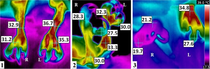
Melero et al. BMC Veterinary Research 2013, 9:149                                                                                                            Page 3 of 7
http://www.biomedcentral.com/1746-6148/9/149

Table 1 Thermal values at different anatomical areas
                                                                              Fingers
                                       Healthy                                              Recovered                                     Required amputation
     Differences of        Within       Equivalent fingers              Within each                  Equivalent fingers              Within          Equivalent fingers
    temperature (°C)     each finger      of both feet1                    finger                      of both feet1               each finger         of both feet1
                                                                                          Right finger IV
                                                                                    2
    Spanish imperial
Melero et al. BMC Veterinary Research 2013, 9:149 Page 4 of 7
http://www.biomedcentral.com/1746-6148/9/149
Figure 1 Thermal analysis of the Spanish imperial eagle (A. adalberti). Case 1. Thermography of feet; Lateral views of the (1) left foot and
(2) the right foot upon admission, and (3) the right foot 35 days after admission. Roman numerals designate each finger (I-IV). A physiological
thermal pattern is observed in the left foot and in right fingers I, II and III. The thermal range is the same for Figure 1(1-3). Thermography of
wings; Ventral view of the wings taken (4) at the time the bandage was removed and (5) at the time of first flight test. A physiological thermal
pattern is observed in the left wing. Initial designated right (R) and left (L) legs and wings. (6) An anatomic diagram of the ventral view of a right
wing, drawn based on a figure in Orosz et al. [18]. The tendon of the tensor propatagialis is shown in green. The area of the proximal right radial
fracture is enclosed by arrows (4, 6) and arrowheads (5).
the neurovascular necrosis, which would have required Necropsy revealed thinness, an oedema in the left wing,
amputation of the limb. Since amputation of the leg is haematoma in ribs and keel, atrophied pectoral muscles,
incompatible with the hunting and feeding habits of this numerous clots distributed by all the vessels in lung and air
species, it was euthanised for humanitarian reasons on sacs veins with yellowish deposits, thickened pericardium,
28 March 2012 based on previous studies recommenda- myocardial greenish stain in the left ventricle, whitish injury
tions [23-25]. Then, necropsy was performed according in the apex, and liver yellowish and congestive encephalon.
to the protocol of Schmidt and Reavill [26,27]. Cold oedemas mainly in the distal wing and coagulation in
Thermal examination upon admission revealed an ab- internal organs have been previously described as common
normally high difference between legs, which deterio- necropsy findings in electrocuted birds [8]. The wing frac-
rated significantly from admission (4.1°C) to the day that ture and haematomas in pectoral muscles and keel were
euthanasia was practiced (13.6°C) (Table 1, Figure 3) and probably secondary to the fall after colliding with power
a high temperature area in the left wing, from the elbow lines.
to the carpus, coincident with the fracture and sur- The most remarkable histopathological findings were in
rounding tissue; and a low temperature in the region the liver. The hepatic parenchyma had broad multifocal
distally from the carpus. The comminute fracture and fragmented areas of coagulative necrosis surrounded by
consequent inflammation increased the temperature be- abundant immature granulation tissue and numerous
tween the elbow and the carpus, while the effect of elec- foamy macrophages and giant multinucleated cells. The vi-
trocution lowered the temperature between the carpus able hepatic parenchyma adjacent to the granulation tissue
and the distal part of the wing. During treatment the was disorganized and contained numerous packed small
thermal pattern in this region become more marked and groups of oval cells arranged in tubules (regeneration) and
sharper boundaries with the surrounding tissue as a re- separated by moderate amounts of mature fibrous tissue,
sult of the animal’s worsened clinical status and of hand- haemosiderin and bile-laden macrophages, heterophils and
ling during surgery. lymphocytes. The hepatic coagulative necrosis was suggest-
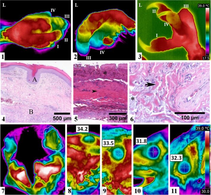
ive of blunt trauma, often associated with electrocutionMelero et al. BMC Veterinary Research 2013, 9:149 Page 5 of 7 http://www.biomedcentral.com/1746-6148/9/149 Figure 2 Thermal analysis of the Lammergeier (G. barbatus). Case 2. (1–3) Thermal images of the left foot, taken (1) of the dorsal view and (2) of the lateral view on 17 April and (3) of the lateral view on 27 June. Initial designated left (L) leg and roman numerals designate each finger (I-IV). A physiological thermal pattern is observed in fingers I and II. (4–6) A histology section of left digit III stained with haematoxylin and eosin showing: (4) epidermis within normal limits (A) and deep dermis with broad linear segments of mixed inflammatory cells, oedema fluid and fragmented collagen fibres (B); (5) full thickness segmental coagulative necrosis with crushed keratinocyte nuclei (asterisk) and dermal collagenolysis (arrow head); (6) detail of mucinous material (asterisk) and smudged collagen fibres (arrow). Thermal images of eyes of two Lammergeiers. (1–3) Clinical case 2 before and during treatment: (1) Frontal view of the head taken on 17 April. The left eye is enclosed by a dotted line. A physiological thermal pattern is observed in the right eye. Lateral view of (2) the left eye and (3) the right eye, both taken on 27 June. (4, 5) Healthy Lammergeier, lateral view of (4) the left eye and (5) the right eye. The thermal range is the same for Figure 1(1-3) and for Figure 2(7-11). Figure 3 Thermal analysis of the osprey (P. haliaetus). Case 3. Thermal images of legs. (1) Dorsal view and (2) right lateral view of the legs during the first examination. (3) Dorsal view on the day that euthanasia was practiced. Initial designated right (R) and left (L) legs. A physiological thermal pattern is observed in the left leg. The thermal range is the same for Figure 3(1-3).
Melero et al. BMC Veterinary Research 2013, 9:149                                                                                                    Page 6 of 7
http://www.biomedcentral.com/1746-6148/9/149

[28-30]. Tissue is damaged due to the direct effects of the                    treatment of the Lammergeier. Mar Melero is the recipient of a PhD student
electrical current as it dissipates through organs [29,30].                    grant from the Complutense University of Madrid.

Moreover, as this injured animal was located close to                          Author details
power lines, electrocution was highly suggestive.                              1
                                                                                VISAVET Centre and Animal Health Department, Veterinary School,
   The anamnesis, the veterinary examinations, and the                         Complutense University of Madrid, 28040 Madrid, Spain. 2GREFA Native
                                                                               Fauna and Their Habitat Rehabilitation Group, 28220 Majadahonda, Spain.
histopathological analyses and necropsies, when per-                           3
                                                                                Wildlife Rehabilitation Centre of Vallcalent, 25003 Lleida, Spain. 4Medicine
formed, confirmed electrocution of these three animals.                        and Surgery Department (Anatomic Pathology), Veterinary School,
Thermographically, temperature reduction caused by elec-                       Complutense University of Madrid, 28040 Madrid, Spain.

trocution was detected since the animals’ admission, and                       Received: 24 September 2012 Accepted: 17 July 2013
prior to macroscopic lesions appearing. Additionally,                          Published: 23 July 2013
the comparison made of the thermographs before and
after physiotherapy revealed that thermography may                             References
help predict tissue damage prognosis and guide reha-                           1. Molina-López RA, Casal J, Darwich L: Causes of morbidity in wild raptor
                                                                                   populations admitted at a wildlife rehabilitation centre in Spain from
bilitation efforts.                                                                1995–2007: a long term retrospective study. PLoS One 2011, 6(9):e24603.
   Thermographically, injured tissues were cooler than                         2. Burfield IJ: The conservation status and trends of raptors and owls in
the corresponding healthy ones and the thermal pat-                                Europe. Ambio 2008, 37(6):401–407.
                                                                               3. González LM, Margalida A, Mañosa S, Sánchez R, Oria J, Molina JI, Caldera J,
tern was altered, which showed that thermal symmetry                               Aranda A, Prada L: Causes and spatio-temporal variations of non-natural
and thermal distribution are more important parameters                             mortality in the vulnerable Spanish imperial eagle Aquila adalberti
than absolute temperature. Mabuchi et al. [11] drew the                            during a recovery period. Oryx 2007, 41(4):495–502.
                                                                               4. Guil F, Fernández-Olalla M, Moreno-Opo R, Mosqueda I, Gómez ME, Aranda
same conclusion when assessing the application of ther-                            A, Arredondo A, Guzmán J, Oria J, González LM, Margalida A: Minimising
mography for clinical diagnoses in humans. However,                                mortality in endangered raptors due to power lines: the importance of
as reference absolute values are currently lacking, each                           spatial aggregation to optimize the application of mitigation measures.
                                                                                   PLoS One 2011, 6(11):e28212.
clinical case should be studied carefully by performing                        5. Bevanger K: Biological and conservation aspects of bird mortality caused
thermography before and during rehabilitation in order                             by electricity power lines: a review. Biol Conserv 1998, 86(1):67–76.
to follow the thermal evolution. Although thermography                         6. Janss GFE, Ferrer M: Avian electrocution on power poles: European
                                                                                   experiences. In In Birds and power lines: collision, electrocution and breeding.
is the most rapid and non-invasive tool for electrocution                          Edited by Ferrer M, Janss GFE. Madrid: Quercus; 1999:145–164.
diagnosis, histopathology should be used to confirm the                        7. Lehman RN, Kennedy PL, Savidge JA: The state of the art in raptor
suspicious cases.                                                                  electrocution research: a global review. Biol Conserv 2007, 136(2):159–174.
                                                                               8. Routh A, Sanderson S: Waterfowl. In In Handbook of Avian Medicine. 2nd
                                                                                   edition. Edited by Tully TN, Dorrestein GM, Jones AK. Oxford:
Conclusions                                                                        Butterworth-Heinemann; 2009:234––265.
Thermography is a rapid, stress-free and objective tool                        9. Üzün I, Akyildiz E, İnanici MA: Histopathological differentiation of skin
                                                                                   lesions caused by electrocution, flame burns and abrasion. Forensic Sci Int
that allows: the early detection of asymmetry tempe-                               2008, 178(2–3):157–161.
rature distribution and lesions consistent with electrocu-                     10. Mc Cafferty DJ: The value of infrared thermography for research on mammals:
tion without necropsy or amputation; the determination                             previous applications and future directions. Mamm Rev 2007, 37(3):177–255.
                                                                               11. Mabuchi K, Chinzei T, Fujimasa I, Haeno S, Motomura K, Abe Y, Yonezawa T:
of areas damaged by the entry and exit of an electric                              Evaluating asymmetrical thermal distributions through image
current; the assessment of the animal’s clinical status;                           processing. IEEE Eng Med Biol Maq 1998, 17(4):47–55.
the diagnosis of pathologies associated with electrocu-                        12. Renkielska A, Nowakowski A, Kaczmarek M, Dobke MK, Grudzinski J,
                                                                                   Karmolinski A, Stojek W: Static thermography revisited – an adjunct
tion, such as fractures and patagium retraction; and the                           method for determining the depth of the burn injury. Burns 2005,
evaluation of how lesions evolve with different treat-                             31(6):768–775.
ments; all of which can contribute to more effective                           13. Tepper M, Neeman R, Milstein Y, David MB, Gannot I: Thermal imaging
                                                                                   method for estimating oxygen saturation. J Biomed Opt 2009,
treatment.                                                                         14(5):054048.
                                                                               14. Dirección General para la Biodiversidad. Ministerio de Medio Ambiente:
Competing interests                                                                Catálogo Nacional de Especies Amenazadas. Madrid: Listado de taxones por
The authors declare that they have no competing interests.                         categorías de amenazas. R.D. 439/1990, R.D. 139/2011; 2011.
                                                                               15. IUCN 2012. IUCN Red List of Threatened Species. www.iucnredlist.org.
Authors’ contributions                                                         16. Morgan PB, Soh MP, Efron N: Potential applications of ocular
MM contributed by taking the thermograms and images analysis and by                thermography. Optom Vis Sci 1993, 70(7):568–576.
writing the manuscript. FG, ON, IL, MAJ and SJS helped draft the manuscript.   17. Colegrave N, Engel J, Plowman AB: Randomisation tests. In In Zoo research
FG, ON and IL contributed with the clinical diagnosis, treatments and              guidelines: Statistics for typical zoo datasets. Edited by Plowman AB. London:
assessments. MAJ contributed by performing the histopathology analysis. SJS        BIAZA; 2006:7–16.
contributed with the physiotherapy diagnosis and treatment at GREFA. JMSV      18. Orosz SE, Ensley PK, Haynes CJ: Avian surgical anatomy: thoracic and pelvic
contributed to coordinating and reviewing the whole process. All the               limbs. Philadelphia: W.B. Saunders; 1992.
authors have read and approved the final manuscript.                           19. Jato S, Otero I, González F, López I, Mendoza JL: Patagium rehabilitation
                                                                                   treatment in wild birds following long-term wing immobilization. Wildl
Acknowledgements                                                                   Rehabilitation Bull 2011, 29(2):33–41.
The authors acknowledge the staff and volunteers of the GREFA and              20. Gross TL, Ihrke PJ, Walder EJ, Affolter VK: Necrotizing diseases of the
Vallcalent rehabilitation centres for their assistance and devotion. We            epidermis. In Skin Diseases of the Dog and Cat, Clinical and Histopathologic
acknowledge the Kirma physiotherapy centre for their collaboration in the          Diagnosis. 2nd edition. Ames: Blackwell Science; 2005:94–97.
Melero et al. BMC Veterinary Research 2013, 9:149 Page 7 of 7
http://www.biomedcentral.com/1746-6148/9/149
21. Thomsen HK, Danielsen L, Nielsen O, Aalund O, Nielsen KG, Karlsmark T,
Genefke IK: Early epidermal changes in heat- and electrically injured pig
skin. I. A light microscopic study. Forensic Sci Int 1981, 17(2):133–143.
22. Danielsen L, Gniadecka M, Thomsen HK, Pedersen F, Strange S, Nielsen KG,
Petersen HD: Skin changes following defibrillation. The effect of high
voltage direct current. Forensic Sci Int 2003, 134(2–3):134–141.
23. Myers DA: Common procedures and concerns with wildlife. Vet Clin North
Am Exot Anim Pract 2006, 9(2):437–460.
24. Baer CK: Guidelines for euthanasia of nondomestic animals. Lawrence:
American Association of Zoo Veterinarians (AAZV); 2006.
25. American Veterinary Medical Association: AVMA Guidelines on Euthanasia. In
Formerly report of the AVMA panel on euthanasia. Schaumburg: AVMA; 2007.
26. Schmidt RE, Reavill DR: A practitioner’s guide to avian necropsy. Necropsy CD,
Zoological Education Network; 2003. ISBN ISBN: 0-9706395-0-3.
27. Rae MA: Practical avian necropsy. Semin Avian Exot Pet Med 2003, 12(2):62–70.
28. Koumbourlis AC: Electrical injuries. Crit Care Med 2002, 30(11 Suppl):424–430.
29. Ritenour AE, Morton MJ, McManus JG, Barillo DJ, Cancio LC: Lightning
injury: a review. Burns 2008, 34(5):585–594.
30. Fish RM, Geddes LA: Conduction of electrical current to and through the
human body: a review. Eplasty 2009, 9:e44.
doi:10.1186/1746-6148-9-149
Cite this article as: Melero et al.: Detection and assessment of
electrocution in endangered raptors by infrared thermography. BMC
Veterinary Research 2013 9:149.
Submit your next manuscript to BioMed Central
and take full advantage of:
• Convenient online submission
• Thorough peer review
• No space constraints or color figure charges
• Immediate publication on acceptance
• Inclusion in PubMed, CAS, Scopus and Google Scholar
• Research which is freely available for redistribution
Submit your manuscript at
www.biomedcentral.com/submitYou can also read