Development of a Multi-Material 3D Printer for Functional Anatomic Models
←
→
Page content transcription
If your browser does not render page correctly, please read the page content below
RESEARCH ARTICLE

Development of a Multi-Material 3D Printer for
Functional Anatomic Models
Laszlo Jaksa1,2*, Dieter Pahr2,3, Gernot Kronreif1, Andrea Lorenz1
1
  Austrian Center for Medical Innovation and Technology (ACMIT Gmbh), Viktor-Kaplan-Strasse 2/A, 2700 Wiener
Neustadt, Austria
2
  Technical University of Vienna, Institute of Lightweight Design and Structural Biomechanics, Object 8, Gumpendorfer
Strasse 7, 1060 Vienna, Austria
3
  Karl Landsteiner University of Health Sciences, Department of Anatomy and Biomechanics, Dr.-Karl-Dorrek-Strasse 30,
3500 Krems an der Donau, Austria

Abstract: Anatomic models are important in medical education and pre-operative planning as they help students or doctors
prepare for real scenarios in a risk-free way. Several experimental anatomic models were made with additive manufacturing
techniques to improve geometric, radiological, or mechanical realism. However, reproducing the mechanical behavior of soft
tissues remains a challenge. To solve this problem, multi-material structuring of soft and hard materials was proposed in this
study, and a three-dimensional (3D) printer was built to make such structuring possible. The printer relies on extrusion to
deposit certain thermoplastic and silicone rubber materials. Various objects were successfully printed for testing the feasibility
of geometric features such as thin walls, infill structuring, overhangs, and multi-material interfaces. Finally, a small medical
image-based ribcage model was printed as a proof of concept for anatomic model printing. The features enabled by this printer
offer a promising outlook on mimicking the mechanical properties of various soft tissues.
Keywords: Silicone 3D printing; Multi-material 3D printing; Anatomic models; Soft tissues

*Correspondence to: Laszlo Jaksa, Austrian Center for Medical Innovation and Technology, Viktor-Kaplan-Strasse 2/A, 2700 Wiener Neustadt,
Austria; laszlo.jaksa.official@gmail.com

Received: July 20, 2021; Accepted: September 1, 2021; Published Online: October 12, 2021
Citation: Jaksa L, Pahr D, Kronreif G, et al., 2021, Development of a Multi-Material 3D Printer for Functional Anatomic Models. Int J
Bioprint, 7(4):420. http://doi.org/10.18063/ijb.v7i4.420

1. Introduction                                                                show that the use of physical anatomic models improves
                                                                               medical education from various aspects due to the
1.1. Anatomic models                                                           additional haptic and spatial information students could
In medical practice and education, anatomic models are                         not receive through books or screen visualizations[5-7].
provided through using human donors, animal models, or                         In the surgical domain, anatomical models can aid the
artificial technical solutions that range from hand-crafted                    planning of complicated surgeries in a wide range of
training models to mass-produced commercial products.                          surgical specialties, since rehearsing the steps of the
In the former case of using real biological tissues,                           operation on a patient-specific model can reveal upcoming
progress is often hindered by the lack of available human                      intra-operative complications[2,8-11]. This can significantly
donors, strict regulations regarding animal and human                          reduce the risk and duration of certain operations, which
testing, and problems in experiment repeatability due                          may result in the lower risk of complication and higher
to the anatomical uniqueness of every human or animal                          patient satisfaction[12]. Moreover, patient-specific models
specimen[1,2]. Using advanced artificial anatomical models                     help the development of various customized implants and
has the potential to ease these problems, especially in case                   other medical instruments[12-14].
of anatomy or surgical education, pre-operative planning,                            Traditionally, artificial anatomical models are
or development of novel medical devices[3,4]. Studies                          mass-produced through casting or molding techniques,
© 2021 Jaksa, et al. This is an Open Access article distributed under the terms of the Creative Commons Attribution License, permitting distribution and
reproduction in any medium, provided the original work is cited.

                                                                                                                                                 145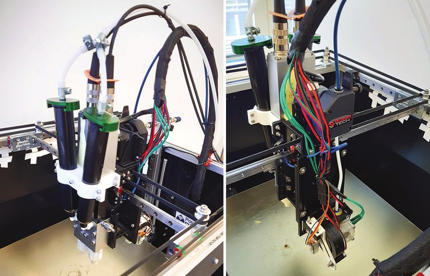
                                                                                                         3D Printer for Anatomic Models

often based on population-averaged geometries[3,13].                     deposited simultaneously[26]. While changing or mixing
The materials used in such models are usually different                  materials in a single droplet generator unit is difficult,
hard and soft polymers, such as thermoplastics, waxes,                   IJP can easily achieve anisotropic properties by creating
or rubbers. With casting and molding techniques, the                     inclusions of various materials[16,17,27], or even seemingly
mechanical properties of various represented tissues can                 gradient composition change[28]. However, IJP is limited
be matched mainly through material selection as these                    in creating hollow and completely closed cavities because
traditional technologies produce fully dense parts. This                 droplets need support underneath them. Therefore, internal
level of matching is often sufficient for certain mass-                  structuring is only possible if the support material can be
produced educational models[6], but the requirements                     washed or cut out after printing without damaging the
of medical product development and testing, as well                      printed object. From the standpoint of anatomic models,
as preoperative planning may benefit from a better                       a relevant IJP printer on the market is the J750 Digital
mechanical fidelity[3,14-17].                                            Anatomy Printer by Stratasys Ltd. (Eden Pairie, MI)
                                                                         [24,29]
                                                                                . This offers an outstanding performance concerning
1.2. Additive manufacturing (AM) of anatomic                             geometric representation and the number of materials and
models                                                                   colors used, including soft materials[30,31]. However, the
AM, also called three-dimensional (3D) printing, has                     mechanical realism of soft tissue representing materials is
become an increasingly influential group of technologies                 still criticized[15].
in the field of anatomic models and other medically                               Using soft materials is an intensely researched
relevant areas in recent years[12,18,19]. Achieving better               direction of AM[32]. Besides IJP, thermoset, photoreactive
geometric and mechanical fidelity is possible with the                   or chemically cured materials like certain silicones, resins
combination of medical imaging technologies and AM[20].                  or hydrogels may be deposited through extrusion as well,
For polymeric materials, the two dominant groups of                      which is also called direct ink writing (DIW)[3,33-35]. This
AM techniques are based on photopolymerization and                       is used to print models of various soft structures[3,36-38].
on extrusion[3]. In an extrusion-based technique called                  Most of these operate with pressurized material reservoirs
fused filament fabrication (FFF), a thermoplastic filament               with controllable valves or syringe extruders to deposit
is pushed into a heated extruder, and deposited through                  soft materials. The rheological properties of the printed
a nozzle[21]. This is mostly used for bone modeling and                  material, such as viscosity or thixotropy, are decisive
mold making in the field of anatomic models[3,9]. FFF                    for maintaining the shape of the printed object. Creating
is cheaper than most other AM technologies due to its                    closed air inclusions is theoretically possible with FFF
relative simplicity and fierce competition between several               and certain DIW techniques[20].
manufacturers. These systems can process a large variety                          Silicone rubbers offer a range of mechanical
of hard thermoplastic filaments but are limited in their                 properties that may be ideal to represent soft tissues
ability to handle soft materials. A large proportion of                  in anatomic models[33]. Certain silicone AM (SAM)
available medical image-based anatomic models are                        technologies are already being applied to anatomical
made of hard plastic using FFF[20].                                      models in some research endeavors and early-stage
      Liquid photopolymers can be deposited and                          commercial services[33,39-47]. These are summarized
solidified in small droplets via material jetting, which                 in Table 1. The collaboration of Wacker Chemie AG
is called inkjet printing (IJP). Among others, it has                    (Munich, Germany) with ACEO (Burghausen, Germany)
been used to create surgical training models of aortic                   led to a droplet-based silicone printing technology, which
aneurysms, kidney tumors, skulls and fetuses[15,22,23].                  relies on curing each layer of silicone with UV light, in a
IJP can also use multi-colored inks to make full-color                   similar fashion to IJP[39,48]. Dow Inc. (Midland, MI) and
objects[9,20] and even use multiple hard and soft materials              German RepRap GmbH (Feldkirchen, Germany) created
in a single print job[14,16,17,24,25]. The deposited droplets            an extrusion-based technology called Liquid AM (LAM)
can be understood as voxels. Given the proper printhead,                 which deposits silicone with extrusion and cures it layer-
this allows multiple voxels of multiple materials to be                  wise using a heat source[40,49]. Another SAM process is

Table 1. Summary of relevant commercial soft material printing technologies
    Group name                                    Process name                 Principle                          Material
    Stratasys Ltd.                                J750                         Droplet jetting                    Photopolymers
    Wacker Chemie AG                              ACEO                         Droplet jetting                    Silicone rubbers
    Dow Inc./GermanRepRap GmbH                    LAM                          Extrusion                          Silicone rubbers
    Fripp Design Ltd.                             Picsima                      Extrusion                          Silicone rubbers
    Spectroplast AG                               Spectroplast                 Vat photo‑polymerization           Silicone rubbers

146                                   International Journal of Bioprinting (2021)–Volume 7, Issue 4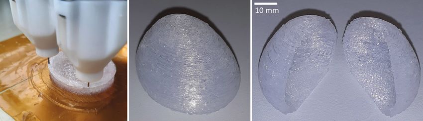
Jaksa, et al.

developed by Spectroplast AG (Zürich, Switzerland), a                  also work with having both the soft matrix and a harder
spinoff company of ETH Zürich. This method uses layer-                 reinforcement being deposited through DIW[51]. In any
wise photopolymerization in a liquid silicone bath[43].                case, this strategy would allow the hardening, toughening
Another method called Picsima by Fripp Design Ltd.                     (further referred to as “up-tuning”) of bulk mechanical
(Rotherham, UK) represents a different bath-based printing             properties compared to the original matrix material. Since
approach, namely extruding the catalyst component of a                 both FFF and extrusion-based DIW can print closed and
two-part silicone into a bath of the base component[41].               empty cavities, the weakening, and softening (further
SAM may also utilize a non-planar coordinate system.                   referred to as “down-tuning”) of mechanical properties
Coulter et al. developed a printing method specialized on              would also be possible[15].
rotating printing surfaces, which offers unique advantages                   Therefore, the main aim of this research was to
in realizing certain geometries[44,45]. Despite the promising          design, build and test a 3D printer based on the concept
development that these technologies represent, almost                  of combining hard and soft materials for printing more
all focus on single-material printing. Therefore, the                  realistic anatomic models. As a proof of concept, the
capabilities to tune mechanical properties are limited to              printer should be capable of printing at least one soft
realizing porous structures with internal cavities[32].                and one hard material, and thus achieve both up-tuning
                                                                       and down-tuning to influence mechanical properties.
1.3. Problems in mechanical realism                                    Moreover, the printer should also realize thin-walled
These AM technologies (IJP, FFF, and DIW) are highly                   structures and closed internal cavities with the soft
applicable to create personalized anatomic models that                 material since these are relevant features in anatomic
are geometrically unique[3]. However, geometric or color               models. In this study, a 3D printer with these features was
fidelity alone do not satisfy all possible needs of medical            built, and its abilities were evaluated through qualitative
device development, surgical education, or preoperative                analysis of various printed proof-of-concept objects,
planning. For more advanced applications, models                       including a small ribcage model based on a medical
should behave realistically under physical manipulation                image. The applicability of the system in the field of
with hands or surgical instruments[20]. To achieve such                anatomic models and the future direction of research are
surgical realism, the materials used to represent various              also discussed.
biological tissues need to have similar mechanical
properties to the tissues, such as density, elastic modulus,           2. Materials and methods
hardness, tensile strength, or viscoelasticity[15-17]. While
matching hard tissues like bone with AM is already a
                                                                       2.1. Technology definition
mature field, there are still many unsolved problems                   The design process of this novel AM system started
regarding soft tissues[20]. Most biological tissues – unlike           with a comparison of various AM technologies and
technical materials – exhibit multi-level hierarchic                   their specifications, as clarifying differences is
structures of various functional building blocks, which                critical for choosing the right printing concept. The
often results in anisotropic and viscoelastic mechanical               fact that IJP, DIW, and FFF can handle different
properties[15,50]. This behavior could be approximated                 materials in the same print is a required feature to
with soft-hard multi-material structures[14,16,17], but to             produce multi-material structures. Other technologies
date, there are no AM technologies available that can                  based on material jetting or vat photopolymerization,
approximate a multitude of tissues[15]. Therefore, two                 such as binder jetting (BJ), stereolithography (SLA),
major areas for improvement could be printing both                     and digital light processing (DLP) all use a single-
hard and soft materials simultaneously, and tuning local               material bath (or “vat”) of liquid resin or powder [21].
mechanical properties through multi-material structuring.              This prevents multi-material printing, and the
These should happen simultaneously to produce high                     creation of closed air inclusions. For IJP, DIW, and
quality anatomic models that resemble real tissues from a              FFF, changing materials simply requires switching
mechanical standpoint[15].                                             to a different filament, cartridge, or printhead.
                                                                       Mimicking the macroscopic mechanical properties
1.4. Research aims                                                     of biological tissues through up- and down-tuning
Combining extrusion-based AM technologies such as                      requires printing both soft and hard materials.
FFF and DIW may be helpful for making more realistic                   Extrusion is the preferred method to create closed
anatomic models. While using FFF to produce the whole                  internal cavities and support structures, if needed. The
model is ineffective regarding mechanical realism,                     mentioned technologies are compared considering
thermoplastics may be used as fiber reinforcement if                   our construction preferences in Table 2. Further
printed into a softer matrix material, like a silicone rubber          descriptions and schematics of these technologies are
that is deposited by a DIW printhead. Such a concept may               available in other literature [21,32].
                                    International Journal of Bioprinting (2021)–Volume 7, Issue 4                           147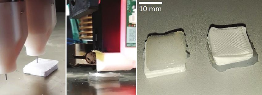
3D Printer for Anatomic Models
Table 2. A survey of features considering various additive manufacturing technologies
Technology Principle Soft Multi‑ Material Support structures Closed
materials material deposition cavities
SLS Powder bed fusion Limited No Spreading Powder or printed No
BJ Material jetting Limited No Spreading and Powder or printed No
droplets
IJP Material jetting Yes Yes Droplets Printed Limited
SLA Vat photo‑polymerization Limited No Spreading Liquid or printed No
DLP Vat photo‑polymerization Limited No Spreading Liquid or printed No
FFF Extrusion Limited Yes Heated extrusion Printed Yes
DIW Extrusion Yes Yes Extrusion or Liquid or printed Yes
droplets
*Ideal for Extrusion Yes Yes Extrusion Printed Yes
anatomic models
*An imaginary technology which we found ideal for anatomic models if mechanical realism is desirable.
Concerning the printing material, some silicone component selection. An FFF printer which does not only
rubbers exhibit ideal mechanical properties to mimic apply Delta or XY-Core kinematics, but also employs
various soft tissues. This also makes them a popular a control board that is open-source and easily extended
casting material for certain anatomic models, where with the chosen Viscotec extruder was considered highly
a casting mold of the desired anatomy is first printed desirable.
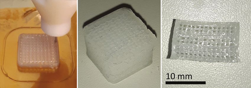
with FFF or SLA, and then filled with a two-component Finally, a Railcore II 300 ZL open-source FFF 3D
addition-cured silicone rubber[7,9]. However, in multi- printer system[53] was chosen and modified (Figure 1).
material printing, the adhesion of the various printing On this printer, the original E3D V6 FFF printhead
materials is important, unlike in casting. Certain single- was extended with the Viscotec Vipro-HEAD 3/3
component condensation-cured silicone rubbers may (Figure 2)[52]. Silicones and other high-viscosity materials
exhibit an adhesive behavior to some thermoplastic can be fed into this screw extruder with pressurized air up
polymer materials, which makes these a promising to 6 bars, from 55 mL cartridges, which are also mounted
combination in a multi-material printing scenario. on the printhead. If necessary, these can be moved to the
Therefore, the printer should employ an FFF printhead frame, which removes their volume and mass limitations,
for printing thermoplastics, and a continuous extrusion- enabling a large material supply to the printhead given
based DIW printhead to print single-component silicone that the feed pressure is sufficient. The silicone printing
rubbers. nozzle is connected to the outlet of the extruder through
a Luer-thread and is secured against unscrewing with a
2.2. Printer system retainer part. These white Luer-adapters and retainers
were custom-made for the extruder (Figure 2A).
For an extrusion-based DIW printhead, various extrusion A nozzle with 0.33 mm outlet diameter was selected for
mechanisms, such as syringes, peristaltic pumps and screw silicone extrusion. The original E3D V6 FFF printhead
extruders, are available. However, for high-viscosity on the other side of the carriage (Figure 2B) is capable of
and high-precision applications, screw extruders were melting and depositing thermoplastic filaments through a
preferred. The final choice fell on a Vipro-HEAD 3/3 two- 0.4 mm diameter nozzle.
component printhead by Viscotec GmbH (Töging am Inn, The printer is controlled by the Duet 2 Wi-Fi control
Germany)[52], which enables processing either one or two electronics, extended with a Duex 5 extension board,
single-component silicones, or a two-component silicone. operating with RepRap v1.18 firmware[54]. The system
In this study, only one single-component silicone rubber can be connected to a personal computer (PC) through
was used. a Wi-Fi network, and print jobs can be started through
Regarding printer mechanics from the standpoint the Duet Web Interface, which is accessible through an
of printing soft and flexible materials, it is important internet browser running on the PC. The general printer
that the building platform only moves in the axis of the configuration, including the printhead definitions and
building direction (usually labeled “Z”), so that it does dosing calibration settings are done by modifying a file
not shake the printed objects horizontally during printing. stored on the Duet 2 Wi-Fi board. The slicing software
The printer kinematics which fulfills this criterion are the used to generate G-codes for printing objects is Prusa
so-called XY-Core and the Delta kinematics. Hardware Slicer (version 2.1), an open-source slicer originally
and software formed another important aspect in the made for filament-based printers[55]. The user can easily
148 International Journal of Bioprinting (2021)–Volume 7, Issue 4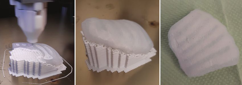
Jaksa, et al.

define a multi-material printhead and generate G-codes                    poly-lactic acid (PLA) filament from Fillamentum
for multi-material FFF-DIW print jobs. No post-                           Manufacturing (Hulín, Czech Republic) was used with the
processing of the generated G-code files is necessary, and                E3D V6 FFF printhead in case of prints that demonstrate
an extrusion correction factor can also be set to fine-tune               the multi-material capabilities. PLA was chosen as it is an
dosing accuracy, when necessary. To start a print job, the                easily accessible and popular FFF material.
generated G-code files must be uploaded to the Duet 2
Wi-Fi board through the Duet Web Interface. This way,                     2.4. Printing tests
the printer is likely also compatible with other popular                  For accurate dosing, the silicone printhead was calibrated
slicing software, such as Cura or Simplify3D.                             for the chosen material using a KERN PES 42002M
                                                                          scale (Kern & Sohn GmbH, Balingen, Germany). After
2.3. Materials                                                            this, 15 × 15 × 10 mm silicone blocks were printed with
The selected silicone material is a high-viscosity single-                various speeds and layer thicknesses to find reasonable
component condensation-crosslinking liquid silicone                       settings for further printing tests. The integrity of the
                                                                          printed cubes was qualitatively evaluated by observing
rubber called Elkem AMSil 20101 (Elkem Silicones,
                                                                          them and then slicing them with a blade to see if there are
Oslo, Norway), which was used with the Viscotec DIW
                                                                          any internal faults (Figure 3).
printhead. This material is intended for cold extrusion;
                                                                                Based on the calibration prints, a printing speed of
therefore, no heating or other means of energy input is
                                                                          15 mm/s and a layer thickness of 0.3 mm were chosen
required during printing. Moreover, a 1.75 mm diameter                    for further trials. Furthermore, every printed object
                                                                          was left on the building platform untouched for 24 h to
                                                                          ensure sufficient crosslinking before any manipulation
                                                                          or inspection. After calibration, the system’s ability to
                                                                          print silicone objects with closed internal cavities, infill
                                                                          structuring and thin walls as well as to combine silicone
                                                                          DIW and thermoplastic FFF was assessed by conducting
                                                                          six printing tests:
                                                                          1. In the first test, a thin-walled shell was printed based
                                                                               on the same 15 × 15 × 10 mm cuboid that was used
                                                                               for the calibrations. In this case, only two lines of
                                                                               outer contour were used, resulting in approximately
                                                                               0.7 mm shell wall thickness (Figure 4). This is
                                                                               relevant to anatomic models in case of printing
                                                                               vessels or membranes, which feature thin walls.
                                                                          2. The second test involved a silicone block of the same
                                                                               dimensions as in the first test, but with 40% volume
                                                                               fraction gyroid infill structuring to simulate down-
Figure 1. The modified Railcore II 300 ZL printer, extended with a
                                                                               tuning (Figure 5).
Viscotec Vipro-HEAD 3/3 extruder.

 A                                B

Figure 2. The Viscotec Vipro-HEAD 3/3 extruder with custom
Luer-compatible endpieces (A), and the original E3D V6 filament           Figure 3. A 15 × 15 × 10 mm test block that was printed after
extruder on the opposite side of the printhead carriage (B).              calibration and cut in half after printing.

                                       International Journal of Bioprinting (2021)–Volume 7, Issue 4                             149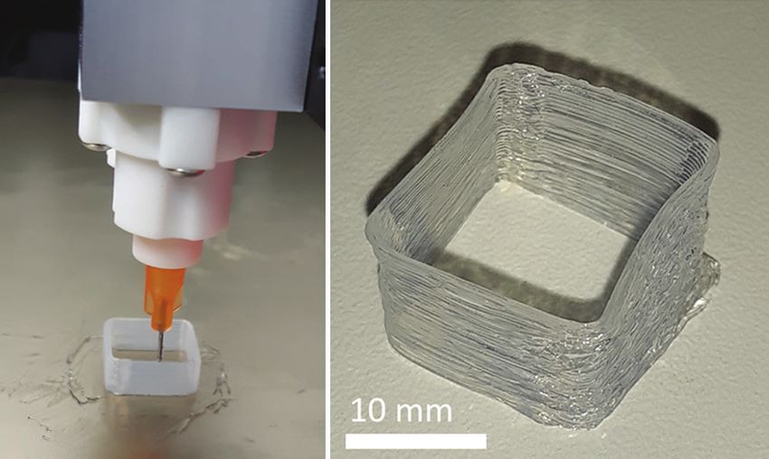
                                                                                                                3D Printer for Anatomic Models

3. In the third test, a 50% downscaled version of a                            simulate a situation of printing hard plastic support
   human bladder was printed based on a 3D model                               under a silicone structure (Figure 7A).
   segmented from a computed tomography (CT) image                          5. In the fifth test, the same model was used as in the
   earlier. To test the feasibility of such a large internal                   fourth test, but now the PLA was printed on top of
   cavity with overhanging areas, no support was used                          the silicone to simulate laying the filament as an
   inside. The envelope dimensions of this downscaled                          inclusion into the silicone matrix (Figure 7B).
   bladder were approximately 35 × 30 × 25 mm, and                          6. The sixth test was planned to give some synthesis of
   the wall thickness was changing between 1.5 and                             the features investigated through the previous tests.
   2 mm (Figure 6).                                                            The ribcage and the surrounding soft tissues were
4. The fourth test involved a pair 15 × 15 × 3 mm square                       segmented from the CT image of a newborn using
   silicone and PLA multi-material chips on top of each                        3D-Slicer, and the model was printed (Figure 8).
   other. The silicone was printed on top of the PLA to                        The ribs and the support structures were printed from
                                                                               PLA and the surrounding soft tissue was printed
    A                        B                                                 from silicone. To mimic bone structure, the ribs were
                                                                               printed with a 30% gyroid infill, while 80% infill was
                                                                               used for the soft tissue.

                                                                            3. Results
                                                                            All test objects were successfully printed. The thin-walled
                                                                            shell of the first test (Figure 4) did not collapse during or
                                                                            after printing, despite having only 0.7 mm wall thickness.
                                                                                  The 40% gyroid block of the second test (Figure 5)
                                                                            was cut in half with a blade after crosslinking to reveal
                                                                            the internal structure (Figure 5C).
Figure 4. Thin-walled silicone rubber shell during (A) and after                  The downscaled bladder in the third test (Figure 6)
printing (B).                                                               had minor material integrity errors at the top due to the

                   A                                B                                    C

Figure 5. Silicone block with 40% volume fraction gyroid infill during (A) and after printing (B), and after slicing with a blade (C) to reveal
the internal structure.

                   A                                B                               C

Figure 6. A downscaled human bladder with no internal support during (A) and after printing (B), and after slicing with a blade (C) to reveal
the internal cavity.

150                                      International Journal of Bioprinting (2021)–Volume 7, Issue 4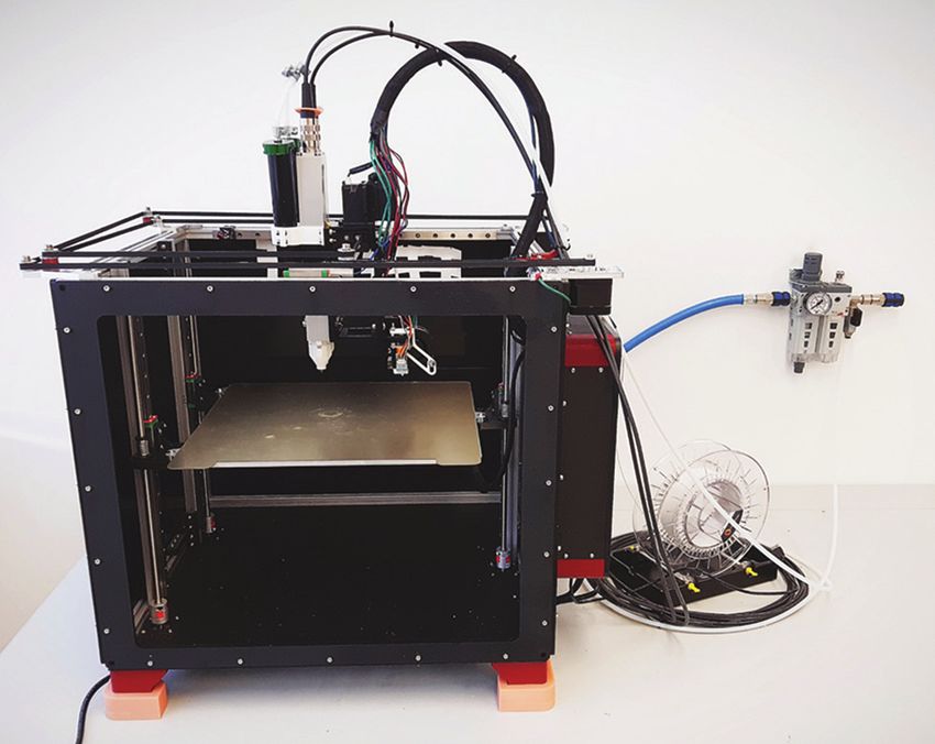
Jaksa, et al.

                  A                         B                             C

Figure 7. Printing silicone rubber on top of PLA (A), then PLA on top of silicone rubber (B), and the resulting multi-material chips from
both tests (C) .

                  A                               B                                      C

Figure 8. Silicone-PLA multi-material ribcage model based on a medical image of a newborn during printing (A), with support after
printing (B), and after support removal (C).

lack of internal support, but the external geometry stayed                anatomic models in the future. The specifications stemming
intact. This bladder was also cut in half after printing to               from this goal were printing multi-material structures
reveal the internal cavity (Figure 6C).                                   out of at least one hard and one soft material, while also
      The multi-material chips from the fourth (Figure 7C)                being capable of printing empty cavities, infill structures
and fifth (Figure 7B) tests were also printable. Moreover,                and thin-walled features. Considering the advantages and
in case of the fifth test, the PLA top was deformed,                      drawbacks of various AM methods and their applicable
presumably due to printing on a soft and unstable silicone                materials, a printer was built that combines FFF and DIW
surface. After printing, the adhesion between the silicone                technologies to print with a single-component silicone
and the PLA in the multi-material chips was evaluated                     rubber and a thermoplastic PLA filament.
by trying to manually separate the materials. The silicone                      The printing trials demonstrated that the established
was considered adhesive enough to resist this manual                      technology is capable of printing objects of both
peeling, since the bulk silicone material was damaged                     materials and can print silicone with a weakened internal
before the interface.                                                     structure (down-tuning) or combine silicone with PLA
      After seeing the success of the five previous                       (up-tuning). An unsupported internal cavity and a thin-
tests, the ribcage model of the sixth test was printed to                 walled structure were also printable with the silicone. It
demonstrate the applicability of the printer to produce                   was shown that the FFF printhead can create hard support
medical image-based anatomic models (Figure 8). No                        structures. The strong adhesion between the PLA and the
complications were experienced during the printing                        silicone that was experienced during the tests suggests
process, although the manual removal of the support                       that this material combination can be applied to create
structures was challenging due to the adhesion between                    more complex multi-material structures, and that the
the silicone and the PLA.                                                 silicone must be cut away from the PLA in case of using
                                                                          PLA for printing support structures under the silicone.
4. Discussion                                                             These assumptions were confirmed through the last test,
                                                                          where the ribcage was indeed printable with PLA support
4.1. Overview of aims and results                                         and sufficient adhesion between the silicone and the PLA.
The aim of this study was to build and test a 3D printer that                   These promising outcomes imply that the technology
enables features necessary for producing more realistic                   could be used to approximate the mechanical properties
                                       International Journal of Bioprinting (2021)–Volume 7, Issue 4                               151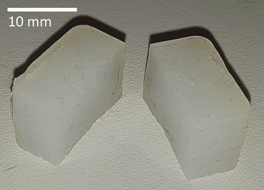
                                                                                                       3D Printer for Anatomic Models

of various soft biological tissues through up- and down-              4.3. Limitations and outlook
tuning strategies. Using the other half of the Viscotec
                                                                      In case of DIW and FFF, spatial resolution, printing
printhead as a third extruder, the system could easily
                                                                      quality, and printing speed are tightly connected process
be extended to introduce a viscous fluid into printed
                                                                      parameters; therefore, the presented technology is slow
internal cavities. Such a method used in parallel with
                                                                      compared to the shower-like droplet generation of IJP or
up- and down-tuning strategies could possibly increase
                                                                      BJ, or the scanning laser or full-layer projector of SLA
viscoelasticity[15] in anatomic models. This means
                                                                      or DLP. Moreover, despite the successful first prints, the
that with the further development of our technology, a
                                                                      system suffers from certain other limitations in its current
significant increase in anatomic model realism may
be delivered, accelerating the development of certain                 state. If used in more complex geometries, the removal
medical instruments and improve medical education and                 of PLA supports might damage the contact surface of
preoperative planning.                                                the silicone. This adverse effect may be minimized by
                                                                      careful support design in the future. The difficulties with
4.2. Comparison with other technologies                               removing the silicone parts from the building platform
                                                                      may be eased by choosing a different printing surface.
From the perspective of down-tuning, the greatest                     It must also be pointed out that the printing abilities
limitation of concurrent technologies such as IJP, BJ,                were only demonstrated with one silicone and one
SLA, and DLP is their difficulty to realize completely                thermoplastic material, and the general applicability to
closed and empty cavities, either because of an inherent              other materials is so far untested.
need for support (IJP) or because of a leveled slurry                       A decisive factor in the compatibility of a silicone-
or powder bath (BJ, SLA, DLP)[21]. This also applies                  thermoplastic combination is the adhesion between them.
to the Picsima silicone printing process[41], as well as              Qualitatively testing the adhesion strength between the
the technology used by Wacker and ACEO[39], and by                    silicone and PLA or other thermoplastics remains an
Spectroplast[43]. In our case, this limitation is overcome            interesting direction for further research, along with the
by utilizing extrusion-based DIW and FFF, even though                 qualitative testing of the effects of infill structuring on
the LAM process of Dow and GermanRepRap[40] is also                   the mechanical properties of silicone objects. Finally, the
free of this problem.                                                 extent of applicability to the field of anatomic models
      Considering up-tuning, IJP has a better resolution              depends not only on mechanical property tuning but also
and a larger variety of applicable materials than our
                                                                      on geometric limitations. To succeed at printing complex
presented technology[14,16,17]. Our process can use up to
                                                                      anatomic geometries, an optimization method should first
three materials and can be extended to handle two more
                                                                      be developed to find the ideal printing parameters. This
without changing the electronics. The LAM process of
                                                                      may be done in a follow-up study by printing various basic
Dow and GermanRepRap[40] along with the one of Wacker
                                                                      features at different printing settings and then analyzing
and ACEO[39] could theoretically also be extended to work
                                                                      the integrity and accuracy of the printed features. In
with multiple materials. Meanwhile, the other methods
                                                                      addition, using the other available DIW extruder to
(BJ, SLA, DLP, Picsima, and Spectroplast) are more
                                                                      deposit a high-viscosity filler liquid into internal cavities
confined to single material printing[21,41,43].
                                                                      may provide a way to modify viscoelastic mechanical
      The various available IJP printer models from
                                                                      properties.
Stratasys (including the J750 dedicated for anatomic
models)[24] are frequently used in literature for producing           5. Conclusions
soft multi-material tissue models[15-17]. However, the
biological realism of the materials that are printable with           In this study, a novel multi-material AM technology
these printers is often criticized, and IJP technologies              targeted at facilitating the production of more realistic
are inherently limited in terms of printing unsupported               anatomical models was established and tested. The
overhangs. We have demonstrated that our system can                   printable features enabled by this technology offer
print steeply overhanging structures, which (combined                 promising possibilities in the field of functional anatomic
with other relevant features) may enable more accurate                models. Analyzing geometric limitations, along with
tissue approximation than what is possible with IJP                   an evaluation of feasible mechanical properties, are
systems.                                                              needed before this technology could make a significant
      Differentiating our system from other self-built                impact in the field of medical education, device testing,
silicone rubber printers, we can note that some are                   and pre-operative planning. However, a medical image-
specialized on printing on curved surfaces[44,45], while              based anatomic model was already successfully printed
others use two-component silicones on a heated building               in this study, implying a long-term applicability for the
platform[46,47] and do not feature additional printheads for          presented system. Moreover, besides anatomic models,
thermoplastics or other fluids.                                       the system may also have potential applications in the
152                                International Journal of Bioprinting (2021)–Volume 7, Issue 4
Jaksa, et al.

field of soft robotics, wearable electronic devices, or                           for Education in Anatomy. Anat Sci Educ., 6:211–5.
sports equipment.                                                           7.    Sulaiman A, Boussel L, Taconnet F, et al., 2008, In vitro
                                                                                  Non-rigid Life-size Model of Aortic Arch Aneurysm for
Funding                                                                           Endovascular Prosthesis Assessment. Eur J Cardiothorac
This work was supported by the Provincial Government                              Surg, 33:53–7.
of Lower Austria (Land Niederösterreich) under grant                              https://doi.org/10.1016/j.ejcts.2007.10.016
assignment number WST3-F2-528983/005-2018.
                                                                            8.    Giesel FL, Hart AR, Hahn HK, et al., 2009, 3D
Conflicts of interest                                                             Reconstructions of the Cerebral Ventricles and Volume
                                                                                  Quantification in Children with Brain Malformations. Acad
Concerning the manufacturing technology described in
                                                                                  Radiol, 16:610–7.
Materials and Methods subsections 2.1, 2.2 and 2.3, a
patent application has been filed by the Austrian Center                          https://doi.org/10.1016/j.acra.2008.11.010
for Medical Innovation and Technology (ACMIT Gmbh)                          9.    Golab A, Smektala T, Kaczmarek K, et al., 2017, Laparoscopic
at the European Patent Office under applicant reference                           Partial Nephrectomy Supported by Training Involving
number 51241. The inventors are Laszlo Jaksa, Andrea                              Personalized Silicone Replica Poured in Three-Dimensional
Lorenz and Dieter Pahr.                                                           Printed Casting Mold. J Laparoendosc Adv Surg Tech A,
                                                                                  27:420–2.
Author contributions
                                                                                  https://doi.org/10.1089/lap.2016.0596
L. J. designing and assembling the 3D-printer, conducting
                                                                            10. Mavili ME, Canter HI, Sağlam-Aydinatay B, et al., 2007,
all tests, writing the manuscript D. P. consulting and
                                                                                  Use of Three-Dimensional Medical Modeling Methods for
supervising the design and publication processes G. K.
consulting and supervising the design and publication                             Precise Planning of Orthognathic Surgery. J Craniofac Surg,
processes A. L. consulting and supervising the design,                            18:740–7.
assembly, testing and publication processes, managing                             https://doi.org/10.1097/scs.0b013e318069014f
component procurement and research budget.                                  11.   Poukens J, Haex J, Riediger D, 2003, The Use of Rapid
                                                                                  Prototyping in the Preoperative Planning of Distraction
References
                                                                                  Osteogenesis of the Cranio-maxillofacial Skeleton. Comput
1.    Ventola CL, 2014, Medical Applications for 3D Printing:                     Aided Surg, 8:146–54.
      Current and Projected Uses. P T, 39:704–11.                                 https://doi.org/10.3109/10929080309146049
2.    Rengier F, Mehndiratta A, von Tengg-Kobligk H, et al., 2010,          12. Tack P, Victor J, Gemmel P, et al., 2016, 3D-Printing
      3D Printing Based on Imaging Data: Review of Medical                        Techniques in a Medical Setting: A Systematic Literature
      Applications. Int J Comput Assist Radiol Surg, 5:335–41.                    Review. Biomed Eng Online, 15:115.
      https://doi.org/10.1007/s11548-010-0476-x                                   https://doi.org/10.1186/s12938-016-0236-4
3.    Wang K, Ho CC, Zhang C, et al., 2017, A Review on the 3D              13. Yan Q, Dong H, Su J, et al., 2018, A Review of 3D Printing
      Printing of Functional Structures for Medical Phantoms and                  Technology for Medical Applications. Engineering, 4:729–42.
      Regenerated Tissue and Organ Applications. Engineering,               14. Qian Z, Wang K, Liu S, et al., 2017, Quantitative Prediction of
      3:653–62.                                                                   Paravalvular Leak in Transcatheter Aortic Valve Replacement
      https://doi.org/10.1016/j.eng.2017.05.013                                   Based on Tissue-Mimicking 3D Printing. JACC Cardiovasc
4.    Pietrabissa A, Marconi S, Negrello E, et al., 2019, An                      Imaging, 10:719–31.
      Overview on 3D Printing for Abdominal Surgery. Surg                         https://doi.org/10.1016/j.jcmg.2017.04.005
      Endosc, 34(1):1–13.                                                   15. Ratinam R, Quayle M, Crock J, et al., 2019, Challenges
      https://doi.org/10.1007/s00464-019-07155-5                                  in Creating Dissectible Anatomical 3D Prints for Surgical
5.    Preece D, Williams SB, Lam R, et al., 2013, Let’s Get                       Teaching. J Anat, 234:419–37.
      Physical: Advantages of a Physical Model Over 3D Computer                   https://doi.org/10.1111/joa.12934
      Models and Textbooks in Learning Imaging Anatomy. Anat                16. Wang K, Wu C, Qian Z, et al., 2016, Dual-material 3D
      Sci Educ., 6:216–24.                                                        Printed Metamaterials with Tunable Mechanical Properties
      https://doi.org/10.1002/ase.1345                                            for Patient-specific Tissue-mimicking Phantoms. Addit
6.    Khot Z, Quinlan K, Norman GR, et al., 2013, The Relative                    Manuf, 12:31–7.
      Effectiveness of Computer-based and Traditional Resources                   https://doi.org/10.1016/j.addma.2016.06.006

                                         International Journal of Bioprinting (2021)–Volume 7, Issue 4                                   153
3D Printer for Anatomic Models
17. Wang K, Zhao Y, Chang YH, et al., 2016, Controlling the for 3D Voxel Printers. Rapid Prototyp J, 16:241–7.
Mechanical Behavior of Dual-material 3D Printed Meta- https://doi.org/10.1108/13552541011049252
materials for Patient-specific Tissue-mimicking Phantoms. 29. Patent, 2019, Additive Manufacturing of Rubber-like
Mater Des, 90:704–12. Materials. Patent US2019224914A1.
https://doi.org/10.1016/j.matdes.2015.11.022 30. Slesarenko VY, 2017, Towards Mechanical Characterization
18. Goh GL, Zhang H, Chong TH, et al., 2021, 3D Printing of of Soft Digital Materials for Multimaterial 3D-Printing. Int J
Multilayered and Multimaterial Electronics: A Review. Adv Eng Sci, 123:62–72.
Electron Mater, 2021:2100445. https://doi.org/10.1016/j.ijengsci.2017.11.011
https://doi.org/10.1002/aelm.202100445 31. Goh GL, Agarwala S, Yong WI, 2016, 3D Printing of
19. Yap YL, Sing SL, Yeong WY, 2020, A Review of 3D Printing Microfludic Sensor for Soft Robots: A Preliminary Study in
Processes and Materials for Soft Robotics. Rapid Prototyp J, Design and Fabrication. In: Proceedings of the 2nd International
26:1345–61. Conference on Progress in Additive Manufacturing (Pro-AM
https://doi.org/10.1108/rpj-11-2019-0302 2016). p177–81.
20. Qiu K, Haghiashtiani G, McAlpine MC, 2018, 3D Printed 32. Truby RL, Lewis JA, 2016, Printing Soft Matter in Three
Organ Models for Surgical Applications. Annu Rev Anal Dimensions. Nature, 540:371–8.
Chem, 11:287–306. https://doi.org/10.1038/nature21003
https://doi.org/10.1146/annurev-anchem-061417-125935 33. Yeo J, Koh J, Wang F, et al., 2020, 3D Printing Silicone
21. Ngo TD, Kashani A, Imbalzano G, et al., 2018, Additive Materials and Devices. In: Silicon Containing Hybrid
Manufacturing (3D Printing): A Review of Materials, Copolymers. Germany: Wiley-VCH Verlag GmbH & Co.
Methods, Applications and Challenges. Compos B Eng, KGaA; 2020, p. 239–63.
143:172–96. https://doi.org/10.1002/9783527823499.ch9
https://doi.org/10.1016/j.compositesb.2018.02.012 34. Zhao Y, Yao R, Ouyang L, et al., 2014, Three-dimensional
22. Pugliese L, Marconi S, Negrello E, et al., 2018, The Clinical Printing of Hela Cells for Cervical Tumor Model In Vitro.
Use of 3D Printing in Surgery. Updates Surg, 70:381–8. Biofabrication, 6:035001.
https://doi.org/10.1007/s13304-018-0586-5 https://doi.org/10.1088/1758-5082/6/3/035001
23. Dorweiler B, Baqué PE, Chaban R, et al., 2021, Quality 35. Lee JM, Sing SL, Yeong WY, 2020, Bioprinting of
Control in 3D Printing: Accuracy Analysis of 3D-Printed Multimaterials with Computer-aided Design/Computer-aided
Models of Patient-Specific Anatomy. Materials, 14:1021. Manufacturing. Int J Bioprint, 6:245.
https://doi.org/10.3390/ma14041021 https://doi.org/10.18063/ijb.v6i1.245
24. Stratasys Ltd. Available from: https://stratasys.com/medical/ 36. Liu W, Zhang YS, Heinrich MA, et al., 2016, Rapid
advanced-medical-models. [Last accessed on 2021 Jan 10]. Continuous Multimaterial Extrusion Bioprinting. Adv Mater,
25. Mirzaali MJ, Nava AH, Gunashekar D, et al., 2020, 29:1604630.
Mechanics of Bioinspired Functionally Graded Soft-hard 37. Hardin JO, Ober TJ, Valentine AD, et al., 2015, Microfluidic
Composites Made by Multi-material 3D Printing. Composit Printheads for Multimaterial 3D Printing of Viscoelastic Inks.
Struct, 237:111867. Adv Mater, 27:3279–84.
https://doi.org/10.1016/j.compstruct.2020.111867 https://doi.org/10.1002/adma.201570145
26. Ionita CN, Mokin M, Varble N, et al., 2014, Challenges and 38. Skylar-Scott MA, Mueller J, Visser CW, et al., 2019,
Limitations of Patient-specific Vascular Phantom Fabrication Voxelated Soft Material Via Multimaterial Multinozzle 3D
Using 3D Polyjet Printing. Proc SPIE Int Soc Opt Eng, Printing. Nature, 575:330–4.
9038: 90380M. https://doi.org/10.1038/s41586-019-1736-8
https://doi.org/10.1117/12.2042266 39. Wacker Chemie AG, 2021, Avaialble from: https://www.
27. Reiter M, Major Z, 2011, A Combined Experimental aceo3d.com/3d-printing [Last accessed on 2021 Jan 10].
and Simulation Approach for Modelling the Mechanical 40. GermanRepRap GmbH, 2021, Avaialble from: https://www.
Behaviour of Heterogeneous Materials Using Rapid germanreprap.com/material-de/SILASTIC-3D-3335.aspx
Prototyped Microcells. Virtual Phys Prototyp, 6:111–20. [Last accessed on 2021 Jan 10].
https://doi.org/10.1080/17452759.2011.586949 41. Fripp Design Ltd., 2021, Avaialble from: https://www.
28. Hiller J, Lipson H, 2010, Tunable Digital Material Properties picsima.com [Last accessed on 2021 Jan 10].
154 International Journal of Bioprinting (2021)–Volume 7, Issue 4Jaksa, et al.
42. Deltatower GmbH, 2021, Avaialble from: https://www. an Object with Use of a 3D-Printing Device, Patent
deltatower.ch/en/home-2 [Last accessed on 2021 Jan 10]. WO2017108208A1.
43. Spectroplast AG, 2021, Avaialble from: https://www. 49. Patent, 2017, 3D Printing Method Utilizing Heat-curable
spectroplast.com [Last accessed on 2021 Jan 10]. Silicone Composition, Patent WO2017040874A1.
44. Coulter F, 2021, Avaialble from: http://www.fergalcoulter.eu 50. Studart AR, 2016, Additive Manufacturing of Biologically-
[Last accessed on 2021 Jan 10]. inspired Materials. Chem Soc Rev, 45:359–76.
45. Coulter F, Schaffner M, Faber J, et al., 2019, Bioinspired Heart https://doi.org/10.1039/c5cs00836k
Valve Prosthesis Made by Silicone Additive Manufacturing. 51. Bakarich SE, Gorkin R, Panhuis M, et al., 2014, Three-
Matter, 1:266–79. Dimensional Printing Fiber Reinforced Hydrogel Composites.
https://doi.org/10.1016/j.matt.2019.05.013 ACS Appl Mater Interfaces, 6:15998–16006.
46. Luis E, Pan HM, Sing SL, et al., 2019, Silicone 3D Printing: https://doi.org/10.1021/am503878d
Process Optimization, Product Biocompatibility, and 52. Viscotec GmbH, 2021, Avaialble from: https://www.viscotec.
Reliability of Silicone Meniscus Implants. 3D Print Addit de/produkte/3d-druckkoepfe [Last accessed on 2021 Jan 10].
Manufact, 6:319–32. 53. White JS, Akens T, 2021, Avaialble from: https://railcore.org
https://doi.org/10.1089/3dp.2018.0226 [Last accessed on 2021 Jan 10].
47. Luis E, Pan HM, Sing SL, et al., 2020, 3D Direct Printing 54. Duet3D Advanced 3D Printing Electronics, 2021, Avaialble
of Silicone Meniscus Implant Using a Novel Heat-Cured from: https://www.duet3d.com/DuetWifi [Last accessed on
Extrusion-Based Printer. Polymers, 12:1031. 2021 Jan 10].
https://doi.org/10.3390/polym12051031 55. Prusa Research, 2021, Avaialble from: https://www.prusa3d.
48. Patent, 2017, 3D-printing Device and Process for Producting com/prusaslicer [Last accessed on 2021 Jan 10].
International Journal of Bioprinting (2021)–Volume 7, Issue 4 155You can also read