Effects of Hypoxic Environment on Periodontal Tissue through the ROS/TXNIP/NLRP3 Inflammasome Pathway - Hindawi.com
←
→
Page content transcription
If your browser does not render page correctly, please read the page content below
Hindawi
BioMed Research International
Volume 2022, Article ID 7690960, 16 pages
https://doi.org/10.1155/2022/7690960

Research Article
Effects of Hypoxic Environment on Periodontal Tissue through
the ROS/TXNIP/NLRP3 Inflammasome Pathway

 Rui Zhu , Xiaohui Mi , and Yongming Li
 Department of Orthodontics, Shanghai Engineering Research Center of Tooth Restoration and Regeneration, School and Hospital
 of Stomatology, Tongji University, Shanghai, China

 Correspondence should be addressed to Xiaohui Mi; xiaomihui@yeah.net and Yongming Li; 1727039279@qq.com

 Received 15 November 2021; Revised 30 November 2021; Accepted 8 December 2021; Published 17 January 2022

 Academic Editor: Bruna Sinjari

 Copyright © 2022 Rui Zhu et al. This is an open access article distributed under the Creative Commons Attribution License, which
 permits unrestricted use, distribution, and reproduction in any medium, provided the original work is properly cited.

 There is low evidence for the possible association between obstructive sleep apnea-hypopnea syndrome (OSAHS) and
 periodontitis, necessitating further research. This study was aimed at investigating this association. For the in vitro study, 8-
 day-old Wistar rats were divided into the unilateral nasal obstruction group (UNO) and the sham surgery group (SHAM).
 Rats in the former group were subjected to UNO by cauterization of the external nostril at the age of 8 days.
 Immunofluorescence analysis, quantitative real-time polymerase chain reaction, and western blot were performed to assess the
 expression of thioredoxin-interacting protein (TXNIP), NLR family pyrin domain-containing 3 (NLRP3) inflammasome-
 associated factors, and interleukin-1β (IL-1β). Throughout the experimental period, the weights of rats in the two groups were
 similar. The mRNA and protein expression of TXNIP and IL-1β was significantly higher in the UNO than in the SHAM
 groups. Compared with SHAM, NLRP3 inflammasome-associated factors were activated in the UNO group. For the in vitro
 study, a cellular hypoxia model was established by treating human periodontal ligament cells (HPDLCs) with cobalt chloride.
 The studies showed that hypoxia can induce an excessive production and accumulation of reactive oxygen species (ROS) in
 HPDLCs and induce abnormal expression of TNXIP, NLRP3 inflammasome-related factors, and IL-1β. More importantly, N-
 acetylcysteine induced reduction of ROS in HPDLCs, downregulated TXNIP expression, inhibited the expression and
 aggregation of NLRP3 inflammasome-related factors, and abrogated the inflammatory response to hypoxia. In conclusion,
 hypoxia-induced ROS can activate the TXNIP/NLRP3 inflammasome signaling pathway in response to oxidative stress,
 resulting in the increased expression of inflammatory factors in HPDLCs. Our findings provide evidence for the mechanism
 underlying the possible association between OSAHS and periodontal disease.

1. Background Periodontal disease is initiated and sustained by the
 imbalance between the oral microbial community in micro-
Obstructive sleep apnea-hypopnea syndrome (OSAHS) is a bial biofilms and the host inflammatory response [8, 9]. It
condition characterized by obstructive sleep apnea and is characterized by its occurrence in the supporting tissues
insufficient ventilation caused by a collapse or blockage of of the teeth, including gingival disease involving only gingi-
the upper airway during sleep. Sleep defects, daytime naps, val tissues and periodontitis affecting deep periodontal
fatigue, and frequent drops in oxygen saturation (SpO2) tissues, thereby causing alveolar bone resorption and ulti-
are associated with this condition. The incidence of OSAHS mately leading to tooth loss [10]. Further, periodontal dis-
in the United States is 2–4% [1]. OSAHS patients are in a ease is associated with a variety of systemic diseases, such
hypoxic state and often have closely related complications, as cardiovascular diseases, diabetes, and inflammatory
such as cardiovascular disease, cognitive decline, and altered bowel disease [11, 12].
cranial and maxillofacial development [2–4]. The incidence The upregulation of inflammatory mediators in peri-
of periodontal disease in patients with OSAHS is higher than odontal tissue can lead to the occurrence and development
that in healthy people [5–7]. of disease. The inflammatory factor IL-1β is involved in
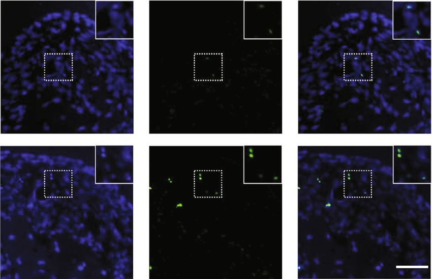
2 BioMed Research International

 Table 1

Gene Forward (5 ′ –3 ′ ) Reverse (5 ′ –3 ′ )
β-Actin (human) CTCGCCTTTGCCGATCC TCTCCATGTCGTCCCAGTTG
NLRP3 (human) CTGGCATCTGGGGAAACCT TTAGGCTTCGGTCCACACAG
TXNIP (human) TCAGTATTGCAGGGCTTGGC GTCTCTTGAGTTGGCTGGCT
ASC (human) ATCCAGGCCCCTCCTCAG AGAGCTTCCGCATCTTGCTT
Caspase-1 (human) ACAAGACCTCTGACAGCACG TTCACTTCCTGCCCACAGAGAC
IL-1β (human) TTCGACACATGGGATAACGAGG TTTTTGCTGTGAGTCCCGGAG
TNF-α (human) CTGGGCAGGTCTACTTTGGG CTGGAGGCCCCAGTTTGAAT
β-Actin (human) GGCACAGTCAAGGCTGAGAAT ATGGTGGTGAAGACGCCAGTA
NLRP3 (rat) CTGCAGAGCCTACAGTTGGG GTCCTGCTTCCACACCTACC
TXNIP (rat) TCCACAGATGGGTGGCAATC AAGTGGGCCAGGTCTGAATG
ASC (rat) GCACAGCCAGAACAGAACATT CCAGGCTGGAGCAAAGCTAA
Caspase-1 (rat) GACCGAGTGGTTCCCTCAAG GACCGAGTGGTTCCCTCAAG
IL-1β (rat) GACCTGTTCTTTGAGGCTGA TCCATCTTCTTCTTTGGGTATTGT

 400

 300
 Body weight (grams)

 200

 100

 0
 8D 2w 3w 4w 5w 6w 7w 8w 9w
 Age in weeks

 SHAM
 UNO
 (a) (b)
 DAPI TUNNEL MERGE Parodontium
 15
 ⁎
 Percentage of TUNEL
 positive nuclei

 10
 SHAM

 5

 0
 SHAM UNO
 UNO

 (c) (d)

Figure 1: Effects of unilateral nasal obstruction (UNO) in rats. (a) Establishment of UNO in Wistar model rats. (b) Changes in body weight
of rats during the experiment (n = 5). (c) TUNEL staining of the periodontal ligament. Scale bars = 50 μm. (d) Percentage of TUNNEL-
positive nuclei in SHAM and UNO. The above data are presented as the mean ± SEM, ∗ p < 0:05.
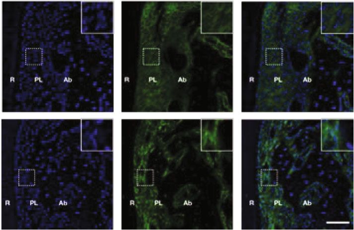
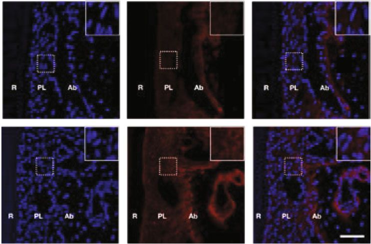
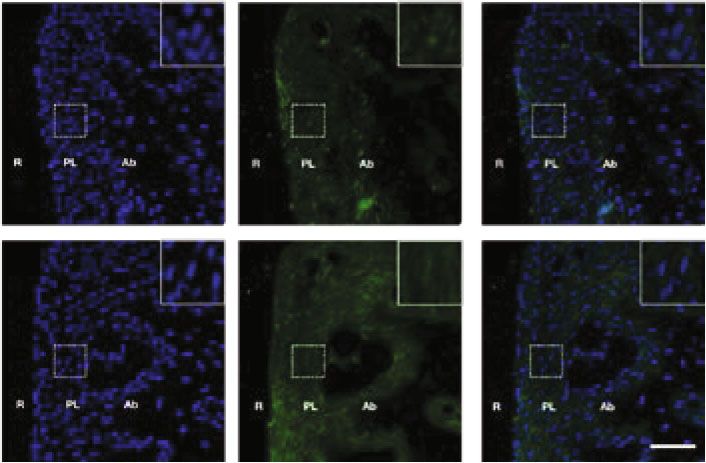
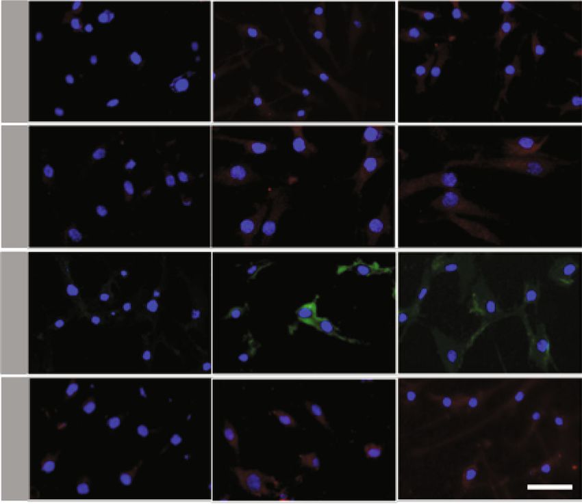
BioMed Research International 3

 DAPI TXNIP MERGE DAPI NLRP3 MERGE

SHAM SHAM

 UNO UNO

 DAPI ASC MERGE
 DAPI CASPASE-1 MERGE

SHAM
 SHAM

 UNO
 UNO

 ⁎
 ⁎
Mean fluorescence intencsity

 Mean fluorescence intencsity

 Mean fluorescence intencsity

 Mean fluorescence intencsity
 1.5 1.5 2.0 1.5
 ns ⁎
 1.5
 1.0 1.0 1.0
 NLRP3
 TXNIP

 Casp-1

 ASC

 1.0
 0.5 0.5 0.5
 0.5

 0.0 0.0 0.0 0.0
 SHAM UNO SHAM UNO SHAM UNO SHAM UNO
 (a)
 8

 ⁎
 6
 mRNA fold change

 ⁎

 4 ⁎ ⁎
 ⁎

 2

 0
 TXNIP NLRP3 ASC Caspase-1 IL-1
 SHAM
 UNO
 (b)

 Figure 2: Continued.
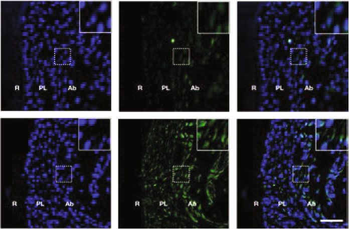
4 BioMed Research International

 2.5
 NLRP3
 ⁎

 Normalized protein level
 2.0
 TXNIP
 Caspase-1 1.5 ⁎ ⁎ ⁎
 ns
 IL-1
 1.0
 ASC
 -actin 0.5
 SHAM UNO
 0.0
 TXNIP NLRP3 ASC Caspase-1 IL-1

 SHAM
 UNO
 (c)

Figure 2: Effects of systemic hypoxia stimulation on the expression of TXNIP/NLRP3 signaling pathway-related factors in periodontal
tissues of rats: (a) immunofluorescence staining (IF) showed that TXNIP, NLRP3, ASC, and caspase-1 in periodontal tissues. Ab:
alveolar; PL: periodontal ligament; R: root. Scale bars = 25 μm. (b) RT-PCR analysis of TXNIP, NLRP3, ASC, caspase-1, and IL-1β in
periodontal tissues (n = 5). (c) Western blotting analysis of TXNIP, NLRP3, ASC, caspase-1, and IL-1β in periodontal tissues (n = 5 for
each group). The above data are presented as the mean ± SEM, ∗ p < 0:05.

the pathogenesis of periodontitis and has been widely stud- titis [23], and the pathophysiological mechanisms underly-
ied because it stimulates the recruitment and differentiation ing the association between the two conditions remain
of osteoclasts in tissues and contributes to bone absorption unclear. We hypothesized that there is a possible correlation
during periodontitis [13]. Inflammasomes are multiprotein between OSAHS and periodontal disease. OSAHS may affect
complexes that can induce inflammatory responses in cells the incidence of periodontal diseases through the ROS/
and play an important role in the inflammatory response of TXNIP/NLRP3 inflammasome signaling pathway.
various tissues. Among them, the nucleotide-binding This study was aimed at elucidating and providing evi-
leucine-rich repeat (NLR) pyrin domain-containing 3 dence for the pathophysiological mechanism underlying
(NLRP3) inflammasome is the most prominent in the NLR the correlation between OSAHS and periodontal diseases.
family and is proven to be involved in innate immune
responses to infection, inflammation, and chronic diseases 2. Materials and Methods
[14]. The NLRP3 inflammasome complex is composed of
NLRP3, recruitment domain (ASC), and caspase-1 [15]. In 2.1. Isolation and Culture of HPDLCs. Healthy periodontal
response to tissue sensing inflammatory stimuli, procaspase- tissue was collected from five healthy candidates (16–26
1 is activated and cleaves IL-1β into its biologically active years old; mean 17.8 years) who underwent tooth extraction
form. Previous studies have shown that hypoxia can regulate as an orthodontic treatment. The teeth were placed in sterile
the NLRP3 inflammasome expression through TXNIP expres- phosphate-buffered saline (PBS; Hyclone, USA) immediately
sion [16, 17]. Meanwhile, overexpression of NLRP3 in gingival after extraction. The periodontal ligament tissues were
tissue and increased NLRP3 salivary levels have been observed scraped from the middle third of the tooth roots and then
in patients with periodontitis [18]. The ROS/TXNIP/NLRP3 digested for 30 min at 37°C in 3 mg/ml collagenase type I
inflammasome signaling pathway may play an important role (Sigma, USA). The resulting cell suspension was seeded in
in the pathogenesis of periodontal disease. Patients with 25 cm2 flasks containing alpha-minimal essential medium
OSAHS are hypoxic, and the tissues often have oxidative (Hyclone, USA) supplemented with 10% fetal bovine serum
stress. When the body is in a hypoxic state, too many reactive (GIBCO, USA) and 1% penicillin/streptomycin. The cells
oxygen species (ROS) in the respiratory chain complex cannot were incubated at 37°C in a humidified 5% CO2 incubator.
be cleared in time, leading to adverse reactions, such as cell Cells between the third and sixth generations were used for
DNA damage, resulting in cell dysfunction and even death. the subsequent experiments. This research protocol was
This response is known as oxidative stress [19]. Previous stud- approved by the Ethical Committee of the Tongji University
ies have shown that upon its separation from thioredoxin, College.
thioredoxin-interacting protein (TXNIP) expression levels
increase, a necessary process leading to oxidative stress in 2.2. Cell Counting Kit-8 Assay (CCK-8). The CCK-8 assay
the body. Increased TXNIP expression levels can activate (Beyotime, China) was used to evaluate the effect of different
NRLP3 inflammasomes directly and eventually induce an concentrations of CoCl2 on cell proliferation. HPDLCs were
inflammatory response [20–22]. seeded in 96-well plates at a density of 2000 cells/well in
 Current studies have shown that there appears to be low 100 μl alpha-minimal essential medium, and then, different
evidence for the association between OSAHS and periodon- concentrations (0, 100, 200, 300, and 400 μM) of CoCl2 were
BioMed Research International 5

 1.5 1.5

 ⁎
 1.0 ns 1.0
 ⁎

 OD450

 OD450
 ⁎ ⁎
 ⁎
 0.5 0.5
 ⁎ ⁎

 0.0 0.0
 0 100 200 300 400 0 100 200 300 400
 Day 1 CoCl2 ( M) Day 2 CoCl2 ( M)

 (a)
 ROS
 8
 ⁎

 Mean fluorescence
 6 ⁎

 intensity
 ROS
 ROS

 4

 2

 0 200 400
 0
 0 200 400
 CoCl2 ( M)
 CoCl2 ( M)

 (b)

 Figure 3: Continued.

6 BioMed Research International
 TXNIP/DAPI
 NLRP3/DAPI
 ASC/DAPI
 CASP1/DAPI

 0 200 400

 CoCl2 ( M)

 150 3 5 15
 ns ⁎
 Mean fluorescence

 Mean fluorescence

 Mean fluorescence
Mean fluorescence

 ⁎ 4 ⁎
 ⁎
 100 2 ns 10
 intensity

 intensity

 intensity
 intensity

 NLRP3

 3
 TXNIP

 Casp-1

 ASC
 2
 50
 ⁎
 1 5 ⁎
 1
 0 0 0 0
 0 200 400 0 200 400 0 200 400 0 200 400
 CoCl2 ( M) CoCl2 ( M) CoCl2 ( M) CoCl2 ( M)

 (c)

 Figure 3: Continued.
BioMed Research International 7

 4 4 4
 ⁎

 3 3 ⁎ 3 ⁎

 NLRP3/ -actin
 TXNIP/ -actin

 ASC/ -actin
 ⁎
 2 ns 2 2

 ⁎
 1 1 1

 0 0 0
 0 200 400 0 200 400 0 200 400
 CoCl2 ( M) CoCl2 ( M) CoCl2 ( M)

 3 3
 ⁎ ⁎
 ⁎
 Caspase-1/ -actin

 ⁎
 IL-1 / -actin
 2 2

 1 1

 0 0
 0 200 400 0 200 400
 CoCl2 ( M) CoCl2 ( M)

 (d)

 Figure 3: Continued.
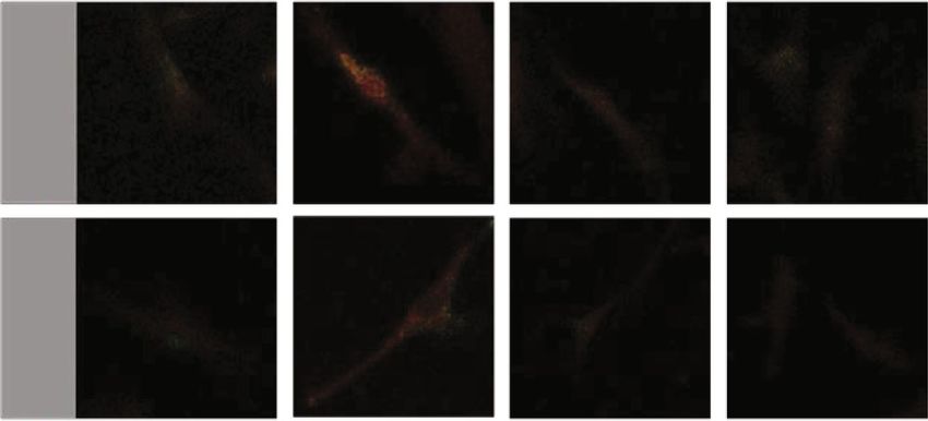
8 BioMed Research International

 NLRP3

 -action

 TXNIP

 Caspase-1

 ASC

 IL-1

 0 200 400

 CoCl2 ( M)

 1.5 1.5 2.0
 Normalized protein level ⁎

 Normalized protein level
 Normalized protein level

 ⁎ ⁎ ns ⁎
 1.5 ⁎
 1.0 1.0
 NLRP3
 TXNIP

 ASC
 1.0
 0.5 0.5
 0.5

 0.0 0.0 0.0
 0 200 400 0 200 400 0 200 400
 CoCl2 ( M) CoCl2 ( M) CoCl2 ( M)

 2.0 ⁎ 1.5
 ⁎
 Normalized protein level

 Normalized protein level

 ⁎
 1.5 ⁎
 1.0
 Caspase-1

 IL-1

 1.0
 0.5
 0.5

 0.0 0.0
 0 200 400 0 200 400
 CoCl2 ( M) CoCl2 ( M)

 (e)

Figure 3: Hypoxic environment induced by CoCl2 activates the ROS/TXNIP/NLRP3 inflammasome pathway in periodontal membrane
fibroblasts. (a) Effects of different concentrations of CoCl2 on the proliferation of periodontal membrane fibroblasts cultured for 24 h and
48 h. (b, c) Immunofluorescence staining (IF) showed that the expressions of ROS, TXNIP, NLRP3, ASC, and caspase-1 were in the cells
treated with different concentrations of CoCl2.. Scale bars = 1 μm. (d) RT-PCR analysis of TXNIP, NLRP3, caspase-1, ASC, and IL-1β in
HPDLCs cells treated with 200 μM and 400 μM CoCl2 for 24 h. (e) Western blotting analysis of TXNIP, NLRP3, caspase-1, ASC, and IL-
1β in HPDLC cells treated with 200 μM and 400 μM for 24 h. The above data are presented as the mean ± SEM, ∗ p < 0:05.

added to stimulate cells for different periods (24 and 48 h). 2.4. Reverse Transcription-Polymerase Chain Reaction (RT-
After 2 h of culture with 10 μl/well CCK-8, the fluorescence PCR). Total RNA from rat periodontal tissues and HPDLCs
intensity was measured at 450 nm. Cell proliferation was was isolated using a TRIzol® reagent (Takara Ltd., Otsu,
plotted relative to the untreated controls. Japan), and cDNA was synthesized using PrimeScript RT
 Master Mix (Takara Ltd.). RT-PCR was conducted using
2.3. CoCl2 Treatment to Mimic Hypoxic Treatment. CoCl2 qPCR SYBR Green Master Mix (Yeasen, China) and a Light-
was dissolved directly in the cell culture medium and filtered Cycler® 96 instrument (Roche, Germany). Primer sequences
by a 0.2 μm filter to produce a concentrated solution of the used for RT-PCR (Sangon, China) are listed in Table 1.
culture medium. To mimic the hypoxic environment
in vivo, the cells were treated with or without CoCl2 at vary- 2.5. Western Blotting. Periodontal tissues were washed with
ing concentrations (200 and 400 μM) for varying times (24 PBS and lysed with radioimmunoprecipitation assay buffer
and 48 h). As part of the experiments, some cells were pre- (Beyotime, China). The tissue lysates were separated on
treated with the ROS inhibitor N-acetylcysteine (NAC; 10% SDS-PAGE gels and transferred to polyvinylidene fluo-
10 mM) for 2 h before CoCl2 (200 μM) exposure for 24 h. ride membranes (Millipore, USA). Membranes were blocked
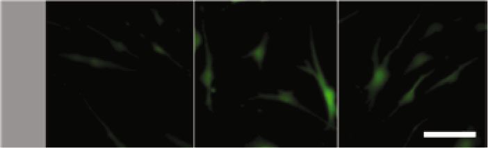
BioMed Research International 9

 ns
 ⁎
 ⁎
 5 ⁎

 4

 Percentage of ROS
 expression
 3
 ROS

 2

 1
 0 200 0+NAC 200+NAC 0
 0 200 0 200
 CoCl2 ( M) +NAC +NAC
 CoCl2 ( M)

 (a)
 NLRP3/ASC
 CSAP1/ASC

 0 200 0+NAC 200+NAC

 CoCl2 ( M)

 ns ns
 ⁎ ⁎
 ⁎ ⁎
 ⁎ ⁎
 1.0 1.0
 Casp-1/SASC (PCC)
 NLRP3/ASC (PCC)

 0.5 0.5

 0.0 0.0
 0 200 0 200 0 200 0 200
 +NAC +NAC +NAC +NAC

 CoCl2 ( M) CoCl2 ( M)

 (b)

 Figure 4: Continued.
10 BioMed Research International

 ⁎
 ⁎ ⁎
 ⁎ ns
 ⁎
 15 ⁎ ⁎
 6 ⁎ 20 ns
 ⁎
 ⁎
 15
 TXNIP/ -actin

 10

 Nrlp3/ -actin
 4

 ASC/ -actin
 10
 5 2
 5

 0 0 0
 0 200 0 200 0 200 0 200 0 200 0 200
 +NAC +NAC +NAC +NAC +NAC +NAC
 CoCl2 ( M) CoCl2 ( M) CoCl2 ( M)
 ⁎ ⁎
 ns 4 ns
 6 ⁎ ⁎
 ⁎ ⁎
 3
 IL-1 / -actin
 Casp-1/ -actin

 4
 2

 2
 1

 0 0
 0 200 0 200 0 200 0 200
 +NAC +NAC +NAC +NAC
 CoCl2 ( M) CoCl2 ( M)

 (c)

 Figure 4: Continued.
BioMed Research International 11

 NLRP3

 TXNIP

 -actin

 Caspase-1

 ASC

 IL-1

 0 200 0+NAC 200+NAC

 CoCl2 ( M)

 ns ns
 ns
 ns ns
 ns
 ⁎ ⁎
 1.5 ns 1.5
 ⁎ 1.5 ⁎
 ⁎

 Normalized protein level
 Normalized protein level

 Normalized protein level

 1.0 1.0 1.0
 TXNIP

 NLRP3

 ASC
 0.5 0.5 0.5

 0.0 0.0 0.0
 0 200 0 200 0 200 0 200 0 200 0 200
 +NAC +NAC +NAC +NAC +NAC +NAC
 CoCl2 ( M) CoCl2 ( M) CoCl2 ( M)

 ns
 ns
 ns
 ns
 ⁎
 1.5 ⁎ ⁎
 1.5 ⁎
 Normalized protein level

 Normalized protein level

 1.0
 1.0
 Casp-1

 IL-1

 0.5 0.5

 0.0 0.0
 0 200 0 200 0 200 0 200
 +NAC +NAC +NAC +NAC

 CoCl2 ( M) CoCl2 ( M)

 (d)

Figure 4: ROS mediates the inflammatory response of periodontal membrane fibroblasts under hypoxia. (a) ROS in HPDLCs after 24 h
treatment with or without 2 mM NAC in the normal group and CoCl2-induced hypoxia environment. (b) Degree of aggregation of
immunofluorescence staining (IF) in periodontal membrane fibroblasts after 24 h treatment with or without 2 mM NAC in the normal
group and CoCl2-induced hypoxia environment. (c) RT-PCR analysis of TXNIP, NLRP3, ASC, caspase-1, and IL-1β in periodontal
membrane fibroblasts after 24 h treatment with or without 2 mM NAC in the normal group and CoCl2-induced hypoxia environment.
(d) Western blotting analysis of TXNIP, NLRP3, ASC, caspase-1, and IL-1β in periodontal fibroblasts treated with or without 2 mM
NAC for 24 h in the normal group and CoCl2-induced hypoxia environment. The above data are presented as the mean ± SEM, ∗ p <
0:05.
12 BioMed Research International

with 5% skimmed milk in TBST for 1 h at room temperature burned. Rats in both groups were raised in the animal facility
and then incubated with the following primary antibodies of Tongji University, Shanghai, China, under specific
overnight at 4°C: TXNIP (Rabbit IgG, Boster, China), pathogen-free (Suzhou Fengshi Laboratory Animal Equip-
NLRP3 (Rabbit IgG, Boster, China), ASC (Rabbit IgG, ment, China) conditions at 22 ± 2° C under a 12 : 12 h light : -
Immunoway, China), and caspase-1 (Rabbit IgG, Immuno- dark cycle, with food and water provided ad libitum. In the
way, China) at 1 : 1000. Thereafter, they were incubated with experimental period, body weights were recorded, and nasal
antirabbit IgG secondary antibody (CST, USA) at 1 : 5000 at obstruction was confirmed every week. Rats whose external
room temperature for 2 h. The internal control was β-actin nostrils remained blocked throughout the experiment were
(Rabbit IgG, Beyotime, China) at 1 : 1000. An Odyssey CLx considered successful models. After 7 weeks, all rats were
Imaging System (LI-COR, USA) was used to detect humanely sacrificed. The periodontal tissue was collected
antibody-bound proteins. Relative protein expression was and processed for analysis as describe below. This research
normalized to β-actin and quantified using the ImageJ protocol was approved by the Ethical Committee of the
software. Tongji University College.

2.6. ROS and Immunofluorescence (IF) Assays. The cells were 2.9. Histological Assessment. Rat periodontal tissue samples
stimulated with CoCl2 for 24 h in a 6-well plate. The level of were fixed in 4% paraformaldehyde, prepared for paraffin-
ROS was detected with a ROS assay kit (Beyotime, China). embedding (Beyotime, China), and sectioned into 4 μm sec-
Images were observed using a fluorescence microscope tions. For IF, the sections were incubated overnight at 4°C with
(Nikon, Japan). The intensity of fluorescence before and primary antibodies (1 : 250) diluted in PBS: TXNIP (Rabbit
after stimulation was measured in real time at 488 nm. For IgG, Boster, China), NLRP3 (Rabbit IgG, Boster, China),
IF, HPDLCs were seeded into cell-climbing slices in a 12- ASC (Rabbit IgG, Immunoway, USA), and caspase-1 (Rabbit
well plate, fixed with 4% paraformaldehyde for 15 min and IgG, Immunoway, China) and then incubated with Alexa
then treated with 0.5% Triton X-100 for permeabilization. Fluor-488 and Alexa Fluor-555 conjugate secondary anti-
After blocking with 5% bovine serum albumin for 30 min, bodies at room temperature for 1 h. For the TUNEL assay, a
the cells were incubated overnight at 4°C with primary anti- One-Step TUNEL Assay Kit (Boster, China) was used. All sec-
bodies (1 : 250) diluted in PBS: TXNIP (Rabbit IgG, Boster, tions were then stained with DAPI (Beyotime, China) for
China), NLRP3 (Rabbit IgG, Boster, China), ASC (Rabbit 5 min in the dark. Sections were captured using a fluorescence
IgG, Santa Cruz, USA), and caspase-1 (Rabbit IgG, Immu- microscope and a confocal laser scanning microscope, and the
noway, China). Finally, the cells were incubated with Alexa fluorescence intensity was quantified using ImageJ software.
Fluor-488 and Alexa Fluor-555 conjugate secondary anti-
bodies (Invitrogen, USA) for 1 h and the nuclei stained with 2.10. Statistical Analysis. All statistical analyses were per-
DAPI for 5 min. Images were captured using a fluorescence formed using GraphPad Prism (GraphPad Software Inc.,
microscope (Nikon, Japan) and a confocal laser scanning USA). The data are expressed as mean ± SEM. Statistical sig-
microscope (Nikon, Japan), and the fluorescence intensity nificance was assessed using one-way ANOVA, and then,
was quantified using ImageJ software. the Bonferroni post hoc test was used to test the selected
 comparison or Dunnett’s multiple comparison post hoc test,
2.7. Confocal Microscopy of Inflammasome Proteins in if each group needed to be compared with the control group.
HPDLCs. Cells were handled as in the IF assay and then Statistically significant difference was determined at ∗ p <
incubated overnight at 4°C with primary antibodies 0:05.
(1 : 250) diluted in PBS: NLRP3 (Rabbit IgG, Boster, China),
ASC (Mouse IgG, Santa Cruz, USA), and caspase-1 (Rabbit 3. Results
IgG, Immunoway, China). Next, the cells were cleaned and
labeled with Alexa Fluor-488 and Alexa Fluor-555 conjugate 3.1. Systemic Changes and Increased Apoptosis of Periodontal
secondary antibodies and then sequentially scanned and Tissue in UNO Rats. The establishment of the UNO rat
visualized using an Olympus laser scanning confocal micro- model was as mentioned above [24–26]. We used the
scope (Olympus, Japan). Image-Pro Plus software (Media peripheral nostril obstruction in rats as the standard for suc-
Cybernetics, USA) was used for colocalization analysis, and cessful modeling. Body weight was used as an indicator of
the Pearson correlation coefficient represented the influence of hypoxia on rats, and the statistical results
colocalization. showed that there was no significant difference in body
 weight between the SHAM and UNO groups (Figures 1(a)
2.8. Animals and Unilateral Nasal Obstruction (UNO) and 1(b)). TUNEL staining was performed to investigate
Model. A UNO rat model was established by cauterizing the effect of hypoxia on periodontal cell apoptosis in UNO
the external nostril [24–26]. Briefly, 8-day-old male Wistar rats after UNO for 8 weeks. During apoptosis, the genomic
rats (n = 30) were randomly divided into two groups: SHAM DNA of cells is fragmented; this is detectable by TUNEL
and UNO. Rats in the two groups were first anesthetized by staining [27, 28]. Results showed that TUNEL-positive cells
hypothermia (10 min at -20°C), and then, the external nostril were increased in UNO rats compared to those in SHAM
of rats in the UNO group was burned using a heated hot rats (Figures 1(c) and 1(d)). Taken together, the results show
container for the gutta-percha point, whereas in the SHAM that apoptosis is a response to oxidative stress and is a prod-
group, the surrounding tissues of the left nostril were uct of the inflammatory response.
BioMed Research International 13

3.2. The Levels of TXNIP/NLRP3 Inflammasome Signaling 4. Discussion
Pathway-Related Factors in Periodontal Tissues of Rats in
the UNO Group Were Changed. The expression of OSAHS causes a decrease in blood oxygen saturation. With
hypoxia-related factors in periodontal tissues of the UNO a decrease in blood saturation, OSHAH patients often
and SHAM groups was detected by RT-PCR to study the develop cardiovascular system and cognitive disorders.
changes in inflammatory factors in periodontal tissues. The Recent literature indicates that the incidence of periodontal
expression of TXNIP, NLRP3, caspase-1, ASC, and the disease is higher in patients with OSHAS than in healthy
inflammatory factor IL-1β was increased in the UNO group individuals. However, the mechanism underlying the corre-
compared with the SHAM group. We then detected the pro- lation between OSAHS and the incidence of periodontal dis-
tein levels of TXNIP, NLRP3, caspase-1, ASC, and IL-1β in ease remains unclear. It is important to determine how
the periodontal tissues of the two groups of rats hypoxia affects the biological behavior of HPDLCs, the prin-
(Figures 2(b) and 2(c)). IF showed that the UNO group cipal cells in the periodontal ligament that help maintain the
had higher TXNIP, ASC, and caspase-1-positive cells than stability of the periodontal system and promote the repair
did the SHAM group, whereas there was no difference in and regeneration of periodontal tissue [29].
NLRP3-positive cells. In addition, western blotting showed Hypoxia can lead to oxidative damage and apoptosis in
higher expression levels of TXNIP, caspase-1, ASC, and IL- various tissues, which is key to maintaining tissue homeosta-
1β in the UNO group than in the SHAM group, whereas sis [30–32]. Many factors can trigger its occurrence, espe-
there was no difference in NLRP3 protein expression cially oxidative stress [33]. Elevated ROS levels and
(Figure 2(a)). oxidative stress were observed in patients with OSAHS. In
 vivo, we observed more positive TUNEL signals in the peri-
 odontal membrane of rats with UNO. These results indicate
3.3. CoCl2 Simulates Hypoxia in HPDLCs, Induces ROS
 that hypoxia can lead to oxidative stress and apoptosis in
Production, Increases TXNIP Expression, and Upregulates
 periodontal tissue. After modeling UNO in rats, rats pre-
NLRP3 Inflammasome-Related Factor Expression. CoCl2-
 sented with oral respiratory symptoms [24–26]. Oral respi-
induced hypoxia is one of the most commonly used models
 ration is one of the main clinical manifestations in patients
in hypoxia studies. To study the effect of hypoxia on
 with OSAHS [1]. Oral respiration is also reported to be asso-
HPDLCs, different concentrations of CoCl2 were used to
 ciated with gingival inflammation [34, 35]. The NLRP3
stimulate cells. After two days of culture at 200 μM and
 inflammasome is a multiprotein complex composed of
400 μM, cell viability decreased significantly. However, there
 NLRP3, ASC, and caspase-1. When the NLRP3 inflamma-
was no significant difference in the cell viability of HPDLCs
 some polymerizes, IL-1β is produced to induce a cellular
treated with 200 μM at 24 h (Figures 3(a)). In addition, the
 inflammatory response. Previous studies have shown that
fluorescence intensity of ROS, TXNIP, NLRP3, ASC, and
 hypoxia is involved in NLRP3 activation and subsequent
caspase-1 differed from those in the normal group
 inflammasome formation by increasing TXNIP expression
(Figures 3(b) and 3(c)). We noted that with a change in
 [36–38], and the occurrence of periodontitis is associated
CoCl2 concentration, NLRP3 expression was not signifi-
 with the NRLP3 inflammasome [39]. Recent studies have
cantly different from that in the control group. At the same
 reported that TXNIP could induce the formation and aggre-
time, one day after 200 μM and 400 μM culture, we per-
 gation of the NLRP3 inflammatory body, leading to dysfunc-
formed RT-PCR and western blotting analysis on TXNIP,
 tion and injury of periodontal membrane fibroblasts,
NLRP3, caspase-1, ASC, and IL-1β in HPDLCs
 resulting in periodontitis [10]. The in vivo results showed
(Figures 3(d) and 3(e)). The results showed that culture with
 that the transcription and expression of TXNIP, ASC, and
200 μM CoCl2 for 24 h significantly increased the transcrip-
 caspase-1 in periodontal tissues of UNO rats were higher
tion and expression of TXNIP, NLRP3, caspase-1, ASC, and
 than those of SHAM rats, and the expression level of the
IL-1β. However, with the increase in concentration, the
 inflammatory factor IL-1β in periodontal tissues of UNO
expression of the above factors did not increase, which could
 rats was upregulated. In IF experiments, high TXNIP, ASC,
result from NLRP3 inflammasome upregulation-regulated
 and caspase-1 expression signals were detected in the peri-
gene expression. Therefore, in our subsequent experiments,
 odontal membrane of UNO rats compared to those of
we used the 200 μM culture for 24 h as a method to construct
 SHAM rats. Interestingly, our experimental results showed
a hypoxic cell environment.
 that compared to that in the SHAM group, the transcription
 level of NLRP3 in periodontal tissues was increased in the
3.4. Inhibition of ROS Production Attenuates CoCL2-Induced UNO group; however, there was no significant difference
TNXIP Expression and NLRP3 Inflammasome Activation. in its protein level. The result that the NRLP3 transcription
NAC inhibited ROS generation (Figure 4(a)). We verified level in periodontal tissues was upregulated but the protein
whether ROS are involved in CoCl2-induced NLRP3 inflam- level was not upregulated in a hypoxic environment in vivo
masome activation and expression by NAC pretreatment was consistent with the results of a study, which demon-
(Figure 4(b)). The results showed that the CoCl2-induced strated that hypoxia mainly leads to the aggregation of
aggregation of NLRP3 inflammasome-related factors and inflammasomes by increasing the nuclear localization of
IL-1β expression decreased significantly with decreased NLRP3 and caspase-1 [40], resulting in increased expression
ROS expression. Interestingly, NAC also inhibited CoCl2- of inflammatory factors in tissues. Meanwhile, it is reported
induced TXNIP expression (Figures 4(c) and 4(d)). that there are no differences in the serum levels of NLRP3
14 BioMed Research International

inflammasome components between OSAHS and controls Conflicts of Interest
[41]. Hypoxia-induced cell death may play a key role in sys-
temic diseases caused by OSAHS. In addition, the expression The authors declare that they have no competing interests.
level of the inflammatory factor IL-1β in the periodontal tis-
sues of UNO rats was upregulated. Authors’ Contributions
 In vitro experiments found that CoCl2 stimulation of
HPDLCs resulted in ROS production and accumulation in RZ, XM, and YL designed the study. RZ conducted the
cells, but the accumulation of ROS in cells was significantly experiments and wrote the manuscript. XM and YL revised
improved after the application of NAC. Meanwhile, NAC the manuscript. All authors have read and approved the
significantly reduced the level of the inflammatory gene IL- manuscript.
1β and the aggregation of NLRP3 inflammasome. Several
studies have shown that targeting ROS can reduce cardiovas- Acknowledgments
cular damage in OSAHS [42]. Therefore, antioxidant ther-
apy may be a new treatment method to reduce the damage We would like to thank Editage (http://www.editage.cn/) for
of periodontal tissue caused by oxidative stress. English language editing. This work was supported by the
 OSAHS is difficult to diagnose, and patients themselves National Natural Science Foundation of China (Program
tend to ignore and underestimate the symptoms [43]. There- Nos. 11402175 and 81970921) and the General Project of
fore, it is necessary to introduce specific sleep disorder ques- Shanghai Municipal Health Commission (Program No.
tions and questionnaires into dental records, which can help 202140355).
clinicians identify patients at risk for OSAHS [44]. The study
suggests that there is a pathological correlation between References
OASHS and the occurrence of periodontal diseases, which
 [1] C. Guilleminault, F. L. Eldridge, and W. C. Dement, “Insomnia
is supported by the above views.
 with sleep apnea: a new syndrome,” Science, vol. 181, no. 4102,
 pp. 856–858, 1973.
5. Conclusions [2] R. K. Malhotra, “Neurodegenerative disorders and sleep,” Sleep
 Medicine Clinics, vol. 13, no. 1, pp. 63–70, 2018.
In summary, our study suggests that OSAHS may increase [3] J. S. Floras, “Sleep apnea and cardiovascular Disease,” Circula-
the expression of inflammatory factors in periodontal tissues tion Research, vol. 122, no. 12, pp. 1741–1764, 2018.
through the ROS/TXNIP/NLRP3 inflammasome signaling [4] N. Peled, M. Kassirer, D. Shitrit et al., “The association of OSA
pathway, thus making periodontal tissues more prone to with insulin resistance, inflammation and metabolic syndrome,”
inflammatory lesions. Respiratory Medicine, vol. 101, no. 8, pp. 1696–1701, 2007.
 [5] T. S. Al-Jewair, R. Al-Jasser, and K. Almas, “Periodontitis and
 obstructive sleep apnea's bidirectional relationship: a system-
6. Limitation atic review and meta-analysis,” Sleep & Breathing, vol. 19,
 no. 4, pp. 1111–1120, 2015.
The animal model of this experiment lacks anatomical fac- [6] B. Tamasas, T. Nelson, and M. Chen, “Oral health and oral
tors of upper airway stenosis, making it significantly differ- health-related quality of life in children with obstructive sleep
ent from human OSAHS. In future experiments, we will apnea,” Journal of Clinical Sleep Medicine, vol. 15, no. 3,
continue to study how to establish animal models that are pp. 445–452, 2019.
closer to the pathogenesis of human OSAHS. The effect of [7] H. Gamsiz-Isik, E. Kiyan, Z. Bingol, U. Baser, E. Ademoglu,
UNO modeling on periodontitis was not determined in this and F. Yalcin, “Does obstructive sleep apnea increase the risk
study. This part of the experiment will be supplemented in for periodontal disease? A case-control study,” Journal of Peri-
future studies to explore whether OSASH can aggravate odontology, vol. 88, no. 5, pp. 443–449, 2017.
the severity of periodontitis. [8] M. G. Balta, E. Papathanasiou, I. J. Blix, and T. E. van Dyke,
 “Host modulation and treatment of periodontal disease,” Jour-
 nal of Dental Research, vol. 100, no. 8, pp. 798–809, 2021.
Data Availability [9] F. Teles, Y. Wang, G. Hajishengallis, H. Hasturk, and J. T.
 Marchesan, “Impact of systemic factors in shaping the peri-
The datasets used and/or analyzed during the current study odontal microbiome,” Periodontology 2000, vol. 85, no. 1,
are available from the corresponding author upon request. pp. 126–160, 2000.
 [10] P. E. Petersen and H. Ogawa, “The global burden of periodon-
 tal disease: towards integration with chronic disease preven-
Ethical Approval tion and control,” Periodontology 2000, vol. 60, no. 1, pp. 15–
 39, 2012.
The animal study was reviewed and approved by the Ethics [11] K. E. Kholy, R. J. Genco, and T. E. Van Dyke, “Oral infections
Committee of the School and Hospital of Stomatology, and cardiovascular disease,” Trends in Endocrinology and
Tongji University (SL2019SR19). The studies involving Metabolism, vol. 26, no. 6, pp. 315–321, 2015.
human participants were reviewed and approved by the [12] K. H. Philips, S. Zhang, K. Moss, K. Ciarrocca, and J. D. Beck,
Ethics Committee of the School and Hospital of Stomatol- “Periodontal disease, undiagnosed diabetes, and body mass
ogy, Tongji University (SL2019DW42). index: implications for diabetes screening by dentists,” Journal
BioMed Research International 15

 of the American Dental Association (1939), vol. 152, no. 1, [28] J. Feng, M. Li, Q. Wei, S. Li, S. Song, and Z. Hua, “Unconju-
 pp. 25–35, 2021. gated bilirubin induces pyroptosis in cultured rat cortical
[13] E. Yavuzyilmaz, N. Yamalik, S. Bulut, S. Özen, F. Ersoy, and astrocytes,” Journal of Neuroinflammation, vol. 15, no. 1,
 Ü. Saatpi, “The gingival crevicular fluid interleukin-1 beta p. 23, 2018.
 and tumour necrosis factor-alpha levels in patients with rap- [29] A. Tomokiyo, N. Wada, and H. Maeda, “Periodontal ligament
 idly progressive periodontitis,” Australian Dental Journal, stem cells: regenerative potency in periodontium,” Stem Cells
 vol. 40, no. 1, pp. 46–49, 1995. and Development, vol. 28, no. 15, pp. 974–985, 2019.
[14] Y. Zhen and H. Zhang, “NLRP3 inflammasome and inflamma- [30] S. C. Chiu, S. Y. Huang, Y. C. Tsai et al., “Poly (ADP-ribose)
 tory bowel disease,” Frontiers in Immunology, vol. 10, p. 276, polymerase plays an important role in intermittent hypoxia-
 2019. induced cell death in rat cerebellar granule cells,” Journal of
[15] M. Haneklaus and L. A. O'Neill, “NLRP3 at the interface of Biomedical Science, vol. 19, no. 1, 2012.
 metabolism and inflammation,” Immunological Reviews, [31] A. C. Racanelli, S. A. Kikkers, A. M. K. Choi, and S. M. Cloo-
 vol. 265, no. 1, pp. 53–62, 2015. nan, “Autophagy and inflammation in chronic respiratory dis-
[16] N. Gupta, A. Sahu, A. Prabhakar et al., “Activation of NLRP3 ease,” Autophagy, vol. 14, no. 2, pp. 221–232, 2018.
 inflammasome complex potentiates venous thrombosis in [32] W. Ding, X. Chen, W. Li, Z. Fu, and J. Shi, “Genistein protects
 response to hypoxia,” Proceedings of the National Academy genioglossus myoblast against hypoxia-induced injury
 of Sciences of the United States of America, vol. 114, no. 18, through PI3K-Akt and ERK MAPK pathways,” Scientific
 pp. 4763–4768, 2017. Reports, vol. 7, no. 1, p. 5085, 2017.
[17] Z. Chen, H. Zhong, J. Wei et al., “Inhibition of Nrf2/HO-1 sig- [33] D. Tang, R. Kang, T. V. Berghe, P. Vandenabeele, and
 naling leads to increased activation of the NLRP3 inflamma- G. Kroemer, “The molecular machinery of regulated cell
 some in osteoarthritis,” Arthritis Research & Therapy, vol. 21, death,” Cell Research, vol. 29, no. 5, pp. 347–364, 2019.
 no. 1, p. 300, 2019. [34] E. Nascimento Filho, M. P. A. Mayer, P. Pontes, A. C. C.
[18] D. M. Isaza-Guzmán, V. M. Medina-Piedrahíta, C. Gutiérrez- Pignatari, and L. L. M. Weckx, “Caries prevalence, levels of
 Henao, and S. I. Tobón-Arroyave, “Salivary levels of NLRP3 mutans streptococci, and gingival and plaque indices in 3.0-
 inflammasome-related proteins as potential biomarkers of to 5.0-year-old mouth breathing children,” Caries Research,
 periodontal clinical status,” Journal of Periodontology, vol. 88, vol. 38, no. 6, pp. 572–575, 2004.
 no. 12, pp. 1329–1338, 2017. [35] E. G. Wagaiyu and F. P. Ashley, “Mouthbreathing, lip seal and
[19] A. J. Kattoor, N. V. K. Pothineni, D. Palagiri, and J. L. Mehta, upper lip coverage and their relationship with gingival inflam-
 “Oxidative stress in atherosclerosis,” Current Atherosclerosis mation in 11-14 year-old schoolchildren,” Journal of Clinical
 Reports, vol. 19, no. 11, 2017. Periodontology, vol. 18, no. 9, pp. 698–702, 1991.
[20] X. Zhang, J. H. Zhang, X. Y. Chen et al., “Reactive oxygen [36] F. R. G. Rocha, A. E. Delitto, J. A. C. de Souza, L. A. González-
 species-induced TXNIP drives fructose-mediated hepatic Maldonado, S. M. Wallet, and C. Rossa Junior, “Relevance of
 inflammation and lipid accumulation through NLRP3 inflam- caspase-1 and Nlrp3 inflammasome on inflammatory bone
 masome activation,” Antioxidants & Redox Signaling, vol. 22, resorption in a murine model of periodontitis,” Scientific
 no. 10, pp. 848–870, 2015. Reports, vol. 10, no. 1, p. 7823, 2020.
[21] D. Lian, L. Dai, Z. Xie et al., “Periodontal ligament fibroblasts [37] Q. Jiang, X. Geng, J. Warren et al., “Hypoxia Inducible Factor-
 migration injury via ROS/TXNIP/Nlrp3 inflammasome path- 1α (HIF-1α) Mediates NLRP3 Inflammasome-Dependent-
 way with _Porphyromonas gingivalis_ lipopolysaccharide,” Pyroptotic and Apoptotic Cell Death Following Ischemic
 Molecular Immunology, vol. 103, pp. 209–219, 2018. Stroke,” Neuroscience, vol. 448, pp. 126–139, 2020.
[22] I. N. Mohamed, S. S. Hafez, A. Fairaq, A. Ergul, J. D. Imig, and [38] S. B. Cheng, A. Nakashima, W. J. Huber et al., “Pyroptosis is a
 A. B. el-Remessy, “Thioredoxin-interacting protein is required critical inflammatory pathway in the placenta from early onset
 for endothelial NLRP3 inflammasome activation and cell preeclampsia and in human trophoblasts exposed to hypoxia
 death in a rat model of high-fat diet,” Diabetologia, vol. 57, and endoplasmic reticulum stressors,” Cell Death & Disease,
 no. 2, pp. 413–423, 2014. vol. 10, no. 12, p. 927, 2019.
[23] D. Lembo, F. Caroccia, C. Lopes, F. Moscagiuri, B. Sinjari, and [39] J. B. de Alencar, J. M. V. Zacarias, P. Y. Tsuneto et al., “Influ-
 M. D’Attilio, “Obstructive sleep apnea and periodontal disease: ence of inflammasome NLRP3, and IL1B and IL2 gene poly-
 a systematic review,” Medicina, vol. 57, no. 6, p. 640, 2021. morphisms in periodontitis susceptibility,” PLoS One, vol. 15,
[24] X. Wang, Y. Cao, Z. Liu et al., “Alveolar bone density reduction no. 1, article e0227905, 2020.
 in rats caused by unilateral nasal obstruction,” Balkan Medical [40] L. M. Yu, W. H. Zhang, X. X. Han et al., “Hypoxia-induced
 Journal, vol. 36, no. 6, pp. 311–319, 2019. ROS contribute to myoblast pyroptosis during obstructive
[25] E. Ren, I. Watari, H. Jui-Chin et al., “Unilateral nasal obstruc- sleep apnea via the NF-κB/HIF-1α signaling pathway,” Oxida-
 tion alters sweet taste preference and sweet taste receptors in tive Medicine and Cellular Longevity, vol. 2019, Article ID
 rat circumvallate papillae,” Acta Histochemica, vol. 121, 4596368, 19 pages, 2019.
 no. 2, pp. 135–142, 2019. [41] T. Tang, Q. Huang, J. Liu et al., “Oxidative stress does not con-
[26] H. Tang, I. Yonemitsu, Y. Ikeda et al., “Effects of unilateral tribute to the release of proinflammatory cytokines through
 nasal obstruction on the characteristics of jaw-closing muscles activating the Nod-like receptor protein 3 inflammasome in
 in growing rats,” The Angle Orthodontist, vol. 89, no. 1, patients with obstructive sleep apnoea,” Sleep & Breathing,
 pp. 102–110, 2019. vol. 23, no. 2, pp. 535–542, 2019.
[27] T. Bergsbaken, S. L. Fink, and B. T. Cookson, “Pyroptosis: host [42] E. Belaidi, J. Morand, E. Gras, J. L. Pépin, and D. Godin-
 cell death and inflammation,” Nature Reviews. Microbiology, Ribuot, “Targeting the ROS-HIF-1-endothelin axis as a thera-
 vol. 7, no. 2, pp. 99–109, 2009. peutic approach for the treatment of obstructive sleep apnea-
16 BioMed Research International

 related cardiovascular complications,” Pharmacology & Thera-
 peutics, vol. 168, pp. 1–11, 2016.
[43] C. R. Laratta, N. T. Ayas, M. Povitz, and S. R. Pendharkar,
 “Diagnosis and treatment of obstructive sleep apnea in adults,”
 Canadian Medical Association Journal, vol. 189, no. 48,
 pp. E1481–e1488, 2017.
[44] M. Santilli, E. Manciocchi, G. D’Addazio et al., “Prevalence of
 obstructive sleep apnea syndrome: a single-center retrospec-
 tive study,” International Journal of Environmental Research
 and Public Health, vol. 18, no. 19, p. 10277, 2021.
You can also read