A robust immune-related lncRNA signature for the prognosis of human colorectal cancer
←
→
Page content transcription
If your browser does not render page correctly, please read the page content below
Bioscience Reports (2022) 42 BSR20220078
https://doi.org/10.1042/BSR20220078

Research Article

A robust immune-related lncRNA signature for the
prognosis of human colorectal cancer
    Gongmin Zhu1,2,* , Lijiao Pei3,* , Fan Yang4 and                                      Chenliang Zhang1
1 Laboratoryof Molecular Targeted Therapy in Oncology, West China Hospital, Sichuan University, Chengdu, Sichuan Province 610041, PR China; 2 Department of Abdominal
Oncology, Cancer Center, West China Hospital, Sichuan University, Chengdu, Sichuan Province 610041, PR China; 3 The State Key Laboratory of Biotherapy, West China Hospital,
Sichuan University, Chengdu, Sichuan Province 610041, PR China; 4 Chengdu Women’s and Children’s Central Hospital, School of Medicine, University of Electronic Science and

                                                                                                                                                                                           Downloaded from http://portlandpress.com/bioscirep/article-pdf/42/7/BSR20220078/935228/bsr-2022-0078.pdf by guest on 09 August 2022
Technology of China, Chengdu, Sichuan Province 611731, China
Correspondence: Chenliang Zhang (zclscu 2010@sina.com)

                                           Background: Colorectal cancer (CRC) is one of the most prevalent malignant cancers world-
                                           wide. Immune-related long non-coding RNAs (IRlncRNAs) are proved to be essential in the
                                           development and progression of carcinoma. The purpose of the present study was to de-
                                           velop and validate a prognostic IRlncRNA signature for CRC patients.
                                           Methods: Gene expression profiles of CRC samples were downloaded from The Cancer
                                           Genome Atlas (TCGA) database. Immune-related genes were obtained from the ImmPort
                                           database and were used to identify IRlncRNA by correlation analysis. Through LASSO Cox
                                           regression analyses, a prognostic signature was constructed. Functional enrichment analy-
                                           sis was performed by gene set enrichment analysis (GSEA). TIMER2.0 web server and tumor
                                           immune dysfunction and exclusion (TIDE) algorithm were employed to analyze the associa-
                                           tion between our model and tumor-infiltrating immune cells and immunotherapy response.
                                           The expression levels of IRlncRNAs in cell lines were detected by quantitative real-time PCR
                                           (qPCR).
                                           Results: A 9-IRlncRNA signature was developed by a LASSO Cox proportional regression
                                           model. Based on the signature, CRC patients were divided into high- and low-risk groups
                                           with different prognoses. GSEA results indicated that patients in high-risk group were asso-
                                           ciated with cancer-related pathways. In addition, patients in low-risk group were found to
                                           have more infiltration of anti-tumor immune cells and might show a favorable response to
                                           immunotherapy. Finally, the result of qPCR revealed that most IRlncRNAs were differently
                                           expressed between normal and tumor cell lines.
                                           Conclusion: The constructed 9-IRlncRNA signature has potential to predict the prognosis
                                           of CRC patients and may be helpful to guide personalized immunotherapy.

                                         Introduction
                                         Colorectal cancer (CRC) is the third most common malignancy worldwide, with almost 1.8 million new
* These authors contributed              cases and approximately 8.6 million deaths in 2018 [1]. Although the diagnosis and treatment of CRC have
equally to this work.                    improved significantly in the past decade, the prognosis is still poor for patients with newly diagnosed
Received: 18 January 2022
                                         CRC presented with distant metastasis, especially liver metastasis [2]. Meanwhile, as a heterogeneous dis-
Revised: 31 March 2022                   ease, the diversity of phenotypes and prognosis of CRC present a huge challenge in making individualized
Accepted: 03 May 2022                    clinical decisions to improve the survival rate of patients [3]. Therefore, it is urgent to establish an effective
                                         risk assessment model to identify patient subgroups with different prognoses, which may also help to find
Accepted Manuscript online:
04 May 2022                              potential therapeutic targets.
Version of Record published:                In recent years, genomic approaches have been applied to investigate the underlying mechanism of can-
20 July 2022                             cer development and explore molecular biomarkers for cancer diagnosis [4,5]. Besides the well-recognized

© 2022 The Author(s). This is an open access article published by Portland Press Limited on behalf of the Biochemical Society and distributed under the Creative Commons Attribution   1
License 4.0 (CC BY).
Bioscience Reports (2022) 42 BSR20220078
                                                                                                                                         https://doi.org/10.1042/BSR20220078

           protein-coding genes in the human genome are proved to be involved in tumorigenesis, accumulating evidence has
           demonstrated that long non-coding RNAs (lncRNAs), a class of non-coding RNA with more than 200 nucleotides in
           length, are also closely associated with the pathogenesis of tumor including cell proliferation, apoptosis, migration,
           and epithelial-to-mesenchymal transition [6,7]. Therefore, these findings indicate that a large number of lncRNAs
           can serve as potential targets and biomarkers for the diagnosis and prognosis of malignant tumors including lung,
           breast, liver, and colorectal cancer [8–11].
              Proverbially, the immune system plays a critical role in the development of tumors. With the advent of immuno-
           logic agents, many cancers have shown positive responses to immunotherapy [12–14]. The previous study has indi-
           cated that numerous lncRNAs show an overwhelming effect on diverse stages of cancer immunity, such as antigen
           release and presentation, immune cell differentiation, migration, and infiltration [15]. Lnc-SNHG1 was proved to
           enhance regulatory T cells (Tregs) differentiation via regulating miR-448/IDO axis, which induced Tregs mediated
           immunosuppression and promoted immune escape in breast cancer [16]. Likewise, LncRNA MIR17HG was reported

                                                                                                                                                                                           Downloaded from http://portlandpress.com/bioscirep/article-pdf/42/7/BSR20220078/935228/bsr-2022-0078.pdf by guest on 09 August 2022
           to directly bind to PD-L1 protein for accumulation in CRC, which blocked T cells activation [17]. Therefore, the dys-
           regulation of these immune-related lncRNAs (IRlncRNAs) may suppress immune response and promote immune
           escape, which contributes to the occurrence and progression of various tumors.
              In the present study, based on immune-related genes from the ImmPort database, we used RNA-seq dataset from
           The Cancer Genome Atlas (TCGA) and two microarray datasets (GSE17536 and GSE38832) from The Gene Expres-
           sion Omnibus (GEO) to develop and validate a 9-IRlncRNA signature for patients with CRC. Next, we constructed a
           nomogram based on the 9-IRlncRNA signature, age, and M stage to evaluate clinical significance. Finally, TIMER2.0
           web server and tumor immune dysfunction and exclusion (TIDE) algorithm were employed to analyze the association
           between our model and tumor-infiltrating immune cells and immunotherapy response.

           Materials and methods
           Data acquisition
           RNA expression data, somatic mutation data, and clinical data of colon adenocarcinoma and rectal adenocarcinoma
           samples were downloaded from the TCGA portal (http://portal.gdc.cancer.gov/projects). 530 samples (488 tumors
           and 42 normal tissues) were contained in CRC patients’ dataset. For clinical data, samples with an overall survival
           time of less than 30 days or any missing data were excluded, and finally, 431 samples were included for subsequent
           study.
              The gene expression profile matrix files of GSE17536 and GSE38832 were downloaded from the GEO database
           (https://www.ncbi.nlm.nih.gov/geo/). Raw microarray expression data were normalized using Robust Multichip Av-
           erage (RMA) and converted to Log2 pattern. Probes were annotated through the Affymetrix Human Genome U133
           Plus 2.0 Array. 177 samples with overall survival (OS) and disease-specific survival (DSS) from GSE17536 and 122
           samples with DSS from GSE38832 were set as external validation. Details of the TCGA cohort and the two testing
           cohorts were shown in Table 1.

           Acquisition of immune-related lncRNAs
           The Ensemble IDs of genes from the TCGA cohort were transformed into gene symbols via the Ensemble database
           (http://asia.ensembl.org/index.html). Next, mRNAs and lncRNAs were extracted from the gene matrix respectively
           according to their biotypes. Then, mRNAs were intersected with immune-related genes (IRGs) obtained from the
           ImmPort database (https://immport.niaid.nih.gov) to get IRGs in colorectal cancer samples [18]. Pearson correlation
           analysis was applied to identify IRlncRNAs via evaluating the correlation between the IRGs and lncRNAs expression
           in colorectal cancer samples (|r| >0.3 and P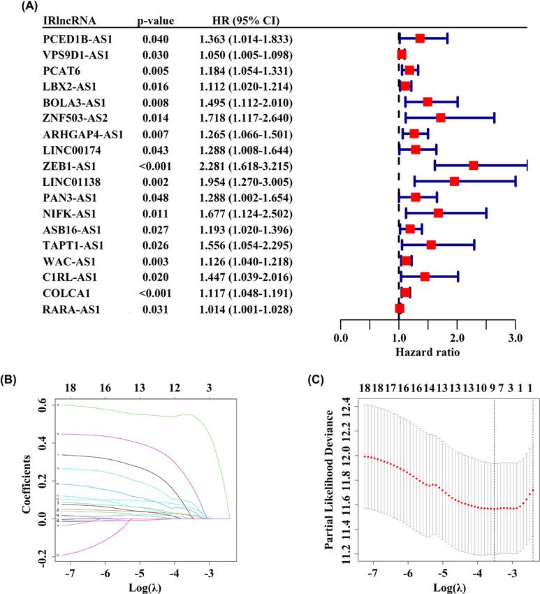
Bioscience Reports (2022) 42 BSR20220078
https://doi.org/10.1042/BSR20220078

Table 1 Clinical information of patients with colorectal cancer in three datasets
  Character                                       TCGA                                            GSE17536                                       GSE38832

  Age (years)
     ≤65                                          192                                             83                                             N/A
     >65                                          239                                             94                                             N/A
  Gender
     Male                                         235                                             96                                             N/A
     Female                                       196                                             81                                             N/A
  Grade
     Low                                          N/A                                             150                                            N/A
     High                                         N/A                                            27                                              N/A
  T stage

                                                                                                                                                                                           Downloaded from http://portlandpress.com/bioscirep/article-pdf/42/7/BSR20220078/935228/bsr-2022-0078.pdf by guest on 09 August 2022
     T1                                           13                                             N/A                                             N/A
     T2                                           78                                             N/A                                             N/A
     T3                                           296                                            N/A                                             N/A
     T4                                           44                                             N/A                                             N/A
  N stage
     N0                                           252                                            N/A                                             N/A
     N1-2                                         179                                            N/A                                             N/A
  M stage
     M0                                           365                                             N/A                                            N/A
     M1                                           66                                              N/A                                            N/A
  AJCC stage
  I-II                                            243                                             81                                             53
  III-IV                                          188                                             96                                             69
  Survival status
     Alive                                        360                                             104                                            94
     Deceased                                     71                                              73                                             28

× expression of lncRNA2) + . . . + (βn × expression of lncRNAn). All patients were divided into a high-risk group
and a low-risk group according to the cut-off value (median risk score).
  To validate the 9-IRlncRNA signature, the risk score was also calculated in two testing datasets (GSE17536 and
GSE38832) and the patients were classified into high- and low-risk groups based on the same cut-off value.

Establishment and evaluation of predictive nomogram
The nomogram containing the 9-IRlncRNA signature and other independent prognostic indicators was plotted using
the “rms” package of R software. The total score of each patient could be calculated via the nomogram, and then was
used to predict the OS rate of 1-, 3- or 5-year. The accuracy of the nomogram was evaluated using the calibration
curves and the time-dependent receiver operating characteristic (ROC) curves. Decision curve analysis (DCA) was
used to compare the reliability of the nomogram with that of age, M stage, or risk group.

Calculation of tumor mutation burden (TMB)
The somatic mutation data of CRC samples were detected using VarScan. To calculate the TMB score of each sample,
all base substitutions and indels in the coding region of targeted genes were counted, and silent mutations failing to
lead to an amino acid change were not counted. Then, the total number of mutations counted was divided by the
exome size (approximate 38 megabases) [19].

Bioinformatics analysis
Principal component analysis (PCA) was employed to reveal the expression pattern of samples in the TCGA
cohort. Gene set enrichment analysis (GSEA) was performed using Broad Institute GSEA software 4.0.1
based on TCGA datasets containing 431 CRC patients classified into high- and low-risk groups [20]. The
GO gene sets “c5.bp.v7.1.symbols.gmt”, KEGG gene sets “c2.cp.kegg.v7.1.symbols.gmt”, Reactome gene sets
“c2.cp.reactome.v7.1.symbols.gmt”, and PID gene sets “c2.cp.pid.v7.1.symbols.gmt” were downloaded from Molec-
ular Signatures Database (http://software.broadinstitute.org/gsea/msigdb/index.jsp). For each gene set analysis, per-
mutations were performed 1000 times to acquire a normalized enrichment score (NES). A normalized P-value <

© 2022 The Author(s). This is an open access article published by Portland Press Limited on behalf of the Biochemical Society and distributed under the Creative Commons Attribution   3
License 4.0 (CC BY).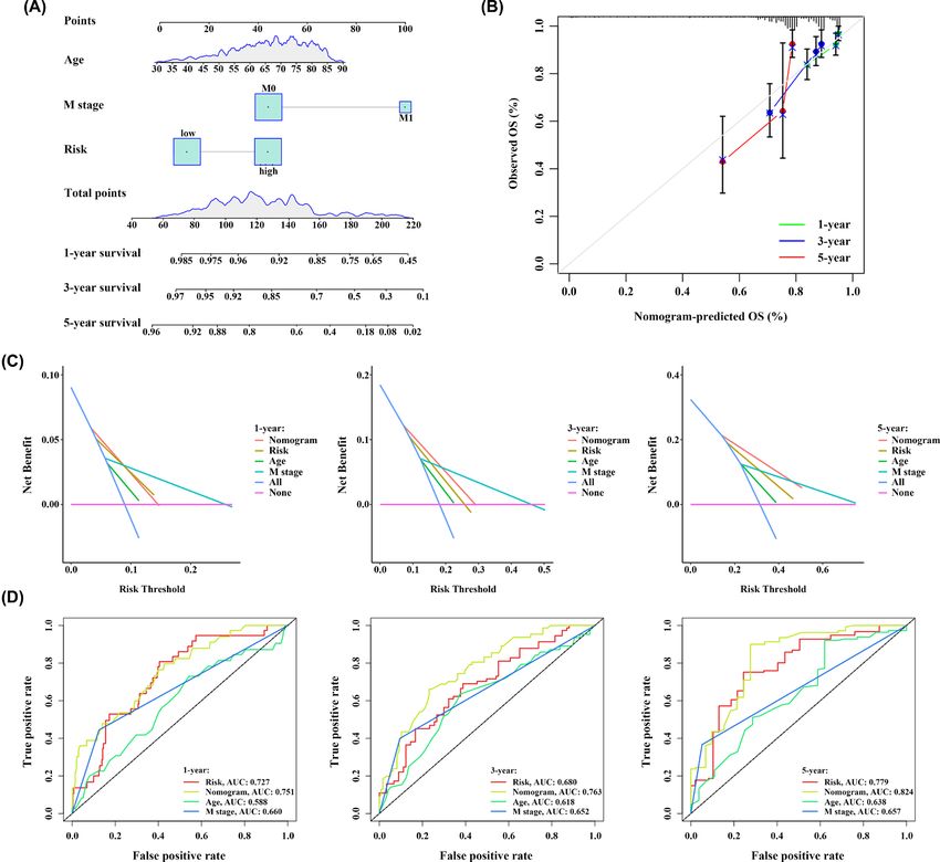
Bioscience Reports (2022) 42 BSR20220078
                                                                                                                                                   https://doi.org/10.1042/BSR20220078

Table 2 List of primers used in the present study
 Gene                                                                                       Primer sequence (5 -3 )

 PCED1B-AS1                                                                                 Forward: TCAAGCCAATCAGCTGACAC
                                                                                            Reverse: AAACAAATGCCCTGCTTGAC
 VPS9D1-AS1                                                                                 Forward: ATGGGTAACCAGGGGTCAAG
                                                                                            Reverse: AGTAACAGTGGTAGAGCCGAC
 PCAT6                                                                                      Forward: ACCCCACTTTCCAGCCTG
                                                                                            Reverse: AGGGAGGCTCACGGACAC
 BOLA3-AS1                                                                                  Forward: ATACCCCTCGTGCTCCTGAT
                                                                                            Reverse: CCGCGTGCTGGACCAT
 ZNF503-AS2                                                                                 Forward: AGGAAACTCACTTCAAAAGCAGC
                                                                                            Reverse: AAAACCGGCACTGAGAGTCC

                                                                                                                                                                                                     Downloaded from http://portlandpress.com/bioscirep/article-pdf/42/7/BSR20220078/935228/bsr-2022-0078.pdf by guest on 09 August 2022
 ZEB1-AS1                                                                                   Forward: TCCCTGCTAAGCTTCCTTCAGTGT
                                                                                            Reverse: GACAGTGATCACTTTCATATCC
 LINC01138                                                                                  Forward: TATTTACGAAAGCTGAAAGCG
                                                                                            Reverse: CTGCATGGGATAGGAGAAAC
 WAC-AS1                                                                                    Forward: GTTCAAGGCAGAAGGCCGTG
                                                                                            Reverse: GGTTCAGCGTTGTTCCCAAG
 COLCA1                                                                                     Forward: ACTCTGATTAGGTCGGGGGA
                                                                                            Reverse: ACCCACTAGCTGCCATGTTC
 GAPDH                                                                                      Forward: TGGTGAAGACGCCAGTGGA
                                                                                            Reverse: GCACCGTAAGGCTGAGAAC

                     0.05 was considered significantly enriched. TIMER2.0 web server (http://timer.cistrome.org), which integrated six
                     state-of-the-art algorithms, including quanTIseq, CIBERSORT, xCell, MCP-counter, TIMER, and EPIC, was used to
                     analyze the composition of tumor-infiltrating immune cells in patients in high- and low-risk groups [21]. The TIDE
                     algorithm was used to evaluate the predictive efficiency of the 9-IRlncRNA signature for the immunotherapy response
                     in CRC [22].

                     Cell culture
                     Human colorectal cancer cell lines (HCT116 and SW480) were purchased from the American Type Culture Collec-
                     tion (Manassas, VA, U.S.A.). Human colon epithelial cell line NCM460 was purchased from the Cell Bank of Type
                     Culture Collection of Chinese Academy of Sciences (Shanghai, China). Dulbecco’s modified Eagle medium (DMEM)
                     containing 10% fetal bovine serum (FBS, Gibco, U.S.A.) and 1% penicillin–streptomycin was used for the cultivation
                     of HCT116 and SW480 cells, and Roswell Park Memorial Institute (RPMI-1640) medium containing 10% FBS and
                     1% penicillin–streptomycin was used for NCM460. All cell lines were allowed to grow in a 37◦ C incubator containing
                     5% CO2 .

                     RNA extraction and quantitative real-time PCR
                     Total RNA was extracted using Cell Total RNA Isolation kit (Foregene, Chengdu, China) following the manufacturer’s
                     protocol, and RNA (1 μg) was reverse-transcribed to cDNA using the PrimeScript RT reagent kit (TaKaRa, Osaka,
                     Japan). Quantitative real-time PCR (qPCR) was conducted using the SYBR Green qPCR Supermixes (Bio-Rad) on
                     the CFX 192 Connect Real-Time PCR system (Bio-Rad, U.S.A.). The qPCR analysis was performed in triplicate with
                     the primers shown in Table 2. The relative expression levels were normalized to GAPDH using the 2−CT method.

                     Statistical analysis
                     Statistical analyses were conducted with R software (version 4.0.1) and GraphPad Prism 5.0 software (San Diego,
                     CA, U.S.A.). Kaplan–Meier curve was employed to reflect the survival difference between high- and low-risk groups,
                     which was assessed by log-rank test. Univariate and multivariate Cox proportional hazard regression models were
                     used to analyze the prognostic significance of 9-IRlncRNA signature. The performance of the 9-IRlncRNA prognostic
                     model was evaluated by area under curve (AUC) value of ROC curve and Harrell’s concordance index (C-index).
                     Two tailed Student’s t test and paired t test were utilized to compare the statistical relevance between two groups.
                     Quantitative data are shown as the mean +  − standard deviation (SD). A two-tailed P-value < 0.05 was regarded as
                     statistically significant.

4             © 2022 The Author(s). This is an open access article published by Portland Press Limited on behalf of the Biochemical Society and distributed under the Creative Commons Attribution
              License 4.0 (CC BY).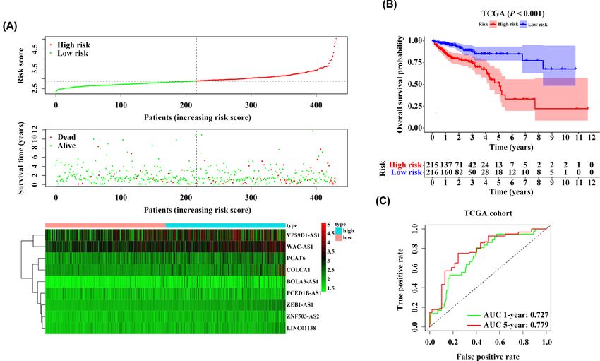
Bioscience Reports (2022) 42 BSR20220078
https://doi.org/10.1042/BSR20220078

Results
Construction of the prognostic IRlncRNAs signature
To make the procedure of our study clearer, a detailed flowchart is illustrated in Figure 1. TCGA dataset was em-
ployed to set as training cohort, and a total of 161 IRlncRNAs were common among all datasets. Then, we conducted
univariate Cox regression analysis to explore the prognosis-related IRlncRNAs and then 18 IRlncRNAs were iden-
tified for subsequent analysis (Figure 2A). Next, LASSO penalized Cox regression was used to establish prognostic
IRlncRNAs signature, and 9 of the 18 IRlncRNAs (PCED1B-AS1, VPS9D1-AS1, PCAT6, BOLA3-AS1, ZNF503-AS2,
ZEB1-AS1, LINC01138, WAC-AS1, and COLCA1) were singled out in training dataset (Figure 2B,C). Risk score of
each patient was calculated according to the expression of 9 IRlncRNAs and their coefficients: risk score = (0.0109 ×
expression of PCED1B-AS1) + (0.0166 × expression of VPS9D1-AS1) + (0.0204 × expression of PCAT6) + (0.0867 ×
expression of BOLA3-AS1) + (0.1806 × expression of ZNF503-AS2) + (0.5488 × expression of ZEB1-AS1) + (0.0448
× expression of LINC01138) + (0.0011 × expression of WAC-AS1) + (0.0587 × expression of COLCA1).

                                                                                                                                                                                           Downloaded from http://portlandpress.com/bioscirep/article-pdf/42/7/BSR20220078/935228/bsr-2022-0078.pdf by guest on 09 August 2022
Analysis of the 9-IRlncRNA signature in the training cohort
Based on the median risk score, we classified the patients with CRC in TCGA cohort into 215 high-risk and 216
low-risk groups. As the risk score increased, both the expression of 9 IRlncRNAs and the mortality of CRC pa-
tients were elevated (Figure 3A). Similarly, Kaplan–Meier curve and log-rank test demonstrated that CRC patients
with high-risk scores showed a worse OS than those with low-risk scores [hazard ratio (HR) = 3.187, 95% confi-
dence interval (CI): 1.993–5.097, P
Bioscience Reports (2022) 42 BSR20220078
                                                                                                                                         https://doi.org/10.1042/BSR20220078

                                                                                                                                                                                           Downloaded from http://portlandpress.com/bioscirep/article-pdf/42/7/BSR20220078/935228/bsr-2022-0078.pdf by guest on 09 August 2022

           Figure 1. Flowchart detailing the development and validation of prognosis-related IRlncRNAs signature

6   © 2022 The Author(s). This is an open access article published by Portland Press Limited on behalf of the Biochemical Society and distributed under the Creative Commons Attribution
    License 4.0 (CC BY).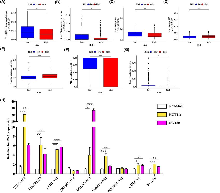
Bioscience Reports (2022) 42 BSR20220078
https://doi.org/10.1042/BSR20220078

                                                                                                                                                                                           Downloaded from http://portlandpress.com/bioscirep/article-pdf/42/7/BSR20220078/935228/bsr-2022-0078.pdf by guest on 09 August 2022

Figure 2. Establishment of prognostic IRlncRNAs signature
(A) Univariate Cox regression analysis identified 18 IRlncRNAs associated with OS. (B, C) A 9-IRlncRNA prognostic model was
constructed by a LASSO regression analysis.

in young (HR = 5.996, 95% CI: 2.448–14.690, P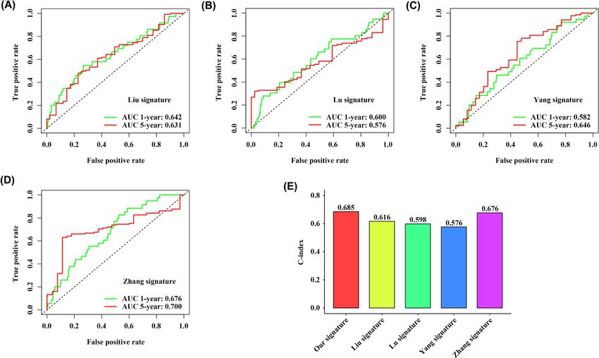
Bioscience Reports (2022) 42 BSR20220078
                                                                                                                                         https://doi.org/10.1042/BSR20220078

                                                                                                                                                                                           Downloaded from http://portlandpress.com/bioscirep/article-pdf/42/7/BSR20220078/935228/bsr-2022-0078.pdf by guest on 09 August 2022
           Figure 3. The 9-IRlncRNA signature predicts prognosis for CRC patients in TCGA training cohort
           (A) The distribution of risk score and survival status of each patient, and the heatmap of the 9 hub lncRNAs expression. (B) Ka-
           plan–Meier survival curve of OS of patients with CRC in high- and low-risk group. (C) Time-dependent ROC curves for predicting
           1- and 5-year OS.

           Obviously, the 9-IRlncRNA signature was superior to the other four models in terms of both the AUC values of 1-
           and 5-year OS prediction and C-index (Figures 3C and 7A–E).

           Construction of predictive nomogram
           To provide a clinical tool to predict the probability of 1-, 3-, and 5-year OS in patients with CRC, three independent
           prognostic factors including age, M stage and the risk score of 9-IRlncRNA signature were employed to construct
           a nomogram (Figure 8A). Their respective point which indicated on the top scale was added up to a total point
           which was corresponding to the 1-, 3-, and 5-year survival rates in the below scale. Calibration plots indicated that
           the nomogram predicted short-term survival (1- and 3-year) better than long-term survival (5-year) (Figure 8B).
           Moreover, DCA curves showed that the nomogram achieved the highest net benefit among the four factors examined
           (age, M stage, risk model, and nomogram) (Figure 8C). Besides, the AUC values of the nomogram at 1-, 3-, and 5-year
           were 0.751, 0.763, and 0.824, respectively, which also displayed the most excellent predictive performance (Figure 8D).

           Analysis of functional enrichment based on 9-IRlncRNA signature
           Compared with all genes and all IRlncRNAs, the 9-IRlncRNA signature could completely distinguish high-risk pa-
           tients from low-risk patients, which indicated good specificity (Figure 9A–C). To further investigate the biological
           process and signaling pathways involved in the 9-IRlncRNA signature, we employed GSEA to explore the pathways
           that were significantly altered between high- and low-risk groups. The results showed that several canonical pathways
           including the mTOR signaling pathway, WNT/β-catenin signaling pathway, Notch signaling pathway, and the TGF-β
           downstream pathway were highly enriched in the high-risk group (Figure 9D–F).

8   © 2022 The Author(s). This is an open access article published by Portland Press Limited on behalf of the Biochemical Society and distributed under the Creative Commons Attribution
    License 4.0 (CC BY).
Bioscience Reports (2022) 42 BSR20220078
https://doi.org/10.1042/BSR20220078
Downloaded from http://portlandpress.com/bioscirep/article-pdf/42/7/BSR20220078/935228/bsr-2022-0078.pdf by guest on 09 August 2022
Figure 4. Validation of the 9-IRlncRNA signature in two testing cohorts
The distribution of risk score and survival status of each patient, and the heatmap of the 9 hub lncRNAs expression in (A) GSE17536
cohort and (B) GSE38832 cohort. (C, D) Kaplan–Meier survival curves of OS and DSS of patients with CRC in high- and low-risk
group in GSE17536 cohort. (E) Kaplan–Meier survival curve of DSS of patients with CRC in high- and low-risk group in GSE38832
cohort. (F) Time-dependent ROC curves for predicting 1- and 5-year OS in GSE17536 cohort. (G) Time-dependent ROC curves for
predicting 1- and 5-year DSS in GSE38832 cohort.
Assessment of immune infiltration and immunotherapy-related markers
with 9-IRlncRNA signature, and detection of 9 IRlncRNAs expression
levels in CRC
We further investigated the differences in immune infiltration between high- and low-risk CRC patients. Based on
quanTIseq and CIBERSORT, the results indicated that the infiltration of CD4+ T cells (non-regulatory), CD4+ mem-
ory activated T cells and M1 macrophages were higher in the low-risk group, and the content of M2 macrophages was
higher in the high-risk group (Figure 10A–D). Moreover, we employed the TIDE algorithm to predict the possibil-
ity of response to immunotherapy. Interestingly, we found that low-risk group had a lower tumor immune exclusion
score than high-risk group, and the microsatellite instability (MSI) score of the low-risk group was significantly higher
© 2022 The Author(s). This is an open access article published by Portland Press Limited on behalf of the Biochemical Society and distributed under the Creative Commons Attribution 9
License 4.0 (CC BY).Bioscience Reports (2022) 42 BSR20220078
https://doi.org/10.1042/BSR20220078
Downloaded from http://portlandpress.com/bioscirep/article-pdf/42/7/BSR20220078/935228/bsr-2022-0078.pdf by guest on 09 August 2022
Figure 5. Cox regression for identifying independent prognostic factors in patients with CRC
(A) Univariate and multivariate analyses of prognostic factors in training cohort. (B) Univariate and multivariate analyses of prognostic
factors in testing cohort (GSE17536).
Figure 6. Stratification analyses
Kaplan–Meier curves showed the OS of the high- and low-risk CRC patients stratified by (A) age, (B) T stage, (C) N stage, (D) M
stage, and (E) AJCC stage.
10 © 2022 The Author(s). This is an open access article published by Portland Press Limited on behalf of the Biochemical Society and distributed under the Creative Commons Attribution
License 4.0 (CC BY).Bioscience Reports (2022) 42 BSR20220078
https://doi.org/10.1042/BSR20220078
Downloaded from http://portlandpress.com/bioscirep/article-pdf/42/7/BSR20220078/935228/bsr-2022-0078.pdf by guest on 09 August 2022
Figure 7. Model comparisons
Comparison of 9-IRlncRNA signature with four previously proposed signatures using (A–D) time-dependent ROC curves for pre-
dicting 1- and 5-year OS and (E) C-index.
than that of the high-risk group (Figure 10E,F). Besides, patients in low-risk group also had a higher TMB (Figure
10G).
Subsequently, we detected the expression levels of these 9 IRlncRNAs in NCM460, HCT116, and SW480 cell lines
using qPCR. The result indicated that the expression levels of WAC-AS1, LINC00138, ZEB1-AS1, BOLA3-AS1,
VPS9D1-AS1, COLCA1, and PCAT6 were significantly up-regulated in CRC cell lines (HCT116 and SW480),
whereas the expression level of ZNF503-AS2 was lowly expressed in CRC cell lines (Figure 10H).
Discussion
Recently, lncRNAs have been proved to play critical roles in the development and progression of many cancers
[23]. In CRC, emerging evidence has indicated that lncRNAs act mostly as signaling molecules in many significant
CRC-related pathways, and are frequently involved in different phases of CRC from precancerous lesions to distant
metastasis [24]. A previous study has found that lncRNAs can also regulate cancer immunity, and concluded that
these IRlncRNAs are a new but essential part of cancer immunotherapy and prognosis [25]. In the present study,
we constructed a 9-IRlncRNAs signature that can successfully divide CRC patients into high- and low-risk groups.
Meanwhile, through multivariate Cox regression analysis, the 9-IRlncRNA signature was proved to be an indepen-
dent OS prognostic factor. In subsequent subgroup analysis, this prognostic signature also showed favorable stability
in different subgroups. Besides, we noted that patients in high-risk group were involved in cancer-related signaling
pathways such as MAPK, WNT/β-catenin, and Notch pathway, which might result in their short OS.
Through our analysis, all the nine IRlncRNAs (PCED1B-AS1, VPS9D1-AS1, PCAT6, BOLA3-AS1, ZNF503-AS2,
ZEB1-AS1, LINC01138, WAC-AS1, and COLCA1) were associated with a dismal prognosis, which was consistent
with their biological functions reported by previous studies. LncRNA PCED1B-AS1 was reported to promote the
proliferation and inhibit the apoptosis of glioma cells via miR-194-5p/PCED1B axis [26]. However, in the present
study, we found that there was no significant difference in the expression level of PCED1B-AS1 between CRC cell
lines and normal colon epithelial cell lines. In non-small cell lung cancer (NSCLC), the expression of VPS9D1-AS1
was higher in NSCLC tissues than in paired adjacent tissues, and the high expression of VPS9D1-AS1 suggested an
adverse prognosis [27]. Similarly, PCAT6 can bind with EZH2 which can bind to the promoter region of LATS2 and
© 2022 The Author(s). This is an open access article published by Portland Press Limited on behalf of the Biochemical Society and distributed under the Creative Commons Attribution 11
License 4.0 (CC BY).Bioscience Reports (2022) 42 BSR20220078
https://doi.org/10.1042/BSR20220078
Downloaded from http://portlandpress.com/bioscirep/article-pdf/42/7/BSR20220078/935228/bsr-2022-0078.pdf by guest on 09 August 2022
Figure 8. Nomogram establishment and evaluation
(A) Nomogram for predicting survival probability at 1-, 3-, and 5-year for CRC patients. (B) Calibration curves for the nomogram.
(C) DCA curves showing the comparison between the nomogram and age, M stage or risk group for predicting 1-, 3-, and 5-year
OS for CRC patients. (D) Time-dependent ROC curves showing the comparison between the nomogram and age, M stage or risk
group for predicting 1-, 3-, and 5-year OS for CRC patients.
inhibit LATS2 expression in NSCLC. LATS2 overexpression can suppress cell proliferation and promote apoptosis.
Therefore, lncRNA PCAT6 exerts an oncogenic function on NSCLC [28]. LncRNA ZEB1-AS1 is a well-recognized
tumor-related lncRNAs and is overexpressed in several malignancies. In CRC, ZEB1-AS1 overexpression is signifi-
cantly related to tumor invasion and distant metastasis, which indicates a poor OS and low recurrence-free survival
rate [29]. Moreover, LINC01138 has been reported as a tumor promoter that can exert its biological functions via the
tumor-related IGF2BP1/IGF2BP3-LINC01138-PRMT5 axis in hepatocellular carcinoma (HCC), which can serve as a
robust biomarker and therapeutic target for HCC [30]. Recent study has reported that lncRNA WAC-AS1 is highly ex-
pressed in liver cancer tissues and cell lines, and verified that WAC-AS1 can regulate ARPP19 by sponging miR-320d
to promote glycolysis and tumor proliferation [31]. Although the function of other lncRNAs remains unknown in
carcinoma, their expression levels are up-regulated in CRC cell lines according to our findings, which may provide
the foundation for the further exploration of the association between these IRlncRNAs and tumorigenesis. More-
over, the identification of additional targets of these nine IRlncRNAs is also a critical step to further explore their
12 © 2022 The Author(s). This is an open access article published by Portland Press Limited on behalf of the Biochemical Society and distributed under the Creative Commons Attribution
License 4.0 (CC BY).Bioscience Reports (2022) 42 BSR20220078
https://doi.org/10.1042/BSR20220078
Downloaded from http://portlandpress.com/bioscirep/article-pdf/42/7/BSR20220078/935228/bsr-2022-0078.pdf by guest on 09 August 2022
Figure 9. PCA and functional enrichment analysis
(A) PCA between high- and low-risk groups based on the whole genes. (B) PCA between high- and low-risk groups based on the
whole immune-related lncRNAs. (C) PCA between high- and low-risk groups based on the 9-IRlncRNA signature. GSEA based on
TCGA cohort to explore the underlying mechanism of the 9-IRlncRNA signature, including gene set of (D) KEGG, (E) PID, and (F)
REACTOME. Normalized P-value < 0.05.
function. With the development of RNA-centric approaches, such as isolation of chromatin by RNA purification [32]
and captured hybridization analysis of RNA targets [33], we will be able to identify the potential interaction targets
of lncRNAs in a native context, which may deepen our understanding of lncRNA-mediated regulation of immune
pathways and improve our insight in lncRNA functions.
Tumor-infiltrating immune cells are reported to be associated with tumor prognosis and have the ability to guide
therapeutics [34]. In our study, patients in low-risk group had more infiltration of CD4+ T cells (non-regulatory),
CD4+ memory activated T cells, and M1 macrophages. CD4+CD25+ regulatory T cells are proved to suppress an-
titumor response and result in tumor immune escape, while non-regulatory CD4+ helper T cells may be beneficial
to the host defense against tumor [35]. CD4+ memory T cells were located in the secondary lymphoid node or-
gans and tissues. When re-exposure to tumor antigen, CD4+ memory T cells undergo fast expansion and induce
more effective and faster immune response against tumor antigen and may prevent tumor relapse [36,37]. Similar-
ity, M1 macrophages were reported to lead to the promotion of inflammation and tumor suppression [38]. There-
fore, the favorable prognosis of patients in low-risk group may be a result of the activation of various anti-tumor
immune cells, and the accumulation of these immune cells may allow patients to benefit from immunotherapy. In
addition to immune cell infiltration, we also found that patients in low-risk group had higher MSI and TMB scores.
In CRC, immune checkpoint therapy received regulatory approval in 2017 to treat heavily mutated tumors that are
mismatch-repair-deficient or harbor high levels of MSI [12]. Similarity, tumors with a higher TMB have a higher like-
lihood of immunotherapy response [39]. Therefore, these results further confirm that patients in low-risk group may
show a favorable response to immunotherapy, and the 9-IRlncRNA signature we constructed may serve as a novel
biomarker for the immunotherapy of CRC patients.
Previous studies have also constructed various lncRNA signatures to predict the prognosis of patients with CRC, but
the 9-IRlncRNA we established shows more excellent performance via higher C-index and AUC values of OS [40–43].
Nevertheless, several limitations of this study should be addressed. First, the prognostic model is constructed using
retrospective data, thus, the results should be further validated using prospective data or clinical trials. Second, due to
the sample size was not large enough in the validation datasets, the accuracy of the prognostic model has decreased.
Conclusion
In conclusion, we constructed a 9-IRlncRNA prognostic model to predict the prognosis of CRC patients based on
the TCGA dataset. The prognostic value of this model was further validated in two external cohorts from the GEO
database. Moreover, the signature was identified as an independent prognostic factor of CRC and was involved in
© 2022 The Author(s). This is an open access article published by Portland Press Limited on behalf of the Biochemical Society and distributed under the Creative Commons 13
Attribution License 4.0 (CC BY).Bioscience Reports (2022) 42 BSR20220078
https://doi.org/10.1042/BSR20220078
Downloaded from http://portlandpress.com/bioscirep/article-pdf/42/7/BSR20220078/935228/bsr-2022-0078.pdf by guest on 09 August 2022
Figure 10. Association of the 9-IRlncRNA signature with tumor-infiltrating immune cells and its ability to predict im-
munotherapy response
(A–D) The infiltration of four immune cells with significant differences in high- and low-risk groups. (E–G) Association of 9-IRlncRNA
signature with several immunotherapy-related markers. (H) The qPCR analysis of expression levels of 9 IRlncRNAs in NCM460,
HCT116 and SW480 cell lines; * PBioscience Reports (2022) 42 BSR20220078
https://doi.org/10.1042/BSR20220078
Funding
This study was supported by grants from the National Natural Science Foundation of China [grant number 32000533]; Sichuan
Science and Technology Program [grant number 2021YFS0215]; and Post-doctor Research Project, West China Hospital, Sichuan
University [grant number 2020HXBH113].
CRediT Author Contribution
Gongmin Zhu: Conceptualization, Data curation, Formal analysis, Supervision, Validation, Visualization, Methodology,
Writing—original draft, Writing—review & editing. Lijiao Pei: Data curation, Formal analysis, Validation, Writing—original draft,
Writing—review & editing. Fan Yang: Data curation, Formal analysis, Writing—review & editing. Chenliang Zhang: Conceptualiza-
tion, Data curation, Funding acquisition, Supervision, Writing—review & editing.
Downloaded from http://portlandpress.com/bioscirep/article-pdf/42/7/BSR20220078/935228/bsr-2022-0078.pdf by guest on 09 August 2022
Abbreviations
AJCC, American Joint Committee on Cancer; AUC, area under curve; C-index, Harrell’s concordance index; CI, confidence in-
terval; CRC, colorectal cancer; DCA, decision curve analysis; DSS, disease-specific survival; GEO, Gene Expression Omnibus;
GSEA, gene set enrichment analysis; HR, hazard ratio; IRG, immune-related gene; IRlncRNA, immune-related long noncod-
ing RNA; LASSO, the least absolute shrinkage and selection operator; MSI, microsatellite instability; OS, overall survival; PCA,
principal component analysis; qPCR, quantitative real-time PCR; ROC, receiver operating characteristic; TCGA, The Cancer
Genome Atlas; TIDE, tumor immune dysfunction and exclusion; TMB, tumor mutation burden; Tregs, regulatory T cells.
References
1 Bray, F., Ferlay, J., Soerjomataram, I., Siegel, R.L., Torre, L.A. and Jemal, A. (2018) Global cancer statistics 2018: GLOBOCAN estimates of incidence
and mortality worldwide for 36 cancers in 185 countries. CA Cancer J. Clin. 68, 394–424, https://doi.org/10.3322/caac.21492
2 Fearnhead, N.S., Wilding, J.L. and Bodmer, W.F. (2002) Genetics of colorectal cancer: hereditary aspects and overview of colorectal tumorigenesis. Br.
Med. Bull. 64, 27–43, https://doi.org/10.1093/bmb/64.1.27
3 Guinney, J., Dienstmann, R., Wang, X., de Reyniès, A., Schlicker, A., Soneson, C. et al. (2015) The consensus molecular subtypes of colorectal cancer.
Nat. Med. 21, 1350–1356, https://doi.org/10.1038/nm.3967
4 Koncina, E., Haan, S., Rauh, S. and Letellier, E. (2020) Prognostic and predictive molecular biomarkers for colorectal cancer: updates and challenges.
Cancers 12, 1–25, https://doi.org/10.3390/cancers12020319
5 Lee, J., Franovic, A., Shiotsu, Y., Kim, S.T., Kim, K.M., Banks, K.C. et al. (2019) Detection of ERBB2 (HER2) gene amplification events in cell-free DNA
and response to anti-HER2 agents in a large asian cancer patient cohort. Front. Oncol. 9, 212, https://doi.org/10.3389/fonc.2019.00212
6 Kim, T. and Croce, C.M. (2018) Long noncoding RNAs: Undeciphered cellular codes encrypting keys of colorectal cancer pathogenesis. Cancer Lett.
417, 89–95, https://doi.org/10.1016/j.canlet.2017.12.033
7 Chi, Y., Wang, D., Wang, J., Yu, W. and Yang, J. (2019) Long non-coding RNA in the pathogenesis of cancers. Cells 8, 1–44,
https://doi.org/10.3390/cells8091015
8 Liu, M., Zhang, H., Li, Y., Wang, R., Li, Y., Zhang, H. et al. (2018) HOTAIR, a long noncoding RNA, is a marker of abnormal cell cycle regulation in lung
cancer. Cancer Sci. 109, 2717–2733, https://doi.org/10.1111/cas.13745
9 Zhou, Q., Guo, J., Huang, W., Yu, X., Xu, C. and Long, X. (2020) Linc-ROR promotes the progression of breast cancer and decreases the sensitivity to
rapamycin through miR-194-3p targeting MECP2. Mol. Oncol. 14, 2231–2250, https://doi.org/10.1002/1878-0261.12700
10 Wang, X., Sun, W., Shen, W., Xia, M., Chen, C., Xiang, D. et al. (2016) Long non-coding RNA DILC regulates liver cancer stem cells via IL-6/STAT3 axis.
J. Hepatol. 64, 1283–1294, https://doi.org/10.1016/j.jhep.2016.01.019
11 Yang, X., Zhang, S., He, C., Xue, P., Zhang, L., He, Z. et al. (2020) METTL14 suppresses proliferation and metastasis of colorectal cancer by
down-regulating oncogenic long non-coding RNA XIST. Mol. Cancer 19, 46, https://doi.org/10.1186/s12943-020-1146-4
12 Ganesh, K., Stadler, Z.K., Cercek, A., Mendelsohn, R.B., Shia, J., Segal, N.H. et al. (2019) Immunotherapy in colorectal cancer: rationale, challenges and
potential. Nat. Rev. Gastroenterol. Hepatol. 16, 361–375
13 Hahn, A.W., Drake, C., Denmeade, S.R., Zakharia, Y., Maughan, B.L., Kennedy, E. et al. (2020) A Phase I Study of
Alpha-1,3-galactosyltransferase-expressing allogeneic renal cell carcinoma immunotherapy in patients with refractory metastatic renal cell carcinoma.
Oncologist 25, e121–e213, https://doi.org/10.1634/theoncologist.2019-0599
14 Lee, J.H., Lee, J.H., Lim, Y.S., Yeon, J.E., Song, T.J., Yu, S.J. et al. (2015) Adjuvant immunotherapy with auto9ogous cytokine-induced killer cells for
hepatocellular carcinoma. Gastroenterology 148, 1383.e1386–1391.e1386, https://doi.org/10.1053/j.gastro.2015.02.055
15 Yu, W.D., Wang, H., He, Q.F., Xu, Y. and Wang, X.C. (2018) Long noncoding RNAs in cancer-immunity cycle. J. Cell. Physiol. 233, 6518–6523,
https://doi.org/10.1002/jcp.26568
16 Pei, X., Wang, X. and Li, H. (2018) LncRNA SNHG1 regulates the differentiation of Treg cells and affects the immune escape of breast cancer via
regulating miR-448/IDO. Int. J. Biol. Macromol. 118, 24–30, https://doi.org/10.1016/j.ijbiomac.2018.06.033
17 Xu, J., Meng, Q., Li, X., Yang, H., Xu, J., Gao, N. et al. (2019) Long noncoding RNA MIR17HG promotes colorectal cancer progression via miR-17-5p.
Cancer Res. 79, 4882–4895, https://doi.org/10.1158/0008-5472.CAN-18-3880
18 Bhattacharya, S., Andorf, S., Gomes, L., Dunn, P., Schaefer, H., Pontius, J. et al. (2014) ImmPort: disseminating data to the public for the future of
immunology. Immunol. Res. 58, 234–239, https://doi.org/10.1007/s12026-014-8516-1
© 2022 The Author(s). This is an open access article published by Portland Press Limited on behalf of the Biochemical Society and distributed under the Creative Commons 15
Attribution License 4.0 (CC BY).Bioscience Reports (2022) 42 BSR20220078
https://doi.org/10.1042/BSR20220078
19 Chalmers, Z.R., Connelly, C.F., Fabrizio, D., Gay, L., Ali, S.M., Ennis, R. et al. (2017) Analysis of 100,000 human cancer genomes reveals the landscape
of tumor mutational burden. Genome Med. 9, 34, https://doi.org/10.1186/s13073-017-0424-2
20 Subramanian, A., Tamayo, P., Mootha, V.K., Mukherjee, S., Ebert, B.L., Gillette, M.A. et al. (2005) Gene set enrichment analysis: a knowledge-based
approach for interpreting genome-wide expression profiles. PNAS 102, 15545–15550, https://doi.org/10.1073/pnas.0506580102
21 Li, T., Fu, J., Zeng, Z., Cohen, D., Li, J., Chen, Q. et al. (2020) TIMER2.0 for analysis of tumor-infiltrating immune cells. Nucleic Acids Res. 48,
W509–W514, https://doi.org/10.1093/nar/gkaa407
22 Jiang, P., Gu, S., Pan, D., Fu, J., Sahu, A., Hu, X. et al. (2018) Signatures of T cell dysfunction and exclusion predict cancer immunotherapy response.
Nat. Med. 24, 1550–1558, https://doi.org/10.1038/s41591-018-0136-1
23 Liz, J. and Esteller, M. (2016) lncRNAs and microRNAs with a role in cancer development. Biochim. Biophys. Acta 1859, 169–176,
https://doi.org/10.1016/j.bbagrm.2015.06.015
24 Wang, L., Cho, K.B., Li, Y., Tao, G., Xie, Z. and Guo, B. (2019) Long noncoding RNA (lncRNA)-mediated competing endogenous RNA networks provide
novel potential biomarkers and therapeutic targets for colorectal cancer. Int. J. Mol. Sci. 20, 1–26, https://doi.org/10.3390/ijms20225758
25 Denaro, N., Merlano, M.C. and Lo Nigro, C. (2019) Long noncoding RNAs as regulators of cancer immunity. Mol. Oncol. 13, 61–73,
Downloaded from http://portlandpress.com/bioscirep/article-pdf/42/7/BSR20220078/935228/bsr-2022-0078.pdf by guest on 09 August 2022
https://doi.org/10.1002/1878-0261.12413
26 Yang, J., Yu, D., Liu, X., Changyong, E. and Yu, S. (2020) LncRNA PCED1B-AS1 activates the proliferation and restricts the apoptosis of glioma through
cooperating with miR-194-5p/PCED1B axis. J. Cell. Biochem. 121, 1823–1833, https://doi.org/10.1002/jcb.29417
27 Tan, J. and Yang, L. (2018) Long noncoding RNA VPS9D1-AS1 overexpression predicts a poor prognosis in non-small cell lung cancer. Biomed.
Pharmacother. = Biomed. Pharmacotherapie 106, 1600–1606, https://doi.org/10.1016/j.biopha.2018.07.113
28 Shi, X., Liu, Z., Liu, Z., Feng, X., Hua, F., Hu, X. et al. (2018) Long noncoding RNA PCAT6 functions as an oncogene by binding to EZH2 and suppressing
LATS2 in non-small-cell lung cancer. EBioMedicine 37, 177–187, https://doi.org/10.1016/j.ebiom.2018.10.004
29 Li, J., Li, Z., Leng, K., Xu, Y., Ji, D., Huang, L. et al. (2018) ZEB1-AS1: a crucial cancer-related long non-coding RNA. Cell Prolif. 51, 1–8,
https://doi.org/10.1111/cpr.12423
30 Li, Z., Zhang, J., Liu, X., Li, S., Wang, Q., Di, C. et al. (2018) The LINC01138 drives malignancies via activating arginine methyltransferase 5 in
hepatocellular carcinoma. Nat. Commun. 9, 1572, https://doi.org/10.1038/s41467-018-04006-0
31 Xia, X., Zhang, H., Xia, P., Zhu, Y., Liu, J., Xu, K. et al. (2021) Identification of glycolysis-related lncRNAs and the novel lncRNA WAC-AS1 promotes
glycolysis and tumor progression in hepatocellular carcinoma. Front. Oncol. 11, 733595, https://doi.org/10.3389/fonc.2021.733595
32 Chu, C., Zhang, Q.C., da Rocha, S.T., Flynn, R.A., Bharadwaj, M., Calabrese, J.M. et al. (2015) Systematic discovery of Xist RNA binding proteins. Cell
161, 404–416, https://doi.org/10.1016/j.cell.2015.03.025
33 Simon, M.D., Pinter, S.F., Fang, R., Sarma, K., Rutenberg-Schoenberg, M., Bowman, S.K. et al. (2013) High-resolution Xist binding maps reveal
two-step spreading during X-chromosome inactivation. Nature 504, 465–469, https://doi.org/10.1038/nature12719
34 Fridman, W.H., Pagès, F., Sautès-Fridman, C. and Galon, J. (2012) The immune contexture in human tumours: impact on clinical outcome. Nat. Rev.
Cancer 12, 298–306
35 Yu, P. and Fu, Y.X. (2006) Tumor-infiltrating T lymphocytes: friends or foes? . Lab. Invest. J. Tech. Methods Pathol. 86, 231–245,
https://doi.org/10.1038/labinvest.3700389
36 Golubovskaya, V. and Wu, L. (2016) Different subsets of T Cells, memory, effector functions, and CAR-T immunotherapy. Cancers 8, 1–12,
https://doi.org/10.3390/cancers8030036
37 Vahidi, Y., Faghih, Z., Talei, A.R., Doroudchi, M. and Ghaderi, A. (2018) Memory CD4(+) T cell subsets in tumor draining lymph nodes of breast cancer
patients: A focus on T stem cell memory cells. Cell. Oncol. (Dordrecht) 41, 1–11, https://doi.org/10.1007/s13402-017-0352-6
38 Lee, H.W., Choi, H.J., Ha, S.J., Lee, K.T. and Kwon, Y.G. (2013) Recruitment of monocytes/macrophages in different tumor microenvironments. Biochim.
Biophys. Acta 1835, 170–179, https://doi.org/10.1016/j.bbcan.2012.12.007
39 Goodman, A.M., Kato, S., Bazhenova, L., Patel, S.P., Frampton, G.M., Miller, V. et al. (2017) Tumor mutational burden as an independent predictor of
response to immunotherapy in diverse cancers. Mol. Cancer Ther. 16, 2598–2608, https://doi.org/10.1158/1535-7163.MCT-17-0386
40 Liu, S., Cao, Q., An, G., Yan, B. and Lei, L. (2020) Identification of the 3-lncRNA signature as a prognostic biomarker for colorectal cancer. Int. J. Mol.
Sci. 21, 1–17, https://doi.org/10.3390/ijms21249359
41 Lu, Y., Wang, W., Liu, Z., Ma, J., Zhou, X. and Fu, W. (2021) Long non-coding RNA profile study identifies a metabolism-related signature for colorectal
cancer. Mol. Med. (Cambridge, Mass.) 27, 83, https://doi.org/10.1186/s10020-021-00343-x
42 Yang, Z.D. and Kang, H. (2020) Exploring prognostic potential of long noncoding RNAs in colorectal cancer based on a competing endogenous RNA
network. World J. Gastroenterol. 26, 1298–1316, https://doi.org/10.3748/wjg.v26.i12.1298
43 Zhang, W., Fang, D., Li, S., Bao, X., Jiang, L. and Sun, X. (2021) Construction and validation of a novel ferroptosis-related lncRNA signature to predict
prognosis in colorectal cancer patients. Front. Genet. 12, 709329, https://doi.org/10.3389/fgene.2021.709329
16 © 2022 The Author(s). This is an open access article published by Portland Press Limited on behalf of the Biochemical Society and distributed under the Creative Commons Attribution
License 4.0 (CC BY).You can also read