Engineered Cardiac Tissues Generated in the Biowire II: A Platform for Human-Based Drug Discovery
←
→
Page content transcription
If your browser does not render page correctly, please read the page content below
TOXICOLOGICAL SCIENCES, 2019, 1–9

                                                                                                     doi: 10.1093/toxsci/kfz168
                                                                                                     Advance Access Publication Date: August 6, 2019
                                                                                                     Research Article

                                                                                                                                                           Downloaded from https://academic.oup.com/toxsci/advance-article-abstract/doi/10.1093/toxsci/kfz168/5543113 by guest on 11 September 2019
Engineered Cardiac Tissues Generated in the Biowire II:
A Platform for Human-Based Drug Discovery
Nicole T. Feric,* Isabella Pallotta,* Rishabh Singh,* Danielle R. Bogdanowicz,*
Marietta M. Gustilo,* Khuram W. Chaudhary,† Robert N. Willette,†
Tim P. Chendrimada,† Xiaoping Xu,† Michael P. Graziano,* and
Roozbeh Aschar-Sobbi*,1
                                                                                                                                 †
*TARA Biosystems Inc, Alexandria Center for Life Sciences, New York, New York 10016; and GlaxoSmithKline,
Collegeville, Pennsylvania 19426
1
 To whom correspondence should be addressed at TARA Biosystems Inc, Alexandria Center for Life Sciences, 430 E 29th Street, Suite 1015, New York, NY
10016. Tel: 646-440-9160; E-mail: rooz@tarabiosystems.com.

ABSTRACT
Recent advances in techniques to differentiate human induced pluripotent stem cells (hiPSCs) hold the promise of an
unlimited supply of human derived cardiac cells from both healthy and disease populations. That promise has been tempered
by the observation that hiPSC-derived cardiomyocytes (hiPSC-CMs) typically retain a fetal-like phenotype, raising concern
about the translatability of the in vitro data obtained to drug safety, discovery, and development studies. The Biowire II
platform was used to generate 3D engineered cardiac tissues (ECTs) from hiPSC-CMs and cardiac fibroblasts. Long term
electrical stimulation was employed to obtain ECTs that possess a phenotype like that of adult human myocardium including
a lack of spontaneous beating, the presence of a positive force-frequency response from 1 to 4 Hz and prominent postrest
potentiation. Pharmacology studies were performed in the ECTs to confirm the presence and functionality of pathways that
modulate cardiac contractility in humans. Canonical responses were observed for compounds that act via the b-adrenergic/
cAMP-mediated pathway, eg, isoproterenol and milrinone; the L-type calcium channel, eg, FPL64176 and nifedipine; and
indirectly effect intracellular Ca2þ concentrations, eg, digoxin. Expected positive inotropic responses were observed for
compounds that modulate proteins of the cardiac sarcomere, eg, omecamtiv mecarbil and levosimendan. ECTs generated in
the Biowire II platform display adult-like properties and have canonical responses to cardiotherapeutic and cardiotoxic agents
that affect contractility in humans via a variety of mechanisms. These data demonstrate that this human-based model can be
used to assess the effects of novel compounds on contractility early in the drug discovery and development process.

Key words: cardiomyocytes; contractility; engineered cardiac tissue; drug discovery; drug safety; in vitro models.

Cardiovascular disease (CVD) remains a leading cause of mor-                    et al., 2014; Onakpoya et al., 2016; Siramshetty et al., 2016). There
bidity and mortality worldwide (Benjamin et al., 2018). Patients                remains the need for both novel therapies to prevent and treat
with heart failure (HF) comprise a growing percentage of the                    HF as well as improved ways to assess the cardiac safety liabili-
CVD population (Benjamin et al., 2018). HF is instigated by both                ties of candidate drug therapies.
genetic, eg, inherited cardiomyopathy and acquired risk factors                     Cardiac contractility is a consequence of a precise series of
eg, coronary heart disease or hypertension (Stienen, 2015).                     events known as excitation-contraction coupling (Eisner et al.,
Cardiotoxicity and HF can also be an unwanted consequence of                    2017). This highly orchestrated process links electrical excita-
treatment with a range of drugs in clinical usage (McNaughton                   tion of the surface membrane (the action potential) and changes

C The Author(s) 2019. Published by Oxford University Press on behalf of the Society of Toxicology.
V
This is an Open Access article distributed under the terms of the Creative Commons Attribution Non-Commercial License (http://creativecommons.org/
licenses/by-nc/4.0/), which permits non-commercial re-use, distribution, and reproduction in any medium, provided the original work is properly cited.
For commercial re-use, please contact journals.permissions@oup.com

                                                                                                                                                       1
2   | FERIC ET AL.

in the cytoplasmic calcium concentration ([Ca2þ]i) to muscle            illuminated using 350 nm excitation. Videos were acquired at a
contraction and relaxation (Eisner et al., 2017). Aberrant contrac-     rate of 100 frames per second using a Zyla 4.2 sCMOS camera
tility is a hallmark of HF and of numerous drugs with cardio-           (Andor, South Windsor, Connecticut) with a 470 nm emission filter
toxic effects and contributes to symptoms including dyspnea,            using NIS-Elements Advanced Research software (Nikon
fatigue, arrhythmia, and ischemia (Guth et al., 2015).                  Instruments Inc, Edgewood, New York). To measure contractile
Historically, contractility measurements have been performed            force, the videos were analyzed using custom software to track
using cardiac tissue isolated from patients or animals. The use         the location of the POMaC wire (Figure 1A). The pixel movements
of isolated tissues poses significant challenges, including lim-        were converted to force as previously described (Zhao et al., 2019).

                                                                                                                                                  Downloaded from https://academic.oup.com/toxsci/advance-article-abstract/doi/10.1093/toxsci/kfz168/5543113 by guest on 11 September 2019
ited availability and variability for the former, and species-to-           The force-frequency relationship (FFR) was determined by
species translation for the latter. These challenges have re-           pacing ECTs at 1 Hz and increasing the stimulation frequency by
stricted the utility of these methods and highlight the need for        1 Hz every 30 s to a final frequency of 4 Hz. Following 4 Hz stimu-
novel models. For in vitro models to faithfully recapitulate this       lation, the postrest potentiation was determined by turning off
process they should demonstrate functional hallmarks of the             the external field stimulation for 10 s and recording the twitch
electrical, calcium handling, and contractile machinery.                transients after resuming field stimulation at 1 Hz. To deter-
     The recent development of robust cardiac differentiation           mine the cross-sectional area of the tissue, ECTs were fixed in
protocols for human induced pluripotent stem cells (hiPSCs)             10% neutral buffered formalin for 2 h followed by 3 washes in
has provided the potential for an unlimited supply of human             phosphate buffered saline. Tissue was cut halfway between the
derived cardiac cells from both healthy and diseased sources,           midsection of the tissue and the POMaC wire (red dotted line in
but this potential has been limited by the observation that             Figure 1A). A photo of the transverse section was acquired, and
hiPSC-derived cardiomyocytes (hiPSC-CMs) typically retain a             the cross-sectional area was determined using ImageJ. Average
more fetal-like phenotype (Denning et al., 2016; Veerman et al.,        tissue cross-sectional area was 0.066 6 0.001 mm2 (n ¼ 6 ECTs).
2015). This raises concerns about the predictability and translat-
ability of results obtained in vitro to the settings of drug safety,    Compound testing. At the end of the electrical stimulation proto-
discovery, and development.                                             col, ECTs were assessed for automaticity (ie, spontaneous beat
     In this study, the Biowire II platform was used to generate        rate), the FFR (active force at 1 Hz through 4 Hz), and postrest poten-
3D engineered cardiac tissues (ECTs) from hiPSC-CMs and car-            tiation. ECTs without spontaneous activity that exhibited a positive
diac fibroblasts and to make nondestructive contractility meas-         FFR and postrest potentiation were used for compound testing.
urements of the ECTs. We present data demonstrating that                    Tests were conducted in a 37 C, 5% CO2 environmental cham-
ECTs produced using this platform and subjected to long term            ber under field stimulation at 1 Hz. Tissues were incubated for
electrical stimulation have adult-like contractile properties. In ad-   30 min in the environmental chamber after which a baseline con-
dition, we show that contractility in the ECTs can be modulated         tractility video was acquired for 30 s. One-third of the tissue culture
by a variety of agents known to effect essential intracellular sig-     medium volume (6 ml total) was pipetted from the well containing
naling pathways present in the human myocardium. These data             the ECTs twice to equilibrate the ECTs to the shear stress induced
demonstrate the utility of the platform as a tool for discovery of      by the procedure. A video of 30 s duration was acquired 10 min af-
novel therapies and testing of drug safety in preclinical develop-      ter the pipetting (baseline). The test article was added to the well
ment, using a human model of myocardial tissue.                         to provide the desired final concentration and one-third of the me-
                                                                        dia volume was pipetted twice to gently mix. The test article stock
                                                                        solutions were prepared in either dimethyl sulfoxide or water as
MATERIALS AND METHODS                                                   appropriate, and serially diluted in medium. Following 10–30 min
Generation of ECTs. ECTs were generated in the Biowire II platform      of incubation a video of 30 s duration was acquired. The same pro-
as described previously (Zhao et al., 2019). Briefly, 3D ECTs were      cedure was followed for all subsequent doses (lowest to highest)
formed in polystyrene microwells containing parallel poly(octa-         such that 1 ECT was incubated with all doses. Contractility videos
methylene maleate (anhydride) citrate) (POMaC) wires. Each ECT          were analyzed using a custom analysis software. The force at each
was comprised of 100 000 viable iCell Cardiomyocytes2 (Cellular         dose was normalized by dividing the force values in the presence
Dynamics International, Madison, Wisconsin) and 10 000 human            of compound by the baseline force values of the same tissue.
ventricular cardiac fibroblasts (Lonza, Allendale, New Jersey) in a
collagen/Matrigel/fibrin gel. Following 7 days in culture, the hiPSC-   Statistics. Statistical analyses were performed using GraphPad
CMs and cardiac fibroblasts self-organized into 3D ECTs and were        Prism (GraphPad Software, Inc, La Jolla, California). Data are pre-
suspended between the POMaC wires (Figure 1A). Custom cham-             sented as mean 6 SEM. Results were considered statistically sig-
bers containing parallel carbon electrodes were used to provide         nificant (#) if p < .05 using one-way ANOVA followed by a
electrical field stimulation using biphasic pulses of 2 ms duration,    Tukey’s post hoc multiple comparison test or where appropriate
at twice the excitation threshold. Stimulation was started at 1 Hz      repeat measure one-way ANOVA followed by Dunnett’s post
and increased by 0.1 Hz increments daily to a maximum of 6 Hz.          hoc multiple comparison test. The replicate numbers are indi-
All ECTs used in this study were subjected to the same stimulation      cated in the figure captions. For EC50/IC50 calculation, the non-
protocol for a minimum of 6 weeks. Compound effects on contrac-         linear fit function of GraphPad Prism (Sigmoidal with 3
tility were similar in tissues stimulated from 6 to 9 weeks.            parameters) was used to find the best fit for the data.

Image-based contractility measurements. ECT contractility was mea-
sured by tracking the deflection of the POMaC wires as a function       RESULTS
of time as previously described (Zhao et al., 2019). Stimulated ECTs
were placed in a custom chamber containing parallel carbon elec-        Cardiac ECTs Generated via the Biowire II Platform Have Adult-Like
trodes to provide external field stimulation in an environmental        Contractile Properties
chamber at 37 C and 5% CO2. ECTs were stimulated at twice the          The functionality of the contractile machinery was assessed by
excitation threshold during acquisition. The POMaC wires were           measuring the frequency-dependent regulation of contractility,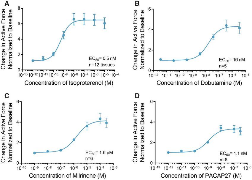
ENGINEERED CARDIAC TISSUES GENERATED IN THE BIOWIRE II PLATFORM                                       |   3

                                                                                                                                                                             Downloaded from https://academic.oup.com/toxsci/advance-article-abstract/doi/10.1093/toxsci/kfz168/5543113 by guest on 11 September 2019
Figure 1. Engineered cardiac tissues (ECTs) generated in the Biowire II platform have adult-like contractile properties. A, A representative image of an ECT suspended
in the Biowire II platform (top left). Twitch force is measured by acquiring a video of the contracting tissues under field stimulation and converting the pixel displace-
ment of the poly(octamethylene maleate (anhydride) citrate) (POMaC) wire (bottom left, red horizontal bars) into a force measurement (Zhao et al., 2019). A time lapse of
the POMaC displacement is shown on the top right panel. Once pixel movements are converted to force, parameters of contractility including maximal twitch amplitude
or active force (Tpeak), time constants and rates of contraction and relaxation are derived (bottom right). B, ECTs stimulated in the Biowire II platform showed positive
force-frequency relationship and (C) prominent postrest potentiation after 6 weeks of electrical stimulation. D, Summary of force-frequency relationship and postrest
potentiation normalized to ECT cross-sectional area. (n ¼ 6) ECTs. Data presented as mean 6 SEM. #p < .05 using one-way ANOVA.

ie, the FFR. FFR is a property of the adult heart where a stepwise                      subsequent release of Ca2þ from the SR (Pieske et al., 1996). Due to
increase in force is observed when the frequency of stimulation is                      spontaneous beating, we could not assess postrest potentiation at
increased within a physiologically relevant range (Buckley et al.,                      1-week postseeding. After 6 weeks of electrical stimulation, we ob-
1972). Prior to electrical stimulation ie, 1-week postseeding, the                      served a significant postrest potentiation of force following 10 s of
ECTs were spontaneously beating at approximately 1.5 Hz and                             rest (Figs. 1C and 1D).
had limited capture at 3 and 4 Hz impairing the ability to assess                           In aggregate the data demonstrate that the ECTs manifest
the FFR. After 7 weeks of culture (6 weeks of electrical stimula-                       contractile parameters that approach that seen in adult human
tion), a positive FFR was observed ie, increased force of contrac-                      myocardium.
tion with increasing stimulation frequencies (Figs. 1B and 1D).
    We further investigated the contractile machinery of the                            ECTs Generated via Biowire II Display Positive Inotropic Responses to
ECTs by assessing postrest potentiation of force, an indicator of                       Drugs That Modulate Messenger 30 -50 -Cyclic Adenosine
the capacity of the sarcoplasmic reticulum (SR) to store and release                    Monophosphate Production and Degradation
Ca2þ. In large mammals and humans, short periods of cardiac rest                        b-adrenergic signaling is an important physiological regulator
give rise to an increased force of contraction of the first beats fol-                  of cardiac contractility (de Lucia et al., 2018). It results in an in-
lowing restimulation. This results from to an increased uptake and                      crease in the production of the intracellular second messenger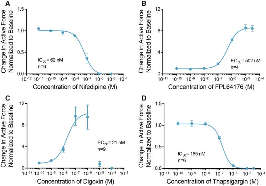
4   | FERIC ET AL.

                                                                                                                                                                              Downloaded from https://academic.oup.com/toxsci/advance-article-abstract/doi/10.1093/toxsci/kfz168/5543113 by guest on 11 September 2019
Figure 2. Engineered cardiac tissues (ECTs) respond to effectors of the cyclic adenosine monophosphate (cAMP) signaling pathway. A, Treatment with isoproterenol in-
duced an increase in contractile force with EC50 of 0.5 nM (n ¼ 12). B, Like isoproterenol, dobutamine induced an increase in contractile force with EC50 of 16 nM (n ¼ 5).
C, Milrinone, a phosphodiesterase-3 (PDE3) inhibitor also increased contractile force with EC50 of 1.6 mM (n ¼ 6). D, Treatment with pituitary adenylate cyclase-activating
peptide (PACAP27) induced an increase in contractile force with EC50 of 1.1 nM (n ¼ 6). Data are presented as mean 6 SEM.

cyclic adenosine monophosphate (cAMP) and a positive inotro-                            concentration (IC50) of 62 nM (Figure 3A, Supplemental Table 2),
pic response (Bers, 2008). ECTs treated with the b-agonist iso-                         whereas treatment with the LTCC activator FPL 64176 induced a
proterenol showed a maximal 6.6 6 0.8-fold increase in                                  maximal 8.5 6 0.6-fold increase in the force of contraction at a
contractile force at a concentration of 10 nM with a half maxi-                         concentration of 10 mM with an EC50 of 502 nM (Figure 3B,
mal effective concentration (EC50) of 0.5 nM (Figure 2A, time con-                      Supplemental Table 2).
stants and rates of contraction and relaxation are summarized                               The Naþ-Ca2þ exchanger (NCX) is the primary means by
in Supplemental Table 1). Similarly, addition of dobutamine, a                          which cardiomyocytes regulate intracellular Ca2þ and hence a
predominantly b1 agonist increased the maximal contractile                              critical modulator of excitation-contraction coupling. To as-
force by 4.4 6 0.6-fold at 360 nM with EC50 of 16 nM (Figure 2B).                       sess the functionality of the NCX in this model we treated
    Further, we investigated the intracellular pathways involved                        ECTs with digoxin. Digoxin is a cardiac glycoside, a class of
in adrenergic signaling, using small molecule modulators that                           compounds that directly inhibits the Naþ/Kþ ATPase and
effect distinct signaling pathways. Degradation of cAMP is me-                          requires a functional NCX for its inotropic effect (Altamirano
diated by a cAMP-dependent enzyme, phosphodiesterase-3                                  et al., 2006; Ozdemir et al., 2008). The inhibition of the Naþ/Kþ
(PDE3). We observed a maximal 4.3 6 1.2-fold increase in the                            ATPase results in increased intracellular Naþ accumulation,
force of contraction when ECTs were treated with the PDE3 in-                           which reduces the electrochemical drive for Ca2þ efflux
hibitor milrinone at a concentration of 100 mM with an EC50 of                          through NCX. In this setting excess intracellular Ca2þ is pri-
1.6 lM (Figure 2C, Supplemental Table 1).                                               marily removed from the cytosol by the sarco/endoplasmic re-
    Pituitary     adenylate     cyclase-activating     polypeptide                      ticulum Ca2þ-ATPase (SERCA), which loads the SR with Ca2þ
(PACAP27) is a 27-amino acid peptide that activates adenylyl cy-                        leading to an increase in cardiac contractility (Ottolia et al.,
clase via binding to its cognate G-protein-coupled receptor. We                         2013). Treatment of ECTs with digoxin induced a maximal
observed a maximal 3.4 6 1.1-fold increase in force of contrac-                         9.7 6 1.7-fold increase in contractile force at a concentration of
tion when ECTs were treated with 10 nM PACAP27 with an EC50                             100 nM with an EC50 of 21 nM (Figure 3C, Supplemental
of 1.1 nM (Figure 2D, Supplemental Table 1).                                            Table 2). Digoxin at concentration of 1 mM induced ectopic ac-
                                                                                        tivity and significantly prolonged the duration of contraction
ECTs Have Functional L-Type Ca21 Channels, SR, and Na1-Ca21                             (Supplemental Figure 1). At concentrations above 10 lM, ECT
Exchangers                                                                              contractility was significantly diminished and contraction
The voltage-gated L-type Ca2þ Channels (LTCCs) are the primary                          could not be elicited in the majority of the ECTs using external
mediators of Ca2þ influx into the cardiomyocyte, which is es-                           field stimulation.
sential for initiation of cardiac excitation-contraction coupling                           Thapsigargin, a noncompetitive inhibitor of SERCA activity
(Benitah et al., 2010; Bers, 2002). Treatment of ECTs with the                          reduces the SR calcium load and leads to increased cytosolic
LTCC blocker nifedipine completely inhibited contraction at a                           calcium and reduced contractility. Treatment of ECTs with
concentration of 1 lM with a half maximal inhibitory                                    thapsigargin      completely       inhibited     contraction    at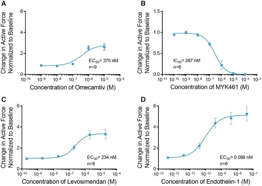
ENGINEERED CARDIAC TISSUES GENERATED IN THE BIOWIRE II PLATFORM                                       |   5

                                                                                                                                                                             Downloaded from https://academic.oup.com/toxsci/advance-article-abstract/doi/10.1093/toxsci/kfz168/5543113 by guest on 11 September 2019
Figure 3. Engineered cardiac tissues (ECTs) respond to calcium handling effectors. A, Treatment with nifedipine decreased contractile force with IC50 of 62 nM (n ¼ 6). B,
Treatment with FPL64176 induced a robust increase in contractile force with EC50 of 502 nM (n ¼ 4). C, Treatment with digoxin induced a robust increase in contractile
force at concentrations below 1 mM (n ¼ 9). Higher concentrations of digoxin showed toxic effects with no contractility observed at doses higher than 10 mM. To calculate
the EC50 for the positive inotropic effects of digoxin, concentration values above 1 mM where not included in the curve fit. D, Thapsigargin completely abolished contrac-
tile force at concentrations above 1 mM (IC50 ¼ 165 nM, n ¼ 6). Data are presented as mean 6 SEM.

concentrations above 1 lM with IC50 of 165 nM (Figure 3D,                               modulation of the intracellular Ca2þ transients and myofila-
Supplemental Table 2).                                                                  ment Ca2þ sensitivity. Treatment of ECTs with ET-1 induced a
                                                                                        maximal 5.2 6 0.7-fold increase in contractile force at 40 nM
ECTs Respond to Sarcomere Modulators                                                    with an EC50 of 98pM (Figure 4D, Supplemental Table 3).
In the cardiac sarcomere, energy derived from myosin-
mediated ATP hydrolysis is used to drive contraction (Malik
et al., 2011). Omecamtiv mecarbil (OM) is a novel positive ino-
                                                                                        DISCUSSION
trope that binds to and stimulates the ATPase activity of myosin                        Here we evaluated the contractile function of human 3D ECTs
resulting in an increased force of contraction in humans                                generated from hiPSC-CMs in the Biowire II platform. We dem-
(Planelles-Herrero et al., 2017). Treatment of ECTs with OM eli-                        onstrate that the platform enables the generation of 3D ECTs
cited a maximal 2.6 6 0.3-fold increase in contractile force at a                       that remain viable and retain their functionality for weeks.
concentration of 10 mM with an EC50 of 370 nM (Figure 4A). OM at                        These ECTs develop properties of the adult myocardium and ex-
10 lM significantly increased the time of contraction and relaxa-                       hibit robust responses to a variety of inotropic compounds with
tion and had no effect on the rates of contraction and relaxation                       distinct mechanisms of action selected to confirm the presence,
(Supplemental Table 3). In contrast, treatment of ECTs with                             and interrogate the function of, pathways known to regulate
MYK-461 (mavacamten), an inhibitor of cardiac myosin ATPase,                            cardiac function.
completely abolished contraction at concentrations above 10 lM                              G-protein coupled receptor signaling pathways play a critical
with IC50 of 287 nM (Figure 4B, Supplemental Table 3).                                  role in functional adaptation in the heart by regulating inotropic
    Cardiac troponin is a trimeric protein complex in the sarco-                        and chronotropic responses. Endothelin-1, a potent vasoactive
mere (Sorsa et al., 2004). It plays a critical role in regulating sen-                  peptide signaling via the phospholipase-C/protein kinase-C
sitivity to Ca2þ. Levosimendan is a positive inotrope that                              pathway, has been shown to regulate cardiac contractility both
modulates troponin-C, 1 protein of the trimer and increases its                         in vivo and in isolated human myocardium (MacCarthy et al.,
sensitivity to regulation by Ca2þ (Pollesello et al., 1994;                             2000; Pieske et al., 1999). ECTs generated a positive inotropic re-
Robertson et al., 2016). Treatment of ECTs with levosimendan in-                        sponse, in a concentration-dependent manner, upon exposure
duced a maximal 3.3 6 0.4-fold increase in contractile force at a                       to ET-1; confirming the presence of a functional ET-1 receptor
concentration of 2 lM with an EC50 of 234 nM (Figure 4C).                               signaling cascade. In patients suffering from HF, dysregulation
                                                                                        of b-adrenergic signaling is a common feature of cardiac patho-
ECTs Respond to Activation of Phospholipase C                                           physiologies and a target of therapy eg, b-adrenergic antago-
Endothelin-1 (ET-1) is a 21-amino acid peptide that binds to the                        nists are a cornerstone of therapy (Bristow et al., 1990). We
ET receptor, a Gq-protein-coupled receptor, and activates phos-                         demonstrated canonical responses to the well-studied b-ago-
pholipase C (Sugden, 2003). Second messenger effects are pro-                           nists isoproterenol and dobutamine. Inotropic responses are ob-
posed to mediate positive inotropic events that involve                                 served for PACAP27, which activates adenylyl cyclase via
6 | FERIC ET AL.
Downloaded from https://academic.oup.com/toxsci/advance-article-abstract/doi/10.1093/toxsci/kfz168/5543113 by guest on 11 September 2019
Figure 4. Engineered cardiac tissues (ECTs) display canonical inotropic responses to sarcomere modulators and activation of phospholipase C. A, Omecamtiv increased
the contractile force with EC50 of 370 nM (n ¼ 9). B, Conversely, increasing doses of MYK461 decreased the contractile force with IC50 of 287 nM (n ¼ 6). C, Treatment with
levosimendan increased the contractile force (n ¼ 9) with EC50 of 234 nM. D, Treatment with endothelin-1 which stimulates the activity of phospholipase C (PLC), in-
creased the contractile force with EC50 of 98 pM (n ¼ 6). Data are presented as mean 6 SEM.
receptors distinct from isoproterenol and milrinone (an inhibi- effect of increasing the contractile force. As such, treatment of
tor of cAMP degradation) indicating that a range of cAMP-medi- the ECTs with the LTCC blocker, nifedipine and with FPL 64176
ated signaling cascades are also present in the ECTs. Although yielded the expected result of decreased and increased contrac-
observation of the stimulatory effects of b-adrenergic agonism tility respectively, indicating the LTCC of the ECT was functional
in engineered human heart tissues is not novel, previous and could be regulated by external stimuli. One of the oldest HF
reports have suggested little to no significant effect on contrac- therapies is digoxin, a cardiac glycoside isolated from the fox-
tility (Hirt et al., 2014), but rather an increase in chronotropy. glove plant. Digoxin induces a positive inotropic effect via its
Additionally, others have observed positive inotropic responses ability to promote Ca2þ loading in the SR. A biphasic increase in
at concentrations far right-shifted and therefore, less physiolog- force was observed when ECTs were treated with digoxin at or
ically relevant to effects observed in the in this study (Zhang below 1 lM demonstrating that contractility can be regulated at
et al., 2013). Interestingly, milrinone has been shown to produce the level of NCX and that the SR modulates Ca2þ levels in a
a positive inotropic response in fibroblast containing ECTs physiologically relevant manner. Treatment of tissues with di-
(Ravenscroft et al., 2016), whereas when tissues were prepared goxin at concentrations above 1 lM showed toxic effects com-
without fibroblasts, there was no inotropic response to milri- monly observed with this compound (Supplemental Figure 1)
none as compared to human heart tissue (Mannhardt et al., (Mannhardt et al., 2017; Ruch et al., 2003).
2017); suggesting maturity in the adrenergic pathway and/or en- The recent discovery of agents that modulate sarcomere
hanced PDE3A expression in fibroblast containing ECTs. We ob- proteins holds great promise for the development of novel
served a significant increase in the magnitude of force treatments for HF. OM is an agent that directly regulates cardiac
generated in response to milrinone in ECTs electrically stimu- myosin and levosimendan is an agent that regulates the func-
lated for 6 weeks as compared to 3 weeks, demonstrating that tion of troponin-C, the Ca2þ sensor of the sarcomere. Data from
long term electrical stimulation is also an important factor that studies in HF patients show OM and levosimendan can promote
contributes to the increased inotropic response to milrinone a positive inotropic effect and hemodynamic improvements
(Supplemental Figure 2). Further studies are required to deter- (Follath et al., 2002; Teerlink et al., 2016b) without a significant
mine the mechanism by which the inotropic response to milri- increase in oxygen consumption (Lilleberg et al., 1998; Shen
none was increased in ECTs generated in the Biowire II et al., 2010; Ukkonen et al., 2000). Consistent with data from clini-
platform. cal and animal studies, both compounds were able to modulate
Calcium channels are both the direct and indirect target of contractility in ECTs eliciting significant increases in contractile
various cardiac therapies. LTCC antagonists are a mainstay in force with concentrations typical for these compounds (Nagy
the treatment of hypertension, cardiac ischemia, and arrhyth- et al., 2015). MYK461, a small molecule inhibitor of myosin also
mias, but can elicit cardio depressant effect. Conversely, com- shows potential in the treatment of HF. Tissue treated with
pounds such as Bay K-8644 and the more potent FPL 64176 were MYK461 showed significant decrease in contractility in the
designed to stimulate LTCC activity and have the consequent range consistent with published results (Green et al., 2016).ENGINEERED CARDIAC TISSUES GENERATED IN THE BIOWIRE II PLATFORM | 7
Downloaded from https://academic.oup.com/toxsci/advance-article-abstract/doi/10.1093/toxsci/kfz168/5543113 by guest on 11 September 2019
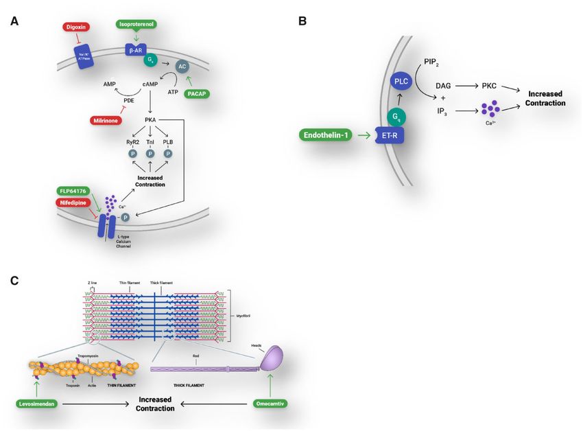
Figure 5. Engineered cardiac tissues (ECTs) generated in Biowire II platform have canonical responses to compounds that affect contractility via physiologically rele-
vant pathways. A, b-adrenergic/cAMP-mediated pathway and the L-type calcium channel. b-AR, b-adrenergic receptor; AC, adenylyl cyclase; PACAP, pituitary adenyl-
ate cyclase-activating peptide; cAMP, cyclic adenosine monophosphate; ATP, adenosine triphosphate; PDE, phosphodiesterase; PKA, protein kinase A; RYR2, ryanodine
receptor; TnI, troponin-I; PLB, phospholamban. B, Gq-protein-coupled receptor signaling. ET-R, endothelin receptor; PLC, phospholipase C; PIP2, phosphatidylinositol 4,
5-bisphosphate; IP3, inositol 1, 4, 5-trisphosphate; DAG, diacylglycerol; PKC, protein kinase C. C, Modulation of proteins of the cardiac sarcomere.
Table 1. Summary of Engineered Cardiac Tissues (ECT) Inotropic Responses
Effective Plasma
ECT Concentration
Compound EC50/IC50 or EC50/IC50 Reference
Isoproterenol 0.5 nM 27 nMa EC50 in isolated human ventricular muscle strip (Flesch et al., 1999)
Dobutamine 16 nM 133–632 nM Plasma levels of 133 nM were associated with significant increase in cardiac index with
linear increase in index up to 632 nM (Leier et al., 1979)
Milrinone 1.6 mM 0.791 mM Plasma concentration associated with 50% increase in cardiac index (Bailey et al., 1994)
PACAP27 1.1 nM 0.5 nMa Recalculated EC50 in dog isolated left ventricle (Hirose et al., 1998)
Nifedipine 62 nM 10–81 nM Steady state of 10 nM (Raemsch and Sommer, 1983), plasma levels above 81 nM were associ-
ated with decreased dp/dtmax (Clifton et al., 1990)
FPL64176 502 nM 600 nMa Recalculated EC50 in guinea pig papillary muscle (Rampe et al., 1993)
Digoxin 21 nM 1.5–2.6 nM Plasma level range in patients with improved fractional shortening (Guyatt et al., 1988)
Thapsigargin 165 nM 300 nMa Complete inhibition of contraction in isolated adult rat cardiomyocytes (Wrzosek et al.,
1992)
Omecamtiv 370 nM 792 nM Maximum plasma level associated with increased stroke volume (Teerlink et al., 2016a)
MYK461 287 nM 180 nMa IC50 in adult rat ventricular cardiomyocytes (Green et al., 2016)
Levosimendan 234 nM 351 nM Plasma level associated with increased stroke volume (Kivikko et al., 2003)
Endothelin-1 0.1 nM 9.1 nMa EC50 in isolated human ventricular trabeculae (Saetrum Opgaard et al., 2000)
The EC50/IC50 for each compound was compared to the effective therapeutic dose of the compound where data were available. For compounds where effective thera-
peutic dose was not available, comparison was made with published results from in vitro or animal models and is indicated by superscript letter (a).8 | FERIC ET AL.
In conclusion, human ECTs created in the Biowire II platform de Lucia, C., Eguchi, A., and Koch, W. J. (2018). New insights in
have adult-like canonical responses to compounds known to af- cardiac beta-adrenergic signaling during heart failure and
fect contractility via an array of physiologically relevant path- aging. Front. Pharmacol. 9, 904.
ways (Figure 5, Summary data presented in Table 1). These Denning, C., Borgdorff, V., Crutchley, J., Firth, K. S., George, V.,
studies can be conducted under external stimulation, providing Kalra, S., Kondrashov, A., Hoang, M. D., Mosqueira, D., Patel,
investigators with a high degree of experimental control, and A., et al. (2016). Cardiomyocytes from human pluripotent
with the ability to assess compounds using a nondestructive stem cells: From laboratory curiosity to industrial biomedical
measurement of contractility. This model can be used to assess platform. Biochim. Biophys. Acta 1863, 1728–1748.
Downloaded from https://academic.oup.com/toxsci/advance-article-abstract/doi/10.1093/toxsci/kfz168/5543113 by guest on 11 September 2019
the effects of novel compounds in a human-based model of Eisner, D. A., Caldwell, J. L., Kistamas, K., and Trafford, A. W.
contractility early in the drug discovery and development pro- (2017). Calcium and excitation-contraction coupling in the
cess. These results suggest the utility of the Biowire II platform heart. Circ. Res. 121, 181–195.
to create disease models via the use of patient derived Flesch, M., Kilter, H., Cremers, B., Laufs, U., Sudkamp, M.,
iPSC-CMs. Ortmann, M., Muller, F. U., and Bohm, M. (1999). Effects of en-
dotoxin on human myocardial contractility involvement of
nitric oxide and peroxynitrite. J. Am. Coll. Cardiol. 33,
SUPPLEMENTARY DATA 1062–1070.
Supplementary data are available at Toxicological Sciences Follath, F., Cleland, J. G., Just, H., Papp, J. G., Scholz, H.,
online. Peuhkurinen, K., Harjola, V. P., Mitrovic, V., Abdalla, M.,
Sandell, E. P., et al. (2002). Efficacy and safety of intravenous
levosimendan compared with dobutamine in severe low-
FUNDING output heart failure (the lido study): A randomised double-
The authors received no financial support for the research, blind trial. Lancet 360, 196–202.
authorship and for the publication of this article. Green, E. M., Wakimoto, H., Anderson, R. L., Evanchik, M. J.,
Gorham, J. M., Harrison, B. C., Henze, M., Kawas, R., Oslob, J.
D., Rodriguez, H. M., et al. (2016). A small-molecule inhibitor
DECLARATION OF CONFLICTING INTERESTS of sarcomere contractility suppresses hypertrophic cardio-
myopathy in mice. Science 351, 617–621.
N.T.F., I.P., R.S., D.R.B., M.G., M.P.G., and R.A.-S. are employees
Guth, B. D., Chiang, A. Y., Doyle, J., Engwall, M. J., Guillon, J. M.,
and shareholders of TARA Biosystems Inc.
Hoffmann, P., Koerner, J., Mittelstadt, S., Ottinger, S., Pierson,
J. B., et al. (2015). The evaluation of drug-induced changes in
REFERENCES cardiac inotropy in dogs: Results from a HESI-sponsored con-
Altamirano, J., Li, Y., DeSantiago, J., Piacentino, V., 3rd, Houser, S. sortium. J. Pharmacol. Toxicol. Methods 75, 70–90.
R., and Bers, D. M. (2006). The inotropic effect of cardioactive Guyatt, G. H., Sullivan, M. J., Fallen, E. L., Tihal, H., Rideout, E.,
glycosides in ventricular myocytes requires Naþ-Caþ Halcrow, S., Nogradi, S., Townsend, M., and Taylor, D. W.
2 ex-
changer function. J. Physiol. 575, 845–854. (1988). A controlled trial of digoxin in congestive heart fail-
Bailey, J. M., Levy, J. H., Kikura, M., Szlam, F., and Hug, C. C., Jr. ure. Am. J. Cardiol. 61, 371–375.
(1994). Pharmacokinetics of intravenous milrinone in Hirose, M., Furukawa, Y., Lakhe, M., and Chiba, S. (1998).
patients undergoing cardiac surgery. Anesthesiology 81, Regional differences in cardiac effects of pituitary adenylate
616–622. cyclase-activating polypeptide-27 in the isolated dog heart.
Benitah, J. P., Alvarez, J. L., and Gomez, A. M. (2010). L-type Ca(2þ) Eur. J. Pharmacol. 349, 269–276.
current in ventricular cardiomyocytes. J. Mol. Cell. Cardiol. 48, Hirt, M. N., Boeddinghaus, J., Mitchell, A., Schaaf, S., Bornchen,
26–36. C., Muller, C., Schulz, H., Hubner, N., Stenzig, J., Stoehr, A.,
Benjamin, E. J., Virani, S. S., Callaway, C. W., Chamberlain, A. M., et al. (2014). Functional improvement and maturation of rat
Chang, A. R., Cheng, S., Chiuve, S. E., Cushman, M., Delling, F. and human engineered heart tissue by chronic electrical
N., Deo, R., et al. (2018). Heart disease and stroke statistics- stimulation. J. Mol. Cell. Cardiol. 74, 151–161.
2018 update: A report from the American heart association. Kivikko, M., Lehtonen, L., and Colucci, W. S. (2003). Sustained he-
Circulation 137, e67–e492. modynamic effects of intravenous levosimendan. Circulation
Bers, D. M. (2002). Cardiac excitation-contraction coupling. 107, 81–86.
Nature 415, 198–205. Leier, C. V., Unverferth, D. V., and Kates, R. E. (1979). The relation-
Bers, D. M. (2008). Calcium cycling and signaling in cardiac myo- ship between plasma dobutamine concentrations and car-
cytes. Annu. Rev. Physiol. 70, 23–49. diovascular responses in cardiac failure. Am. J. Med. 66,
Bristow, M. R., Hershberger, R. E., Port, J. D., Gilbert, E. M., 238–242.
Sandoval, A., Rasmussen, R., Cates, A. E., and Feldman, A. M. Lilleberg, J., Nieminen, M. S., Akkila, J., Heikkila, L., Kuitunen, A.,
(1990). Beta-adrenergic pathways in nonfailing and failing Lehtonen, L., Verkkala, K., Mattila, S., and Salmenpera, M.
human ventricular myocardium. Circulation 82, I12–I25. (1998). Effects of a new calcium sensitizer, levosimendan, on
Buckley, N. M., Penefsky, Z. J., and Litwak, R. S. (1972). haemodynamics, coronary blood flow and myocardial sub-
Comparative force-frequency relationships in human and strate utilization early after coronary artery bypass grafting.
other mammalian ventricular myocardium. Pflugers Arch. Eur. Heart J. 19, 660–668.
332, 259–270. MacCarthy, P. A., Grocott-Mason, R., Prendergast, B. D., and
Clifton, G. D., Booth, D. C., Hobbs, S., Boucher, B. A., Foster, T. S., Shah, A. M. (2000). Contrasting inotropic effects of endoge-
McAllister, R. G., Jr., and DeMaria, A. N. (1990). Negative ino- nous endothelin in the normal and failing human heart:
tropic effect of intravenous nifedipine in coronary artery dis- Studies with an intracoronary ET(A) receptor antagonist.
ease: Relation to plasma levels. Am. Heart J. 119, 283–290. Circulation 101, 142–147.ENGINEERED CARDIAC TISSUES GENERATED IN THE BIOWIRE II PLATFORM | 9
Malik, F. I., Hartman, J. J., Elias, K. A., Morgan, B. P., maturity in stem cell derived cardiomyocyte microtissues.
Rodriguez, H., Brejc, K., Anderson, R. L., Sueoka, S. H., Lee, Toxicol. Sci. 152, 99–112.
K. H., Finer, J. T., et al. (2011). Cardiac myosin activation: A Robertson, I. M., Pineda-Sanabria, S. E., Yan, Z., Kampourakis, T.,
potential therapeutic approach for systolic heart failure. Sun, Y. B., Sykes, B. D., and Irving, M. (2016). Reversible cova-
Science 331, 1439–1443. lent binding to cardiac troponin C by the Ca2þ-sensitizer lev-
Mannhardt, I., Eder, A., Dumotier, B., Prondzynski, M., Kramer, osimendan. Biochemistry 55, 6032–6045.
E., Traebert, M., Sohren, K. D., Flenner, F., Stathopoulou, K., Ruch, S. R., Nishio, M., and Wasserstrom, J. A. (2003). Effect of car-
Lemoine, M. D., et al. (2017). Blinded contractility analysis in diac glycosides on action potential characteristics and con-
Downloaded from https://academic.oup.com/toxsci/advance-article-abstract/doi/10.1093/toxsci/kfz168/5543113 by guest on 11 September 2019
hiPSC-cardiomyocytes in engineered heart tissue format: tractility in cat ventricular myocytes: Role of calcium
Comparison with human atrial trabeculae. Toxicol. Sci. 158, overload. J. Pharmacol. Exp. Ther. 307, 419–428.
164–175. Saetrum Opgaard, O., Moller, S., de Vries, R., Edvinsson, L., and
McNaughton, R., Huet, G., and Shakir, S. (2014). An investigation Saxena, P. R. (2000). Positive inotropic responses mediated by
into drug products withdrawn from the EU market between endothelin ET(A) and ET(B) receptors in human myocardial
2002 and 2011 for safety reasons and the evidence used to trabeculae. Clin. Sci. (Lond.) 99, 161–168.
support the decision-making. BMJ Open 4, e004221. Shen, Y. T., Malik, F. I., Zhao, X., Depre, C., Dhar, S. K., Abarzua,
Nagy, L., Kovacs, A., Bodi, B., Pasztor, E. T., Fulop, G. A., Toth, A., P., Morgans, D. J., and Vatner, S. F. (2010). Improvement of
Edes, I., and Papp, Z. (2015). The novel cardiac myosin activa- cardiac function by a cardiac myosin activator in conscious
tor omecamtiv mecarbil increases the calcium sensitivity of dogs with systolic heart failure. Circ. Heart Fail. 3, 522–527.
force production in isolated cardiomyocytes and skeletal Siramshetty, V. B., Nickel, J., Omieczynski, C., Gohlke, B. O.,
muscle fibres of the rat. Br. J. Pharmacol. 172, 4506–4518. Drwal, M. N., and Preissner, R. (2016). WITHDRAWN—a re-
Onakpoya, I. J., Heneghan, C. J., and Aronson, J. K. (2016). source for withdrawn and discontinued drugs. Nucleic Acids
Worldwide withdrawal of medicinal products because of ad- Res. 44, D1080–1086.
verse drug reactions: A systematic review and analysis. Crit. Sorsa, T., Pollesello, P., and Solaro, R. J. (2004). The contractile ap-
Rev. Toxicol. 46, 477–489. paratus as a target for drugs against heart failure: Interaction
Ottolia, M., Torres, N., Bridge, J. H., Philipson, K. D., and of levosimendan, a calcium sensitiser, with cardiac troponin
Goldhaber, J. I. (2013). Na/Ca exchange and contraction of the C. Mol. Cell. Biochem. 266, 87–107.
heart. J. Mol. Cell. Cardiol. 61, 28–33. Stienen, G. J. (2015). Pathomechanisms in heart failure: The con-
Ozdemir, S., Bito, V., Holemans, P., Vinet, L., Mercadier, J. J., tractile connection. J. Muscle Res. Cell Motil. 36, 47–60.
Varro, A., and Sipido, K. R. (2008). Pharmacological inhibition Sugden, P. H. (2003). An overview of endothelin signaling in the
of Na/Ca exchange results in increased cellular Ca2þ load at- cardiac myocyte. J. Mol. Cell. Cardiol. 35, 871–886.
tributable to the predominance of forward mode block. Circ. Teerlink, J. R., Felker, G. M., McMurray, J. J., Solomon, S. D.,
Res. 102, 1398–1405. Adams, K. F., Jr., Cleland, J. G., Ezekowitz, J. A., Goudev, A.,
Pieske, B., Beyermann, B., Breu, V., Loffler, B. M., Schlotthauer, K., Macdonald, P., Metra, M., et al. (2016a). Chronic oral study of
Maier, L. S., Schmidt-Schweda, S., Just, H., and Hasenfuss, G. myosin activation to increase contractility in heart failure
(1999). Functional effects of endothelin and regulation of (COSMIC-HF): A phase 2, pharmacokinetic, randomised,
endothelin receptors in isolated human nonfailing and fail- placebo-controlled trial. Lancet 388, 2895–2903.
ing myocardium. Circulation 99, 1802–1809. Teerlink, J. R., Felker, G. M., McMurray, J. J. V., Ponikowski, P.,
Pieske, B., Sutterlin, M., Schmidt-Schweda, S., Minami, K., Meyer, Metra, M., Filippatos, G. S., Ezekowitz, J. A., Dickstein, K.,
M., Olschewski, M., Holubarsch, C., Just, H., and Hasenfuss, G. Cleland, J. G. F., Kim, J. B., et al. (2016b). Acute treatment with
(1996). Diminished post-rest potentiation of contractile force omecamtiv mecarbil to increase contractility in acute heart
in human dilated cardiomyopathy. Functional evidence for failure: The ATOMIC-AHF study. J. Am. Coll. Cardiol. 67,
alterations in intracellular Ca2þ handling. J. Clin. Invest. 98, 1444–1455.
764–776. Ukkonen, H., Saraste, M., Akkila, J., Knuuti, J., Karanko, M., Iida,
Planelles-Herrero, V. J., Hartman, J. J., Robert-Paganin, J., Malik, F. H., Lehikoinen, P., Nagren, K., Lehtonen, L., and Voipio-
I., and Houdusse, A. (2017). Mechanistic and structural basis Pulkki, L. M. (2000). Myocardial efficiency during levosimen-
for activation of cardiac myosin force production by ome- dan infusion in congestive heart failure. Clin. Pharmacol. Ther.
camtiv mecarbil. Nat. Commun. 8, 190. 68, 522–531.
Pollesello, P., Ovaska, M., Kaivola, J., Tilgmann, C., Lundstrom, K., Veerman, C. C., Kosmidis, G., Mummery, C. L., Casini, S., Verkerk,
Kalkkinen, N., Ulmanen, I., Nissinen, E., and Taskinen, J. A. O., and Bellin, M. (2015). Immaturity of human stem-cell-
(1994). Binding of a new Ca2þ sensitizer, levosimendan, to re- derived cardiomyocytes in culture: Fatal flaw or soluble prob-
combinant human cardiac troponin C. A molecular model- lem? Stem Cells Dev. 24, 1035–1052.
ling, fluorescence probe, and proton nuclear magnetic Wrzosek, A., Schneider, H., Grueninger, S., and Chiesi, M. (1992).
resonance study. J. Biol. Chem. 269, 28584–28590. Effect of thapsigargin on cardiac muscle cells. Cell Calcium 13,
Raemsch, K. D., and Sommer, J. (1983). Pharmacokinetics and 281–292.
metabolism of nifedipine. Hypertension 5, II18–II24. Zhang, D., Shadrin, I. Y., Lam, J., Xian, H. Q., Snodgrass, H. R., and
Rampe, D., Anderson, B., Rapien-Pryor, V., Li, T., and Dage, R. C. Bursac, N. (2013). Tissue-engineered cardiac patch for ad-
(1993). Comparison of the in vitro and in vivo cardiovascular vanced functional maturation of human ESC-derived cardio-
effects of two structurally distinct Caþþ channel activators, myocytes. Biomaterials 34, 5813–5820.
BAY K 8644 and FPL 64176. J. Pharmacol. Exp. Ther. 265, Zhao, Y., Rafatian, N., Feric, N. T., Cox, B. J., Aschar-Sobbi, R.,
1125–1130. Wang, E. Y., Aggarwal, P., Zhang, B., Conant, G., Ronaldson-
Ravenscroft, S. M., Pointon, A., Williams, A. W., Cross, M. J., and Bouchard, K., et al. (2019). A platform for generation of
Sidaway, J. E. (2016). Cardiac non-myocyte cells show en- chamber-specific cardiac tissues and disease modeling. Cell
hanced pharmacological function suggestive of contractile 176, 913–927.Supplemental Table 1
Isoproterenol TTPT (ms) RT90 (ms) TW50 (ms) +dT/dt (µN/s) ‐dT/dt (µN/s)
(nM)
0 60 ± 4 133 ± 11 103 ± 8 75 ± 10 ‐29 ± 5
1 61 ± 4 101 ± 10# 94 ± 8# 307 ± 52# ‐179 ± 35#
10 63 ± 4 108 ± 9# 93 ± 8# 382 ± 63# ‐205 ± 35#
Milrinone TTPT (ms) RT90 (ms) TW50 (ms) +dT/dt (µN/s) ‐dT/dt (µN/s)
(µM)
0 58 ± 2 137 ± 6 103 ± 2 59 ± 14 ‐22 ± 5
1 61 ± 1 129 ± 1 110 ± 1 #
151 ± 36 #
‐62 ± 16#
10 62 ± 1 121 ± 2# 108 ± 1 236 ± 62# ‐107 ± 30#
Dobutamine TTPT (ms) RT90 (ms) TW50 (ms) +dT/dt (µN/s) ‐dT/dt (µN/s)
(nM)
0 67 ± 5 154 ± 7 128 ± 6 68 ± 11 ‐25 ± 5
# #
35.5 61 ± 1 102 ± 3 99 ± 2 252 ± 33 #
‐138 ± 22#
355 64 ± 2 99 ± 2# 100 ± 4# 312 ± 43# ‐187 ± 30#
PACAP27 TTPT (ms) RT90 (ms) TW50 (ms) +dT/dt (µN/s) ‐dT/dt (µN/s)
(nM)
0 70 ± 7 151 ± 6 125 ± 10 55 ± 6 ‐22 ± 1
1 64 ± 3 131 ± 3# 112 ± 4 124 ± 17# ‐53 ± 8#
10 61 ± 3 98 ± 3# 96 ± 4# 212 ± 30# ‐119 ± 1#
Supplemental Table 1. Change in contractility parameters of ECTs with effectors of the cAMP
signaling pathway. TTPT, time to maximal twitch amplitude; RT90, relaxation time from maximal
twitch amplitude to 90% of twitch amplitude; TW50, twitch width at 50% twitch amplitude; +dT/dt,
maximal rate of contraction; ‐dT/dt, maximal rate of relaxation. Data presented as mean ± SEM. # P <
0.05 using repeat measure one‐way ANOVA. Isoproterenol (n=12), milrinone (n=6), dobutamine
(n=5), PACAP27 (n=6) ECTs.Supplemental Table 2
Nifedipine TTPT (ms) RT90 (ms) TW50 (ms) +dT/dt (µN/s) ‐dT/dt (µN/s)
(µM)
0 66 ± 2 138 ± 4 121 ± 4 145 ± 15 ‐60 ± 8
# #
0.1 54 ± 6 114 ± 10 89 ± 8 95 ± 7 ‐42 ± 5
1 23 ± 10# 53 ± 24# 39 ± 18# 28 ± 13# ‐11 ± 5#
FPL64176 TTPT (ms) RT90 (ms) TW50 (ms) +dT/dt (µN/s) ‐dT/dt (µN/s)
(µM)
0 59 ± 1 145 ± 2 104 ± 3 56 ± 6 ‐20 ± 2
# # # #
0.1 68 ± 1 158 ± 3 127 ±3 117 ± 12 ‐43 ± 4#
1 89 ± 1# 209 ± 6# 169 ± 4# 253 ± 27# ‐92 ± 9#
Digoxin TTPT (ms) RT90 (ms) TW50 (ms) +dT/dt (µN/s) ‐dT/dt (µN/s)
(µM)
0 50 ± 3 124 ± 7 98 ± 8 53 ± 10 ‐16 ± 2
# # # #
1 169 ± 30 266 ± 33 292 ± 40 152 ± 24 ‐82 ± 13#
10 70 ± 35 102 ± 51 119 ± 59 14 ± 7# ‐9 ± 5
Thapsigargin TTPT (ms) RT90 (ms) TW50 (ms) +dT/dt (µN/s) ‐dT/dt (µN/s)
(µM)
0 53 ± 3 125 ± 5 98 ± 3 93 ± 13 ‐35 ± 6
0.1 60 ± 1# 143 ± 10 114 ± 5# 55 ± 13 ‐24 ± 2
# # # #
1 106 ± 3 175 ± 4 180 ± 5 22 ± 3 ‐12 ± 2
Supplemental Table 2. Change in contractility parameters of ECTs in response to calcium
handling effectors. TTPT, time to maximal twitch amplitude; RT90, relaxation time from maximal
twitch amplitude to 90% of twitch amplitude; TW50, twitch width at 50% twitch amplitude;
+dT/dt, maximal rate of contraction; ‐dT/dt, maximal rate of relaxation. Data presented as mean
± SEM. # P < 0.05 using repeat measure one‐way ANOVA. Nifedipine (n=6), FPL64176 (n=4),
digoxin (n=9), thapsigargin (n=6) ECTs.Supplemental Table 3
Omecamtiv TTPT (ms) RT90 (ms) TW50 (ms) +dT/dt (µN/s) ‐dT/dt (µN/s)
(µM)
0 63 ± 3 144 ± 5 111 ± 5 65 ± 8 ‐24 ± 3
# # #
1 95 ± 3 208 ± 6 191 ± 7 147 ± 18 #
‐59 ± 8#
10 195 ± 3# 473 ± 8# 358 ± 6# 66 ± 7 ‐24 ± 3
MYK461 TTPT (ms) RT90 (ms) TW50 (ms) +dT/dt (µN/s) ‐dT/dt (µN/s)
(µM)
0 64 ± 2 143 ± 5 117 ± 4 77 ± 15 ‐30 ± 7
0.1 57 ± 1# 132 ± 4# 103 ± 4# 78 ± 18 ‐30 ± 7
#
1 35 ± 11 90 ± 28 62 ± 20 20 ± 7 ‐7 ± 3#
Levosimendan TTPT (ms) RT90 (ms) TW50 (ms) +dT/dt (µN/s) ‐dT/dt (µN/s)
(µM)
0 62 ± 3 149 ± 6 113 ± 5 50 ± 6 ‐19 ± 2
0.2 61 ± 3 125 ± 6# 108 ± 5 132 ± 16# ‐58 ± 8#
2 63 ± 2 122 ± 5# 108 ± 4 185 ± 8# ‐84 ± 8#
Endothelin‐1 TTPT (ms) RT90 (ms) TW50 (ms) +dT/dt (µN/s) ‐dT/dt (µN/s)
(nM)
0 53 ± 2 142 ± 5 113 ± 8 52 ± 6 ‐16 ± 1
#
0.4 69 ± 1 141 ± 3 125 ± 2 166 ± 23 #
‐72 ± 12#
4 71 ± 1# 139 ± 3 125 ± 2 182 ± 26# ‐82 ± 14#
Supplemental Table 3. Change in contractility parameters of ECTs in response to sarcomere
modulators and activation of phospholipase C. TTPT, time to maximal twitch amplitude; RT90,
relaxation time from maximal twitch amplitude to 90% of twitch amplitude; TW50, twitch width
at 50% twitch amplitude; +dT/dt, maximal rate of contraction; ‐dT/dt, maximal rate of
relaxation. Data presented as mean ± SEM. # P < 0.05 using repeat measure one‐way ANOVA.
Omecamtiv (n=9), MYK461 (n=6), levosimendan (n=9), endothelin‐1 (n=6) ECTs.Supplemental Figure 1
A 1 M Digoxin
100 nM Digoxin
500 ms
5 N
500 ms
5 N
500 ms
5 N
500 ms
5 N
B
Normalized TTPT
Supplemental Figure 1. Digoxin increased ectopic activity. A. Sample traces showing transients
at 100 nM and 1 µM digoxin in 4 ECTs. 1 µM digoxin prolonged the duration of contraction and
induced ectopic activity. B. Summary of contraction parameters normalized to baseline showing
increase in time to maximal force generation (time to maximal twitch amplitude) and relaxation
time (relaxation time from maximal twitch amplitude to 90% of twitch amplitude) at 1 µM
digoxin (n=9). Data is presented as mean ± SEM. # pSupplemental Figure 2 Supplemental Figure 2. Improved inotropic response to milrinone after 6 weeks of electrical stimulation. Summary of fold change in force normalized to baseline after addition of 100 µM milrinone. 6 weeks of electrical stimulation (n=9) increased the magnitude of force generated in response to milrinone as compared to 3 weeks of stimulation (n=6). Data is presented as mean ± SEM. # p
You can also read