Epidermal chloroplasts are defense-related motile organelles equipped with plant immune components
←
→
Page content transcription
If your browser does not render page correctly, please read the page content below
ARTICLE
                  https://doi.org/10.1038/s41467-021-22977-5                 OPEN

                  Epidermal chloroplasts are defense-related motile
                  organelles equipped with plant immune
                  components
                  Hiroki Irieda        1✉   & Yoshitaka Takano2
1234567890():,;

                  In addition to conspicuous large mesophyll chloroplasts, where most photosynthesis occurs,
                  small epidermal chloroplasts have also been observed in plant leaves. However, the func-
                  tional significance of this small organelle remains unclear. Here, we present evidence that
                  Arabidopsis epidermal chloroplasts control the entry of fungal pathogens. In entry trials,
                  specialized fungal cells called appressoria triggered dynamic movement of epidermal chlor-
                  oplasts. This movement is controlled by common regulators of mesophyll chloroplast pho-
                  torelocation movement, designated as the epidermal chloroplast response (ECR). The ECR
                  occurs when the PEN2 myrosinase-related higher-layer antifungal system becomes ineffec-
                  tive, and blockage of the distinct steps of the ECR commonly decreases preinvasive nonhost
                  resistance against fungi. Furthermore, immune components were preferentially localized to
                  epidermal chloroplasts, contributing to antifungal nonhost resistance in the pen2 background.
                  Our findings reveal that atypical small chloroplasts act as defense-related motile organelles
                  by specifically positioning immune components in the plant epidermis, which is the first site
                  of contact between the plant and pathogens. Thus, this work deepens our understanding of
                  the functions of epidermal chloroplasts.

                  1 Academic Assembly, Institute of Agriculture, Shinshu University, Nagano, Japan. 2 Graduate School of Agriculture, Kyoto University, Kyoto, Japan.
                  ✉email: irieda@shinshu-u.ac.jp

                  NATURE COMMUNICATIONS | (2021)12:2739 | https://doi.org/10.1038/s41467-021-22977-5 | www.nature.com/naturecommunications                              1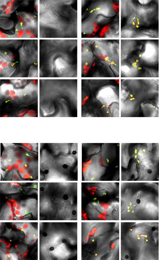
ARTICLE                                                                 NATURE COMMUNICATIONS | https://doi.org/10.1038/s41467-021-22977-5

T
        he epidermis of multicellular organisms acts as a boundary           in the absence of the PEN2-based antifungal defense system,
        with the environment to protect against a wide variety of            suggesting that the ECR is newly activated when PEN2-related
        biotic and abiotic stresses. Although the plant epidermis is         defense becomes ineffective. Our data revealed that CHUP1 and
the site of primary pathogen recognition, it can also be attacked            JAC1, regulators of chloroplast photorelocation movement in
by pathogens, resulting in infection. For instance, the anthracnose          mesophyll cells, also control the ECR. Importantly, blocking the
fungi Colletotrichum species and the rice blast fungus Magna-                distinct steps of the ECR via mutation or overexpression of the
porthe oryzae develop a specialized dome-shaped fungal cell                  CHUP1 gene significantly reduced preinvasive NHR against
called an appressorium, which is highly pigmented with melanin               nonadapted fungi in epidermal cells. Furthermore, we found that
and is used in direct fungal entry into plant epidermis1. The                multiple immune components that are preferentially localized to
melanized appressorium produces a penetration peg, followed by               epidermal chloroplasts contribute to preinvasive NHR in the
the extension of the invasive hyphae. However, if the plant is a             lower layer of PEN2-related immunity. We propose that atypical
nonhost, a robust and broad-spectrum defense response termed                 small chloroplasts in the epidermis function as defense-related
nonhost resistance (NHR) is developed to effectively block the               motile organelles involved in Arabidopsis NHR.
infection of a vast number of nonadapted pathogens2. More
specifically, epidermal immune responses are of great importance
in terminating the entry of nonadapted fungal pathogens.                     Results
   PEN2 myrosinase is known as a positive regulator of epidermal             Epidermal chloroplasts emerged on the upper periclinal sur-
NHR in Arabidopsis thaliana against multiple fungal pathogens,               face in response to the entry trial by nonadapted Colleto-
including Blumeria graminis f. sp. hordei, Colletotrichum tropicale          trichum fungi. To investigate the relationship between epidermal
(Ctro), and Magnaporthe oryzae3–7. Together with the ATP                     chloroplasts and preinvasive NHR against the appressorium-
binding cassette transporter PEN3, PEN2 regulates the                        mediated aggression of fungal pathogens in A. thaliana, we used
tryptophan-derived secondary metabolite-mediated defense                     three nonadapted Colletotrichum strains: C. fioriniae CC1 (Cfio), C.
pathway3–5,8,9. In addition, EDR1 protein kinase has a positive              siamense MAF1 (Csia), and Corb, which are cosmos, apple, and
effect on the preinvasive NHR to nonadapted Ctro, whereas it has             cucurbit anthracnose fungi, respectively. We also used the adapted
negative effects on immunity to the adapted fungus Golovino-                 strain C. higginsianum Abr1-5 (Chig), a brassica anthracnose fungus.
myces cichoracearum10,11. However, pen2 and edr1 mutants retain              Cfio, Csia, or Corb did not form any lesions in wild-type Arabidopsis
normal resistance against the nonadapted fungus Colletotrichum               (Col-0), whereas Chig developed severe lesions (Fig. 1a). Interest-
orbiculare 104-T (Corb)7,11. Therefore, multiple immune com-                 ingly, microscopic observation revealed that chlorophyll-containing
ponents, including as-yet unknown factors, support the deploy-               epidermal chloroplasts emerged on the upper periclinal wall (sur-
ment of epidermal NHR against the entry of nonadapted fungal                 face) of pavement cells after inoculation with nonadapted Colleto-
pathogens in Arabidopsis.                                                    trichum fungi, especially Cfio and Csia (Fig. 1b, c). The degree of this
   In plant immunity, chloroplasts play an important role in the             response varied according to the fungal strain, but the response was
biosynthesis of defense-related molecules12. Secondary messengers            detected at 1 day post-inoculation (dpi) and tended to increase until
such as reactive oxygen species (ROS) and calcium (Ca2+) are                 3 dpi. This phenomenon was also observed in a transgenic plant line
produced and released by chloroplasts, respectively; precursors of           expressing the plastid-cyan fluorescent protein (CFP) marker (Sup-
phytohormones such as salicylic acid (SA), jasmonic acid, and                plementary Fig. 1). We named this phenomenon the ECR, because
abscisic acid are also synthesized in chloroplasts13–16. It has been         the epidermal chloroplasts are usually positioned at the lower peri-
proposed that immune signals triggered by pathogen-associated                clinal (bottom) and anticlinal walls in a steady state. The ECR against
molecular patterns (PAMPs) are transmitted from the cell surface             Chig was not clearly detectable, suggesting that the ECR is a specific
to chloroplasts via intracellular Ca2+ relay, followed by                    response to nonadapted fungi (Fig. 1c).
chloroplast-to-nucleus retrograde ROS signaling in A. thaliana17.               Since Corb activated the ECR on Col-0 at a lower level
In chloroplast-to-nucleus ROS signaling in Nicotiana benthami-               compared with Cfio and Csia, we hypothesized that some
ana in response to viral and bacterial pathogens, the involvement            preinvasive defenses in NHR may take priority over the ECR.
of tubular extensions from the chloroplast (called stromules) has            Consistent with this idea, we found that pen2 plants showed
been proposed18.                                                             increased ECR against Cfio, Csia, and Corb compared to Col-0
   In higher plants, it has been widely recognized that no chlor-            plants (Fig. 1c). This suggests that the ECR occurs preferentially
oplasts exist in epidermal cells other than guard cells, with the            in the absence of PEN2-related immunity. Indeed, pen2 increased
exception of some plants (e.g., tobacco)19–22. A large population            epidermal invasion (at 4 dpi) and leaf lesion formation (at 7 dpi)
of chloroplasts is highly differentiated in leaf mesophyll cells for         by Cfio and Csia, indicating compromised NHR in pen2 plants
efficient photosynthesis, while guard cell chloroplasts regulate              (Fig. 1a, d, e). Quantitative real-time PCR (RT-qPCR) analysis
stomatal movements in response to CO2 and light23. However,                  also showed that many defense-related genes were induced only
chlorophyll-containing chloroplasts in the epidermal pavement                in pen2 at 1 dpi (Supplementary Fig. 2). This suggests the
cells of A. thaliana have been recognized, although their size and           presence of a preinvasive defense system that is activated in the
number are smaller than those of mesophyll cells24,25. The                   absence of PEN2, further supporting that the PEN2-related
number and thickness of grana and the chlorophyll content of                 pathway is a higher-layer preinvasive defense system against
epidermal chloroplasts are also low, leading to the plausible                Colletotrichum fungi. Remarkably, the impact of pen2 mutation
assumption that the photosynthetic contribution of these small               on Corb-induced ECR was drastic, while Corb was not able to
organelles is lower than that of mesophyll cells24. Hence, the               invade pen2 (Fig. 1d, e). Since the ECR is not accompanied by
functional significance of small epidermal chloroplasts remains               fungal invasion, this may imply that the ECR contributes to
unclear.                                                                     preinvasive NHR.
   Here, we report that epidermal chloroplasts dynamically                      Interestingly, we detected only a slight increase in the
respond to Colletotrichum and Magnaporthe fungi that are not                 frequency of the ECR in edr1 mutants (Fig. 1c). Indeed, unlike
adapted to Arabidopsis. We showed that epidermal chloroplasts                the pen2 mutant, the preinvasive NHR of the edr1 mutant against
emerge at the upper periclinal wall in response to the entry trial of        appressorium-mediated Cfio and Csia entry did not decrease,
these nonadapted fungi (hereafter referred to as the epidermal               with no induction of defense-related genes (Fig. 1e, Supplemen-
chloroplast response, ECR). The frequency of the ECR increased               tary Fig. 2); this result strengthens the link between the

2                NATURE COMMUNICATIONS | (2021)12:2739 | https://doi.org/10.1038/s41467-021-22977-5 | www.nature.com/naturecommunications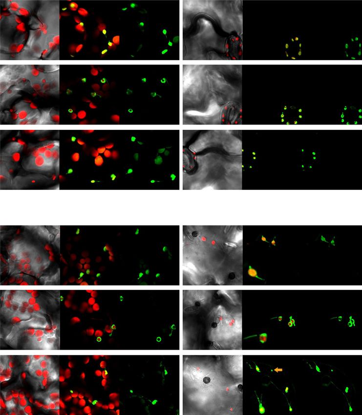
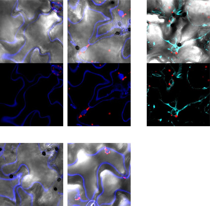
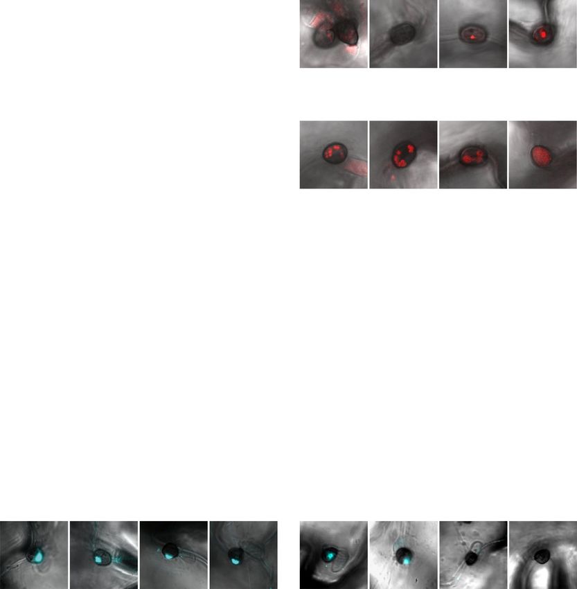
NATURE COMMUNICATIONS | https://doi.org/10.1038/s41467-021-22977-5                                                                        ARTICLE

Fig. 1 The ECR occurs at the lower layer of PEN2-related immunity and is critical for NHR against Colletotrichum fungi. a Pathogenicity of Colletotrichum
fungi on Arabidopsis mutants. A conidial suspension of adapted Chig and nonadapted Cfio, Csia, and Corb was inoculated onto leaves of wild-type Arabidopsis
(Col-0), pen2-1, and edr1-1 and incubated for 7 days. b ECR after Cfio inoculation. The epidermal surface of the Cfio-inoculated cotyledon of Col-0 was
investigated at 3 days post-inoculation (dpi). The chloroplasts were visualized based on chlorophyll autofluorescence. The DIC images were captured by
confocal microscopy. DW was used as a control. c The ratio of epidermal cells with surface chloroplasts was investigated at 1, 2, and 3 dpi. A total of 100
cells in contact with the melanized appressorium were observed. N.D.: not determined due to damages of epidermal cell by fungal invasion. d, e Fungal
invasion of Arabidopsis epidermis. Cfio, Csia, and Corb were inoculated onto cotyledons of the indicated Arabidopsis. The entry ratio was quantified at 4 dpi.
A total of 100 melanized appressoria were investigated. The arrowheads and arrows indicate melanized appressoria and invasive hyphae, respectively.
Scale bar, 10 µm. The means and SE were calculated from three independent plants. Means not sharing the same letter are significantly different (P < 0.05,
two-way ANOVA with Tukey’s HSD test).

NATURE COMMUNICATIONS | (2021)12:2739 | https://doi.org/10.1038/s41467-021-22977-5 | www.nature.com/naturecommunications                                  3

ARTICLE                                                                 NATURE COMMUNICATIONS | https://doi.org/10.1038/s41467-021-22977-5

breakdown of higher-layer preinvasive defenses such as the                   photosynthesis (accumulation response). Under strong light, they
PEN2-related pathway and ECR activation.                                     migrate to the anticlinal wall to reduce photodamage (avoidance
                                                                             response). CHUP1 generates a chloroplast-actin-based motive
                                                                             force through the polymerization of chloroplast-actin; therefore,
The ECR was correlated with fungal secretion activity during
                                                                             CHUP1 is essential for both accumulation and avoidance
the entry trial. We focused on the triggers of the ECR. One
                                                                             responses40–42. On the other hand, JAC1 plays a role in the
candidate trigger is pathogen-derived PAMPs. The immune
                                                                             appearance and disappearance of chloroplast-actin filaments and
kinases BAK1, BIK1, and PBL1 function in PAMP-triggered
                                                                             is required for the accumulation response41.
immunity26–29. However, bak1-5 and bik1 pbl1 mutants showed
                                                                                To gain new insight into the relationship between these
normal ECR (Supplementary Fig. 3). Thus, we analyzed fungal
                                                                             components and the ECR, we examined how the levels of CHUP1
factors other than PAMPs that may induce the ECR. In Colleto-
                                                                             and JAC1 proteins affected the ECR (Fig. 3). We generated
trichum fungi, the yeast STE12 gene homolog CST1 and tetra-
                                                                             transgenic plants overexpressing CHUP1 (CHUP1ox) or JAC1
spanin gene PLS1 are involved in appressorium-mediated
                                                                             (HA-JAC1ox) and analyzed them with chup1 and jac1 mutants.
penetration30,31. The cst130 and pls131 mutants could form mel-
                                                                             The growth of these plants was normal, except that HA-JAC1ox
anized appressoria, but were defective in penetration peg for-
                                                                             exhibited a semi-curly leaf phenotype (Fig. 3a, Supplementary
mation. In contrast, the sterol glucosyltransferase gene ATG26
                                                                             Fig. 5). Intriguingly, CHUP1 overexpression completely sup-
and isocitrate lyase gene ICL1 were not involved in penetration
                                                                             pressed the ECR (Fig. 3b, c). In contrast, the chup1 mutant
peg formation, but were involved in subsequent invasive hyphal
                                                                             constitutively displayed epidermal chloroplasts at the surface with
development in planta32,33; therefore, atg2632 and icl133 mutants
                                                                             or without fungal inoculation (Fig. 3b, c). An inoculum with high
were defective in invasive hyphal development. We first inocu-
                                                                             concentration of fungal conidia on the chup1 mutant did not
lated pen2 plants with these Corb mutants (pls1 was generated in
                                                                             significantly increase the population of ECR-activated cells, but
this study, Supplementary Fig. 4) and found that atg26 and icl1
                                                                             the number of surface chloroplasts was slightly increased (Fig. 3d).
could induce the ECR, while cst1 and pls1 could not (Fig. 2a).
                                                                             Therefore, the chup1 mutant showed constitutive positioning of
This suggests that fungal penetration peg formation is essential
                                                                             the epidermal chloroplasts at the surface, but the mutant is
for triggering the ECR (Fig. 2a). We examined the possibility that
                                                                             deficient in ECR that is newly triggered by the inoculation of
plant damage caused by penetration peg formation activated the
                                                                             nonadapted Colletotrichum fungi (Fig. 3b–d). Thus, both over-
ECR. The wound-induced endogenous peptide Pep1 and its
                                                                             expression and mutation of CHUP1 commonly cause impair-
paralogs are recognized by the damage-associated molecular
                                                                             ments in the intracellular movement of epidermal chloroplasts in
pattern (DAMP) receptors PEPR1 and PEPR2 in Arabidopsis34,35.
                                                                             the ECR. On the contrary, JAC1 overexpression resulted in a
However, we detected normal ECR in pepr1 pepr2 mutants
                                                                             chup1-like phenotype, although the jac1 mutant showed near-
(Supplementary Fig. 3). Combined with the fact that BAK1 and
                                                                             normal ECR (Fig. 3b, c).
BIK1 also function in Pep1 DAMP signaling36,37, Pep1 and its
                                                                                Mutations in hydrophilic amino acid residues in CHUP1
paralogs might be not a cue of ECR.
                                                                             (R4A&S12A&R20A) cause them to change location from the
   Next, we examined whether fungal secretions from peg
                                                                             perichloroplasts to the cytosol42. We generated CHUP1-
triggered the ECR. We inoculated pen2 plants with the Corb
                                                                             R4A&S12A&R20Aox lines and found that this point-mutated
expressing mCherry-labeled effector CoDN338 and found that the
                                                                             CHUP1 exhibited decreased endogenous CHUP1 proteins,
effector signal at the penetration pore was stronger than that on
                                                                             thereby resulting in a chup1-like dominant negative phenotype;
Col-0 (Fig. 2b). Similar results were observed on pen3, implying
                                                                             epidermal chloroplasts were constitutively detected at the surface
the possible involvement of tryptophan-derived secondary
                                                                             without fungal inoculation, and this phenotype was correlated
metabolites in suppressing fungal effector delivery. A gene
                                                                             with the levels of endogenous CHUP1 proteins (Fig. 3a, e,
disruption mutant of the membrane traffic component SEC22
                                                                             Supplementary Fig. 5). In brief, the dominant negative effects of
in Corb, Δsec22, showed decreased effector secretion, and the
                                                                             these mutant lines were partial; therefore, the ECR still occurred
effector signals were abnormally retained and dispersed inside
                                                                             in response to Cfio (Fig. 3e). These results further suggest that
fungal cells when Δsec22 was inoculated on host cucumber38. As
                                                                             CHUP1 negatively regulates the ECR.
expected, the focal accumulation of the effector at the penetration
                                                                                Chloroplast photorelocation is mediated by the blue light
pore, located in the center at the bottom of the appressorium, was
                                                                             receptors phototropin 1 (phot1) and 2 (phot2)43–45. Since phot2
attenuated in Δsec22 when Δsec22 was inoculated on nonhost
                                                                             is involved in both accumulation and avoidance responses, we
Arabidopsis (Fig. 2b). Additionally, the effector exhibited
                                                                             evaluated the ECR of the phot2 and phot1 phot2 mutants. We
abnormal retention inside the appressorium (Fig. 2b). Intrigu-
                                                                             found that the ECR occurred normally, except for a slight
ingly, the ECR significantly correlated with fungal secretion
                                                                             increase in surface chloroplasts in phot1 phot2 without fungal
activity at the penetration pore (Fig. 2b, c), whereas the Corb
                                                                             inoculation (Fig. 3f). These findings suggest that chloroplast
Δsec22 mutant induced sufficient papillary callose deposition at
                                                                             photorelocation and the ECR have different stimulus recognition
the entry trial sites of the fungus; papillary callose deposition is
                                                                             systems, although they might share downstream components.
one of the defense responses of the plant against fungal entry
trials (Fig. 2d). Since penetration peg-defective cst1 and pls1
mutants hardly induced papilla (Fig. 2e), these results imply that
                                                                             The ECR contributes to NHR against Colletotrichum fungi. To
A. thaliana recognizes pathogen-derived secretions from the peg
                                                                             clarify whether the ECR is involved in plant immunity, we gen-
and activates the ECR. However, there is still a possibility that the
                                                                             erated CHUP1ox and chup1 plants in the pen2 mutant back-
slight differences in the degree of fungal progression in the
                                                                             ground (Fig. 4). The fungal entry rates on these generated plants
penetration attempt, which is not reflected by papilla formation,
                                                                             were higher than those on pen2, while the CHUP1ox and chup1
affects ECR activation.
                                                                             single mutants retained normal resistance (Fig. 4a). Transgenic
                                                                             pen2 chup1 lines expressing CHUP1 under its own promoter
The ECR shares common regulators with mesophyll chlor-                       showed complementation of the ECR and antifungal immunity
oplast photorelocation movement. In mesophyll cells, typical                 (Fig. 4b–d). Intriguingly, we found that the edr1 pen2 double
large chloroplasts exhibit photorelocation movement39. Under                 mutant exhibited an increased entry rate compared with the pen2
weak light, they move toward the periclinal walls to promote                 single mutant, suggesting that the effect of edr1 mutation on

4                NATURE COMMUNICATIONS | (2021)12:2739 | https://doi.org/10.1038/s41467-021-22977-5 | www.nature.com/naturecommunications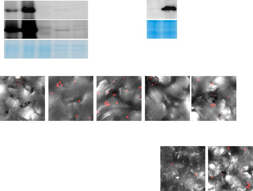
NATURE COMMUNICATIONS | https://doi.org/10.1038/s41467-021-22977-5                                                               ARTICLE

preinvasive NHR became apparent in the pen2 background                          immunity. RT-qPCR analysis showed a significant decrease or
(Fig. 4a). Moreover, the effects of ECR impairment by CHUP1                     decreasing trend in the expression of some Cfio-induced genes in
overexpression and mutation on preinvasive NHR were greater in                  pen2 CHUP1ox and pen2 chup1 compared to that in pen2, which
edr1 pen2 double mutants than in pen2 single mutants. These                     suggests the possible involvement of the ECR in regulating these
results demonstrate that the ECR contributes to preinvasive NHR                 defense-related genes (Fig. 4e).
against Colletotrichum fungi at the lower layer of PEN2-related                   When nonadapted Colletotrichum fungi invade Arabidopsis,
immunity, while the breakdown of additional EDR1-related                        postinvasive NHR responses, including plant cell death, are
preinvasive defense has a greater impact on ECR-mediated                        activated46. Therefore, we examined the possible involvement of

NATURE COMMUNICATIONS | (2021)12:2739 | https://doi.org/10.1038/s41467-021-22977-5 | www.nature.com/naturecommunications                      5
ARTICLE                                                                      NATURE COMMUNICATIONS | https://doi.org/10.1038/s41467-021-22977-5

Fig. 2 Fungal peg formation and penetration pore-mediated secretion correlate with plant ECR during entry trial. a ECR after inoculation of Corb
mutants. Wild-type (WT) Corb, atg26, icl1, cst1, and pls1 were inoculated onto cotyledons of pen2-1 plants. The ECR was quantified at 1, 2, and 3 dpi. DW
was used as a control. b Focal accumulation of Corb effector at the penetration pore was abolished by disruption of the SEC22 gene in Corb. Corb expressing
CoDN3-mCherry was inoculated onto the indicated Arabidopsis. At 2 dpi, the images were captured by confocal microscopy, and the ratio of the effector
signals at the penetration pore was quantified. The arrows indicate effector accumulation at the penetration pore. c ECR after inoculation of Corb WT and
Δsec22 at 2 dpi. d, e Papilla formation at the entry trial site of Corb WT, Δsec22, atg26, icl1, cst1, and pls1. Corb-inoculated plants were subjected to staining
with aniline blue fluorochrome for visualization of callose deposition. At 1 dpi, the images were captured, and the ratio of papillary callose deposition was
investigated. For all experiments, 100 melanized appressoria were observed, and the means and SE were calculated from three independent plants. Scale
bar, 10 µm. Means not sharing the same letter are significantly different (P < 0.05, one-way (b–e) or two-way (a) ANOVA with Tukey’s HSD test). n.s. not
significant.

the ECR in postinvasive NHR. Trypan blue (TB) staining                            while CAS-sfGFP was also detected in the epidermal plastid with
indicated the induction of cell death in the epidermal pavement                   no chlorophyll (Fig. 5). These localization patterns are different
and palisade mesophyll (not invaded) cells inoculated with Cfio                    from those of CHUP1, which localizes to both epidermal and
(Fig. 4f), which is consistent with a previous report46. The                      mesophyll chloroplasts54. Z-stack and sectional images clearly
invasive hyphae were confined to the dead epidermal cell,                          showed preferential localization of GSH1, EDS5, and CAS to the
suggesting that postinvasive defense in ECR-defective plants                      small epidermal chloroplasts (Supplementary Fig. 8, Supple-
functioned normally (Fig. 4f).                                                    mentary Movies 2–7). Altogether, we concluded that the immune
   Several preinvasive NHR components, such as PEN1, PEN2,                        components GSH1, EDS5, and CAS preferentially localized to
PEN3, and PEN4/PCS1, are known to localize at pathogen entry                      small epidermal chloroplasts in preinvasive NHR.
trial sites3,7,8,47,48. We also detected PEN2-GFP at the entry trial                 To further strengthen the link between the ECR and epidermal
sites of Cfio, Csia, and Corb (Supplementary Fig. 6). However,                     chloroplast-localized GSH1, EDS5, and CAS, we generated
epidermal chloroplasts did not show specific accumulation at the                   fluorescent marker plant lines of these immune components in
entry trial sites (Figs. 1b, 3b, Supplementary Fig. 6); instead, we               the CHUP1ox and chup1 backgrounds (Fig. 6). We found that
frequently observed rapidly moving chloroplasts on the upper                      their localization patterns were strongly correlated with epidermal
surface (Supplementary Fig. 7, Supplementary Movie 1).                            chloroplasts and the ECR (Figs. 5 and 6, Supplementary Movie 8).
Although epidermal chloroplasts were occasionally observed near                   These observations suggested that the ECR changes the
the appressoria, the majority of the population in the surface                    intracellular locations of the immune components GSH1, EDS5,
chloroplasts was scattered.                                                       and CAS.
                                                                                     We also detected fluorescent signals of GSH1-GFP, EDS5-
Immune components GSH1, EDS5, and CAS preferentially                              sfGFP, and CAS-sfGFP in stromules (Figs. 5 and 6a), where the
localize to epidermal chloroplasts and stromules in preinvasive                   plastid-CFP marker was observed, but chlorophyll autofluores-
NHR. The immune components GSH1, EDS5, and CAS have                               cence was not (Supplementary Fig. 1). In the immune response of
been reported to localize to small epidermal chloroplasts46,49,50.                N. benthamina, induced stromules are proposed to be a
In previous reports, EDS5-GFP preferentially localized around                     chloroplast-to-nucleus route for ROS signaling and the transport
epidermal chloroplasts compared to mesophyll chloroplasts when                    of NRIP1, which is a protein required for the recognition of the
expressed under both its own and cauliflower mosaic virus                          tobacco mosaic virus effector p5018. N. benthamiana NRIP1,
(CaMV) 35S promoters49. When only expressed under the CaMV                        which is exogenously expressed in A. thaliana, also localizes to
35S promoter, mesophyll chloroplasts had weak EDS5-GFP sig-                       stromules18. Therefore, the stromules possibly link to the
nals, but the chloroplast periphery-specific pattern was main-                     regulation of the localization of Arabidopsis immune-related
tained in two types of chloroplasts49. CAS-GFP driven by the                      proteins via ECR.
CaMV 35S promoter was detected in both epidermal and
mesophyll chloroplasts, with no difference in their localization                  GSH1, EDS5, and CAS contribute to preinvasive NHR against
patterns50. The localization of GSH1-GFP driven by its own                        Colletotrichum fungi. To evaluate the contribution of GSH1,
promoter in the mesophyll has yet to be confirmed46. GSH1 γ-                       EDS5, and CAS to preinvasive NHR against Colletotrichum fungi
glutamylcysteine synthetase contributes to NHR against Ctro via                   in epidermal cells, we quantified the entry rates of Cfio, Csia, and
glutathione biosynthesis46,51. EDS5 is a MATE family transporter                  Corb. Hiruma et al. reported that gsh1 mutants showed a sub-
that is required for the transport of the SA precursor outside the                stantial decrease in preinvasive defense against appressorium-
chloroplasts52,53. CAS is a Ca2+-sensing receptor involved in                     independent atypical entry of Ctro46. On the other hand, the gsh1
transient Ca2+ signaling in chloroplasts during plant immunity17.                 mutant showed a slight reduction in epidermal NHR against
To analyze the subcellular localization of these components in                    appressorium-mediated Cfio entry, and the effect was weaker
ECR-activated cells, we used gsh1 plants expressing GSH1-GFP46                    than that in pen2. Meanwhile, Csia and Corb barely invaded the
and generated transgenic Col-0 plants expressing EDS5-super                       gsh1 mutant (Fig. 7a). The eds5 and cas mutants retained normal
folder GFP (sfGFP) or CAS-sfGFP under the control of the                          resistance against these Colletotrichum fungi (Fig. 7a). Further-
CaMV 35S promoter. In a steady state, these three components                      more, the gsh1, eds5, and cas mutants exhibited normal ECR
localized to the small epidermal chloroplasts positioned at the                   (Supplementary Fig. 9). Remarkably, these three mutations sig-
bottom of the epidermal cells (Fig. 5 upper), which is consistent                 nificantly reduced the preinvasive NHR against Cfio and Csia in
with previous reports49,50. At the surface area, we could detect                  the pen2 background (Fig. 7a). In particular, GSH1 was of
fluorescent signals of these components only on the guard cell                     paramount importance for preventing fungal entry in the pen2
chloroplasts. Importantly, after Cfio inoculation, GSH1, EDS5,                     background. These results demonstrated the contribution of these
and CAS localized to ECR-activated surface chloroplasts (Fig. 5                   components to preinvasive NHR and the priority of PEN2-related
lower). With or without Cfio inoculation, all the fluorescent sig-                  defense against Colletotrichum fungi.
nals of these components in small epidermal chloroplasts were                        The Cfio-induced expression of the defense-related genes PR1
much stronger than those in the large mesophyll chloroplasts,                     and FRK1 in pen2 gsh1, pen2 eds5, and pen2 cas double mutants

6                   NATURE COMMUNICATIONS | (2021)12:2739 | https://doi.org/10.1038/s41467-021-22977-5 | www.nature.com/naturecommunications
NATURE COMMUNICATIONS | https://doi.org/10.1038/s41467-021-22977-5                                                                 ARTICLE

showed a greater decrease compared to that in pen2, suggesting                  expression are consistent with the fact that EDS5 and CAS play a
the involvement of the epidermal chloroplast-localized proteins                 role in SA accumulation17,52,53,57. We also found that a mutation
GSH1, EDS5, and CAS in the expression of these genes (Fig. 7b).                 of SID2 involved in SA biosynthesis increased Colletotrichum
It was reported that PR1 and FRK1 expression is regulated by the                entry into the epidermis in the pen2 background (Supplementary
SA signaling pathway55,56. Therefore, our data on PR1 and FRK1                  Fig. 10). Interestingly, Rekhter et al. previously provided images

NATURE COMMUNICATIONS | (2021)12:2739 | https://doi.org/10.1038/s41467-021-22977-5 | www.nature.com/naturecommunications                        7

ARTICLE                                                                   NATURE COMMUNICATIONS | https://doi.org/10.1038/s41467-021-22977-5

Fig. 3 CHUP1 and JAC1 regulate the ECR in a phototropin-independent manner. a Levels of CHUP1 and HA-JAC1 proteins in the indicated Arabidopsis.
Protein extracts were analyzed by immunoblot with anti-CHUP1 and anti-HA antibodies. CBB staining was used as a loading control. b, e Effects of CHUP1
and JAC1 on Cfio-induced ECR. Cfio was inoculated onto cotyledons of CHUP1ox and jac1, and the surface of the epidermis was observed. DW was spotted
onto chup1, CHUP1-R4A&S12A&R20Aox lines, HA-JAC1ox, and jac1 as a control. The epidermal chloroplasts were visualized based on chlorophyll
autofluorescence. The DIC images were captured by confocal microscopy at the indicated time point. Scale bar, 10 µm. c–e Quantification of ECR in
CHUP1ox, chup1, HA-JAC1ox, jac1, and CHUP1-R4A&S12A&R20Aox plants. Cfio, Csia, and Corb (5 × 10⁵ conidia/mL) were inoculated onto cotyledons of the
indicated Arabidopsis and examined at 1, 2, and 3 dpi (c, e). For more fungal inoculum on the chup1 mutant, Cfio (1.5 × 10⁶ conidia/mL) was inoculated (d). A
total of 100 cells in contact with the melanized appressorium were observed. As a control, DW was spotted. f Cfio-induced ECR in phot2 and phot1 phot2
mutants. As a control, DW was spotted. The means and SE were calculated from three independent plants. Means not sharing the same letter are
significantly different (P < 0.05, two-way ANOVA with Tukey’s HSD test). n.s. not significant.

showing preferential localization of ICS1/SID2 in small epidermal              preinvasive defenses such as PEN2-related pathway are ineffec-
chloroplasts compared to large mesophyll chloroplasts, although                tive. To elucidate the role of the ECR in Arabidopsis under natural
they did not refer to this point in the text53.                                condition, we searched for nonadapted Colletotrichum fungi with
   Notably, the double and triple mutants tested above retained                higher invasion ability than Cfio. We found that C. nymphaeae
sufficient preinvasive defense against Corb (Figs. 4a, 7a). To                  PL1-1-b (Cnym), isolated from a Japanese flowering cherry,
clarify the involvement of these components, we systematically                 developed small visible lesions on wild-type Arabidopsis Col-0
generated multiple mutant plants. Remarkably, the preinvasive                  compared to Cfio, but its progression was tightly restricted
NHR against Corb was abolished in correlation with the number                  (Fig. 8a). Thus, Cnym is not adapted to Arabidopsis in compar-
of mutations; the invasive hyphae were clearly observed (Fig. 7c               ison to the pathogenicity of adapted Chig, while Cnym could
left, 7d upper). Along with the higher entry rates of Cfio and Csia             partly invade epidermal cells (Fig. 8a, b). Consistent with the
in multiple mutant plants (Supplementary Fig. 11), these results               degree of lesion formation in Col-0, the entry rate of Cnym was
suggest that the epidermal chloroplast-related factors ECR, GSH1,              higher than that of Cfio (Figs. 1a, e and 8a, c). Importantly, Cnym
EDS5, and CAS are common NHR determinants that contribute                      induced the ECR at a high frequency in Col-0, indicating that the
to preinvasive immunity against Colletotrichum fungi in Arabi-                 ECR strongly occurs against the nonadapted fungus Cnym in
dopsis. On the other hand, many plants with multiple mutations                 wild-type Arabidopsis (Fig. 8d). Defense-related genes were also
showed enhanced susceptibility to Chig, suggesting that these                  induced in Col-0 by Cnym inoculation, which contrasts with the
factors also contribute to basal resistance against adapted                    results observed after Cfio inoculation (Supplementary Figs. 2
Colletotrichum fungus (Supplementary Fig. 12).                                 and 14). Cnym-induced ECR was moderately elevated in the pen2
   Our data also suggested that the ECR had somewhat additive                  mutant with increased fungal entry (Fig. 8c, d), suggesting that
effects with GSH1, EDS5, and CAS on NHR against Corb (Fig. 7d                  PEN2-related immunity was still partially working against Cnym.
upper). For this reason, we speculate that epidermal chloroplasts              Importantly, CHUP1ox and chup1 single mutants exhibited a
of Arabidopsis might carry other components engaged in                         significant decrease and decreasing trend, respectively, in pre-
additional immune pathway(s) that are effective against highly                 invasive defense against Cnym compared to Col-0; meanwhile,
incompatible Corb. It is also possible that GSH1, EDS5, and CAS                the ECR-normal phot2 mutant showed resistance comparable to
can partially function in preinvasive NHR against Corb in the                  that of Col-0 (Fig. 8c, d). Collectively, these results strongly
absence of the ECR. Nevertheless, we still expected the presence               suggest that the ECR is highly activated and contributes to pre-
of functional redundancy with GSH1, EDS5, and CAS with the                     invasive NHR, at least against nonadapted fungus Cnym in wild-
ECR: GSH1, EDS5 and CAS partially have overlapping functions                   type Arabidopsis plants.
with the ECR for preinvasive NHR, because we here revealed that
(i) the ECR regulates the intracellular localization of GSH1, EDS5,            The ECR and immune components GSH1, EDS5, and CAS are
and CAS, and (ii) both the ECR and these immune components                     involved in NHR against M. oryzae. To assess the function of the
are required for preinvasive NHR against Corb. The ECR might                   ECR in plant immunity against appressorium-mediated entry of
exhibit functional redundancy with GSH1, EDS5, and CAS on                      other fungi, we inoculated A. thaliana with nonadapted M. oryzae
NHR against Cfio and Csia; however, their entry rates on many                   (Fig. 9). The ECR also occurred in response to the entry trial of
multiple mutants of Arabidopsis reached 80% or higher                          M. oryzae, and this response increased in the pen2 mutant, sug-
(Supplementary Fig. 11). To clarify this point, we searched for                gesting a similar epidermal response to that observed for Colle-
additional Colletotrichum fungi and found the Csia strain COC4,                totrichum fungi (Fig. 9a, b). As has been previously reported6, M.
which was isolated from the hau tree and had low invasion ability              oryzae invaded the pen2 mutant to a slight degree, whereas the
into Arabidopsis mutants. Similar to Corb, this Csia (low-invasive             CHUP1ox plant showed comparable resistance to Col-0 (Fig. 9c).
strain) induced lower levels of the ECR in Col-0 (Supplementary                Moreover, both pen2 CHUP1ox and pen2 chup1 plants exhibited
Fig. 13), but showed a higher entry rate on multiple mutants than              a higher reduction in preinvasive defense compared to pen2; the
Corb (Fig. 7c, d). Importantly, ECR-deficient mutation exhibited                pathogen sufficiently developed invasive hyphae through mela-
redundancy with gsh1, eds5, and cas mutations in the context of                nized appressorium but was confined in the dead epidermal
preinvasive NHR against the low-invasive Csia strain (e.g., edr1               pavement cell, both of which were clearly visualized by TB
pen2 gsh1 eds5 cas vs. edr1 pen2 gsh1 eds5 cas chup1). These                   staining (Fig. 9b–d). In contrast, the apoplastic bacterial pathogen
results indicate a functional link between the ECR and these                   Pseudomonas syringae, which cannot penetrate the epidermis but
immune components in NHR (Fig. 7d lower).                                      enters through natural openings (e.g., stomata) and multiplies in
                                                                               the apoplastic space, did not induce the ECR (Fig. 9e, Supple-
The ECR strongly occurs and contributes to NHR against                         mentary Fig. 15). These findings further support the idea that the
specific nonadapted Colletotrichum fungus in wild-type Ara-                     trigger for the ECR is specific to the fungal penetration stage.
bidopsis. The ECR was strongly induced and contributed to NHR                  Consistent with this, the susceptibility to P. syringae was not
in the pen2 mutant (Figs. 1c and 4), implying that the ECR                     affected in ECR-defective plants (Fig. 9f). We also investigated the
ensures preinvasive immunity in NHR when higher-layer                          effects of gsh1, eds5, and cas mutations on preinvasive NHR

8                  NATURE COMMUNICATIONS | (2021)12:2739 | https://doi.org/10.1038/s41467-021-22977-5 | www.nature.com/naturecommunications
NATURE COMMUNICATIONS | https://doi.org/10.1038/s41467-021-22977-5                                                                    ARTICLE

against M. oryzae and found that M. oryzae showed a greater                     Epidermal chloroplasts support the intracellular movement of
increase in entry into all the double mutants with pen2 mutation                other organelles in ECR-activated cells. We frequently observed
(Fig. 9c). These results suggest that A. thaliana deploys similar               that several chloroplasts clustered around the presumed nucleus
immune responses via epidermal chloroplasts to prevent the                      at the surface of ECR-activated cells (Figs. 1b, 3b, 5 and 9a). In the
appressorium-mediated entry of Colletotrichum and Magna-                        mesophyll and epidermal pavement cells of Arabidopsis, the
porthe fungi.                                                                   nuclei move together with chloroplasts in a phototropin-

NATURE COMMUNICATIONS | (2021)12:2739 | https://doi.org/10.1038/s41467-021-22977-5 | www.nature.com/naturecommunications                            9
ARTICLE                                                                   NATURE COMMUNICATIONS | https://doi.org/10.1038/s41467-021-22977-5

Fig. 4 The ECR contributes to Arabidopsis NHR against Colletotrichum fungi. a Fungal entry rate into the epidermis of ECR-defective Arabidopsis. A total
of 100 melanized appressoria were examined at 4 dpi. b CHUP1 protein levels in the indicated plants. Protein extracts were analyzed by immunoblot with
anti-CHUP1 antibody. CBB staining was used as a loading control. c ECR of pen2-1 chup1 and CHUP1-complementation lines. A total of 100 cells in contact
with the melanized appressorium were examined at 2 dpi. DW was used as a control. d Entry rate of Cfio and Csia into the epidermis of pen2-1 chup1 and
CHUP1-complementation lines at 4 dpi. e Induced gene expression of PAD3, CYP79B2, PDF1.2a, PR1, FRK1, and NHL10 by inoculation of Cfio. Cfio was
inoculated onto cotyledons of the indicated Arabidopsis. Gene expression was assayed at 24 hpi by RT-qPCR. DW was used as a control. f Epidermal and
mesophyll cell death caused by appressorium-mediated entry of Cfio at 4 dpi. The inoculated plants were subjected to TB staining and observed
macroscopically (upper) and microscopically using x40 (middle) and x10 (lower) objective lenses. The arrowhead and arrows indicate melanized
appressorium and invasive hyphae, respectively. Scale bar, 20 µm. For all quantification analyses except RT-qPCR, the means and SE were calculated from
three independent plants. The means and SE of RT-qPCR results were calculated from three independent experiments. Means not sharing the same letter
are significantly different (P < 0.05, two-way ANOVA with Tukey’s HSD test).

Fig. 5 Epidermal chloroplasts preferentially position immune components GSH1, EDS5, and CAS. Localization patterns of GSH1, EDS5, and CAS proteins
in Arabidopsis. The transgenic gsh1 mutant expressing GSH1-GFP46 under its own promoter and Col-0 plants expressing EDS5-sfGFP and CAS-sfGFP under
the CaMV 35S promoter were incubated with or without Cfio and observed at 2 dpi. Chloroplasts were visualized using chlorophyll autofluorescence. The
lower left dashed-line square regions show a magnified image of the dashed box. The white arrowheads and arrows indicate melanized appressoria and
stromules, respectively. The orange arrow indicates CAS localized in the epidermal plastid without chlorophyll. Scale bar, 10 µm. Similar results were
obtained from three independent experiments.

dependent manner54,58. Moreover, perinuclear clustering of                     inoculated Arabidopsis (Fig. 10). DAPI-stained nuclei with several
chloroplasts, stromule induction, and stromule-mediated                        chloroplasts emerged at the epidermal surface in response to Cfio,
chloroplast-to-nucleus ROS signaling in response to viral effec-               whereas no nuclei were observed at the epidermal surface without
tors have been reported in N. benthamiana18,59,60. To confirm                   fungal inoculation (Fig. 10a). We also observed CFP-labeled
whether nuclei showed an ECR-like response after fungal attack,                endoplasmic reticulum (ER) around the presumed nuclei with a
we simultaneously visualized the nuclei and chloroplasts of Cfio-               cluster of chloroplasts in the ECR-activated cells (Fig. 10b). These

10                 NATURE COMMUNICATIONS | (2021)12:2739 | https://doi.org/10.1038/s41467-021-22977-5 | www.nature.com/naturecommunications
NATURE COMMUNICATIONS | https://doi.org/10.1038/s41467-021-22977-5                                                                      ARTICLE

Fig. 6 Localization pattern of GSH1, EDS5, and CAS tightly linked to epidermal chloroplasts and the ECR. a Localization pattern of GSH1, EDS5, and CAS
proteins in CHUP1ox and chup1 plants. The transgenic gsh1 CHUP1ox and gsh1 chup1 plants expressing GSH1-GFP under its own promoter, and CHUP1ox and
chup1 plants expressing EDS5-sfGFP and CAS-sfGFP under the CaMV 35S promoter were incubated with Cfio and observed at 2 dpi. DW was used as a
control. The chloroplasts were visualized based on chlorophyll autofluorescence. The white arrowheads and arrows indicate melanized appressoria and
stromules, respectively. Scale bar, 10 µm. b Quantification of epidermal cells with surface fluorescence of GFP/sfGFP-labeled immune components. Cfio-
inoculated cotyledons of the indicated Arabidopsis were analyzed at 2 dpi. A total of 100 cells in contact with the melanized appressorium were observed.
As a control, DW was spotted. The means and SE were calculated from three independent plants. Means not sharing the same letter are significantly
different (P < 0.05, two-way ANOVA with Tukey’s HSD test).

results indicate that epidermal chloroplasts and nuclei sur-                    epidermal surface with Cfio inoculation. These results indicate
rounded by the ER dynamically respond to the fungal entry trial.                that epidermal chloroplasts also function as critical factors
   It has been reported that perinuclear chloroplast movement is                supporting the movement of other organelles via the ECR during
required to generate the motive force of nuclear movement in                    the antifungal defense response.
Arabidopsis epidermis54. Consistent with this, the nuclei of the
CHUP1ox and chup1 plants that exhibited ineffective ECR
showed a similar behavior to that of perinuclear chloroplasts                   Discussion
(Fig. 10c, d). Since CHUP1 localizes only to the epidermal and                  Typical large mesophyll chloroplasts are important plant orga-
mesophyll chloroplast envelopes and not to the nuclear                          nelles that are responsible for photosynthesis. Guard cell chlor-
envelope54, it is suggested that CHUP1-dependent ECR repres-                    oplasts have recently been reported to regulate stomatal
sion results in the inhibition of nuclear movement to the                       movement in response to CO2 and light23. However, although
                                                                                small chloroplasts in the epidermal pavement cells of A. thaliana

NATURE COMMUNICATIONS | (2021)12:2739 | https://doi.org/10.1038/s41467-021-22977-5 | www.nature.com/naturecommunications                              11
ARTICLE NATURE COMMUNICATIONS | https://doi.org/10.1038/s41467-021-22977-5 have been recognized24,25, their functional significance remains and Magnaporthe fungi. We designated this phenomenon as the unknown. ECR. The ECR was activated in the lower layer of PEN2-related In this study, we discovered a dynamic response of epidermal immunity (Fig. 1c, Supplementary Fig. 13); therefore, it may not chloroplasts in response to fungal pathogens (Supplementary be the first layer of preinvasive NHR. We concluded that the ECR Fig. 16). More precisely, the response occurred against is programmed to be activated when higher-layer preinvasive appressorium-mediated entry trials of nonadapted Colletotrichum NHR, such as the PEN2-related pathway, is not effective; this is 12 NATURE COMMUNICATIONS | (2021)12:2739 | https://doi.org/10.1038/s41467-021-22977-5 | www.nature.com/naturecommunications
NATURE COMMUNICATIONS | https://doi.org/10.1038/s41467-021-22977-5                                                                          ARTICLE

Fig. 7 Epidermal chloroplast-localized immune components are critical for preinvasive NHR against Colletotrichum fungi. a Fungal entry rate into
Arabidopsis epidermis. Cfio, Csia, and Corb were inoculated onto cotyledons of the indicated Arabidopsis. b Induced gene expression of PAD3, CYP79B2,
PDF1.2a, PR1, FRK1, and NHL10 by inoculation of Cfio. Cfio was inoculated onto cotyledons of the indicated Arabidopsis, and gene expression was assayed at
24 hpi by RT-qPCR. DW was used as a control. c, d Invasions of Corb and Csia (low-invasive strain) into Arabidopsis epidermis at 4 dpi. Corb and Csia (low-
invasive strain) were inoculated onto cotyledons of the indicated Arabidopsis with multiple mutations. The arrowheads and arrows indicate melanized
appressoria and invasive hyphae, respectively. Scale bar, 10 µm. For quantification of fungal entry of Cfio, Csia, and Corb, 100 melanized appressoria were
investigated at 4 dpi. For quantification of fungal entry of Csia (low-invasive strain), 300 melanized appressoria were investigated at 4 dpi. For all
quantification analyses except RT-qPCR, the means and SE were calculated from three independent plants. The means and SE of RT-qPCR results were
calculated from three independent experiments. Means not sharing the same letter are significantly different (P < 0.05, one-way (d) or two-way (a, b)
ANOVA with Tukey’s HSD test).

Fig. 8 The ECR strongly occurs and contributes to NHR against specific nonadapted Colletotrichum fungus in wild-type Arabidopsis. a Pathogenicity of
Colletotrichum fungi on wild-type Arabidopsis (Col-0). A conidial suspension of adapted Chig and nonadapted Cnym and Cfio was inoculated onto cotyledons
of Col-0 and incubated for 7 and 9 days. b, c Cnym invasion into epidermis of Col-0 at 4 dpi. The arrowhead and arrow indicate melanized appressorium
and invasive hypha, respectively. Scale bar, 10 µm. For quantification of fungal entry of Cnym, 300 melanized appressoria were investigated at 4 dpi. d
Quantification of ECR in Col-0, CHUP1ox, chup1, pen2-1, and phot2 plants after inoculation of Cnym at 1, 2, and 3 dpi. A total of 100 cells in contact with the
melanized appressorium were observed. For all quantification analyses, the means and SE were calculated from three independent plants. Means not
sharing the same letter are significantly different (P < 0.05, two-way ANOVA with Tukey’s HSD test). The asterisks indicate significant difference (*P <
0.05, one-way ANOVA with Dunnett’s test).

because a specific nonadapted fungus, which partly overcomes                     many types of fungal pathogens, plant cell wall damage caused by
higher-layer preinvasive defense(s), induces the greatest ECR in                plant cell wall-degrading enzymes (PCWDEs), which are com-
the wild-type Arabidopsis background (Fig. 8).                                  monly secreted by fungi, or such PCWDEs themselves might
   The trigger for the ECR remains unknown. We originally                       trigger the ECR. Therefore, it is possible that unknown PAMP or
considered PAMPs or DAMPs as candidates; indeed, almost all                     DAMP signaling independent of BAK1, BIK1, PBL1, PEPR1, and
Cfio-induced genes in pen2 plants (Supplementary Fig. 2) are                     PEPR2 is involved in this process.
PAMP- or DAMP-inducible5,34,61–63. However, the ECR did not                        Five nonadapted Colletotrichum strains displayed different
depend on the PAMP and DAMP signaling kinases BAK1, BIK1,                       abilities to trigger the ECR; Cnym induced the strongest ECR, and
and PBL1, nor on the DAMP receptors PEPR1 and PEPR2                             Cfio induced the ECR more strongly than Csia (Figs. 1c and 8d).
(Supplementary Fig. 3). Instead, we found that fungal peg for-                  Meanwhile, Csia (low-invasive strain) and Corb almost never
mation and secretion activity were strongly correlated with the                 induced the ECR, in Col-0 (Fig. 1c, Supplementary Fig. 13). All
ECR, suggesting penetration stage-specific pathogen recognition                  these fungi induced a rather strong ECR in pen2 (Figs. 1c and 8d,
by A. thaliana (Fig. 2). Colletotrichum effectors are secreted from             Supplementary Fig. 13). This might reflect their differential ability
the penetration pore formed at the base of the appressorium38,64.               to overcome the higher-layer preinvasive defenses of Arabidopsis.
A specific effector conserved at least in Colletotrichum and                     Cnym partly invaded Col-0 and Cfio invaded Col-0 to a slight
Magnaporthe is a candidate trigger for the ECR, although the                    degree but invaded pen2 more, Csia could invade pen2 but not
recognition of pathogen effectors usually induces a hypersensitive              Col-0, and Csia (low-invasive strain) and Corb could not invade
response. However, if the ECR is a more universal process against               pen2 (Figs. 1e, 7d and 8c). Remarkably, Cfio induced the

NATURE COMMUNICATIONS | (2021)12:2739 | https://doi.org/10.1038/s41467-021-22977-5 | www.nature.com/naturecommunications                                   13
ARTICLE                                                                   NATURE COMMUNICATIONS | https://doi.org/10.1038/s41467-021-22977-5

Fig. 9 The ECR contributes to preinvasive NHR against M. oryzae. a ECR after inoculation of M. oryzae. M. oryzae was inoculated onto cotyledons of pen2-
1 plants, and the ECR was investigated at 2 dpi. The chloroplasts were visualized based on chlorophyll autofluorescence. b Quantification of ECR against M.
oryzae. ECR was investigated at 1, 2, and 3 dpi. A total of 100 cells in contact with the melanized appressorium were observed. N.D.: not determined due to
damages of epidermal cell by fungal invasion. c Entry rate of M. oryzae into epidermis at 3 dpi. A total of 100 melanized appressoria were investigated. d
Epidermal cell death caused by appressorium-mediated entry of M. oryzae at 3 dpi. The inoculated plants were subjected to TB staining and observed
microscopically using x40 objective lens. The arrowheads and arrows indicate melanized appressoria and invasive hyphae, respectively. Scale bar, 20 µm.
The means and SE were calculated from three independent plants. Means not sharing the same letter are significantly different (P < 0.05, one-way (c) or
two-way (b) ANOVA with Tukey’s HSD test). e Quantification of ECR against Pseudomonas syringae pv. tomato (Pst) DC3000. Cotyledons of Col-0 were
drop-inoculated with Pst DC3000, and ECR was investigated at 1, 2, and 3 dpi. A total of 100 epidermal pavement cells were observed. The means and SE
were calculated from three independent plants. f Growth of Pst DC3000 in Arabidopsis cotyledons. Cotyledons of indicated Arabidopsis were drop-
inoculated with Pst DC3000, and incubated for 4 days. The number of bacteria in eight cotyledons obtained from four independent plants was plotted on a
log10 scale. The means and SE were calculated from three independent experiments. n.s. not significant (*P < 0.05, one-way (f) or two-way (e) ANOVA
with Tukey’s HSD test).

14                 NATURE COMMUNICATIONS | (2021)12:2739 | https://doi.org/10.1038/s41467-021-22977-5 | www.nature.com/naturecommunications
NATURE COMMUNICATIONS | https://doi.org/10.1038/s41467-021-22977-5                                                                     ARTICLE

Fig. 10 The nucleus dynamically responds to fungal entry trial in an ECR-dependent manner. a Simultaneous observation of chloroplasts and nuclei in
ECR-activated epidermis. The surface of Cfio-inoculated cotyledons of Col-0 was examined at 3 dpi. The chloroplasts and nuclei were visualized by
chlorophyll autofluorescence and DAPI staining, respectively. DW was used as a control. The arrows indicate nuclei. b Simultaneous observation of
chloroplasts and ER around nuclei in ECR-activated cells. The surface of Cfio-inoculated cotyledons of the transgenic plant line ER-ck expressing ER-CFP
was observed at 3 dpi. The arrows indicate perinuclear ER. c Simultaneous observation of chloroplasts and nuclei in ECR-defective plants at 3 dpi. Cfio-
inoculated cotyledons of CHUP1ox plants were investigated. DW was spotted onto chup1 mutant as a control. The arrows indicate nuclei. d The ratio of
epidermal cells with surface nuclei was investigated at 3 dpi. A total of 100 cells in contact with the melanized appressorium were observed. Scale bar,
10 µm. The means and SE were calculated from three independent plants. Means not sharing the same letter are significantly different (P < 0.05, two-way
ANOVA with Tukey’s HSD test).

expression of defense-related genes in pen2 but not in Col-0 at 24              regulators of chloroplast photorelocation in mesophyll cells
h post-inoculation (hpi) (Supplementary Fig. 2). Most of these                  (Supplementary Fig. 16). The excess or depletion of CHUP1
genes were also not induced in Col-0 at 72 hpi of Cfio, while                    proteins resulted in impairments of the ECR (Figs. 3, 8d and 9b).
Cnym moderately induced these genes at 72 hpi (Supplementary                    Furthermore, these plants showed decreased preinvasive NHR
Figs. 2 and 14). These results suggest that the threshold at which              against the entry of nonadapted fungi in the pen2 background
the ECR occurs in each epidermal cell is lower than that for the                (Figs. 4 and 9c). CHUP1 is a critical factor for the ECR and
induction of defense-related genes. However, the population of                  avoidance and accumulation responses in the mesophyll photo-
ECR-activated epidermal cells gradually increased throughout the                relocation system, whereas the ECR and avoidance response of
pathogen-inoculated leaf during incubation with the pathogen,                   jac1 were near-normal and normal, respectively (Fig. 3)40,41.
which might activate the expression of defense-related genes                    This indicates that the ECR has a regulatory mechanism similar
(Fig. 1b, c, Supplementary Fig. 2). Thus, the ECR could be a good               to the mesophyll chloroplast avoidance response. Since only the
indicator to evaluate whether fungal pathogens overcome higher-                 photorelocation movement of large mesophyll chloroplasts has
layer preinvasive defense(s) even in a slight degree.                           been well-studied39, our findings revealed that another type of
   Importantly, we demonstrated that the ECR is an actual pre-                  chloroplast in the epidermis also moves under fungal attack.
invasive NHR determinant against fungal pathogens. The                          Although the signal input system is distinct, light and pathogen
ECR was under the control of CHUP1 and JAC1, which are                          recognition, respectively, the sharing of downstream components

NATURE COMMUNICATIONS | (2021)12:2739 | https://doi.org/10.1038/s41467-021-22977-5 | www.nature.com/naturecommunications                             15
ARTICLE                                                                 NATURE COMMUNICATIONS | https://doi.org/10.1038/s41467-021-22977-5

implies the versatility of the optical response system for plant             Since stromules are considered facilitators of ROS signaling and
immunity.                                                                    immune protein transport from chloroplasts to the nucleus in N.
   We also aimed to explore the mechanism underlying the                     benthamiana, the same is probably true for Arabidopsis18.
involvement of the ECR in antifungal NHR. We showed that the                 Therefore, our data indicate the potential link between immune-
immune components GSH1, EDS5, ICS1/SID2, and CAS con-                        related Arabidopsis proteins and stromules. Stromules have also
tributed to preinvasive NHR (Figs. 7 and 9c, Supplementary                   been reported to be associated with other organelles and sub-
Figs. 10 and 11). Importantly, these components were pre-                    cellular structures74. Further studies are required to investigate
ferentially localized to epidermal chloroplasts, regardless of the           how stromules contribute to ECR-mediated immunity by trans-
fungal entry trials (Fig. 5, Supplementary Fig. 8, Supplementary             porting many components. Intriguingly, the Arabidopsis chup1
Movies 2–7)53. Although a previous report has shown that CAS-                mutant exhibited constitutive stromule formation and an
GFP expressed constitutively also localized to mesophyll chlor-              increased hypersensitive response cell death in response to bac-
oplasts, the levels of CAS proteins showed large variation50.                terial effectors as a defense response18. Thus, the decreased pre-
Therefore, the expression levels of CAS-sfGFP in our experiment              invasive NHR against Colletotrichum fungi caused by chup1
might have been lower than those in the previous study. Alter-               mutation with constitutive stromule formation further supported
natively, sfGFP, with its more improved folding efficiency and                the importance of the ECR itself for antifungal immunity (Figs. 4
higher fluorescence intensity in planta than other GFP variants65,            and 8c, d).
might make a difference in the localization pattern of CAS pro-                 Collectively, our findings shed light on the dynamic responses
tein in our experiment. Remarkably, gsh1, eds5, sid2, and cas                of atypical small chloroplasts in the plant epidermis. We propose
mutations exhibited an increased fungal entry rate, especially in            that Arabidopsis epidermal cells deploy an ECR-centered
the absence of PEN2-related defense (Fig. 7a, Supplementary                  dynamic network, wherein the intracellular traffic of small sig-
Fig. 10). Plants with multiple mutations in these components                 naling molecules and immune-related proteins might easily occur
showed more severe breakdown of preinvasive NHR (Fig. 7d,                    via the ECR, and possibly stromules and other organelle move-
Supplementary Fig. 11). More importantly, we showed that the                 ments, to support intracellular signaling and hence antifungal
ECR-deficient mutation acted redundantly with gsh1, eds5, and                 NHR. The plant epidermal cell is the primary stronghold that
cas mutations in the preinvasive NHR against the low-invasive                combats environmental pathogens; therefore, another type of
Csia strain (Fig. 7d lower). These results suggest that the move-            chloroplast in the epidermis may have evolved as a defense-
ment of these immune components via the ECR is a key factor for              related organelle. Future studies on this system in the ECR-
epidermis-specific antifungal NHR (Supplementary Fig. 16). In                 activated epidermis will expand our understanding of plant
contrast, rice SNARE protein OsVAMP714 localizes to both                     defense measures at the point of contact with fungal pathogens.
mesophyll chloroplasts and the epidermal vacuolar membrane,
and mesophyll localization is more important for the resistance              Methods
against adapted M. oryzae66. Thus, the functional differences                Fungal strains and media. Colletotrichum higginsianum Abr1-5 (MAFF305635),
between epidermal and mesophyll chloroplasts in plant immunity               Colletotrichum nymphaeae PL1-1-b (MAFF240037), Colletotrichum fioriniae CC1
                                                                             (MAFF306550), Colletotrichum siamense MAF1 (MAFF243010), COC4
remain to be elucidated.                                                     (MAFF243696), and Magnaporthe oryzae Ai79-142 (MAFF101520) were obtained
   The involvement of CHUP1-mediated chloroplast movement                    from the Ministry of Agriculture, Forestry and Fisheries GenBank, Japan. The wild-
to the host–pathogen interface in immunity to the oomycete                   type Colletotrichum orbiculare (syn. C. lagenarium) strain 104-T (MAFF240422)
pathogen Phytophthora infestans was proposed in N.                           was stored at the Laboratory of Plant Pathology, Kyoto University. C. orbiculare
benthamiana67. Thus, partially similar immune mechanisms                     transgenic strains, 104-T expressing CoDN3::CoDN3-mCherry, and Δsec22
                                                                             expressing CoDN3::CoDN3-mCherry38, and the disruption mutants, cst130, atg2632,
involving chloroplast movement may exist in A. thaliana and N.               and icl133, have been described previously. Cultures of all fungal isolates of Col-
benthamiana. In contrast, we occasionally observed epidermal                 letotrichum were maintained on 2.5% or 3.9% (w/v) PDA medium (Nissui Pharma
chloroplasts near the appressoria in ECR-activated cells, and the            or BD Difco) at 24 °C in the dark. M. oryzae culture was maintained on oatmeal
majority of the population in the surface chloroplasts was scat-             agar medium at 24 °C in the dark. For conidiation, C. nymphaeae, C. fioriniae, and
                                                                             M. oryzae were cultured under a 16 h black light/8 h dark cycle.
tered. This is slightly different from the observations in N.
benthamiana67. Moreover, unlike Arabidopsis ECR (Supplemen-
                                                                             Plant lines and growth. Arabidopsis thaliana seeds were sown on rockwool
tary Fig. 3), P. infestans-induced stromule formation in N. ben-             (Grodan, Roermond, The Netherlands) and grown at 22 °C with 16 h of illumi-
thamiana depended on BAK1-mediated signaling67, implying the                 nation per day in nutrient medium. A. thaliana accession Col-0 was used as the
presence of a different pathogen recognition system from Ara-                wild-type. pen2-13, pen2-1 + PEN2::PEN2-GFP3, pen3-18, edr1-110, edr1-1 pen2-111,
bidopsis ECR. In addition, we revealed that epidermal                        gsh1-1 + GSH1::GSH1-GFP46, pen2-1 gsh1-146, bak1-575, bik1-1 pbl1-1
                                                                             (SALK_005291, SAIL_1236_D07)29, pepr1-1 pepr2-1 (SALK_059281,
chloroplast-associated nuclei surrounded by ER also showed an                SALK_036564)35 and sid2-276 mutants have been described previously. gsh1-1
ECR-like response in an ECR-dependent manner (Fig. 10). It is                (CS3804), chup1 (SALK_129128), jac1 (SAIL_574_B09), phot1 (SALK_088841),
known that many plant organelles, including the nucleus, move to             phot2 (SALK_142275), eds5-1 (CS3735) and cas (SALK_070416) mutants, and pt-
the site of attempted penetration by fungal pathogens, including             ck (CS16265) and ER-ck (CS16250) organelle marker lines were obtained from
Colletotrichum fungi68–71. Although we occasionally observed                 Arabidopsis Biological Resource Center (Columbus, OH, USA).
nuclear translocation associated with epidermal chloroplasts near
the fungal appressoria in ECR-activated cells, ECR-dependent                 Plasmid construction. Escherichia coli strain DH5α was used as the host for DNA
                                                                             manipulation. All primers used for plasmid construction are listed in Supple-
intracellular nuclear movement may also be related to antifungal             mentary Table 1. All plasmids used for generating A. thaliana transgenic lines were
NHR. Meanwhile, there is a possibility that the ECR supports the             derived from pRI101-AN, a vector for protein expression under the control of the
movement of other epidermal immune-related organelles, in                    cauliflower mosaic virus (CaMV) 35S promoter (3262, TaKaRa Bio, Japan). The
addition to the nuclei and ER. The link between chloroplasts and             CaMV 35S promoter sequence was removed from pRI-AtCHUP1p-AtCHUP1 to
                                                                             express CHUP1 under its own promoter. For overexpression of CHUP1 protein, a
EDR1 has not yet been reported. However, EDR1 localizes to the               fragment of CHUP1 amplified from the A. thaliana genome was digested with NdeI
nucleus, ER, cytoplasm, and trans-Golgi network/early                        and SmaI, and introduced into the corresponding site of pRI101-AN, resulting in
endosomes11,72,73; therefore, EDR1 might be associated with the              pRI101-AtCHUP1. To generate CHUP1 with point mutations, the fragment of
ECR through the movement of other organelles.                                CHUP1-R4A&S12A&R20A was amplified and digested with NdeI and SmaI, and
                                                                             introduced into the corresponding site of pRI101-AN, resulting in pRI101-
   The immune components GSH1, EDS5, and CAS expanded                        AtCHUP1-R4A&S12A&R20A. For complementation of the chup1 mutant, the
their location from the epidermal chloroplasts to stromules                  fragment containing the CHUP1 gene with its upstream region, including the
(Figs. 5 and 6, Supplementary Fig. 8, Supplementary Movies 2–7).             promoter sequence, was amplified from the A. thaliana genome, digested with PstI

16               NATURE COMMUNICATIONS | (2021)12:2739 | https://doi.org/10.1038/s41467-021-22977-5 | www.nature.com/naturecommunications
You can also read


