Epidermal Hyaluronan in Barrier Alteration-Related Disease
←
→
Page content transcription
If your browser does not render page correctly, please read the page content below
cells
Review
Epidermal Hyaluronan in Barrier Alteration-Related Disease
Céline Evrard, Catherine Lambert de Rouvroit and Yves Poumay *
Research Unit for Molecular Physiology (URPhyM), Department of Medicine, Namur Research Institute for Life
Sciences (NARILIS), University of Namur, B-5000 Namur, Belgium; celine.evrard@unamur.be (C.E.);
catherine.lambert@unamur.be (C.L.d.R.)
* Correspondence: yves.poumay@unamur.be; Tel.: +32-(0)81-72-42-57
Abstract: In skin, although the extracellular matrix (ECM) is highly developed in dermis and
hypodermis, discrete intercellular spaces between cells of the living epidermal layers are also filled
with ECM components. Herein, we review knowledge about structure, localization and role of
epidermal hyaluronan (HA), a key ECM molecule. HA is a non-sulfated glycosaminoglycan non-
covalently bound to proteins or lipids. Components of the basal lamina maintain some segregation
between the epidermis and the underlying dermis, and all epidermal HA is locally synthesized and
degraded. Functions of HA in keratinocyte proliferation and differentiation are still controversial.
However, through interactions with partners, such as the TSG-6 protein, HA is involved in the
formation, organization and stabilization of the epidermal ECM. In addition, epidermal HA is
involved in the formation of an efficient epidermal barrier made of cornified keratinocytes. In atopic
dermatitis (AD) with profuse alterations of the epidermal barrier, HA is produced in larger amounts
by keratinocytes than in normal skin. Epidermal HA inside AD lesional skin is located in enlarged
intercellular spaces, likely as the result of disease-related modifications of HA metabolism.
Keywords: hyaluronan; epidermal extracellular matrix; atopic dermatitis
Citation: Evrard, C.; Lambert de
Rouvroit, C.; Poumay, Y. Epidermal
Hyaluronan in Barrier
Alteration-Related Disease. Cells 2021, 1. Introduction
10, 3096. https://doi.org/10.3390/ Hyaluronan, also called hyaluronic acid (HA), is a linear non-sulfated glycosamino-
cells10113096 glycan (GAG) present in extracellular matrices (ECM) of vertebrates [1]. In a 70 kg human
body, HA represents around 15 g [2]. First identified in the vitreous humor, HA is also
Academic Editor: Michèle T. Martin present in skin, muscles, skeleton, and synovial fluid where it plays a role in shock ab-
sorption. The skin contains 50% of total HA [3,4]. While the majority of cutaneous HA is
Received: 14 October 2021
localized in the dermis, a significant amount is found between keratinocytes of basal and
Accepted: 8 November 2021
spinous layers of epidermis [5]. In the epidermis, HA is involved in the establishment of a
Published: 9 November 2021
competent barrier and plays controversial roles in keratinocyte proliferation and differenti-
ation. Some of these functions are made possible by its binding to the membrane receptor
Publisher’s Note: MDPI stays neutral
CD44 (see Section 3.5 below for further details on CD44) [6]. Other protein partners are
with regard to jurisdictional claims in
involved in HA functions and ECM organization. In particular, the TSG-6 protein appears
published maps and institutional affil-
to be essential for the retention of HA in the epidermal ECM and its organization. The
iations.
importance of HA in the skin has been reviewed by Kavasi et al., 2017 [7], and Muto et al.,
2019 [8]. The present review focuses on HA in the epidermal extracellular matrix and its
potential implication in atopic dermatitis, a disease associated with an altered epidermal
barrier.
Copyright: © 2021 by the authors.
Licensee MDPI, Basel, Switzerland. 2. The Epidermal Extracellular Matrix
This article is an open access article
In skin, the ECM is most abundant in dermis and hypodermis. However, a discrete
distributed under the terms and
ECM is, nonetheless, present in the epidermis between cells of the living basal, spinous,
conditions of the Creative Commons
Attribution (CC BY) license (https://
and granular layers, where keratinocytes are separated by intercellular spaces approxi-
creativecommons.org/licenses/by/
mately 15–20 nm thick. Thereby, the epidermal ECM can solely be directly observed by
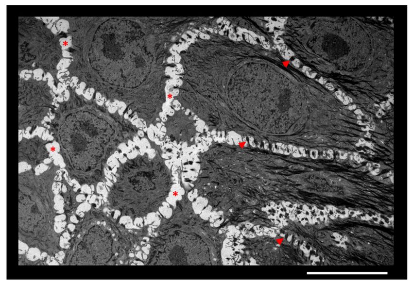
4.0/). transmission electron microscopy (Figure 1).
Cells 2021, 10, 3096. https://doi.org/10.3390/cells10113096 https://www.mdpi.com/journal/cells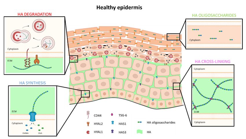
Cells 2021, 10, 3096                                                                                                   2 of 17

                       Figure 1. Intercellular spaces between keratinocytes of epidermis. Granular layer of an in vitro recon-
                       structed human epidermis observed by transmission electron microscopy. Adjacent keratinocytes are
                       separated by ECM (*) and anchored to each other by desmosomes (arrowheads). Scale bar = 10 µm.

                             The sugar-rich composition of epidermal ECM was first revealed in 1951 using Alcian
                       blue staining [9]. The epidermal ECM is accordingly largely composed of carbohydrate
                       macromolecules called glycosaminoglycans (GAGs). Among GAGs, those with covalent
                       link to serine residues in core-proteins give rise to proteoglycans. Heparan sulfate and
                       chondroitin sulfate, for instance, account, respectively, for 60% and 20% of such GAGs in
                       the epidermis [10], where they participate in formation of proteoglycans, such as glypican,
                       syndecans (1 to 4), and versican [11–13]. Hyaluronan, the only non-sulfated GAG and the
                       only GAG without link to proteins, is also found in the epidermal ECM [5]. An in vitro
                       study of human keratinocytes has shown that HA accounts for 54% of GAGs released in
                       culture medium by this cell type [14]. In human skin, the epidermal ECM contains 25 µg
                       of HA/g of fresh epidermis, while the dermis contains between 120 and 200 µg of HA/g
                       of fresh dermis [3,15]. Together, GAG and proteoglycans create a specialized intercellular
                       environment favorable to the diffusion of nutrients, growth factors, and cytokines [12,16],
                       as well as for eventual upcoming immune cells. Furthermore, some types of chondroitin
                       sulfate proteoglycans (e.g., MCSP) are expressed in an age-dependent manner by epidermal
                       cells that exhibit stem cell properties [17].
                             The ECM can be divided into two zones: the pericellular matrix and the intercellular
                       matrix. The pericellular matrix is in close contact with neighboring cells, allowing direct
                       interaction with cellular receptors [18]. In pericellular matrix, HA can interact with its
                       trans-membrane receptor CD44, and cytokines and growth factors can rapidly bind their
                       receptors. The intercellular matrix is found between cells without direct contact [18]. In
                       the epidermis, intercellular spaces are very thin. Nevertheless, they can increase in certain
                       pathologies, such as atopic dermatitis, which is characterized by an increased epidermal
                       HA production able to retain a large amount of water, thereby creating spongiosis [19].
                       These increased spaces notably promote the migration of immune cells into the epithelial
                       tissue.

                       3. Epidermal Hyaluronan
                       3.1. Molecular Structure of HA
                            HA is composed of repeated disaccharide units made of D-glucuronic acid (GlcUA)
                       and D-N-acetylglucosamine (GlcNAc). These saccharides are bound by β-1.3 and β-1.4
                       linkages to form long linear polymers [β1.4-GlcUA-β1.3-GlcNAc-]n of high molecular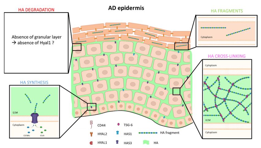
Cells 2021, 10, 3096                                                                                            3 of 17

                       weights, usually ranging from 105 to 107 Daltons, and from 2 to 25 µm in length [20,21].
                       HA polymers interact with proteins [3], although they never bind through covalent link.
                       Specific HA-interacting proteins are called hyaladherins or HA-binding proteins (HABP).
                       Some of them, such as CD44 and RHAMM, are membrane-bound, and recognized as two
                       HA receptors on cell plasma membrane. Other hyaladherins are secreted into the ECM,
                       such as the TNFα-stimulated-gene-6 (TSG-6) protein involved in extracellular cross-linking
                       of HA [22]. Some proteoglycans, such as versican, are also able to bind HA via their
                       N-terminal domain, thereby creating complex molecular networks in the ECM [2,16,23].
                       Moreover, since HA carries negative charges, this macromolecule can retain a large amount
                       of water (up to 70% of its weight) through its remarkable hydrophilicity. Such characteristics
                       of HA confer viscoelastic properties to the HA-containing tissues and spaces [4,21].

                       3.2. Synthesis of HA
                            HA is synthesized by enzymes located at the inner side of the plasma membrane
                       and simultaneously exported as polymers directly in the ECM. These enzymes, called HA
                       synthases (HAS), are glycosyltransferases [24]. The substrates utilized by HAS enzymes are
                       UDP-α-N-acetyl-D-glucosamine (UDP-GlcNAc) and UDP-α-D-glucuronate (UDP-GlcUA)
                       originating from cellular metabolism of glucose [25]. In mammals, three HAS isoenzymes
                       have been identified: HAS1, HAS2, and HAS3. Encoded by genes located on different chro-
                       mosomes (19q13.3 for HAS1, 8q24.12 for HAS2 and 16q22.1 for HAS3), HAS enzymes share
                       55 to 70% identity [26]. They contain, between N-terminal and C-terminal regions, a central
                       region harboring 75–87% similarity [27]. All three enzymes have seven transmembrane
                       domains [24] and have distinct binding domains for the two substrates [25]. Although
                       structurally similar, the three HAS enzymes are characterized by very different temporal
                       expression patterns, by the sizes of generated HA polymers, and by different enzymatic
                       activities [27]. HAS activity is partially dependent on the concentration of available cellular
                       UDP-GlcNAc. While their Vmax do not show any significant differences, the Km of HAS1
                       enzyme is more elevated than the Km of HAS2 and HAS3 enzymes, showing that HAS1
                       exhibits a lower affinity for its substrate than HAS2 and HAS3 [27]. Consequently, HAS1
                       enzyme is unable to form HA polymers if the amount of available UDP-GlcNAc is low. On
                       the other hand, the activity of HAS2 appears poorly influenced by substrate concentration,
                       and the activity of HAS3 remains independent of UDP-GlcNAc concentration [28].
                            HAS1 mRNA is expressed during early stages of embryonic development (gastrula-
                       tion and neurulation) and during cell differentiation. This synthase has a low enzymatic
                       activity since the amount of product formed per minute is low. HAS1 enzyme uses large
                       amounts of UDP-GlcNAc, allowing the production of high molecular weight HA poly-
                       mers [27,28]. In the epidermis, an in vitro study showed that HAS1 mRNA is expressed
                       during keratinocyte differentiation [29]. HAS2 enzyme is essential during embryonic
                       development [30]. While HAS2 mRNA is very weakly expressed in adult tissues, its
                       overexpression causes skin changes. In particular, Has2 mRNA overexpression induces
                       an increased amount of HA with increased thickness of the skin, as in the skin of the
                       Sharpei dog breed [31]. Similarly, naked mole rats carry mutations in Has2 gene that
                       cause an abnormally enhanced synthesis of high molecular weight HA [32]. Conversely,
                       in elderly humans, the typical thinning of the skin is accompanied by decreased HAS2
                       mRNA expression [33]. HAS2 is characterized by a slightly higher activity than HAS1 and
                       generates in vitro HA polymers of comparable molecular weights. However, HA polymers
                       generated by HAS2 in vivo in transfected cells exhibit higher molecular weights than those
                       made by HAS1 [27]. HAS3 is the most active enzyme and synthesizes, therefore, low
                       molecular weight polymers (100–1000 kDa) [27]. This enzyme is predominantly expressed
                       in inflammatory processes (reviewed in Tavianatou et al., 2019 [34]), including inflamed
                       skin [35]. Accordingly, Malaisse et al. (2014) [29] highlighted that HAS3 mRNA is overex-
                       pressed in lesional atopic dermatitis skin, while HAS1 mRNA was preferentially expressed
                       in healthy epidermis. However, in a different study, divergent data indicate abundant
                       mRNA expression of HAS3 in healthy keratinocytes [36].
Cells 2021, 10, 3096 4 of 17
HAS activity can be modulated by growth factors and cytokines, such as keratinocyte
growth factor (KGF), epidermal growth factor (EGF), tumor necrosis factor α (TNF-α),
transforming growth factor β (TGF-β), and bone morphogenetic protein (BMPs), as well
as by extracellular UTP [37–39]. Post-translational modifications of HAS enzymes can
occur [40]. Phosphorylation [41,42] and O-GlcNAcylation [43] of HAS enzyme increases
their activities. However, the link between post-translational modifications and changes in
enzymatic activity remains unknown. Finally, some mono-ubiquitination of HAS2 could
be involved in its enzymatic activity, as mutated lysine residue 190, the acceptor site for
ubiquitin, is responsible for HAS2 enzyme inactivation [44].
3.3. Degradation of HA
The half-life of HA is relatively short in the epidermis, ranging from 1 to 2 days [3,45].
Enzymes responsible for HA degradation are endoglycosidases and exoglycosidases.
Hyaluronidases (HYAL) are members of the endoglycosidase family and specifically cleave
the β-1.4 bonds in HA polymers. The residual oligosaccharides are then hydrolyzed by
exoglycosidases into β-D-glucuronate and β-N-acetyl-D-hexosamine [46]. In humans,
five hyaluronidase-like genes and one pseudogene (HYALP1) have been identified. They
share 40% similarity. HYAL1, 2, and 3 are located on chromosome 3p21.3 and have similar
genomic structure with four exons. HYAL4, HYAL5 (PH20), and HYALP1 are located on
chromosome 7q31.3 and present one extra exon compared to HYALs encoded on chromo-
some 3. Beside the hyaluronidase-like genes, another enzyme implicated in HA catabolism
has been identified. KIAA1199, also named CEMIP (cell-migration inducing protein) or
HYBID (HA-binding protein involved in HA depolymerization), is located on chromosome
15q25.1 and presents no significant similarity with other hyaluronidases [47]. HYAL1,
HYAL2, KIAA1199, and PH20 enzymes are able to cleave HA polymers [47,48]. While
PH20 has an activity restricted to spermatozoa, KIAA1199, HYAL1, and HYAL2 are active
in various somatic tissues. KIAA1199 enzyme is notably involved in the regulation of
infection-related dermis inflammation as its absence has been shown to increase inflam-
mation and antimicrobial activity [49]. It is also implicated in photoaging skin through
its action on fibroblasts [50]. It degrades HA by means of endocytosis of clathrin-coated
membrane regions [50]. Only HYAL1 and HYAL2 are expressed by keratinocytes [51,52].
Hyaluronidase 1 is responsible for the cleavage of HA polysaccharides into di- or
tetra-saccharides [47]. HYAL1 is active in the skin and more specifically in keratinocytes
within the granular layer [51]. In terms of subcellular localization, HYAL1 is specifically
localized in lysosomes, an acidic compartment (pH about 3.7) necessary for its activity, as
the optimal pH for HYAL1 is 3-4 [53,54]. Hyaluronidase 2 is structurally similar to HYAL1
but has a different activity and localization. HYAL2 protein is anchored to the plasma
membrane by a glycosylphosphatidylinositol (GPI) anchor and generates fragments of HA
around 20kDa of molecular weights [55,56]. HYAL2 can be active at acidic (4) and neutral
(7.5) pH [48]. In the epidermis, HYAL2 enzyme is anchored in cell membranes of viable
layers [52].
A cellular model of HA degradation based on the involvement of both HYAL1 and
HYAL2 enzymes was proposed in the early 2000s [45,57]. Pericellular HA is believed
to bind its CD44 receptor at the plasma membrane. HYAL2 protein is also anchored to
the cell membrane, close to CD44, creating intimate vicinity between membrane-bound
HA and HYAL2 protein, resulting in enzymatic degradation of HA polymers into 20kDa
oligosaccharides, which then become internalized in endosomes, along with the enzyme.
When HA fragments-containing endosomes fuse with lysosomes, the low pH inside the
resulting compartment initiates the HYAL1-driven digestion of HA fragments into di- or
tetra-saccharides.
At epidermal tissue levels, HA detected using procedures based on HABP appears
mainly localized between keratinocytes that constitute basal and spinous layers, but it is
not detected in granular and cornified layers [29]. While CD44 and HYAL2 proteins are
anchored in the membrane of living layers [52,58], HYAL1 enzyme is active in lysosomalCells 2021, 10, 3096 5 of 17
compartment of granular layer [51]. Over the course of differentiation, some residual
intracellular HA oligosaccharides seem to end up inside cornified keratinocytes where
they most likely play a role in skin hydration together with natural moisturizing factors
(NMF) [20,51] (Figure 2). During keratinocyte migration towards the most superficial layers
and as a result of the acidification of the upper layers, filaggrin becomes degraded into small
free metabolites and amino acids, thereby forming NMF essential to skin hydration, barrier
homeostasis and superficial desquamation. Skin hydration is a crucial factor involved in
tissue resistance to mechanical stress and contributes to its resilience [59].
Figure 2. Model of HA metabolism in healthy human epidermis. HA is synthetized and extruded by HAS1 and, to a
lesser extent, by HAS3 in ECM of basal and spinous layers. In ECM, TSG-6 protein binds and cross-links HA to trap it
locally and form a dense matrix. In the granular layer, extracellular HA binds CD44. Simultaneously, HA is degraded
into oligosaccharides by HYAL2 and internalized in endosomes which fuse with lysosomes. At acidic pH of lysosomes,
HYAL1 enzyme is active and degrades HA in di- or tetra-saccharides. These low molecular weight HA are then found in
corneocytes, where they participate in cutaneous hydration.
One might wonder whether HA present in the ECM of epidermis is exclusively
metabolized by keratinocytes or whether exchanges occur between the dermis and the
epidermis. Studies based on in vitro reconstructed epidermal models have shown that
epidermal HA synthesized by keratinocytes is subsequently found under the epidermal
tissue (either in medium [29], or in the supporting collagen matrix [60]), suggesting that
HA molecules can escape the epidermis and diffuse into sub-epidermal spaces and, by
extension, into the dermis. The skin epidermal-dermal junction is organized by basement
membrane (BM) components that create the essential structure between epidermal and
dermal HA. Indeed, when the epidermis of elderly people is exposed to sunlight, the BM is
somehow damaged by UV exposure, and smaller amounts of HA are observed in exposed
epidermis than in unexposed tissue. In addition, the inhibition of metalloproteinases and
heparanases, in an in vitro model of full thickness skin equivalent exposed to UV, protects
the BM integrity and retains 25% additional HA when compared to HA amount in control
epidermis [61]. Consistently, in in vitro rat epidermis model, the setting up of a BM between
the epidermis and the collagen matrix was shown to retain approximately 75% HA in the
epidermis, whereas 80% of HA was lost in absence of BM [62,63]. In reconstructed human
epidermis (RHE) models on polycarbonate filters, cells in the basal layer secrete basement
membrane components (lamina densa and lamina lucida) and form hemidesmosomes.
Structures organized by RHE on the filter can only be considered as BM-like structures
since they lack collagen VII and fibronectin, for instance, in contrast to normal BM [64].Cells 2021, 10, 3096 6 of 17
Nevertheless, the proportion of HA retained in RHEs is approximately 95%, since a very
small amount of epidermal HA appears released into the underlying medium [29]. In
addition to the importance of BM in the retention of epidermal HA, the TSG-6 protein in
the extracellular matrix is involved in some cross-linking with HA chains and seems able to
prevent HA from “sinking” into the underlying compartment [65] (Figure 2). In summary,
a large amount of HA synthesized by keratinocytes does remain in the epidermal matrix
and is locally degraded, but about 10% of epidermal HA diffuses into the dermis through
the BM and reaches local lymph and blood vessels, in which HA then circulates towards
lymph nodes, or the liver, where it is degraded.
3.4. Functions of HA in the Epidermis
3.4.1. Regulation of Keratinocyte Proliferation
In keratinocytes, interaction of HA with its CD44 receptor activates signaling pathways
that induce proliferation [66].
Three decades ago, the detection of HA on the surface of newly divided keratinocytes
firstly indicated a potential link between elevated HA production and cell division [67].
Indeed, another study incubated in vitro rat epidermis with EGF (Epidermal Growth
Factor) to induce keratinocyte proliferation and reported simultaneous increased HA
production [60]. Consistent with those studies, topical application of HA bound to phos-
phatidylethanolamine on aged mouse skin (allowing HA diffusion through the hydropho-
bic cornified barrier) was found able to induce keratinocyte proliferation and associated
epidermal thickening [68]. Moreover, injection of HA into a human cutaneous wound
model was shown to accelerate re-epithelialization without altering inflammation [69].
When keratinocytes are treated with 4-methylumbelliferone (4-MU), an inhibitor of HA syn-
thesis, an expected decrease in HA production was found in this cell type, accompanied by
inhibited proliferation and decreased cell migration related to perturbed cytoskeleton [70].
However, our group more recently observed that 4-MU was likely not suitable to investi-
gate keratinocyte proliferation since the inhibitor affects this process in a HA-independent
manner [71]. Despite this limitation, studies have demonstrated some involvement of HA
in cell proliferation without the use of 4-MU. Notably, addition of exogenous HA to an
organotypic tissue culture provoked an induction of keratinocyte proliferation and related
epidermal thickening. In addition, expression of some molecules involved in the epidermal-
dermal junction was also reported as increased by the addition of HA [72]. More recently,
a treatment with 1-ethyl-β-N-acetylglucosamine (β-NAG2), a substrate converted into
UDP-N-acetylglucosamine by cellular metabolism, was found to stimulate HA production
in a cultured epidermis, together with increased proliferation of basal cells [73].
However, despite many studies that indicated links between HA production and
keratinocyte proliferation, our effort to identify a causal link between these two events
remained unsuccessful. Indeed, our group reported that neither a treatment of recon-
structed epidermis with an exogenous hyaluronidase activity from a Streptomyces bacterium
(StrepH), nor an inhibited HA synthesis caused by knocking down UGDH gene expression
(UDP-glucose 6-dehydrogenase) in keratinocytes produced any significant consequences
on keratinocyte proliferation [71]. In summary, the eventual ability of HA to directly
modulate keratinocyte proliferation is a question that remains controversial.
3.4.2. Regulation of Keratinocyte Differentiation
When studying the role of HA in epidermal differentiation, studies have reported
HA as being a repressor of the process. In 2003, for instance, a study carried out on rat
keratinocytes illustrated that treatment of those cells with KGF (Keratinocyte Growth
Factor) was inducing an accumulation of HA by increased HAS2 and HAS3 expression.
Simultaneously, the expression levels for differentiation markers, such as the keratin 10 or
filaggrin, were conversely reduced [74]. When reconstructed rat epidermis were treated
with the exogenous StrepH hyaluronidase, the expression of keratin 10 and filaggrin were
found increased as a consequence of HA degradation [63]. Still, another study usingCells 2021, 10, 3096 7 of 17
StrepH on mouse epidermis to degrade HA led to similar conclusions as the treatment with
StrepH was reducing the epidermal thickness by half its normal value. In such conditions,
the expression of keratin 10 and filaggrin appear higher than in tissues cultured with
intact HA [75]. Those results can be explained if keratinocytes treated with exogenous
hyaluronidase are committed earlier towards terminal differentiation, impeding to reach
the normal tissue thickness during epidermal reconstruction.
Conversely, other studies have puzzlingly demonstrated an activating effect of HA
on epidermal differentiation. However, since different sizes of HA polymers may induce
different, or even opposite effects on signaling pathways, HA of different sizes may differ-
entially regulate differentiation of epidermal keratinocytes. An increase in HA amounts
production was found to trigger expression of markers, such as claudin and occludin,
involved in the formation of tight junctions in differentiated granular keratinocytes [76].
Experiments studying human epidermis treated with 1-ethyl-β-N-acetylglucosamine (β-
NAG2) indicated, with increased HA production, some concomitant increase in filaggrin
and transglutaminase 1 expression. However, this accelerated differentiation can be slowed
down to basal levels by blocking the β-NAG2 substrate converting enzyme [73].
Interestingly, the stimulation of differentiation by HA seems also mediated by its
interaction with the CD44 receptor. Bourguignon et al. (2004) demonstrated that HA-
CD44 interaction was involved in cytoskeletal functions that are essential for keratinocyte
adherence and differentiation [77]. Moreover, the addition of exogenous HA was shown to
increase the expression of keratin 10, involucrin, and profilaggrin under the dependence
of CD44 receptor [78], and the absence of CD44 in the epidermis consistently resulted
in decreased keratin 10 and filaggrin expression [79]. Again, another study performed
regarding the addition of small HA (tetra-saccharide) molecules to keratinocyte cultures
has reported an increased expression of keratin 10 when phosphorylation of CD44 and
increased cytosolic calcium concentration happen [80].
Finally, studies conducted by Malaisse et al. (2016) in our laboratory concluded that
HA production by keratinocytes and their differentiation process are independent events.
Indeed, human reconstructed epidermis incubated with 4-MU or StrepH reveal no impact
on the expression of either keratin 10, filaggrin, or involucrin. In addition, even further,
keratinocytes deficient for the UGDH enzyme reveal no alteration in their differentiation
process [71]. In summary, the relation between HA and keratinocyte differentiation is once
again not yet elucidated.
To conclude, possible roles of HA during keratinocyte proliferation or differentiation
and their overall importance for epidermal physiology are still a matter of debate. However,
although the role of HA in keratinocyte proliferation remains unresolved, its CD44 receptor
is a marker of cell stemness [81]. CD44 is a marker for cancer stem cells in several epithelia,
but also of keratinocytes enriched for epidermal stem cells with long-term repopulating
ability. On the other hand, the maintenance of stem cells and their property for self-renewal
are favored when the environment is enriched in HA [82], suggesting involvement of HA
and CD44 in the maintenance of an epidermal stem cell character.
3.4.3. Involvement in the Epidermal Barrier Settings and Integrity
The final step in keratinocyte differentiation is keratinization, which means production
of the impermeable cornified layer, the most superficial layer of the epidermis. During this
late stage of epidermal differentiation, cells become embedded in a lipid ceramide-rich ma-
trix which seals intercellular spaces between the fully differentiated cornified keratinocytes.
This process maintains a waterproof barrier on the skin surface [83]. During this pro-
cess, as early as in the granular layer, lamellar bodies elaborated by the Golgi apparatus
are triggered to fuse with the apical plasma membrane of keratinocytes and discharge
their content (e.g., sphingomyelin, glycosphingolipids, phospholipids, cholesterol), as
well as several acid hydrolases (serine protease, phospholipase A2, sphingomyelinase, β-
glucocerebrosidase) in the extracellular matrix, ready to cleave substrates at low pH [84,85].
Between the shedding of lamellar bodies in the stratum granulosum and the establishmentCells 2021, 10, 3096 8 of 17
of a dense matrix in the stratum corneum, lipids undergo maturation, thanks to enzymes
released jointly by the lamellar bodies. These enzymes are indeed involved in the cleav-
age of glycosphingolipids into ceramides, and phospholipids into fatty acids. Ceramides
account for 50% of stratum corneum lipids and are covalently bound to envelope proteins,
mainly to involucrin [86]. Cholesterol is another major matrix constituent and accounts for
25% of matrix lipids [86,87]. HA and its CD44 receptor are involved in the establishment of
this lipid barrier.
HA and CD44 are involved in formation of lamellar bodies and in the polarization
of their secretion. In Cd44 knock-out mice, content and polarized secretion of lamellar
bodies are clearly disturbed [78]. While, in normal conditions, lipids are exclusively se-
creted at the apical pole of granular layer cells, at the interface with the cornified cells, the
absence of Cd44 results in dual apical and basolateral secretion of lamellar bodies, indi-
cating unpolarized secretion. In addition, HA-mediated cholesterol synthesis is blocked
in those conditions. Indeed, whereas HA increases cholesterol concentration in the epi-
dermis, Cd44-/- keratinocytes treated with HA do not produce cholesterol. As a major
consequence, defects in lipid production and polarized secretion impair the formation of
the epidermal barrier [78]. In agreement with those observations, the addition of small
HA molecules (tetra-saccharide) increases the expression level of enzymes responsible for
ceramide synthesis, as well as the intracellular ceramide-content in human keratinocytes
in vitro. This HA-mediated enhancement of lipid content improves the water retention in
stratum corneum of mice following cutaneous UVA irradiation, as assessed by decreased
transepithelial water loss [88]. The absence of CD44 also impacts the organization and,
thus, the function of epidermal tight junctions. During development, Cd44-/- mice exhibit
on day 18.5 of embryogenesis a transient 1-day delay in skin barrier establishment. Such
a delay corresponds with alteration in availability and organization of tight junction pro-
teins [89]. In summary, the HA-CD44 interaction is involved in the maintenance of the
epidermal barrier integrity and both molecules influence each other. HA influences CD44
expression [90]. Conversely, in the absence of CD44, a smaller amount of HA is detected in
the epidermis, which then appears thinner [78].
3.5. Epidermal Hyaladherins
In the ECM, HA is able to interact through non-covalent binding with a large number
of molecules called hyaladherins or HA-binding proteins. These HA partners are extracel-
lularly localized or attached to the cell membrane [21]. Among cellular partners, the best
known are the CD44 receptor and the RHAMM receptor.
CD44 (Cluster of Differentiation 44) is a 19 exons-encoded transmembrane glycopro-
tein which exhibits numerous isoforms produced as a consequence of alternative splicing
and post-translational modifications [6] that modulate CD44 affinity for HA [91,92]. In the
epidermis, 18 different transcripts encoding CD44 isoforms have been identified [58]. All
variants of CD44 share an extracellular domain able to bind HA, named LINK domain.
Other molecules, such as GAGs (heparan sulfate, dermatan sulfate, chondroitin sulfate),
are also bound by CD44 receptors, depending on the type of isoforms. GAG-CD44 inter-
action has been found able to enhance the presentation of growth factors to their specific
receptors [58]. HA-CD44 interaction allows the formation of a pericellular matrix [93]
involved in the regulation of keratinocyte differentiation [6] and in the development of
skin barrier [89]. Actually, HA-CD44 binding induces several various signaling pathways,
depending on the size of HA polymers. Indeed, high molecular weight HA molecules
bind to the CD44 receptor by mostly irreversible hydrogen bonds and activate RhoA-like
RhoGTPase signaling pathways that lead to cell proliferation and migration. In contrast,
low molecular weight HA form weaker, more reversible hydrogen bonds, and activate
Rac1-like pathways that favor cell adhesion and differentiation [6,34]. The variable effects
produced by different HA molecules that exhibit different molecular weight were recently
presented in detail and reviewed by Tavianatou et al., 2019 [34].Cells 2021, 10, 3096 9 of 17
RHAMM (Receptor for HA-mediated mobility) is another HA receptor characterized
by several isoforms that localize at the cell surface, in the cytoplasm, in the nucleus, and
even in the ECM. Poorly expressed in physiological conditions, RHAMM receptor is
overexpressed in cancer cells and in inflammatory conditions, such as during skin scarring.
This receptor is able to regulate keratinocyte migration. Indeed, in Rhamm-/- mice, the
migration of keratinocytes during wound healing was considerably disturbed [94].
The Case of Epidermal TSG-6 Protein
In the category of extracellular hyaladherins, there exist some proteoglycans, especially
versican in the epidermis, but we will emphasize here an anti-inflammatory protein named
TSG-6 (TNFα stimulated gene 6). This TSG-6 protein harbors a LINK domain very similar to
the LINK domain characterized in the CD44 receptor and responsible for HA-binding [95].
However, interactions between HA and the TSG-6 protein appear stronger than those
observed between HA and the CD44 receptor [96]. Interestingly though, binding HA to
the TSG-6 protein increases the affinity of HA for its CD44 receptor [97,98]. A possible
explanation can be found in the observation that HA binding induces dimerization of
TSG-6 protein, causing cross-linking of HA chains, matrix condensation, and increased
physical proximity of HA polymers to CD44 receptors [99]. Cross-linking of HA chains and
their consequences on the organization of the ECM further occur through inter-α-trypsin
inhibitor (ITI), a protein with an even higher affinity for HA than TSG-6. ITI is a plasma
glycoprotein belonging to a family of protease inhibitors composed of two heavy chains
(HC) linked by ester bonds to chondroitin sulfate and to a bikunin. When ITI localizes at the
same site as HA and TSG-6 protein, it can catalyze the formation of a covalent HC-HA bond.
This reaction takes place in two steps. Firstly, one ITI heavy chain is transferred to TSG-6
protein by a first transesterification reaction. Secondly, the heavy chain is transferred to an
acetylglucosamine residue of HA [100]. Such HC-HA complexes form a more widespread
and hydrated HA network than the network resulting from HA-TSG-6 complexes [97]. Six
heavy chains have been identified, and HC1 and HC2 have been detected in the normal
epidermis [101]. The predominantly expressed heavy chain in human skin (at the dermal
level) is HC5, whose expression is increased in inflammatory skin conditions, such as
allergic contact dermatitis [102]. HC5 has been shown to bind and stabilize HA, and
to participate in matrix formation [103]. The TSG-6 protein, although possessing anti-
inflammatory properties, is expressed and secreted in the epidermis under physiological
conditions [101]. We recently found that the epidermal TSG-6 protein is involved in the
retention of HA between keratinocytes, in the thin intercellular spaces between epidermal
cells [65]. The HA maintained between keratinocytes by TSG-6 protein creates space and
reduces cohesion between keratinocytes, allowing the diffusion of cytokines and growth
factors [104]. Furthermore, the TSG-6 protein is widely expressed and secreted in skin
under inflammatory conditions related to a proper epidermal wound healing [105,106]. It is
also secreted by keratinocytes in response to epidermal infection by dermatophytes [107], or
when epidermal cells are challenged by Th2-immune conditions [65,108]. Simultaneously,
HA is massively produced in inflammatory conditions resulting in increased intercellular
spaces in which cells can migrate [104]. Of course, the TSG-6 protein is expressed and
secreted by many other cell types than epidermal keratinocytes. It may, therefore, play
other roles than those discussed in the epidermis, as it is detailed by Day and Milner, in
their recent 2019 review on this protein and its function [22].
4. Epidermal HA in Atopic Dermatitis
4.1. Atopic Dermatitis
As one well-characterized pathological condition observed in skin, atopic dermatitis
can exemplify how much HA can be involved when some abnormal tissue conditions
occur. Atopic dermatitis (AD) is a chronic inflammatory skin disease characterized by
xerosis, spongiosis, and hypogranulosis, as well as altered skin barrier [109]. The etiology
is controversial since many factors are involved: environmental factors, genetic predisposi-Cells 2021, 10, 3096 10 of 17
tion, defective barrier, and immune dysregulation [110]. There are two main hypotheses
proposed regarding the development of AD. According to the “outside-inside” hypothe-
sis, patients have a defective epidermal barrier that potentially allows allergens and/or
pathogens entry in the skin, leading to inflammatory and immune responses. Conversely,
the “inside-outside” hypothesis suggests that a preexisting immune dysregulation initially
impacts the barrier. Indeed, any triggered release of Th2 cytokines (such as interleukins
4, 13, 25) exhibits proven ability to weaken the epidermal barrier. In respect to both hy-
potheses, the situation created during atopic dermatitis actually leads to some vicious
circle which reinforces the diseased state [19]. Available review papers, e.g., De Vuyst et al.,
2017 [111], and Huet et al., 2018 [112], propose models to study atopic dermatitis based on
in vitro reconstructed human epidermis. The inside-outside condition can be mimicked by
creating inflammatory conditions in the models, for instance, when RHE are incubated with
mixtures of cytokines typically elevated in AD (IL-4, -13, -31, -22, -25, and/or TNFα). The
presence of Th2 cytokines results in decreased expression of late differentiation markers,
such as keratin 10, filaggrin, involucrin, and loricrin, along with a reduction in ceramides
and free fatty acids content. Together, such alterations in the epidermal differentiation
program that is responsible to create and maintain the epidermal barrier contribute to the
impairment of its function. According to the outside-inside hypothesis, genetic factors in
this case may be involved in barrier defects. In favor of this hypothesis, RHE produced by
filaggrin-deficient keratinocytes display increased permeability and increased sensitivity
to Th2 cytokines treatment. In addition, many other environmental factors, such as abuse
of detergent products on the skin, affect the lipid-based epidermal barrier in vivo [110].
4.2. Implication of HA in Atopic Dermatitis
Remaining low in normal conditions, the mRNA expression of HAS3 has been re-
ported as upregulated in AD skin conditions, simultaneously with well-recognized AD
markers, i.e., CA2 and NELL2 [29,113]. Indeed, differences in HAS expression profiles
have been reported between healthy epidermis, mainly expressing HAS1 mRNA, and the
epidermis of AD patients where elevated HAS3 expression is found (HAS2 remains scarcely
expressed in adult epidermis) [29] (Figure 3). Similarly, when inflammatory conditions
related to UVB exposure are imposed to keratinocytes, HAS3 mRNA expression is upregu-
lated [36,114], and HAS3 mRNA is also overexpressed upon stimulation by interferon γ,
whilst diminished following TGFβ challenge [115]. Consistently, HAS3 enzyme exhibits
high activity and produces low molecular weight HA fragments [27]. Such low molecular
weight fragments participate to tissue inflammation because they behave in the epidermis
as DAMPs (Damage Associated Molecular Pattern) and bind Toll-Like Receptor (TLR) 2
and TLR 4 (for more details, see the review by Tavianatou et al., 2019 [34]). TLRs, through
activation of NFκB, are the initiators of release of pro-inflammatory cytokines, as well as
inducers of HAS3 overexpression, thereby amplifying local synthesis of low molecular
weight HA and reinforcing the inflammatory response as a consequence [116]. In addition,
the heparin-binding EGF-like growth factor (HB-EGF) released from keratinocytes in atopic
dermatitis also increases HAS3 expression [29,117].
Overexpression of HAS3 in a model mimicking atopic dermatitis causes massive
accumulation of HA in the intercellular spaces between keratinocytes of the living epider-
mal layers [19]. Increase in the amount of intercellular HA is consistent with enhanced
detection of HA in ECM of severe eczema skin in parallel with HAS3 overexpression [118].
Accumulation of HA in intercellular spaces can also be related to spongiosis, typical of
AD skin. In severe eczema, some reduced expression of epidermal cadherins results in
water entry in the tissue and spongiosis [118]. The presence of HA in larger amounts than
in normal skin might be responsible for water retention between keratinocytes and for
spongiosis. Alternatively, the enlargement of intercellular spaces due to reduced cadherin-
driven junctions might favor accumulation of HA in the available space. Of interest, as
HA is particularly retained in intercellular spaces by TSG-6 protein [65] (Figure 3), and
TSG-6 protein is increasingly produced in vivo in AD skin, such as in vitro in reconstructedCells 2021, 10, 3096 11 of 17
epidermis challenged with Th2 cytokines, this anti-inflammatory protein might favor the
accumulation of HA in pathologies, such as AD [65].
Figure 3. Model of HA metabolism in atopic dermatitis epidermis. Low molecular weight HA fragments are synthetized
and extruded by HAS3 enzyme in ECM of basal and spinous layers, while HAS1 mRNA is less expressed. In inflammatory
conditions, TSG-6 protein is more expressed in parallel with an increase HA production, allowing a strong cross-linking. As
atopic dermatitis is characterized by hypogranulosis, expression and activity of HYAL1 are probably disturbed, and HA
fragments are found in corneocytes.
In inflammatory epidermis, overproduction of HA is accompanied by overexpression
of the CD44 receptor. Abundance of this receptor is thought to play a role in recapture of
HA released in excess, in order to finally reduce and resolve inflammation [35]. In parallel
with HA endocytosis, hyaluronidases 1 and 2 enzymes degrade HA, according to the
model proposed by Tammi et al., 2001 [57].
AD is an inflammatory condition characterized by an absent granular layer, where
HYAL1 enzyme is located and active in physiological conditions [51]. According to this
model, HYAL1 may be less active in AD lesional epidermis (Figure 3). In line with this
hypothesis, Hyal1-/- mouse epidermis is characterized by high-molecular-weight HA con-
tained in cornified keratinocytes [51], whereas solely fragments below 6 × 10⁴ Daltons are
detected in cornified cells of wild-type epidermis [20]. Consistently, the typical parakera-
totic superficial layer during contact dermatitis contains HA of molecular weight superior
than 6 × 10⁴ Daltons [119]. While low molecular weight fragments would contribute to
hydration, such as NMF [20], high molecular weight HA in excess in the cornified layer
would have a negative impact on properties of this layer and, therefore, on its barrier
function. Indeed, high molecular weight HA are able to efficiently retain larger overall
content of water in comparison with small fragments of HA, counteracting the formation
of the normally hydrophobic cornified layer and leading to some potentially increased
epidermal permeability.
In epidermal ECM, HA can interact with other matrix components. Notably, versican,
a proteoglycan secreted in the ECM of the basal layer of epidermis, is able to interact
with HA [12,120]. This proteoglycan is involved in the formation of a provisional matrix
required by leukocytes during inflammatory response, such as the one observed in AD [23].
Decorin, another proteoglycan, is secreted between cells of the suprabasal layers of the
epidermis [121]. Decorin mRNA expression was significantly decreased when the HA
extracellular matrix is disorganized, due to the lack of the TSG-6 protein [65]. In contact
dermatitis, decorin-deficient mice exhibit a weaker inflammatory response than wildtypeCells 2021, 10, 3096 12 of 17
mice as a consequence of impaired leukocyte recruitment [122]. Those studies highlight
the importance of the organization of the epidermal matrix composed of GAGs (HA) and
proteoglycans (versican and decorin) in the regulated recruitment of immune cells in an
inflammatory context. Moreover, when HA chains are properly cross-linked and organized
by the TSG-6 protein, certain pro-inflammatory growth factors and cytokines (such as
IL-8) can be sequestered [123,124], thereby limiting the presentation of the ligands to their
respective receptors and, thus, regulating the attraction of neutrophils toward the inflamed
site [106].
Atopic dermatitis is characterized by hyperplasia. Epidermal thickening is apparently
a consequence of overexpressed EGF receptor (EGFR) [125] and HB-EGF [29,126]. HB-EGF
is rapidly cleaved and available as a soluble ligand in the ECM during the inflammatory
setting [127]. HB-EGF can then bind EGFR and activate downstream pathways that can
induce epidermal proliferation and elevated HAS3 mRNA expression [29,117,128]. In
parallel, stimulation of EGFR induces the expression of pro-inflammatory cytokines, such
as IL-8, that recruit leukocytes [125]. Interestingly, EGFR is localized in plasma membranes
cholesterol-rich lipid rafts, close to CD44 [60]. It has been shown, in cancer cell lines, that
EGFR and CD44 receptors interact to auto-regulate HA synthesis [117].
In summary, HA and other ECM components (proteoglycans, cytokines, growth
factors) appear to support HA production in order to generate an organized matrix for the
efficient and controlled recruitment of inflammatory cells in cutaneous pathologies.
5. Conclusions
HA is a glycosaminoglycan found in intercellular spaces. Although secreted in many
vertebrate tissues, 50% of HA is localized in the skin. Predominantly detected in the dermis,
HA is, nonetheless, also located between keratinocytes in living layers of the epidermis,
where it appears involved in development and maintenance of a competent epidermal
barrier. In addition, HA plays controversial roles in proliferation and differentiation of
epidermal keratinocytes. The enzyme HAS1 seems mainly responsible for the production
of HA in the epidermis in normal conditions, but a switch in expression of the different
HAS enzymes in the pathological epidermis, characterized by overexpressed HAS3, alters
the molecular characteristics of epidermal HA in skin diseases. For its degradation, HA is
processed via a CD44-HYAL2-HYAL1 axis, suggesting that HA is potentially internalized
and degraded into very low molecular weight fragments when it reaches the granular cell
layer of the epidermis. This review discusses the recently noticed involvement of HA and
its metabolism in barrier alteration-related diseases, such as atopic dermatitis. Further
studies are still awaited to fully understand the role played by HA in the epidermis, either
under physiological conditions, but also in the context of skin diseases.
Author Contributions: C.E., C.L.d.R. and Y.P. wrote the manuscript. All authors have read and
agreed to the published version of the manuscript.
Funding: This research received no external funding.
Institutional Review Board Statement: Not applicable.
Informed Consent Statement: Not applicable.
Acknowledgments: The authors acknowledge Emilie Faway (University of Namur) and Marc Thiry
(University of Liège) for providing TEM pictures of RHE.
Conflicts of Interest: The authors declare no conflict of interest.
References
1. Meyer, K.; Palmer, J.W. The polysaccharide of the vitreous humor. J. Biol. Chem. 1934, 107, 629–634. [CrossRef]
2. Laurent, T.C.; Fraser, E. Hyaluronan. FASEB J. 1992, 6, 2397–2404. [CrossRef] [PubMed]
3. Fraser, J.R.; Laurent, T.C.; Laurent, U.B. Hyaluronan: Its nature, distribution, functions and turnover. J. Intern. Med. 1997, 242,
27–33. [CrossRef] [PubMed]Cells 2021, 10, 3096 13 of 17
4. Nusgens, B. Hyaluronic acid and extracellular matrix: A primitive molecule? Ann. De Dermatol. Et De Vénéréologie 2010, 137
(Suppl. 1), S3–S8. [CrossRef]
5. Tammi, R.; Ripellino, J.A.; Margolis, R.U.; Tammi, M. Localization of epidermal hyaluronic acid using the hyaluronate binding
region of cartilage proteoglycan as a specific probe. J. Investig. Derm. 1988, 90, 412–414. [CrossRef]
6. Bourguignon, L.Y. Matrix hyaluronan-activated CD44 signaling promotes keratinocyte activities and improves abnormal epider-
mal functions. Am. J. Pathol. 2014, 184, 1912–1919. [CrossRef]
7. Kavasi, R.M.; Berdiaki, A.; Spyridaki, I.; Corsini, E.; Tsatsakis, A.; Tzanakakis, G.; Nikitovic, D. HA metabolism in skin homeostasis
and inflammatory disease. Food Chem. Toxicol. 2017, 101, 128–138. [CrossRef]
8. Muto, J.; Sayama, K.; Gallo, R.L.; Kimata, K. Emerging evidence for the essential role of hyaluronan in cutaneous biology. J. Derm.
Sci. 2019, 94, 190–195. [CrossRef]
9. Montagna, W.; Chase, H.; Melaragno, H. Histology and cytochemistry of human skin. I. Metachromasia in the mons pubis. J.
Biophys. Biochem. Cytol. 1951, 1, 591–597.
10. Tammi, R.H.; Hyyryläinen, A.M.H.; Maibach, H.I.; Tammi, M.I. Ultrastructral localization of keratinocyte surface associated
heparan sulphate proteoglycans in human epidermis. Histochemistry 1987, 87, 243–250. [CrossRef]
11. Malgouries, S.; Thibaut, S.; Bernard, B.A. Proteoglycan expression patterns in human hair follicle. Br. J. Derm. 2008, 158, 234–242.
[CrossRef]
12. Sandjeu, Y.; Haftek, M. Desmosealin and other components of the epidermal extracellular matrix. J. Physiol. Pharm. 2009, 60
(Suppl. 4), 23–30.
13. Le Bitoux, M.-A.; Callejon, S.; Rodriguez Aller, M.; Denis, A.; Pernet, I.; Haftek, M. Syndecans and CD44 in Normal Human
Keratinocyte Cultures: Modulation with Medium Composition and All-trans Retinoic Acid. Open Dermatol. J. 2009, 3, 32–41.
[CrossRef]
14. Brown, K.W.; Parkinson, E.K. Glycoproteins and glycosaminoglycans of cultured normal human epidermal keratinocytes. J. Cell
Sci. 1983, 61, 325–338. [CrossRef] [PubMed]
15. Mier, P.D.; Wood, M. The acid mucopolysaccharides of mammalian skin. Br. J. Derm. 1969, 81, 528–533. [CrossRef] [PubMed]
16. Toole, B.P. Hyaluronan in morphogenesis. Semin. Cell Dev. Biol. 2001, 12, 79–87. [CrossRef] [PubMed]
17. Giangreco, A.; Goldie, S.J.; Failla, V.; Saintigny, G.; Watt, F.M. Human skin aging is associated with reduced expression of the
stem cell markers beta1 integrin and MCSP. J. Investig. Derm. 2010, 130, 604–608. [CrossRef] [PubMed]
18. Theocharis, A.D.; Skandalis, S.S.; Gialeli, C.; Karamanos, N.K. Extracellular matrix structure. Adv. Drug Deliv. Rev. 2016, 97, 4–27.
[CrossRef] [PubMed]
19. De Vuyst, E.; Giltaire, S.; Lambert de Rouvroit, C.; Malaisse, J.; Mound, A.; Bourtembourg, M.; Poumay, Y.; Nikkels, A.F.; Chretien,
A.; Salmon, M. Methyl-beta-cyclodextrin concurs with interleukin (IL)-4, IL-13 and IL-25 to induce alterations reminiscent of
atopic dermatitis in reconstructed human epidermis. Exp. Derm. 2018, 27, 435–437. [CrossRef]
20. Sakai, S.; Yasuda, R.; Sayo, T.; Ishikawa, O.; Inoue, S. Hyaluronan exists in the normal stratum corneum. J. Investig. Derm. 2000,
114, 1184–1187. [CrossRef] [PubMed]
21. Toole, B.P. Hyaluronan: From extracellular glue to pericellular cue. Nat. Rev. Cancer 2004, 4, 528–539. [CrossRef] [PubMed]
22. Day, A.J.; Milner, C.M. TSG-6: A multifunctional protein with anti-inflammatory and tissue-protective properties. Matrix Biol.
2019, 78–79, 60–83. [CrossRef]
23. Iozzo, R.V.; Schaefer, L. Proteoglycan form and function: A comprehensive nomenclature of proteoglycans. Matrix Biol. 2015, 42,
11–55. [CrossRef] [PubMed]
24. Weigel, P.H.; Hascall, V.C.; Tammi, M. Hyaluronan synthases. J. Biol. Chem. 1997, 272, 13997–14000. [CrossRef]
25. Weigel, P.H.; DeAngelis, P.L. Hyaluronan synthases: A decade-plus of novel glycosyltransferases. J. Biol. Chem. 2007, 282,
36777–36781. [CrossRef] [PubMed]
26. Spicer, A.P.; McDonald, J.A. Characterization and molecular evolution of a vertebrate hyaluronan synthase gene family. J. Biol.
Chem. 1998, 273, 1923–1932. [CrossRef]
27. Itano, N.; Sawai, T.; Yoshida, M.; Lenas, P.; Yamada, Y.; Imagawa, M.; Shinomura, T.; Hamaguchi, M.; Yoshida, Y.; Ohnuki, Y.; et al.
Three isoforms of mammalian hyaluronan synthases have distinct enzymatic properties. J. Biol. Chem. 1999, 274, 25085–25092.
[CrossRef]
28. Rilla, K.; Oikari, S.; Jokela, T.A.; Hyttinen, J.M.; Karna, R.; Tammi, R.H.; Tammi, M.I. Hyaluronan synthase 1 (HAS1) requires
higher cellular UDP-GlcNAc concentration than HAS2 and HAS3. J. Biol. Chem. 2013, 288, 5973–5983. [CrossRef]
29. Malaisse, J.; Bourguignon, V.; de Vuyst, E.; Lambert de Rouvroit, C.; Nikkels, A.F.; Flamion, B.; Poumay, Y. Hyaluronan metabolism
in human keratinocytes and atopic dermatitis skin is driven by a balance of hyaluronan synthases 1 and 3. J. Investig. Derm. 2014,
134, 2174–2182. [CrossRef]
30. Camenisch, T.D.; Spicer, A.P.; Brehm-Gibson, T.; Biesterfeldt, J.; Augustine, M.L.; Calabro, A.; Kubalak, S.; Klewer, S.E.; McDonald,
J.A. Disruption of hyaluronan synthase-2 abrogates normal cardiac morphogenesis and hyaluronan-mediated transformation of
epithelium to mesenchyme. J. Clin. Investig. 2000, 106, 349–360. [CrossRef]
31. Zanna, G.; Docampo, M.J.; Fondevila, D.; Bardagi, M.; Bassols, A.; Ferrer, L. Hereditary cutaneous mucinosis in shar pei dogs
is associated with increased hyaluronan synthase-2 mRNA transcription by cultured dermal fibroblasts. Vet. Derm. 2009, 20,
377–382. [CrossRef]Cells 2021, 10, 3096 14 of 17
32. Tian, X.; Azpurua, J.; Hine, C.; Vaidya, A.; Myakishev-Rempel, M.; Ablaeva, J.; Mao, Z.; Nevo, E.; Gorbunova, V.; Seluanov, A.
High-molecular-mass hyaluronan mediates the cancer resistance of the naked mole rat. Nature 2013, 499, 346–349. [CrossRef]
33. Terazawa, S.; Nakajima, H.; Tobita, K.; Imokawa, G. The decreased secretion of hyaluronan by older human fibroblasts under
physiological conditions is mainly associated with the down-regulated expression of hyaluronan synthases but not with the
expression levels of hyaluronidases. Cytotechnology 2015, 67, 609–620. [CrossRef]
34. Tavianatou, A.G.; Caon, I.; Franchi, M.; Piperigkou, Z.; Galesso, D.; Karamanos, N.K. Hyaluronan: Molecular size-dependent
signaling and biological functions in inflammation and cancer. FEBS J. 2019, 286, 2883–2908. [CrossRef] [PubMed]
35. Barnes, L.; Carraux, P.; Saurat, J.H.; Kaya, G. Increased expression of CD44 and hyaluronate synthase 3 is associated with
accumulation of hyaluronate in spongiotic epidermis. J. Investig. Derm. 2012, 132, 736–738. [CrossRef] [PubMed]
36. Kakizaki, I.; Itano, N.; Kimata, K.; Hanada, K.; Kon, A.; Yamaguchi, M.; Takahashi, T.; Takagaki, K. Up-regulation of hyaluronan
synthase genes in cultured human epidermal keratinocytes by UVB irradiation. Arch. Biochem. Biophys. 2008, 471, 85–93.
[CrossRef] [PubMed]
37. Stern, R.; Asari, A.A.; Sugahara, K.N. Hyaluronan fragments: An information-rich system. Eur. J. Cell Biol. 2006, 85, 699–715.
[CrossRef] [PubMed]
38. Pienimaki, J.P.; Rilla, K.; Fulop, C.; Sironen, R.K.; Karvinen, S.; Pasonen, S.; Lammi, M.J.; Tammi, R.; Hascall, V.C.; Tammi, M.I.
Epidermal growth factor activates hyaluronan synthase 2 in epidermal keratinocytes and increases pericellular and intracellular
hyaluronan. J. Biol. Chem. 2001, 276, 20428–20435. [CrossRef] [PubMed]
39. Jokela, T.; Karna, R.; Rauhala, L.; Bart, G.; Pasonen-Seppanen, S.; Oikari, S.; Tammi, M.I.; Tammi, R.H. Human Keratinocytes
Respond to Extracellular UTP by Induction of Hyaluronan Synthase 2 Expression and Increased Hyaluronan Synthesis. J. Biol.
Chem. 2017, 292, 4861–4872. [CrossRef]
40. Tammi, R.H.; Passi, A.G.; Rilla, K.; Karousou, E.; Vigetti, D.; Makkonen, K.; Tammi, M.I. Transcriptional and post-translational
regulation of hyaluronan synthesis. FEBS J. 2011, 278, 1419–1428. [CrossRef] [PubMed]
41. Bourguignon, L.Y.; Gilad, E.; Peyrollier, K. Heregulin-mediated ErbB2-ERK signaling activates hyaluronan synthases leading to
CD44-dependent ovarian tumor cell growth and migration. J. Biol. Chem. 2007, 282, 19426–19441. [CrossRef]
42. Kasai, K.; Kuroda, Y.; Takabuchi, Y.; Nitta, A.; Kobayashi, T.; Nozaka, H.; Miura, T.; Nakamura, T. Phosphorylation of Thr(328)
in hyaluronan synthase 2 is essential for hyaluronan synthesis. Biochem. Biophys. Res. Commun. 2020, 533, 732–738. [CrossRef]
[PubMed]
43. Moretto, P.; Karousou, E.; Viola, M.; Caon, I.; d’Angelo, M.L.; de Luca, G.; Passi, A.; Vigetti, D. Regulation of hyaluronan synthesis
in vascular diseases and diabetes. J. Diabetes Res. 2015, 2015, 167283. [CrossRef] [PubMed]
44. Karousou, E.; Kamiryo, M.; Skandalis, S.S.; Ruusala, A.; Asteriou, T.; Passi, A.; Yamashita, H.; Hellman, U.; Heldin, C.H.; Heldin,
P. The activity of hyaluronan synthase 2 is regulated by dimerization and ubiquitination. J. Biol. Chem. 2010, 285, 23647–23654.
[CrossRef] [PubMed]
45. Stern, R. Devising a pathway for hyaluronan catabolism: Are we there yet? Glycobiology 2003, 13, 105R–115R. [CrossRef] [PubMed]
46. Linker, A.; Meyer, K.; Weissmann, B. Enzymatic Formation of Monosaccharides from Hyaluronate. J. Biol. Chem. 1955, 213,
237–248. [CrossRef]
47. Csoka, A.; Frost, G.; Stern, R. The six hyaluronidase-like genes in the human and mouse genomes. Matrix Biol. 2001, 20, 499–508.
[CrossRef]
48. Kaul, A.; Short, W.D.; Wang, X.; Keswani, S.G. Hyaluronidases in Human Diseases. Int. J. Mol. Sci. 2021, 22, 3204. [CrossRef]
[PubMed]
49. Dokoshi, T.; Zhang, L.J.; Li, F.; Nakatsuji, T.; Butcher, A.; Yoshida, H.; Shimoda, M.; Okada, Y.; Gallo, R.L. Hyaluronan
Degradation by Cemip Regulates Host Defense against Staphylococcus aureus Skin Infection. Cell Rep. 2020, 30, 61–68.e64.
[CrossRef] [PubMed]
50. Yoshida, H.; Okada, Y. Role of HYBID (Hyaluronan Binding Protein Involved in Hyaluronan Depolymerization), Alias
KIAA1199/CEMIP, in Hyaluronan Degradation in Normal and Photoaged Skin. Int. J. Mol. Sci. 2019, 20, 5804. [CrossRef]
51. Malaisse, J.; Evrard, C.; Feret, D.; Colombaro, V.; Dogne, S.; Haftek, M.; de Rouvroit, C.L.; Flamion, B.; Poumay, Y. Hyaluronidase-
1 Is Mainly Functional in the Upper Granular Layer, Close to the Epidermal Barrier. J. Investig. Derm. 2015, 135, 3189–3192.
[CrossRef] [PubMed]
52. Kurdykowski, S.; Mine, S.; Bardey, V.; Danoux, L.; Jeanmaire, C.; Pauly, G.; Brabencova, E.; Wegrowski, Y.; Maquart, F.X.
Ultraviolet-B irradiation induces differential regulations of hyaluronidase expression and activity in normal human keratinocytes.
Photochem. Photobiol. 2011, 87, 1105–1112. [CrossRef] [PubMed]
53. Boonen, M.; Puissant, E.; Gilis, F.; Flamion, B.; Jadot, M. Mouse liver lysosomes contain enzymatically active processed forms of
Hyal-1. Biochem. Biophys. Res. Commun. 2014, 446, 1155–1160. [CrossRef] [PubMed]
54. Puissant, E.; Gilis, F.; Dogne, S.; Flamion, B.; Jadot, M.; Boonen, M. Subcellular trafficking and activity of Hyal-1 and its processed
forms in murine macrophages. Traffic 2014, 15, 500–515. [CrossRef]
55. Harada, H.; Takahashi, M. CD44-dependent intracellular and extracellular catabolism of hyaluronic acid by hyaluronidase-1 and
-2. J. Biol. Chem. 2007, 282, 5597–5607. [CrossRef]
56. Girish, K.S.; Kemparaju, K. The magic glue hyaluronan and its eraser hyaluronidase: A biological overview. Life Sci. 2007, 80,
1921–1943. [CrossRef]You can also read