Forager and farmer evolutionary adaptations to malaria evidenced by 7000 years of thalassemia in Southeast Asia - Nature
←
→
Page content transcription
If your browser does not render page correctly, please read the page content below

www.nature.com/scientificreports
OPEN Forager and farmer evolutionary
adaptations to malaria evidenced
by 7000 years of thalassemia
in Southeast Asia
Melandri Vlok1*, Hallie R. Buckley1, Justyna J. Miszkiewicz2, Meg M. Walker2, Kate Domett3,
Anna Willis7, Hiep H. Trinh4, Tran T. Minh4, Mai Huong T. Nguyen4, Lan Cuong Nguyen4,
Hirofumi Matsumura5, Tianyi Wang6, Huu T. Nghia4 & Marc F. Oxenham2*
Thalassemias are inherited blood disorders that are found in high prevalences in the Mediterranean,
Southeast Asia and the Pacific. These diseases provide varying levels of resistance to malaria and
are proposed to have emerged as an adaptive response to malaria in these regions. The transition to
agriculture in the Holocene has been suggested to have influenced the selection for thalassemia in the
Mediterranean as land clearance for farming encouraged interaction between Anopheles mosquitos,
the vectors for malaria, and human groups. Here we document macroscopic and microscopic skeletal
evidence for the presence of thalassemia in both hunter-gatherer (Con Co Ngua) and early agricultural
(Man Bac) populations in northern Vietnam. Firstly, our findings demonstrate that thalassemia
emerged prior to the transition to agriculture in Mainland Southeast Asia, from at least the early
seventh millennium BP, contradicting a long-held assumption that agriculture was the main driver
for an increase in malaria in Southeast Asia. Secondly, we describe evidence for significant malarial
burden in the region during early agriculture. We argue that the introduction of farming into the region
was not the initial driver of the selection for thalassemia, as it may have been in other regions of the
world.
Thalassemias, a group of inherited hemoglobin blood d isorders1, are highly prevalent in present day Southeast
Asia and the Pacific. These diseases stand testament to a deep history of genetic adaptation to the parasitic disease
malaria2,3. However, it is unknown when thalassemias or malaria originated in the region. The two forms of the
disease, alpha and beta thalassemia, cause disruptions to the synthesis of the alpha or beta hemoglobin chains
respectively. Malformation of hemoglobin results in an excess of the opposing hemoglobin chain which is the
cause of disease in the body4,5. Today, malaria afflicts approximately 8 million people in Southeast Asia. Eradica-
tion efforts are sporadic and reliant on access to treatment at a community level to be effective6. Thalassemias,
like other hemoglobin disorders, disrupt the mechanism for malarial parasite-binding to red blood c ells2. In
Southeast Asia where thalassemia genes are observed in high frequencies, in as much as over 75% of the popula-
tion, malaria is also highly endemic, particularly the most lethal form Plasmodium falciparum3.
Although most variants of alpha thalassemia provide resistance to malaria with little clinical complication,
most homozygous beta thalassemia variants (thalassemia major and intermedia) can have significant effects on
health. Complications of beta thalassemia include gross changes to the skeleton and death from infection or iron
overload3,7,8. In Southeast Asia specifically, a milder form of beta thalassemia, the hemoglobin-E (HbE) variant,
is also present in high frequencies, with the gene present in up to 30–50% of the population in some geographi-
cal areas9. Homozygous HbE is not associated with clinical symptoms. However, complications can occur with
co-inheritance of HbE and classical beta thalassemia variants (HbE beta thalassemia)7,10. HbE beta and classic
beta thalassemias are frequently found co-inherited with alpha thalassemia in Southeast Asia and result in a wide
1
Department of Anatomy, University of Otago, Dunedin, New Zealand. 2School of Archaeology and Anthropology,
The Australian National University, Canberra, Australia. 3College of Medicine and Dentistry, James Cook University,
Townsville, Australia. 4Institute of Archaeology, Hanoi, Vietnam. 5School of Health Sciences, Sapporo Medical
University, Sapporo, Japan. 6Department of Archaeology, University of Cambridge, Cambridge, UK. 7College of
Arts, Society & Education, James Cook University, Townsville, Australia. *email: Melandri.vlok@otago.ac.nz;
marc.oxenham@anu.edu
Scientific Reports | (2021) 11:5677 | https://doi.org/10.1038/s41598-021-83978-4 1
Vol.:(0123456789)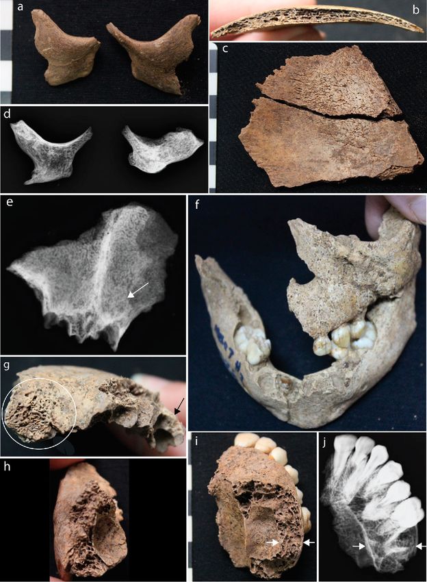
www.nature.com/scientificreports/
Figure 1. Map of Southeast Asia with sites important for this study. Khok Phanom Di in Thailand provides
the only prehistoric evidence of thalassemia in the region. Our study includes research from Man Bac and Con
Co Ngua in northern Vietnam. Modified from image by Koba-chan (https://upload.wikimedia.org/wikipedia/
commons/1/15/Topographic30deg_N0E90.png) created from DEMIS Mapserver (http://www2.demis.nl/world
map/mapper.asp). Published under CC BY-SA 3.0 (https://creativecommons.org/licenses/by-sa/3.0/deed.en).
spectrum of symptoms and s everity11. Modern day research demonstrates that variants within Southeast Asia
and the Pacific can vary across short distances, and relate to the ecological ranges of Anopheles mosquitos, the
vectors for malarial p arasites3. Given the co-evolutionary relationship that continues to drive high frequencies
of thalassemia in the Southeast Asian region today, it can be hypothesized that the identification of thalassemia
in the prehistoric record can be, at least in part, related to a prior selection pressure of malaria.
To date, the only archaeological evidence for hereditary anemia in the region dates from approximately
4000–3500 BP from Khok Phanom Di, a Neolithic site in central Thailand (Fig. 1)12. In the Mediterranean
region, where beta thalassemia is similarly frequent, skeletal evidence in prehistoric assemblages supports the
emergence of the disease with the transition to agriculture (the Neolithic) from approximately 7000 years a go13.
It is hypothesized that agricultural practices such as land clearance encouraged contact between human groups
and Anopheles mosquitos13,14. Did these conditions then emerge alongside the spread of agriculture in Southeast
Asia as is proposed to be the case in the Mediterranean?
The emergence of farming in Mainland Southeast Asia (MSEA) occurred much later than the Mediter-
ranean, from only after 4500 years ago15, with most Neolithic sites post-dating 4,000 BP. Prior to this time the
region was occupied by indigenous Pre-Neolithic foragers descended from the first people out of Africa and
into Asia16. By the early seventh millennium BP some forager groups in northern Vietnam and southern China
developed large sedentary settlements, at the same time as agriculture was practiced to the north in C hina17. The
Scientific Reports | (2021) 11:5677 | https://doi.org/10.1038/s41598-021-83978-4 2
Vol:.(1234567890)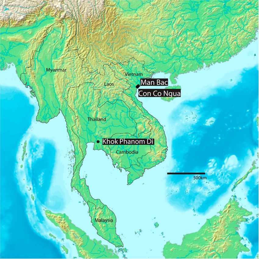
www.nature.com/scientificreports/
Individual ID MB07H1M8 MB07H1M12* MB07H2M26 MB05M12 MB05M3 MB07H1M1
Age (years) 30–39 0 1.5 2 0.5 12
Sex Male N/A N/A N/A N/A N/A
Diagnostic criteria
Marrow hyperplasia of the facial bones: maxillae-leading to ventral displacement of
central incisors, zygomatic bones-leading to orbital displacement, and/or mandible A N/A P P P P
(rodent facies deformity) (SD)
Radiographic: “rib-within-a-rib” appearance. Radiographically defined sclerotic
P A P P A
bands within the ribs due to extramedullary hematopoiesis (SD)
Poor or lack of pneumatization of the paranasal and cranial sinuses sparing the
A N/A P A** A P
ethmoid sinuses (D)
Enlarged tubular bones of the hands and feet due to marrow hyperplasia (infants)
sometimes associated with enlarged nutrient foramina or Radiographic: coarse
P N/A N/A N/A A N/A
trabecular patterns of the hands or feet, sometimes associated with cyst-like lucencies
due to focal collection of hyperplasic marrow (D)
Premature fusion of epiphyseal plates particularly of the proximal humerus and distal
A A A A A N/A
femur, often causing short long bone maximum length (D)
Widening of entire rib, or widening of the rib head and neck with pronounced bul-
bous appearance posteriorly (costal osteomas). Associated with radiograph appear- P A A A P A
ance of erosion of the inner cortex (D)
Diagnosis Probable Possible Probable Probable Probable Probable
Table 1. Macroscopic and radiographic diagnosis of thalassemia in Man Bac individuals. P = present,
A = absent, N/A = skeletal element missing/unobservable. *MB07H1M12 presented with multiple suggestive
lesions, but no diagnostic (D) or strongly diagnostic (SD) lesions (see Supplementary Table S1). **Frontal
sinuses were not preserved, absence of pneumatization based on preservation of maxillary sinuses only.
subsequent adoption of agriculture in MSEA during the Neolithic (4500–3100 BP) was associated with multiple
migration events of farmers from what is now geo-politically southern China, coming into contact with these
forager groups15,18,19. These subsistence transition and migration events significantly altered the demography
and genetics of this region from this point forward. The aim of this research is to investigate, for the first time,
whether thalassemia, as a proxy for malaria burden, was present prior to agriculture in northern Vietnam, and
to what degree the transition to agriculture may have contributed to the emergence of thalassemia in Southeast
Asia. Additionally, if thalassemia is observed we aim to further investigate which variants may have been present.
We applied a diagnostic protocol based on macroscopically observed dry bone lesions for thalassemia to
two archaeological human skeletal assemblages. The Pre-Neolithic site of Con Co Ngua radiocarbon dated to at
minimum 6200–6700 cal BP17,18 represented a pre-agricultural but sedentary forager community (n = 155; Fig. 1).
The Man Bac site dating to 3,906–3,523 cal BP was occupied during the agricultural transition of Southeast Asia
(n = 70). This site captures co-habitation of indigenous forager and migrant farmers during the early stages of
the agricultural transition18. To support the strength of the disease diagnosis using the macroscopic methods,
we also conducted histological analysis on three Con Co Ngua individuals to assess microscopic pathological
changes in affected bone.
Results
Macroscopic results present strong evidence for thalassemia at Man Bac. Five children aged
between 6 months and 12 years and one adult presented with skeletal changes that are strongly diagnostic
(pathognomonic) for thalassemia (Table 1, Supplementary Text S1; Supplementary Table S1; Figs. 2, 3). These
changes included rodent facies, a skeletal condition specific to thalassemia, where expansion of the marrow
results in a bulbous face and mandible. Radiographs confirm marrow expansion, a clinical consequence of severe
thalassemia (Fig. 2). Radiographic ‘rib-within-a-rib sign’, again pathognomonic for thalassemia, was identified
in three individuals. This condition results from extensive marrow expansion within the shafts of the ribs. Fur-
ther evidence for thalassemia included a lack of the development of facial and cranial sinuses, commonly caused
by extensive marrow expansion of the face since infancy. Two children also presented with severe porosity of
the orbits and the endocranium, likely due to marrow expansion through the cortical margins of the skull, as a
result of thalassemia. Additionally, a newborn with multiple lesions suggestive of possible thalassemia, including
evidence of marrow expansion throughout the skeleton was identified (Table 1, Figs. 2, 3).
Macroscopic results present evidence suggestive but not diagnostic for thalassemia at Con Co
Ngua. We identified seven adults and adolescents at Con Co Ngua presenting with macroscopic and radio-
graphic ‘bone-within-a-bone’ changes of the limb bones (Supplementary Fig. S1; Supplementary Table S1). We
observed a mixture of enlargement and restriction of the medullary canal areas in these individuals. In thalas-
semia, this skeletal change is caused by marrow expansion within the long bones perforating the outer cortex.
As such, these pathologies are suggestive of thalassemia but alone are not diagnostic for thalassemia, as they
can also be found in a number of other chronic c onditions20 (Supplementary text S1). However, these skeletal
changes were similar to those with strong diagnostic evidence of thalassemia at Man Bac. We extracted bone
samples from three of the CCN individuals for further microscopic analysis.
Scientific Reports | (2021) 11:5677 | https://doi.org/10.1038/s41598-021-83978-4 3
Vol.:(0123456789)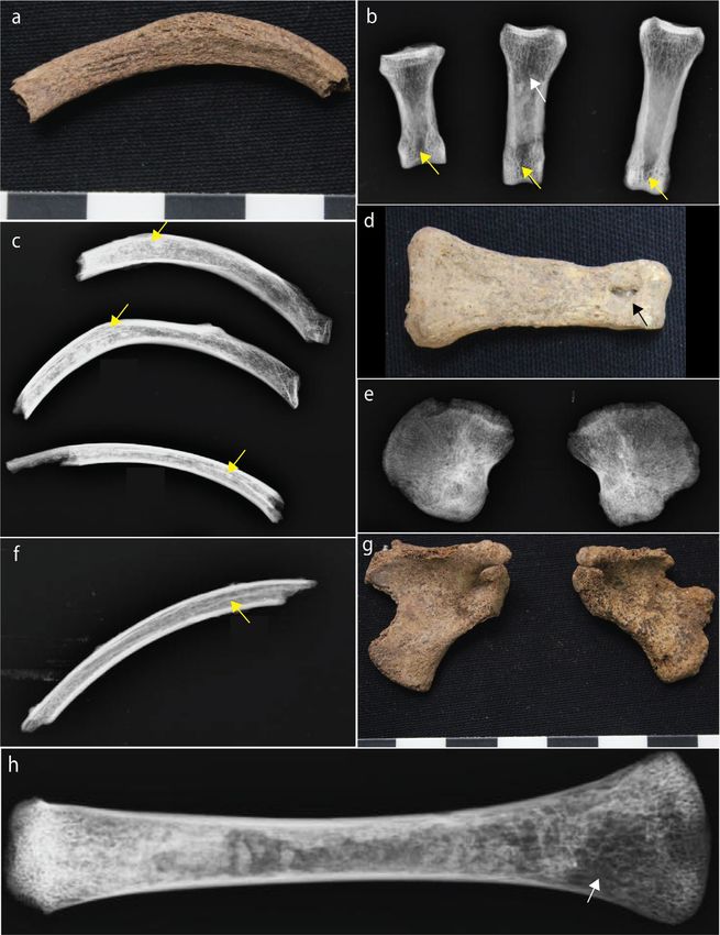
www.nature.com/scientificreports/

                                            Figure 2.  Cranial evidence for thalassemia at Man Bac. (a) Anterior protrusion of the zygomatic bones
                                            consistent with rodent facies (MB05M3, approx. 6 months old, antero-lateral aspect). (b) and (c) Diploic
                                            expansion of the cranial vault. There is no porosity on the ectocranium but hair-on-end formations are present
                                            on the endocranium (MB05M12, approx. 2 years, lateral aspect). (d) Marrow hyperplasia of the zygomatic
                                            bones (MB05M12, antero-posterior view). (e) Lack of pneumatization of the frontal sinus (MB07H1M1, approx.
                                            12 years, antero-posterior view). (f) Rodent facies of the maxilla, mandible and zygoma (MB07H2M26, approx.
                                            1.5 years, antero-superior aspect). (g) Severe cribra orbitalia (white circle) and diploic expansion of the crania
                                            (black arrow, antero-lateral aspect) (MB07H1M1). (h) Marrow hyperplasia of the left zygoma (MB07H1M1,
                                            lateral aspect). (i) and (j) Marrow hyperplasia of the maxilla (MB07H1M1, superior-inferior view). The expanse
                                            of the marrow hyperplasia is indicated by the white arrows.

          Scientific Reports |   (2021) 11:5677 |                https://doi.org/10.1038/s41598-021-83978-4                                                  4

Vol:.(1234567890)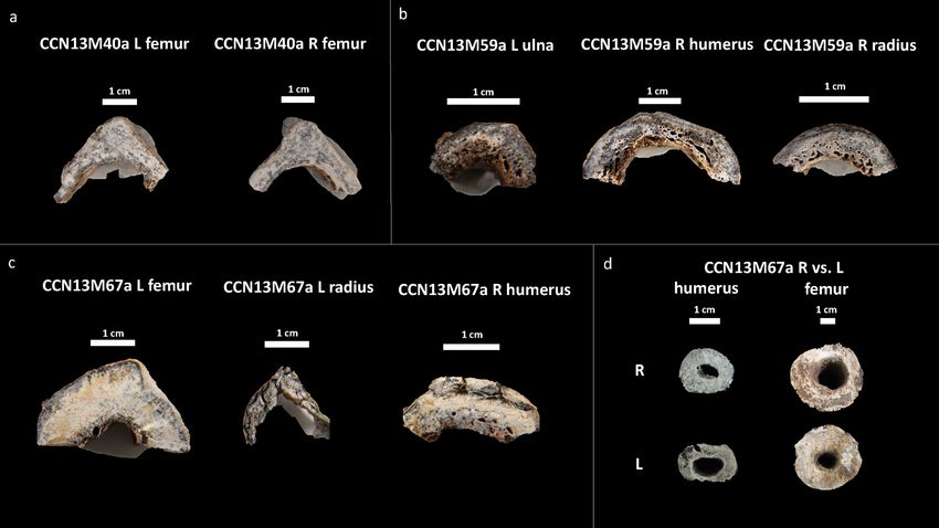
www.nature.com/scientificreports/

                                  Figure 3.  Postcranial evidence for thalassemia at Man Bac. (a) Enlarged rib (MB05M3, approx. 6 months old,
                                  superior aspect). (b) and (d) Expanded foramina of the phalanges (yellow arrows) with marrow hyperplasia
                                  (white arrow, MB07H1M8, middle aged adult, antero-posterior view). (c) “Rib-within-a-rib” sign (yellow
                                  arrows, MB07H1M8, supero-inferior view). (e) Alteration of the trabecular structure of the ilia. Note the
                                  radiating pattern (MB07H1M12, neonate, antero-posterior view). (f) “Rib-within-a-rib” sign (yellow arrow,
                                  MB07H2M26, approx. 1.5 years, supero-inferior view). (g) Enlargement of the scapular spines (MB07H1M12,
                                  neonate, posterior aspect). (h) Marrow hyperplasia of the humerus (white arrow, MB05M12, approx. 2 years,
                                  antero-posterior view).

Scientific Reports |   (2021) 11:5677 |               https://doi.org/10.1038/s41598-021-83978-4                                                5

                                                                                                                                         Vol.:(0123456789)
www.nature.com/scientificreports/
Figure 4. Cross section of microscopic samples from Con Co Ngua. (a) Femora sections from CCN13M40a.
There is increased cortical thickness, and the cortical width of right femur is asymmetrically wider than the left.
(b) Upper limb sections from CCN13M40a. Large endocortical pores are evident. Medullary canal widening is
most distinct on the humerus. (c) Upper and lower limb sections from CCN13M67a. While the femur presents
with extreme cortical thickness and restriction of the medullary canal area, the upper limb sections present with
severe porosity of the endocortical surfaces. (d) Asymmetry of the medullary canals of the humeri and femora
of CCN13M67a.
Combined microscopic and macroscopic results strongly suggest thalassemia at Con Co
Ngua. We assessed bone samples of one adolescent (> 16 years) of unknown sex (CCN13M67a), and two
adults: CCN13M40a (young adult female), CCN13M59a (old adult male) (Fig. 4). We focused specifically on the
endocortex (cortical bone located on the inner third portion of the cross-section) as preliminary macroscopic
assessment of the samples prior to microscopy determined this region to be particularly important (Fig. 4).
Widespread pores were evident on the endocortical surfaces of samples from the old adult male (CCN13M59a).
The porosity consists of ‘giant’ pores that appear to have been created as a result of adjacent coalescing pores
(Figs. 4, 5). All three bones of this individual showed a “trabecularization effect”, which essentially transformed
cortical bone into a trabeculae-like matrix of bone as a result of prolonged resorption, and possibly marrow
expansion.
In contrast, the bone samples in the young adult female (CCN13M40a), showed denser and enlarged endo-
cortical surfaces. The adolescent (CCN13M67a) presented with a mix of large porous regions of the endocortex
(right humerus) and regions of increased cortical density (femur). Possible beginnings of the ‘trabecularization
effect’ such as those observed in the old adult male (CCN13M59a) were found in the right humerus of the old
adult. The extent of medullary bone size reduction can be seen, particularly in the adolescent femur section where
the cortical wall is unusually enlarged (Fig. 4d). This femur showed evidence for secondary bone remodeling
confirming cyclical replacement of old bone with new bone tissue, possibly driven by increased metabolic bone
needs (Fig. 6). There was a clear pattern whereby samples from the femur did not show evidence for widespread
porosity and coalescing of bone pores, but were observed in the bones of the upper limb (ulna, humerus, and
radius) (Fig. 6).
We compared the microscopic results to various diseases of infectious and metabolic etiologies known to
cause the macroscopic ‘bone-within-a-bone’ sign (Supplementary text S1). The microscopic outcomes of Con
Co Ngua are only consistent with clinical bone histological observations in cases of beta thalassemia. Increased
osteoclast-mediated resorptive activity, and decreased osteoblastic activity have both been described as under-
lying processes that increase porosity of the endocortical margins of the long bones in beta thalassemia21. In
thalassemia, skeletal changes can be localized, due to focal deposits of iron during iron overload which is con-
sistent with the variation between the upper and lower limbs we observed across the samples21–23. The overall
histological bone pattern observed in the Con Co Ngua individuals therefore supports localized metabolic
changes consistent with thalassemia.
Scientific Reports | (2021) 11:5677 | https://doi.org/10.1038/s41598-021-83978-4 6
Vol:.(1234567890)www.nature.com/scientificreports/
Figure 5. Macroporosity observed in the arm bones of CCN13M59a. The porosity is large and coalescing.
Discussion
What hemoglobinopathy variants were present in prehistoric Vietnam? Our results demonstrate
skeletal evidence for thalassemia from the early seventh millennium BP in northern Vietnam. The gross skeletal
changes in all but one Man Bac individual are consistent with beta thalassemia4. The clinical consequences,
including bone changes, of beta thalassemia only develop following replacement of fetal gamma hemoglobin
with adult beta hemoglobin in the months following birth. However, the possible case diagnosed in a Man Bac
Scientific Reports | (2021) 11:5677 | https://doi.org/10.1038/s41598-021-83978-4 7
Vol.:(0123456789)www.nature.com/scientificreports/
Figure 6. Regions of bone captured from the endocortical surface in the adolescent (CCN13M67a), allowing to examine the
degree of bone modelling and remodeling. (a–d) The femur: Secondary osteons (SO), primary osteons (PO), Haversian canals
(HC), and osteocyte lacunae (OL) can be seen. (a) White arrows point to endosteal lamellar layers which are typical for this bone
region. (c) White arrows point to a cement line of a secondary osteon that indicates a remodeling event of a fragmentary osteon
(FO) underneath, confirming cyclical replacement of old bone with new bone tissue, possibly driven by increased metabolic bone
needs. (e) White arrows point to primary lamellar bone layers and an isolated HC. (f) A SO amongst primary bone.
Scientific Reports | (2021) 11:5677 | https://doi.org/10.1038/s41598-021-83978-4 8
Vol:.(1234567890)www.nature.com/scientificreports/
newborn may be consistent with alpha thalassemia major (Bart’s hydrops fetalis)24. Alpha thalassemia major,
caused by deletion of all of the four alpha alleles, is a common variant in Southeast Asia today25. There is little
clinical description of the skeletal complications of alpha thalassemia major due to its fatal outcome. However,
severe bone marrow expansions occur in utero during growth detected as early as 18 fetal weeks7, and skeletal
complications similar to beta thalassemia major can be e xpected24. Severe growth stunting is a common out-
come of alpha thalassemia major, and skeletal deformities have been clinically documented, but not d escribed5,7.
Evidence for co-occurrence of both alpha and beta thalassemia variants at Man Bac is of evolutionary sig-
nificance. The gross skeletal pathologies of Man Bac post-birth individuals are commonly observed in patients
with beta thalassemia major, a severe form that requires removal of excess iron and blood transfusions in order
to survive past infancy26. However, 57% (4/7) of individuals macroscopically diagnosed with thalassemia at
Man Bac were older than 1-year of age, having survived infancy without treatment. Alpha and beta thalassemia
co-inheritance is known to result in a milder form of disease than beta thalassemia major due to a balance of
depleted alpha and beta hemoglobins27. Additionally, the Southeast Asian variant HbE beta thalassemia com-
monly results in severe forms of thalassemia that do not require blood transfusions to survive (known clinically
as thalassemia intermedia)28. It is possible that co-inheritance of beta thalassemia with alpha and/or HbE may
account for the severe skeletal changes in individuals at Man Bac who survived past infancy.
It is not possible to determine whether the Con Co Ngua individuals had beta or alpha thalassemia. While
there is clear clinical recognition of the skeletal changes of beta t halassemia29, HbH alpha thalassemia caused
by deletion of three of the four alpha alleles are reported to cause mild to moderate skeletal deformities in some
patients5. Significant facial deformity was not recorded at Con Co Ngua. However, infants at this site were very
poorly preserved, so more severe forms of thalassemia at Con Co Ngua cannot be ruled out.
Evidence for deep antiquity of thalassemia and malaria in Mainland Southeast Asia. Based on
our macroscopic and microscopic observations we suggest that thalassemia was potentially a considerable bur-
den for Southeast Asian populations prior to the adoption of farming. This contrasts with our current knowledge
on the emergence of thalassemias in the Mediterranean and demonstrates that the agricultural transition was
not the defining factor in the emergence of this disease in Southeast Asia. We note here that the one adult with
probable thalassemia from Man Bac had dental and skeletal affinity to Australo-Papuan populations, such as
those from Con Co Ngua (and all foraging groups from Vietnam prior to agriculture). The inhabitants from the
Neolithic central Thailand site of Khok Phanom Di where thalassemia was previously reported represent mixed
Australo-Papuan and East Asian dental affinities30. Therefore, a deep antiquity for thalassemia in the region prior
to the Neolithic appears highly probable. However, the complexity of gene flow and stabilizing selection in the
region exceeds the capabilities of skeletal morphometric data.
The heterogeneity of mutations that cause beta, HbE beta and alpha thalassemia in Southeast Asia support
multiple instances of independent emergence within the region likely tied to a consistent threat of malaria
within MSEA31,32. Alpha thalassemia variants in Southeast Asia populations have similarly been identified in
modern indigenous Papuan and Austroasiatic (modern Southeast Asian) speaking populations33. The widespread
geography of these variants today, and the compounded effect of multiple migration and genetic admixture
events since prehistory, mean it is not possible to determine the origins of these variants from the distribution
in modern populations. However, different haplotypes do indicate separate founder effects in Australo-Papuan
(indigenous Australian and New Guinean Highlanders) and Austroasiatic (Thai) groups33. At least in the case of
alpha thalassemia, a pre-agricultural deep history of this disease in MSEA is consistent with present day genetic
data. In contrast, selection models of one HbE variant from a Thai population indicates an emergence between
4400 and 1240 years ago, consistent with agricultural intensification between the Neolithic to Iron Age periods
(4500–1500 BP) in M SEA34. HbE emergence is consistent with the time period of the Man Bac individuals with
thalassemia. Multiple independent events of thalassemia emergence may have occurred due to different social
transitions in Southeast Asia’s prehistory.
The presence of thalassemia in prehistoric MSEA, as is the case today, indicates a constant selection pressure
of malaria stemming from deep antiquity. It is expected that the gene frequencies of thalassemias in the absence
of malaria would not have been maintained given its mortal cost when inherited in its severe f orms14. While P.
falciparum has a considerably higher mortality rate than P. vivax, thereby incurring a stronger selection pressure,
both Plasmodium species are endemic to the region and thought to have had possible selection effects on thalas-
semia in Southeast Asia35. Both P. falciparum and P. vivax malarias share African origins. It is thought that P.
vivax emerged out of Africa with human groups prior to 30,000 years a go36 consistent with the first anatomically
modern humans who inhabited MSEA as early as 60,000 years a go16. P. falciparum is thought to have emerged
in Africa approximately 60,000–40,000 years ago, and estimated to have undergone a bottle neck approximately
6,000–4,000 years ago that likely favored human infection37. This proposed bottle neck event post-dates the
skeletal evidence for thalassemia at Con Co Ngua and pre-dates that of Man Bac. Rather than provide resist-
ance, alpha thalassemia has been proposed to increase susceptibility to infection by P. vivax35. This susceptibility
may be an adaptive mechanism to cross vaccinate against P. falciparum35 which signals the complex connection
between Plasmodium infections and the emergence of thalassemia. The association between thalassemias and P.
vivax morbidity and mortality, however, remains unknown and is a necessary component to understanding the
relationship between malarial types and the emergence of thalassemias in the r egion38. Rarer still, Plasmodium
variants commonly found in Southeast Asian forest macaques have also been known to transfer to h umans39,
an important local reservoir in communities in V ietnam40. Potential human to non-human primate interactions
may also factor into the relationship between malaria and thalassemia in the prehistory of the region. Both Man
Bac and Con Co Ngua inhabitants exploited non-human primates as a food source17,41.
Scientific Reports | (2021) 11:5677 | https://doi.org/10.1038/s41598-021-83978-4 9
Vol.:(0123456789)www.nature.com/scientificreports/
Lowland flood plains in Vietnam, such as where Man Bac and Con Co Ngua are located have low reported
malarial cases and primary vectors today, as is the case with coastal and estuarine environments42. However,
the original ecology of the region surrounding these sites in antiquity included movement within fringe forests,
brackish water, and riverine areas that may have placed the inhabitants at risk of a number of different Anopheles
mosquito species that carry both human and simian Plasmodium species42. In tropical forested areas of MSEA,
malarial vectors are common. Tropical forested regions expanded substantially from approximately 14,000 years
ago following the Last Glacial Maximum, which would have been opportunistic for the spread of species A. dirus
and A. minimus, the primary mosquito vectors of malaria in MSEA42. The tropical climate would have enabled
year-round transmission even in seasonal areas such as northern Vietnam that experience dry and wet s easons43.
While pre-agricultural, the complex hunter-gathers of Con Co Ngua existed in large sedentary populations,
thereby encompassing a suite of factors that have been argued to be associated with the emergence of malaria
and thalassemia in the M editerranean44. Sedentary residential mobility would have enabled completion of the
cycle of the disease within one host population42, increasing the selection pressure on these populations. In
Southeast Asia today, an increased risk is observed in people who live in villages but actively exploit the forest
for resources45. This risk appears to be related to the early night feeding times of these vectors. The exposure
to forest Anopheles mosquitos and sedentary residence alone would likely have been sufficient to increase the
probability of contracting malaria in the absence of agricultural land clearing.
The agricultural transition and gene flow variation. While Con Co Ngua individuals were less well
preserved than those from Man Bac, 10% (7/70) of the latter assemblage presented evidence for thalassemia.
Man Bac is contemporaneous with Khok Phanom Di indicating these alleles were widespread through the region
at this point in time. Therefore, the transition to agriculture may have considerably altered the gene flow or
increased the selection pressure of thalassemia mutations, and shaped the relationship between human groups
and malarial vectors in the region. Wet rice agriculture (irrigation) was only developed during the Iron Age
(2500–1500 BP) in Southeast Asia46. This form of agriculture has been previously, erroneously, argued to be
linked to increased malarial vectors compared to dry rice agriculture in M SEA47. Our results indicate a strong
relationship between human groups and malaria prior to wet rice farming. Present day reports of differences
in malarial vector prevalence in irrigated versus non-irrigated farming localities vary and depends on other
ecological dynamics such as seasonality48. In Mainland Southeast Asia, A. dirus is found in significantly higher
densities in forested areas than in villages, and reside in shallow pools42 rather than in deep waters. Therefore,
the moated settlements of the Iron Age in Thailand may have made the ecology around villages less attractive
to primary malarial vectors or attracted different Anopheles mosquitos such as A. barbirostris with significantly
lower vector potential49. Instead, the reliance of dry rice farming on mild seasonal flooding may have increased
the exposure of Neolithic inhabitants to malarial vectors42, particularly as they continued to exploit forested
areas through mixed farmer-foraging p ractices50. While Pre-Neolithic foragers in northern Vietnam and south-
ern China were arguably primarily sedentary, an increase in sedentism and significant population growth dur-
ing the transition to agriculture in MSEA51,52 would have increased the transmission potential within human
populations.
Possible interaction between Man Bac inhabitants and other nearby contemporaneous sites further inland,
where higher vector densities have been r eported42, may have also contributed to the presence of thalassemia at
Man Bac. Alternatively, we also recognize that gene flow of thalassemia alleles from other inland Neolithic sites
may be responsible, and a direct relationship between thalassemia and malaria presence at Man Bac cannot be
determined. A similar argument can be made for Con Co Ngua, as contemporaneous forager sites of northern
Vietnam are also archaeologically documented to have maintained interactions through exchange53. Nevertheless,
it is apparent that mobility and interactions within and between groups were essential for the spread of thalas-
semia variants in the prehistory of MSEA, and the prevalence of this disease can be tied directly or indirectly to
malarial endemicity in prehistoric MSEA.
Conclusion
Through a combined macroscopic and microscopic approach we demonstrate that thalassemia has been present
in northern Vietnam from at least the early seventh millennium BP. This finding indicates that agriculture was
not a crucial factor in the emergence of thalassemia in response to malaria in Southeast Asia as appears to be the
case in the Mediterranean. In the context of large sedentary forager populations exploiting forested resources,
such as the inhabitants of Con Co Ngua, we propose a pre-agricultural origin for the emergence of thalassemia
in MSEA as an adaptive response to the threat of malaria. However, the agricultural transition approximately
4500–3500 years ago likely encouraged the spread of malarial vectors, increasing the gene frequencies of thalas-
semias. The outcomes of our research indicate a deep history of thalassemias which are endemic in MSEA today.
Methods and materials
Ethics and approvals. Paleopathological observation and collection and analysis of samples for histologi-
cal investigation was approved by the Vietnam Institute of Archaeology in Hanoi, Vietnam on October 12, 2018.
Bone samples were exported to the Australian National University in Canberra under approval by Dr. Nguyen
Gia Doi (Director), and are currently housed at this institution until further notice. As the samples are of an
archaeological nature, no ethical approvals were required for this study.
The sample. We macroscopically assessed 70 individuals from Man Bac and 155 individuals from Con Co
Ngua for pathology. All skeletal elements with pathological change with the potential for diagnosis were radio-
graphed. Three individuals from Con Co Ngua were sampled for histological analysis.
Scientific Reports | (2021) 11:5677 | https://doi.org/10.1038/s41598-021-83978-4 10
Vol:.(1234567890)www.nature.com/scientificreports/
Pathology Diagnostic strength Differential diagnosis References
Marrow hyperplasia of the facial bones: maxillae- leading to ventral
29,66–70
displacement of central incisors, zygomatic bones-leading to orbital SD
displacement, and/ or mandible (rodent facies deformity)
Radiographic: “rib-within-a-rib” appearance. Radiographically defined 29,66,68,71
SD Sickle cell anemia, osteomyelitis, leukemia*
sclerotic bands within the ribs due to extramedullary hematopoiesis
Poor or lack of pneumatization of the paranasal and cranial sinuses Neoplasms, Paget’s disease, trauma, hypopituitarism, hypothyroidism, 29,68
D
sparing the ethmoid sinuses osteopetrosis, sickle cell anemia
Widening of entire rib, or widening of the rib head and neck with pro-
29,66,68,71
nounced bulbous appearance posteriorly (costal osteomas). Associated D Neuroblastoma, Nieman-Pick disease, Leukemia
with radiograph appearance of erosion of the inner cortex
Enlarged tubular bones of the hands and feet due to marrow hyper-
plasia (infants) sometimes associated with enlarged nutrient foramina
or Radiographic: coarse trabecular patterns of the hands or feet, D Treponemal disease, leprosy, tuberculosis 12,29,66,68
sometimes associated with cyst-like lucencies due to focal collection of
hyperplasic marrow
Premature fusion of epiphyseal plates particularly of the proximal Scurvy, hypervitaminosis A, trauma, achondroplasia, Morquio’s
12,29,66,68,72
humerus and distal femur, often causing short long bone maximum D disease, Ellis-van Creveld disease, peripheral dysostosis, poliomyelitis,
length mucopolysaccharidosis, rickets, osteomalacia
Extensive marrow proliferation of the long bones leading to expansion
of the medullary canal, associated with thin cortices (and in extreme Other hemolytic anemia, scurvy, rickets, metaphyseal dysplasia, 66,68,73
S
circumstances honeycomb-like porosity) resulting in swollen appear- Gaucher’s disease, osteomyelitis
ance or metaphyseal flasked shaped deformities
After Lagia74: hemolytic anemias and red cell enzyme disorders
Severe porotic hyperostosis/ diploic expansions of vault and maxilla (including sickle cell disease, iron deficiency anemia and G-6-PD defi- 29,66,68,74
S
with “hair-on-end” appearance and/ or cribra orbitalia ciency), cancers (including leukemia, multiple myeloma, meningioma,
metastases and secondary to kidney cancers), and polycythemia
66
Wide dental spacing S Skeletal dysplasias, normal variation
Spiculated or scalloped proliferation of subperiosteal reactive new Infectious diseases, rickets, scurvy, hypertrophic osteoarthropathy, 12,69,75
S
bone on the shafts of the limb bones and the clavicles Gaucher’s diseases, Paget’s disease
Marked osteoporosis and cortical thinning of the vertebrae, with 29
S Age related osteoporosis, osteomalacia, scurvy, trauma
compression fractures in severe cases
76
Bone infarction S Osteomyelitis, sickle cell anemia, osteosarcoma
Enlargement and alteration of the trabecular pattern in flat bones 70
S Other hemolytic anemia, leukemia
(pelvis and scapula)
Table 2. Criteria for diagnosis of thalassemia in dry bone (SD = strongly diagnostic, D = diagnostic, and
S = suggestive). A probable case is defined as an individual exhibiting at minimum one strongly diagnostic
pathology or two diagnostic pathologies. A possible case is defined as an individual exhibiting at minimum
one diagnostic or two suggestive pathologies. *A strongly diagnostic lesion is one that is considered
pathognomonic for that disease and alone stands as evidence of probable disease. In extremely rare instances
these pathologies can occur in other diseases which are listed here in the differential diagnosis.
The skeletal assemblage of Man Bac was well preserved. While post-mortem breaks were frequent, the skel-
etons were easily reconstructed. Surfaces were excellently preserved. For most skeletons both adult and subadult
were either complete or near c omplete54. In contrast, the individuals from Con Co Ngua presented with erosion
of the cortical surfaces, but not to the degree that pathologies could not be observed. Post-mortem breakage
was common, and a large proportion of the assemblage was at most partially complete. Subadults were com-
monly poorly preserved, with infants highly fragmented. Methods for age and sex of the individuals from Man
Bac and Con Co Ngua were previously reported and briefly summarized h ere50,55,56. Non-metric traits of the
os coxae, cranial morphology, and sample-specific post-cranial functions were used to estimate adult sex57–60.
Adult age-at-death was estimated by way of pubic symphyseal m orphology61 and/or sample-specific molar wear
functions developed by O xenham60. Subadult age-at-death was estimated using dental eruption and calcification
standards62,63, and/or skeletal maturity s chedules64.
Paleopathological differential diagnosis. The paleopathological diagnosis of thalassemia was based on
macroscopic and radiographic observations of pathologies in dry bone. A standardized ‘threshold’ diagnostic
protocol for thalassemia was produced following clinical diagnostic standards (Table 2). A possible case was
diagnosed when an individual exhibited one diagnostic or two suggestive lesions. A probable case was diagnosed
when an individual exhibited one strongly diagnostic or two diagnostic lesions. Such recording methods are
consistent with a number of previously published diagnostic protocols in paleopathology, and ensure diagnostic
rigour18,65.
Gross skeletal manifestations of thalassemia are restricted to beta thalassemia only, except in the case of
alpha thalassemia major (also called Bart’s hydrops fetalis) a fatal form, where infants die prior to or immedi-
irth7. Severe porotic hyperostosis leading to a ‘hair-on-end’ appearance is a common skeletal
ately following b
manifestation of thalassemia (as is the case in other anemias) and has been reported particularly in cases of
thalassemia major68,77.
Extensive marrow hyperplasia of the medullary canal and within cancellous bone regions cause consider-
able thinning of the bone cortex, and expansion of the area of the medullary c anal68. The extent of the marrow
Scientific Reports | (2021) 11:5677 | https://doi.org/10.1038/s41598-021-83978-4 11
Vol.:(0123456789)www.nature.com/scientificreports/
hyperplasia causes thinning trabeculae. However, subperiosteal new bone response to the trabecular destruction
instigates the coarsening of trabeculae which is observed r adiographically68. In extreme circumstances cortical
thinning can progress to destruction of the cortex and subsequent honeycomb appearance of the bone surface70.
A distinct facial deformity called rodent facies occurs in severe cases particularly in thalassemia major
whereby extensive marrow proliferation of the maxillae and zygomatic bones creates anterior bulging of the
face, often associated with anterior displacement of the central incisors, and lack of pneumatization of the
maxillary, sphenoidal and frontal s inuses29,66–68. This pathological lesion is strongly diagnostic for thalassemia,
particularly of thalassemia major (Table 2)29,68. The marrow proliferation also causes anterior teeth protrusion
and malocclusion of the remaining t eeth66. A strongly diagnostic radiographic feature of this disorder is the ‘rib-
within-in-a-rib’ sign. This feature is produced by extensive marrow hypertrophy perforating the thin cortex of the
rib associated with a radiodense line in the middle of the medullary c anal29,66,69,78. Hypertrophic changes to the
hands and feet of infants (with associated expanded foramina in the phalanges) is also characteristic of the dis-
ease, as this lesion indicates an extreme form of anemia unlikely to be associated with non-genetic e tiologies12,68.
Marrow perforation of the cortex known as extramedullary hematopoiesis occurs. In certain circumstances
subperiosteal new bone response similar to the “hair-on-end” appearance of the skull, surrounding thin cortices
of postcranial bones, can result as a consequence of marrow p erforation69,75. This skeletal response has been
documented to lead to premature fusion of the epiphyses of long bones particularly of the proximal humerus
and distal femur68,75. The extent of prematurity is dependent on the individual. This lesion is more common in
cases of thalassemia intermedia75. Bone infarction, while less common in thalassemia than sickle cell anemia,
has also been reported. In this pathological condition, bone death occurs followed by inflammatory new bone
response around the region of necrosis, and has a distinct appearance on radiographs76.
Histological sectioning. All sectioning procedures followed standard protocols79,80 and were completed
at the Institute of Archaeology, Hanoi, Vietnam. A Dremel Variable-Speed Rotary Tool 3000 with associated
Dremel blades was used to cut into the bone shafts. This involved making two longitudinally oriented parallel
(along the long bone axis) cuts, followed by a transversely oriented (perpendicular to the longitudinal cuts) cut,
so that sections freely detached themselves from the bone. Permissions obtained from the collection curators
strictly stipulated that sections were only to be removed from the bone regions already affected by post-mortem
breaks to avoid further damage to the specimens. For example, where the long bone was broken in half or
had epiphyseal ends missing due to taphonomic issues. Sampling in those regions avoided further damage to
the specimens. However, we ensured that the bone sections still derived from the long bone shafts so that we
could assess the histological changes on the endocortical surfaces. The three individuals sampled for histological
analysis met these criteria. We sampled the left and right femur of CCN13M40a (Fig. 4). The right humerus, left
ulna and left radius of CCN13M59a were sampled as they were the only available long bones for this individual
(the lower limb only consisted of a fragmented right fibula). The right humerus, left radius, and left femur of the
subadult CCN13M67a were sectioned as both the upper and lower limb bones showed a degree of endocortical
thickening. The lower (distal direction) third of the left femoral shaft, approximately 4 cm below the linea aspera,
and the midshaft of the right femur, were sampled in CCN13M40a. The upper limb bones of CCN13M59a were
sectioned at the distal end of the left ulna and the right radius, and the midshaft of the right humerus. The left
femur of CCN13M67a was sectioned at the middle of the upper (proximal direction) third of shaft, the distal
third of the shaft in the left radius, and the upper (proximal direction) third shaft of the right humerus. We note
that the left radius in this individual shows a well remodeled and healed fracture at midshaft (Supplementary
Fig. S2), which clearly resulted in a deformation of the bone. This will need to be borne in mind when interpret-
ing the appearance of histology in thin section. A total of eight, approximately 1 cm thick, roughly C-shaped,
sections of bone from the posterior aspect were removed for the entire sample (Fig. 4).
Prior to histological preparation, external size measurements of the bones and sections were recorded using
standard digital calipers and a soft measuring tape. All measurements were repeated at least two times from
which an average was computed and reported here. The m easurements79 were: shaft circumference (Circ) at point
of sectioning (mm), anterior–posterior diameter in mm (AP dm), medial–lateral diameter in mm (ML dm),
cortical width (Ct.W) of the extracted sample measured from both the proximal and distal surfaces in the lateral
direction (from the endosteal, referring to the endosteum lining the medullary cavity, to the periosteal surfaces).
Histopathology. No prior information on age, sex or macropathology was provided for the samples to
ensure an objective observation. All samples were processed into thin sections following standard methods
applicable for histological analysis of archaeological human bone81,82. The processing was completed at the Hard
Tissue Histology laboratory in the School of Archaeology and Anthropology at the Australian National Univer-
sity (Canberra, Australia), which is also where the resulting thin sections are curated until further notice. Each
sample was embedded in Buehler epoxy resin, cut on a low-speed saw equipped with a diamond wafering blade,
and attached to microscope glass slides using Stuck epoxy glue. The attached samples were further trimmed on
a cutting saw, and later lapped on a series of grinding pads of various grit sizes that increased from 400 to 1200
in coarseness. The thickness of each section was controlled using a handheld Buehler 28 × 48 mm target holder,
allowing to produce sections of approximately 90–100 μm thickness. The sections were polished using a Buehler
MicroPolish powder, 0.05 µm, until all scratches created during grinding were removed. This was followed by
cleaning the sections in an ultrasonic bath, dehydrating them in a series of ethanol baths, and clearing using
xylene for histopathology. The samples were finally covered with glass cover slips glued to the bone surface with
DPX.
Scientific Reports | (2021) 11:5677 | https://doi.org/10.1038/s41598-021-83978-4 12
Vol:.(1234567890)www.nature.com/scientificreports/
Imaging and histomorphological analysis. Each thin section was imaged using transmitted and polar-
ized light under a high powered Olympus BX53 microscope equipped with a DP74 high resolution c amera81.
We used a series of objective lenses, ranging from 4 × to 40 × (40 × to 400 × total magnification), to examine his-
tological features82. Each section was first examined for any overall diagenetic damage, and second for patterns
in, and areas of, abnormal bone matrices and micro-organization. This was followed by a targeted examination
of bone regions on the endocortical surfaces, which is where we hypothesized abnormal changes would occur as
per the macroscopic manifestation (Fig. 4). As the border between intra-cortical and endocortical bone relies on
a somewhat arbitrary division, the endocortical area examined in our samples was, on average, 1.5–1.7 cm once
the entire section surface was sub-divided into four equal segments (one sub-periosteal, two intra-cortically,
and one endocortically). We captured individual regions of interest (ROIs) from within the endocortical areas.
It is also where we recorded a full endocortical strip of bone using an automated stitching tool of the Olympus
CellSens software.
The microscopic examination revealed an overall moderately good preservation of the s amples83. Our histo-
logical examination was based on inspecting bone regions for: bone matrix type (woven, lamellar)84; bone growth
stage (primary, secondary/ Haversian)85; bone remodeling indicators (poorly remodeled with singular secondary
osteon units, well remodeled with several generations of fragmentary and intact secondary osteons)80,85, ‘giant’
resorption canals (porosity effect indicating prolonged osteoclast-mediated resorption, tabularization effect)86,87.
Where necessary, we also undertook histomorphometric measurements using ImageJ ‘straight line’ tool to obtain
maximum diameter of pores in cases of abnormal porosity.
Received: 10 November 2020; Accepted: 8 February 2021
References
1. Oxenham, M. F. In The Encyclopedia of Archaeological Sciences (ed López Varela, S. L.) 1–4 (Wiley Online, 2018).
2. Weatherall, D. Genetic variation and susceptibility to infection: the red cell and malaria. Br. J. Haematol. 141, 276–286 (2008).
3. Weatherall, D. J. The evolving spectrum of the epidemiology of thalassemia. Hematol. Oncol. Clin. 32, 165–175 (2018).
4. Aydinok, Y. Thalassemia. Hematology 17, s28–s31 (2012).
5. Galanello, R. & Cao, A. Alpha-thalassemia. Genet. Med. 13, 83–88 (2011).
6. World Health Organization. World Malaria Report 2019. (2019).
7. Fucharoen, S. & Winichagoon, P. Thalassemia in Southeast Asia: problems and strategy for prevention and control. Southeast Asian
J. Trop. Med. Public Health 23, 647–647 (1992).
8. Vichinsky, E. Non-transfusion-dependent thalassemia and thalassemia intermedia: epidemiology, complications, and management.
Curr. Med. Res. Opin. 32, 191–204 (2016).
9. Antonarakis, S. E. et al. Evidence for multiple origins of the beta E-globin gene in Southeast Asia. Proc. Natl. Acad. Sci. 79,
6608–6611 (1982).
10. Fucharoen, S. & Weatherall, D. J. The hemoglobin E thalassemias. Cold Spring Harbor Perspect. Med. 2, a011734 (2012).
11. Winichagoon, P., Fucharoen, S., Weatherall, D. & Wasi, P. Concomitant inheritance of α-thalassemia in β°-thalassemia/Hb E
disease. Am. J. Hematol. 20, 217–222 (1985).
12. Tayles, N. Anemia, genetic diseases, and malaria in prehistoric mainland Southeast Asia. Am. J. Phys. Anthropol. 101, 11–27 (1996).
13. Hershkovitz, I. et al. Possible congenital hemolytic anemia in prehistoric coastal inhabitants of Israel. Am. J. Phys. Anthropol. 85,
7–13 (1991).
14. Viganó, C., Haas, C., Rühli, F. J. & Bouwman, A. 2000 Year old β-thalassemia case in sardinia suggests malaria was endemic by the
Roman period. Am. J. Phys. Anthropol. 164, 362–370 (2017).
15. Bellwood, P. & Oxenham, M. in The Neolithic Demographic Transition and Its Consequences (eds Bocquet-Appel, J. P. & Bar-Yosef,
O.) 13–34 (Springer Science, 2008).
16. Oxenham, M. & Buckley, H. in The Routledge Handbook of Bioarchaeology in Southeast Asia and the Pacific. (eds Fredrick Oxenham,
M. & Buckley, H.) 9–23 (Routledge, 2016).
17. Oxenham, M. F. et al. Between foraging and farming: strategic responses to the Holocene thermal maximum in Southeast Asia.
Antiquity 92, 940–957. https://doi.org/10.15184/aqy.2018.69 (2018).
18. Vlok, M. et al. Two probable cases of infection with Treponema pallidum during the neolithic period in Northern Vietnam (ca.
2000–1500B.C.). Bioarchaeol. Int. 4, 15–39 (2020).
19. McColl, H. et al. The prehistoric peopling of Southeast Asia. Science 361, 88–92. https://doi.org/10.1126/science.aat3628 (2018).
20. Williams, H., Davies, A. & Chapman, S. Bone within a bone. Clin. Radiol. 59, 132–144 (2004).
21. Perisano, C. et al. Physiopathology of bone modifications in β-thalassemia. Anemia 2012, 320737 (2012).
22. Thongchote, K. et al. Bone microstructural defects and osteopenia in hemizygous βIVSII-654 knockin thalassemic mice: sex-
dependent changes in bone density and osteoclast function. Am. J. Physiol. Endocrinol. Metabol. 309, E936–E948 (2015).
23. Wong, P., Fuller, P. J., Gillespie, M. T. & Milat, F. Bone disease in thalassemia: a molecular and clinical overview. Endocr. Rev. 37,
320–346 (2016).
24. Tan, S. L., Tseng, A. P. & Thong, P. W. Bart’s hydrops fetalis—clinical presentation and management—an analysis of 25 cases. Aust.
N. Z. J. Obstet. Gynaecol. 29, 233–237 (1989).
25. Chik, K.-W. et al. Treatment of hemoglobin Bart’s hydrops with bone marrow transplantation. J. Pediatr. 132, 1039–1042 (1998).
26. Jaffe, H. L. Metabolic, Degenerative, and Inflammatory Diseases of Bones and Joints. (Lea and Febiger, Washington, DC, 1972).
27. Winichagoon, P., Fucharoen, S., Chen, P. & Wasi, P. Genetic factors affecting clinical severity in β-thalassemia syndromes. J. Pediatr.
Hematol. Oncol. 22, 573–580 (2000).
28. Fessas, P. in Radiology of Thalassemia (eds Papavasiliou, C., Cambouris, T. & Fessas, P.) 1–6 (Springer Science & Business Media,
2012).
29. Tunacı, M. et al. Imaging features of thalassemia. Eur. Radiol. 9, 1804–1809 (1999).
30. Matsumura, H. & Oxenham, M. Demographic transitions and migration in prehistoric East/Southeast Asia through the lens of
nonmetric dental traits. Am. J. Phys. Anthropol. 155, 45–65. https://doi.org/10.1002/ajpa.22537 (2014).
31. Wong, C. et al. On the origin and spread of beta-thalassemia: recurrent observation of four mutations in different ethnic groups.
Proc. Natl. Acad. Sci. 83, 6529–6532 (1986).
32. Fucharoen, G., Fucharoen, S., Jetsrisuparb, A. & Fukumaki, Y. Molecular basis of HbE-β-thalassemia and the origin of HbE in
Northeast Thailand: identification of one novel mutation using amplified DNA from buffy coat specimens. Biochem. Biophys. Res.
Commun. 170, 698–704 (1990).
Scientific Reports | (2021) 11:5677 | https://doi.org/10.1038/s41598-021-83978-4 13
Vol.:(0123456789)www.nature.com/scientificreports/
33. Charoenwijitkul, T. et al. Molecular characteristics of α+-thalassemia (3.7 kb deletion) in Southeast Asia: molecular subtypes,
haplotypic heterogeneity, multiple founder effects and laboratory diagnostics. Clini. Biochem. 71, 31–37 (2019).
34. Ohashi, J. et al. Extended linkage disequilibrium surrounding the hemoglobin E variant due to malarial selection. Am. J. Hum.
Genet. 74, 1198–1208 (2004).
35. Weatherall, D. Thalassaemia and malaria, revisited. Ann. Trop. Med. Parasitol. 91, 885–890 (1997).
36. Liu, W. et al. African origin of the malaria parasite plasmodium vivax. Nat. Commun. 5, 3346 (2014).
37. Otto, T. D. et al. Genomes of all known members of a plasmodium subgenus reveal paths to virulent human malaria. Nat. Microbiol.
3, 687–697 (2018).
38. Douglas, N. M. et al. The anaemia of plasmodium vivax malaria. Malar. J. 11, 135 (2012).
39. Hartmeyer, G. N. et al. Plasmodium cynomolgi as cause of malaria in tourist to Southeast Asia, 2018. Emerg. Infect. Dis. 25, 1936
(2019).
40. Chinh, V. D. et al. Prevalence of human and non-human primate Plasmodium parasites in anopheline mosquitoes: a cross-sectional
epidemiological study in Southern Vietnam. Trop. Med. Health 47, 1–6 (2019).
41. Jones, R. K. et al. Shifting subsistence patterns from the terminal pleistocene to late holocene: a regional Southeast Asian analysis.
Quatern. Int. 529, 47–56 (2019).
42. Poolsuwan, S. Malaria in prehistoric Southeastern Asia. Southeast Asian J. Trop. Med. Public Health 26, 3–22 (1995).
43. Babiker, H. A., Lines, J., Hill, W. G. & Walliker, D. Population structure of Plasmodium falciparum in villages with different malaria
endemicity in East Africa. Am. J. Trop. Med. Hyg. 56, 141–147 (1997).
44. Angel, J. Porotic hyperostosis in the Eastern Mediterranean. MCV/Q Med. Coll. Va. Q. 14, 10–16 (1978).
45. Durnez, L. et al. Outdoor malaria transmission in forested villages of Cambodia. Malar. J. 12, 329 (2013).
46. Castillo, C. et al. Social responses to climate change in iron age north-east Thailand: new archaeobotanical evidence. Antiquity 92,
1274–1291 (2018).
47. King, C. L., Halcrow, S. E., Tayles, N. & Shkrum, S. Considering the palaeoepidemiological implications of socioeconomic and
environmental change in Southeast Asia. Archaeol. Res. Asia 11, 27–37 (2017).
48. Koudou, B. et al. Malaria transmission dynamics in central Côte d’Ivoire: the influence of changing patterns of irrigated rice
agriculture. Med. Vet. Entomol. 19, 27–37 (2005).
49. Thongsahuan, S. et al. Susceptibility of Anopheles campestris-like and Anopheles barbirostris species complexes to Plasmodium
falciparum and Plasmodium vivax in Thailand. Memórias do Instituto Oswaldo Cruz 106, 105–112 (2011).
50. Oxenham, M. F., Matsumura, H. & Kim Dung, N. Man Bac: The Excavation of a Neolithic Site in Northern Vietnam The Biology,
Terra Australis 33. (ANU ePress, 2011).
51. Huffer, D. & Oxenham, M. F. Investigating activity and mobility patterns during the mid-Holocene in Northern Vietnam. In The
Routledge Handbook of Bioarchaeology in Southeast Asia and the Pacific Islands, 110 (2015).
52. McFadden, C., Buckley, H., Halcrow, S. E. & Oxenham, M. F. Detection of temporospatially localized growth in ancient Southeast
Asia using human skeletal remains. J. Archaeol. Sci. 98, 93–101. https://doi.org/10.1016/j.jas.2018.08.010 (2018).
53. Matsumura, H., Hung, H. -C., Zhen, L. & Shinoda, K. in Bio-anthropological Studies of Early Holocene Hunter-Gatherer Sites at
Huiyaotian and Liyupo in Guangxi, China. (National Museum of Nature and Science Tokyo, 2017).
54. Buikstra, J. E. & Ubelaker, D. H. Standards for Data Collection from Human Skeletal Remains: Proceedings of a Seminar at the Field
Museum of Natural History, Arkansas Archaeological Survey Research Series No. 44., (Arkansas Archaeological Survey, 1994).
55. Scott, R. M. et al. Domestication and large animal interactions: skeletal Trauma in Northern Vietnam during the Hunter-gatherer
Da But Period. PLoS One 14, e0218777 (2019).
56. Domett, K. M. & Oxenham, M. F. in Man Bac: The Excavation of a Neolithic Site in Northern Vietnam, The Biology. Terra Australis
33 (eds Fredrick Oxenham, M., Matsumura, H., & Kim Dung, N.) 9–20 (ANU ePress, 2011).
57. Phenice, T. W. A newly developed visual method of sexing the Os Pubis. Am. J. Phys. Anthropol. 30, 297–301 (1969).
58. McFadden, C. & Oxenham, M. F. Revisiting the Phenice technique sex classification results reported by MacLaughlin and Bruce
(1990). Am. J. Phys. Anthropol. 159, 182–183 (2016).
59. Walrath, D. E., Turner, P. & Bruzek, J. Reliability test of the visual assessment of cranial traits for sex determination. Am. J. Phys.
Anthropol. 125, 132–137 (2004).
60. Oxenham, M. F. Bioarchaeology of Ancient Northern Vietnam. Vol. 2781 (Archaeopress, Oxford, 2016).
61. Brooks, S. & Suchey, J. M. Skeletal age determination based on the Os Pubis: a comparison of the Acsádi-Nemeskéri and Suchey-
Brooks Methods. Hum. Evol. 5, 227–238. https://doi.org/10.1007/BF02437238 (1990).
62. Moorrees, C. F., Fanning, E. A. & Hunt, E. E. Jr. Age variation of formation stages for ten permanent teeth. J. Dent. Res. 42,
1490–1502 (1963).
63. Ubelaker, D. H. Human Skeletal Remains: Excavation, Analysis, Interpretation 3rd edn, (Taraxacum Press, Cambridge, 1989).
64. Scheuer, L. & Black, S. Developmental Juvenile Osteology. (Academic Press, Cambridge, 2000).
65. Snoddy, A. M. E. et al. Macroscopic features of scurvy in human skeletal remains: a literature synthesis and diagnostic guide. Am.
J. Phys. Anthropol. 167, 876–895. https://doi.org/10.1002/ajpa.23699 (2018).
66. Lewis, M. Thalassaemia: its diagnosis and interpretation in past skeletal populations. Int. J. Osteoarchaeol. 22, 685–693 (2012).
67. Scutellari, P., Franceschini, F. & O’rzincolo, C. D. J. W. in Radiology of Thalassemia (eds Papavasiliou, C., Cambouris, T., & Fessas,
P.) 50–61 (Springer, 1989).
68. Cambouris, T. in Radiology of Thalassemia (ed Papavasiliou, C.) 21–43 (Springer Science & Business Media, 1989).
69. Adamopoulos, S. G. & Petrocheilou, G. M. Skeletal Radiological Findings in Thalassemia Major. Journal of Research and Practice
on the Musculoskeletal System (in press).
70. Ortner, D. J. Identification of Pathological Conditions in Human Skeletal Remains. 2nd edn, (Academic Press, Cambridge, 2003).
71. Lawson, J. P., Ablow, R. C. & Pearson, H. A. The ribs in thalassemia. II. The pathogenesis of the changes. Radiology 140, 673-679
(1981).
72. Currarino, G. & Erlandson, M. Premature fusion of epiphyses in Cooley’s anaemia. Radiology 93, 656–664 (1964).
73. Lawson, J. P., Ablow, R. C. & Pearson, H. A. Premature fusion of the proximal humeral epiphyses in thalassemia. Am. J. Roentgenol.
140, 239–244 (1983).
74. Lagia, A., Eliopoulos, C. & Manolis, S. Thalassemia: macroscopic and radiological study of a case. Int. J. Osteoarchaeol. 17, 269–285
(2007).
75. Colavita, N., Orazi, C., Danza, S., Falappa, P. & Fabbri, R. Premature epiphyseal fusion and extramedullary hematopoiesis in
thalassemia. Skeletal Radiol. 16, 533–538 (1987).
76. Kanthawang, T., Pattamapaspong, N. & Louthrenoo, W. Acute bone infarction: a rare complication in thalassemia. Skeletal Radiol.
45, 1013–1016 (2016).
77. Sfikakis, P. in Radiology of Thalassemia (eds Papavasiliou, C., Cambouris, T., & Fessas, P.) 44–49 (Springer, 1989).
78. Lewis, M. E. Paleopathology of Children: Identification of Pathological Conditions in the Human Skeletal Remains of Non-Adults.
(Academic Press, Cambridge, 2017).
79. Miszkiewicz, J. J. & Mahoney, P. Ancient human bone microstructure in medieval England: comparisons between two socio-
economic groups. Anatom. Rec. 299, 42–59 (2016).
80. Miszkiewicz, J. J. Investigating histomorphometric relationships at the human femoral midshaft in a biomechanical context. J.
Bone Miner. Metab. 34, 179–192 (2016).
Scientific Reports | (2021) 11:5677 | https://doi.org/10.1038/s41598-021-83978-4 14
Vol:.(1234567890)You can also read