Id4 Suppresses the Growth and Invasion of Colorectal Cancer HCT116 Cells through CK18-Related Inhibition of AKT and EMT Signaling - Hindawi.com

Page created by Megan Chen
 
CONTINUE READING
Id4 Suppresses the Growth and Invasion of Colorectal Cancer HCT116 Cells through CK18-Related Inhibition of AKT and EMT Signaling - Hindawi.com
Hindawi
Journal of Oncology
Volume 2021, Article ID 6660486, 9 pages
https://doi.org/10.1155/2021/6660486

Research Article
Id4 Suppresses the Growth and Invasion of Colorectal
Cancer HCT116 Cells through CK18-Related Inhibition
of AKT and EMT Signaling

          Hui-Jing Chen ,1,2 Yue Yu,3 Yan-Xia Sun,3 Chuan-Zhong Huang,1,2 Jie-Yu Li,1,2
          Fang Liu,1,2 Guo-Xiang Guo,3 and Yun-Bin Ye 1,2,3
          1
            Laboratory of Immuno-Oncology, Fujian Cancer Hospital and Fujian Medical University Cancer Hospital,
            Fuzhou 350014, China
          2
            School of Basic Medical Sciences, Fujian Medical University, Fuzhou 350122, China
          3
            Fujian Provincial Key Laboratory of Translational Cancer Medicine, Fuzhou 350014, China

          Correspondence should be addressed to Yun-Bin Ye; yeyb@fjzlhospital.com

          Received 28 December 2020; Revised 19 March 2021; Accepted 2 April 2021; Published 14 April 2021

          Academic Editor: Xiangya Ding

          Copyright © 2021 Hui-Jing Chen et al. This is an open access article distributed under the Creative Commons Attribution License,
          which permits unrestricted use, distribution, and reproduction in any medium, provided the original work is properly cited.
          Id4 is one of the inhibitors of DNA-binding proteins (Id) and involved in the pathogenesis of numerous cancers. The specific
          mechanism underlying the Id4-mediated regulation of proliferation, invasion, and metastasis of colorectal cancer (CRC) cells is
          still largely unclear. In the present study, results showed CRC cells had a lower baseline Id4 expression than normal intestinal
          epithelial NCM460 cells. In order to explore the role of Id4 in the tumorigenicity, CRC HCT116 cells with stable Id4 expression
          were used, and results showed Id4 overexpression arrested the cell cycle at the G0/G1 phase, inhibited the cell proliferation and the
          colony formation, as well as suppressed the migration and invasion. In the in vivo model, Id4 overexpression inhibited the tumor
          growth and metastasis in the nude mice. Furthermore, Id4 overexpression upregulated the expression of proteins associated with
          cell proliferation, inhibited the PI3K/AKT pathway, and suppressed epithelial-mesenchymal transition (EMT) of HCT116 cells.
          Moreover, Id4 significantly decreased cytokeratin 18 (CK18) expression, but CK18 overexpression in Id4 expressing HCT116-Id4
          cells rescued the activation of AKT, p-AKT, MMP2, MMP7, and E-cadherin. Collectively, our study indicated Id4 may inhibit
          CRC growth and metastasis through inhibiting the PI3K/AKT pathway in a CK18-dependent manner and suppressing EMT. Id4
          may become a target for the treatment of CRC.

1. Introduction                                                          DNA-binding domain [3]. The Id proteins form a hetero-
                                                                         dimer with bHLH transcription factors, which covers their
Colorectal cancer (CRC) is one of the most common ma-                    DNA-binding domain, and then inhibits its transcriptional
lignancies worldwide with more than 1 million new cases                  activity. Id family members are overexpressed in some
and 551 thousand CRC-related deaths in 2018 [1]. About                   cancers such as CRC and have been shown not only to
25% of CRC patients present with metastases at initial di-               promote tumor growth, invasion, and metastasis but also to
agnosis [2]. The activation of oncogenes and inactivation of             maintain the self-renewal of embryonic stem cells. Unlike
tumor suppressor genes have been shown to be related to the              other known Id family members, Id4 contains a highly
occurrence and development of many cancers, including                    conserved HLH motif in the dominant negative HLH (called
CRC, but the molecular mechanisms underlying the path-                   dnHLH) proteins. The other three regions of Id4 protein are
ogenesis and metastasis of CRC are still poorly understood.              similar to those in other members of dnHLH protein family,
    Inhibitors of DNA-binding (also called inhibitors of cell            but less conservative [4]. Previous studies have shown that
differentiation, Id) belong to the family of classic basic helix-         Id4 expression is dysregulated in some human cancers, and
loop-helix (bHLH) transcription factors, but they lack a                 Id4 may act as a tumor suppressor to inhibit cell
Id4 Suppresses the Growth and Invasion of Colorectal Cancer HCT116 Cells through CK18-Related Inhibition of AKT and EMT Signaling - Hindawi.com
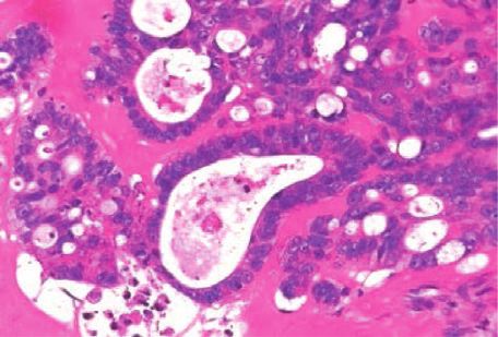
2                                                                                                         Journal of Oncology

proliferation and increase cell apoptosis in prostate cancer     2.4. Colony Formation Assay. In brief, 8 × 10² HCT116-Id4
[5]. On the contrary, Id4 functions as an oncogene in the        cells or control HCT116 cells were seeded in 60 mm plates,
growth and differentiation of breast cancer cells and hep-        followed by incubation at 37°C in an environment with 5%
atoma cancer cells [6, 7]. There is evidence showing that Id4    CO2 for 5–21 days. Surviving colonies containing 50 or more
promoter is hypermethylated in about 30% of primary              cells were stained with crystal violet, counted, and then
gastric cancers, and Id4 expression is downregulated in most     photographed under a QImaging MicroPublisher 5.0 RTV
gastric cancer cell lines due to the hypermethylation in its     microscope.
promoter region [8]. Although Id4 plays different roles in
pathogenesis of various cancers, little is known about the
functions of Id4 and its regulatory effects on the progression    2.5. Cell Cycle Analysis. Cell cycle analysis was performed to
of CRC.                                                          measure the DNA content in different phases by flow
    This study aimed to investigate the effects of Id4 on the     cytometry (FCM). In brief, cells were harvested and then
proliferation, invasion, and metastasis of CRC cells and         washed with phosphate-buffered saline (PBS) by centrifu-
explore the potential mechanisms.                                gation at 1000×g for 5 min at room temperature. The cells
                                                                 were stained with cell cycle analysis kit at 37°C for 30 min in
                                                                 dark after fixation with 70% ethanol at 4°C, washed with PBS,
2. Materials and Methods                                         and then subjected to FCM. The experiments were per-
                                                                 formed in triplicate.
2.1. Cell Lines. The normal colon mucosal cells (NCM460)
and CRC HCT116 cells were maintained in McCoy’s 5A
medium (Invitrogen, CA, USA) at 37°C in 5% CO2, the CRC          2.6. Wound Healing Assay and Cell Migration/Invasion Assay.
SW480 cells and SW620 cells were maintained in L15               HCT116-Id4 cells or control HCT116 cells were seeded in
medium (Invitrogen, CA, USA) at 37°C without CO2, and            60 mm plates and grew overnight until about 90% conflu-
the CRC HCT8 cells were grown in RPMI1640 medium                 ence was observed. On the following day, a wound was made
(Invitrogen, CA, USA) at 37°C in a humidified environment         in the cell monolayer by scratching. Cells were observed
with 5% CO2. The medium was supplemented with 10% fetal          under a light microscope at 0 and 48 h to determine the
bovine serum (FBS).                                              wound healing of the cell monolayer.
                                                                      For migration assay, 5 × 10⁴ HCT116-Id4 cells or
                                                                 HCT116 cells in the serum-free medium were seeded into
2.2. Plasmid Transfection and Generation of Stably Transduced    the upper chamber of a Transwell insert (8 mm pore size, BD
Cell Lines. Id4 overexpressing and empty lentiviral vectors      Bioscience). For invasion assay, the insert was precoated
were provided by the Genechem Biotechnology (Shanghai,           with Matrigel (BD Bioscience), and 8 × 10⁵ cells were seeded
China). The stable Id4 overexpressing HCT116 cells               into the insert. The medium containing 20% FBS was added
(HCT116-Id4) were screened with puromycin at 1.5 μg/ml           into the lower chamber as a chemoattractant. Cells that
(Sigma) for at least one week. The Id4 expression was            remained on the upper chamber after incubation for 48 h
confirmed by quantitative PCR and Western blotting. The           were removed by a cotton swab, and the insert was sub-
primer of Id4 gene was as follows: forward 5’TCGTA               sequently stained with 0.1% crystal violet in 20% methanol,
AAACCCAGAGCGACC 3’, reverse 5’ GCTGACTTTCTTG                     followed by cell counting after photographing under a
TTGGGCG 3’. pcDNA3.3-CK18 was constructed by                     QImaging MicroPublisher 5.0 RTV microscope.
inserting a CK18 gene that was generated from PCR product
to the BamH1 and Xho I sites (reagents were from New
England BioLabs, Beverly, MA, USA) of the plasmid                2.7. Western Blotting. After washing with cold PBS, HCT116
pcDNA3.3-CS2.0-N-flag (Invitrogen). The primers of CK18           cells in 6 cm dishes were lysed with cell lysis buffer (50 mM
gene were as follows: forward, 5’-TAGAGAATTCGGATCC               HEPES, pH 7.4, 250 mM NaCl, 1% Nonidet P-40, 1 mM
ATGAGCTTCACCACTCGCTC-3’, reverse, 5’-GCTTCCA                     EDTA, 1 mM Na3VO4, 1 mM NaF, 1 mM PMSF, and 1 mM
TGGCTCGAGTTAATGCCTCAGAACTTTGGTG-3’. HC                           dithiothreitol and protease inhibitor cocktail from Roche)
T116 cells were transfected with pcDNA3.3-CK18 or empty          for 30 min at 4°C for Western blotting or coimmunopre-
vector pcDNA3.3 with Xtreme GENE HP DNA Transfection             cipitation (IP). The protein concentration was determined
Reagent (Roche Diagnostics) according to the manufac-            by using the BCA protein Assay Kit (Pierce, USA), and then,
turer’s instructions.                                            proteins of the same amount were separated by 12% or 15%
                                                                 SDS-PAGE. Then, proteins were transferred onto PVDF
                                                                 membranes (GE Healthcare), which were blocked in Tris-
2.3. Cell Proliferation Assay. Cell proliferation was assessed   buffered saline containing 5% bovine serum albumin (BSA)
by using the WST method. The Id4 overexpressing or empty         for 1 h at room temperature. The primary antibodies used in
vector-transfected cells were seeded into 96-well plates at      this study included anti-Id4 (1 : 200; Santa Cruz Biotech-
5 × 10³ cells/well and then maintained for 24, 48, or 72 h at    nology, Santa Cruz, CA, USA), anti-P21 (1 : 1000, Cell Sig-
37°C in a humidified environment with 5% CO2. Then, 10 μl         naling Technology, USA), anti-P27 (1 : 1000, Cell Signaling
of WST solution was added to each well, followed by in-          Technology, USA), anti-PI3K (1 : 1000, Cell Signaling
cubation at 37°C for 2 h. The absorbance of each well was        Technology, USA), anti-P-PI3K (1 : 1000, Cell Signaling
determined at 450 nm in a microplate reader.                     Technology, USA), anti-AKT (1 : 1000, Cell Signaling
Id4 Suppresses the Growth and Invasion of Colorectal Cancer HCT116 Cells through CK18-Related Inhibition of AKT and EMT Signaling - Hindawi.com
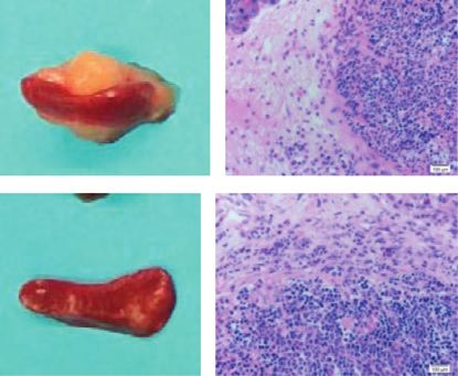
Journal of Oncology                                                                                                            3

Technology, USA), anti-P-AKT (1 : 1000, Cell Signaling          protein expression reduced significantly in these 4 CRC cell
Technology, USA), anti-GAPDH (1 : 1000, Cell Signaling          lines as compared to the NCM460 cells (Figure 1(a)).
Technology, USA), anti-MMP2 (1 : 5000, Abcam, USA),                 To examine the effects of Id4 on the biological charac-
anti-MMP9 (1 : 2000, Abcam, USA), anti-MMP7 (1 : 5000,          teristics of CRC, HCT116 cells with stable Id4 expression
Abcam, USA), anti-Twist (1 : 1000, Cell Signaling Technol-      (HCT116-Id4 cells) were established, and the Id4 expression
ogy, USA), anti-slug (1 : 1000, Cell Signaling Technology,      was confirmed by Western blotting and real-time RT-PCR
USA), anti-β-catenin (1 : 1000, Cell Signaling Technology,      (Figures 1(b) and 1(c), P < 0.05). Subsequently, the cell
USA), anti-snail (1 : 1000, Cell Signaling Technology, USA),    proliferation of HCT116-Id4 cells and control cells was de-
anti-TIMP1 (1 : 1000, Cell Signaling Technology, USA), anti-    termined. As shown in Figure 1(d), the proliferation of the
TIMP2 (1 : 1000, Cell Signaling Technology, USA), and anti-     HCT116-Id4 cells was significantly inhibited as compared to
CK18 (1 : 3000, Abcam, USA). After incubation with              the control group (P < 0.05), indicating that Id4 might play a
horseradish peroxidase-conjugated secondary antibodies,         key role in the growth of HCT116 cells. The colony-forming
the membranes were visualized by using the enhanced             assay confirmed the inhibitory effect of Id4 on the prolifer-
chemiluminescence method (ECL Plus, USA).                       ation of the HCT116 cells (Figure 1(e)). To investigate if Id4 is
                                                                involved in cell cycle progression of HCT116 cells, FCM was
                                                                performed to detect the percentage of cells in different cell
2.8. Coimmunoprecipitation (Co-IP) Assay. For the Co-IP         phases. Results showed that the proportion of HCT116 cells in
assay, the HCT16-Id4 cells were lysed, and the soluble          the G1/G0 phase increased significantly following Id4
proteins were precleared with 100 μl of 50% slurry of protein   transfection (P < 0.01), but the proportions of HCT116 cells in
A agarose (Invitrogen). The clear lysates were then mixed       the S phase and G2/M phase markedly decreased (P < 0.01)
with 4 μg of antibodies and 100 μl of 50% slurry of protein A   (Figure 1(f)). These results demonstrated that Id4 might
agarose. The immunoprecipitated complexes were analyzed         regulate cell cycle progression of HCT116 cells. Moreover,
by Western blotting.                                            markedly smaller tumors were found in nude mice after
                                                                HCT116-Id4 cells inoculation as compared to the control
2.9. Animal Experiments. Specific pathogen-free (SPF)            group (Figure 1(g), P < 0.05), which illustrated that Id4 could
BALB/C nude mice aged 4–6 weeks were purchased from             significantly inhibit the growth of xenograft tumor in the
Shanghai SLAC Laboratory Animal Co. Ltd. All experiments        nude mice. Collectively, these findings suggested that Id4
were performed in accordance with Animal Care Committee         might be a novel tumor suppressor in the CRC.
of Fujian Medical University, China. For in vivo tumor              Furthermore, to investigate the underlying mechanisms
growth assessment, 1 × 10⁶ HCT116-con cells and HCT116-         underlying the regulatory effects of Id4 on the CRC cells, the
Id4 cells in 100 μl of PBS were subcutaneously injected into    expression of proliferation-related markers was detected in
the left and right flank of mice, respectively. The tumor        the HCT116-Id4 cells. Results showed that upregulation of
volume was measured once every 4 days and calculated as         Id4 expression markedly increased the expression of P21,
follows: (length × width)2/2. Two weeks later, the tumor        P27, CDK4, and CDK2 but significantly decreased the ex-
weight was obtained after the mice were sacrificed.              pression of PI3K, P-PI3K, AKT, p-AKT (upstream mole-
    For the establishment of the liver metastasis model,        cules of P21 and P27), and cyclin E in the HCT116-Id4 cells
5 × 10⁶ cells were injected into the spleen of each mouse. At   as compared to the control HCT116 cells (Figure 1(h),
days 28, the mice were sacrificed, and the tumor samples         P < 0.05). These results demonstrated that Id4 upregulated
were collected from the site of the tumor injection and from    the expression of proliferation-related proteins and
the liver and calculated. All the tissues were fixed in 4%       inhibited the PI3K/AKT pathway in the CRC cells.
formalin and then embedded with paraffin for further
pathological examination.
                                                                3.2. Id4 Inhibits the Migration, Invasion, and Metastasis of
                                                                CRC Cells In Vitro and In Vivo. In order to explore the effects
2.10. Statistical Analysis. Data are expressed as mean-         of Id4 on the migration of CRC cells, a wound healing/
s ± standard deviation (SD) from at least 3 independent         scratch assay was performed. As shown in Figure 2(a), Id4
examinations, and statistical analysis was performed with       upregulation remarkably attenuated the wound healing as
Prism7 (GraphPad Software, La Jolla, CA, USA). Com-             compared to the control HCT 116 cells. The invasive po-
parisons were performed with Student’s t-test between           tential of HCT116-Id4 cells and control cells was assessed by
groups or one-way analysis of variance (ANOVA) among            a modified Boyden chamber invasion assay. Results showed
groups. A value of P < 0.05 was considered statistically        Id4 overexpression significantly reduced the HCT116-Id4
significant.                                                     cells invading the Matrigel layer as compared to the control
                                                                HCT 116 cells (P < 0.01, Figures 2(b) and Figure 2(c)). To
3. Results                                                      assess the metastatic ability of HCT116-Id4 cells in vivo,
                                                                2 × 10⁶ cells were injected into the spleens of nude mice. Four
3.1. Id4 Overexpression Inhibits the Proliferation of HCT116    weeks later, all the mice in the control group developed liver
Cells. The Id4 expression was detected in the four CRC cell     metastases (5/5). In contrast, only 1 mouse (1/5) in the
lines (HCT116, SW480, SW620, and HCT8) and the normal           HCT116-Id4 group developed liver metastases (P < 0.05).
colon mucosal cells (NCM460). Results showed the Id4            The number of nodules formed in the liver of the mice
Id4 Suppresses the Growth and Invasion of Colorectal Cancer HCT116 Cells through CK18-Related Inhibition of AKT and EMT Signaling - Hindawi.com
4                                                                                                                                                                                                                                                                                                                             Journal of Oncology

                       NCM460

                                                                                                                                                                                                                                                               HCT116-con

                                                                                                                                                                                                                                                                                     HCT116-Id4
                                         HCT116

                                                                      SW480

                                                                                                SW620

                                                                                                                                  HCT-8
                                                                                                                                              Id4
                                                                                                                                                                                                                                                                                                     Id4
                                                                                                                                              β-Actin
                                                                                                                                                                                                                                                                                                     β-Actin

                                                                              (a)                                                                                                                                                                                                  (b)
                                                               4                                                                                                                                                                                         300
                                         Id4 mRNA relative

                                                                                                         ∗

                                                                                                                                                                                                                       Relative growth
                                          expression (log)

                                                               3                                                                                                                                                                                         200

                                                                                                                                                                                                                           rate (%)
                                                               2                                                                                                                                                                                                                                                        ∗
                                                                                                                                                                                                                                                         100                                             ∗
                                                               1
                                                                                                                                                                                                                                                           0
                                                               0
                                                                                                                                                                                                                                                                 0                 24                    48            72
                                                                               HCT116-control

                                                                                                        HCT116-Id4

                                                                                                                                                                                                                                                                                  HCT116-Id4
                                                                                                                                                                                                                                                                                  HCT116-con
                                                                                                                                                                                                                                                                                  HCT116

                                                                              (c)                                                                                                                                                                                                  (d)
                                                                                                                                    800                                                                                                                                                                                                         100
                                                                                                               Colony formation

                                                                                                                                                                                                                                                                                                                             Distribution (%)
                                                                                                                                                                                                                G0/G1: (45.87±0.44)%                                                               G0/G1:(62.78±0.55)%
                                                                                                                                    600                                                                         S: (37.45±2.04) %                                                                  S: (28.56±0.70)%
                                                                                                                                                                                           200                  G2/M: (16.68±1.13) %                                        280                    G2/M: (8.60±1.32) %

                                                                                                                                    400
                                                                                                                                                                              Number

                                                                                                                                                                                                                                                           Number
                                                                                                                                                                                           150                                                                              210                                                                  50
                                                                                                                                                                  ∗
                                                                                                                                    200                                                    100                                                                              140
          HCT116-con                                   HCT116-Id4                                                                                                                           50                                                                               70
                                                                                                                                          0                                                                                                                                                                                                      0
                                                                                                                                                                                            0                                                                                 0
                                                                                                                                                    HCT116-con

                                                                                                                                                                 HCT116-Id4

                                                                                                                                                                                                                                                                                                                                                             HCT116-con

                                                                                                                                                                                                                                                                                                                                                                           HCT116-Id4
                                                                                                                                                                                                     0 50 100 150 200                                                             0 50 100 150 200
                                                                                                                                                                                                      Channels (PLA)                                                                Channels (PLA)
                                                                                                                                                                                                       HCT116-con                                                                    HCT116-Id4

                                                                                                                                                                                                                                                                                                                                                                          G2M
                                                                                                                                                                                                                                                                                                                                                                          S
                                                                                                                                                                                                                                                                                                                                                                          G0/G1

                                                                              (e)                                                                                                                                                                                                  (f )
                                                                                                                                                                              HCT116-con

                                                                                                                                                                                                 HCT116-Id4

                      HCT116-con

                      HCT116-Id4
                                                                                                                                                                                                              Id4
                600
                                                                                                                                                                                                              P21                                        1.5
                400                                                                                                                                                                                           P27
                                                                                                                                                                                                                               Relative protein levels
       Volume

                                                                                                                                                                                                              P13K                                       1.0
                200
                                                              ∗                                                                                                                                               P-PI3K
                  0                                                                                                                                                                                           AKT                                        0.5
                            HCT116-con

                                                             HCT116-Id4

                                                                                                                                                                                                              P-AKT
                                                                                                                                                                                                                                                           0
                                                                                                                                                                                                              CDK4
                                                                                                                                                                                                                                                                            P21

                                                                                                                                                                                                                                                                                   P27

                                                                                                                                                                                                                                                                                                  P13K

                                                                                                                                                                                                                                                                                                              p-P13K

                                                                                                                                                                                                                                                                                                                       AKT

                                                                                                                                                                                                                                                                                                                                     p_AKT

                                                                                                                                                                                                                                                                                                                                                      CDK4

                                                                                                                                                                                                                                                                                                                                                                    CDK2

                                                                                                                                                                                                                                                                                                                                                                                    Cyclin E

                                                                                                                                                                                                              CDK2

                                                                                                                                                                                                              Cyclin E
                                                                                                                                                                                                                                                                                  HCT116-control
                                                                                                                                                                                                              GAPDH                                                               HCT116-Id4

                                                                              (g)                                                                                                                                                                                                  (h)

Figure 1: Id4 overexpression reduces cell proliferation and arrests cell cycle at the G0/G1 phase. (a) Id4 protein expression in the CRC. (b, c)
Real-time RT-PCR and Western blotting confirmed stable Id4 overexpression in the HCT116 cells. β-Actin was used as a loading control. (d)
WST assay was used to assess the effect of Id4 on the proliferation of HCT116 cells. (e) Colony formation assay was used to assess the effect of
Id4 on the colony-forming ability of the HCT116 cells. (f ) Flow cytometry was performed to detect the cell cycle after Id4 overexpression. (g)
Photographs of the xenograft tumors in the nude mice at day 14 after implantation, and tumor weight was measured. (h) Protein expression
of molecules in the PI3K/AKTpathway and proliferation-related proteins in the HCT116-Id4 cells (Western blotting). GAPDH was used as a
loading control. Data are expressed as mean ± SD. ∗ P < 0.05.
Id4 Suppresses the Growth and Invasion of Colorectal Cancer HCT116 Cells through CK18-Related Inhibition of AKT and EMT Signaling - Hindawi.com
Journal of Oncology                                                                                                                                                                                                                                       5

                    0h                       48h                                                                                                                                                                  250
                                                                                                                                                              Migration
                                                                                  100
   HCT116-con

                                                                                                                                                                                               number per field
                                                                                                                                                                                                                  200

                                                                                                                                                                                                Migration cell
                                                                                                                                                     HCT116-con        HCT116-Id4

                                                              Width injury line
                                                                                      80                                                                                                                          150                         ∗

                                                                 (% of oh)
                                                                                      60
                                                                                                                                   ∗                                                                              100
                                                                                      40                                                                                                                           50
   HCT116-Id4

                                                                                      20                                                                                                                            0
                                                                                       0

                                                                                                                                                                                                                            HCT116-con

                                                                                                                                                                                                                                             HCT116-Id4
                                                                                                        HCT116-con

                                                                                                                                  HCT116-Id4
                                               (a)                                                                                                                               (b)
                             Invasion                                   100                                                                                                                                                 Spleen

                                                                                                                                                                                          HCT116-
                HCT116-con              HCT116-Id4
                                                     number per field

                                                                            80

                                                                                                                                                                                            con
                                                       Invased cell

                                                                            60                                        ∗                        HCT116-con
                                                                            40

                                                                                                                                                                                          HCT116-
                                                                            20                                                                 HCT116-Id4

                                                                                                                                                                                            Id4
                                                                                  0
                                                                                                                                                              Liver metastasis
                                                                                           HCT116-con

                                                                                                                     HCT116-Id4

                                                                                                                                                                                                       20
                                                                                                                                                                                                                                         ∗
                                                                                                                                               HCT116-con

                                                                                                                                                                                       Visible tumor
                                                                                                                                                                                                       15

                                                                                                                                                                                          nodules
                                                                                                                                                                                                       10
                                                                                                                                               HCT116-Id4
                                                                                                                                                                                                          5

                                                                                                                                                                                                          0
                                                                                                                                                                                                                        HCT116- HCT116-
                                                                                                                                                                                                                          con     Id4

                                               (c)                                                                                                                               (d)

Figure 2: Id4 regulated the migration, invasion, and metastatic potentials of CRC cells. (a) Wound healing assay of CRC cells. (b) Migration
assay of CRC cells (Boyden chamber assay). (c) Invasion assay of CRC cells (Boyden chamber assay). (d) Intrasplenic implantation of
HCT116-Id4 cells or control cells. The number of tumors in the spleens and liver metastases of the HCT116-Id4 group decreased
significantly.

implanted with HCT116-Id4 cells remarkably reduced as                                                                                           3.4. Id4 Regulates CK18 Expression in the CRC and Mediates
compared to mice implanted with the control cells                                                                                               the Migration and Invasion through CK18. To explore the
(Figure 2(d)). Collectively, these results suggested an im-                                                                                     downstream effectors of Id4 in the HCT116 cells, proteins
portant role of Id4 on the progression of CRC.                                                                                                  which were likely to interact with Id4 were captured by
                                                                                                                                                immunoprecipitation assay, and then, the proteins were
                                                                                                                                                separated by using the SDS-PAGE method and identified by
3.3. Id4 Suppresses the Epithelial-Mesenchymal Transition of                                                                                    MALDI-TOF-MS (Figure 4(a)). Seven differentially
HCT116 Cells. Epithelial-mesenchymal transition (EMT)                                                                                           expressed proteins were identified in the HCT116-Id4 cells
plays a crucial role in the metastasis of many cancers. To                                                                                      as compared to control cells. Of which, the cytokeratin 18
investigate the phenotypic changes of HCT116 cells after Id4                                                                                    (CK18) was selected for further confirmation. An in vivo
overexpression, the expression of EMT-associated molecules                                                                                      coimmunoprecipitation (Co-IP) assay was performed in the
was detected in the HCT116 cells by Western blotting. At the                                                                                    HCT116-Id4 cells and HCT116 cells to investigate the po-
same time, the expression of MMP2, MMP7, and MMP9                                                                                               tential interaction between Id4 and CK18 in the CRC cells.
(factors in the matrix metalloproteinases family and in-                                                                                        As shown in Figure 4(b), Id4 was able to efficiently
volved in the invasion of cancers) was detected in the control                                                                                  coprecipitate with endogenous CK18 and vice versa. These
cells and HCT116-Id4 cells. Western blotting revealed that                                                                                      results revealed that Id4 could interact with CK18 in vivo.
the protein expression of β-catenin, twist, slug, snail, MMP2,                                                                                      Subsequently, to assess the functional consequence of the
MMP7, and MMP9 significantly decreased, while the ex-                                                                                            interaction between Id4 and CK18, the following experi-
pression of E-cadherin, TIMP1, and TIMP2 was markedly                                                                                           ments were performed to explore whether Id4 could affect
upregulated in the HCT116-Id4 cells as compared to control                                                                                      the CK18 protein expression. As shown in Figure 4(c), Id4
cells (Figure 3(a), P < 0.05). Microscopy showed that Id4                                                                                       overexpression significantly decreased the CK18 protein
overexpressing cells acquired more characteristics of epi-                                                                                      expression in the HCT116-Id4 cells. Thus, we hypothesized
thelial morphology than the control cells (Figure 3(b)).                                                                                        that Id4 attenuates the motility, invasion, and migration of
These revealed a possible molecular mechanism about the                                                                                         CRC cells through CK18. To further validate this hypothesis,
Id4-mediated cell migration and invasion.                                                                                                       Id4 was compensated by its exogenous protein in the
Id4 Suppresses the Growth and Invasion of Colorectal Cancer HCT116 Cells through CK18-Related Inhibition of AKT and EMT Signaling - Hindawi.com
6                                                                                                                                                                                       Journal of Oncology

                                 HCT116-con
                                                                                                    1.5

                                              HCT116-Id4

                                                                          Relative protein levels
                                                                                                    1.0        ∗

                                                             Id4                                                                                                          ∗       ∗
                                                                                                    0.5                                                            ∗
                                                             E-cadherin                                                                                     ∗

                                                                                                                            ∗        ∗       ∗      ∗
                                                             β-catenin                              0.0

                                                                                                          E-cadherin
                                                                                                                       β-catenin
                                                                                                                                   Twist
                                                                                                                                           Slug

                                                                                                                                                          MMP2
                                                                                                                                                                 MMP7
                                                                                                                                                                        TIMP1
                                                                                                                                                                                TIMP2
                                                                                                                                                  Snail
                                                             Twist

                                                             Slug

                                                                                                                       HCT116-con
                                                             Snail
                                                                                                                       HCT116-Id4
                                                             MMP2

                                                             MMP7

                                                             TIMP1

                                                             TIMP2

                                                             GAPDH

                                                                                                          (a)

                                                           HCT116-control                                                                  HCT116-Id4
                                                                                                          (b)

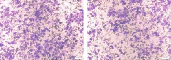
Figure 3: Id4 regulated the expression of EMT-related markers in the HCT116 cells. (a) The expression of EMT-related molecules in the
HCT116-Id4 cells and control cells (Western blotting). (b) Cellular morphology of HCT116-Id4 cells and control cells under microscopy.

HCT116-Id4 cells as a genetic rescue experiment. As shown                                                              clinical behaviours [11, 12]. In our previous study, results
in Figure 4(d), CK18 expression in the HCT116-Id4 cell was                                                             showed Id1 expression increased significantly in the CRC
restored to the control level after transfection with the CK18                                                         tissues than in the normal mucosal tissues, and Id1 expression
overexpressing vector. Consequently, HCT116-Id4/CK18                                                                   was positively related to poor differentiation of CRC cells [13].
overexpressing cells showed the upregulated expression of                                                              Moreover, knock-down of Id1 gene not only inhibits the
MMP2, MMP7, PI3K, P-PI3K, and p-AKT, but the                                                                           growth of HCT116 cells but also suppresses the hepatic
E-cadherin expression was downregulated as compared to                                                                 metastasis in vivo in a CXCR4-dependent manner [14]. These
the HCT116-Id4 cells (Figure 4(d)). Taken together, our                                                                results underscore the importance of Id-mediated tumori-
findings suggested Id4 affects the CK18 expression and the                                                               genesis, cell proliferation, and metastasis of cancers.
PI3K/AKT pathway via CK18.                                                                                                 Despite the structural similarity, Id4 still has different
                                                                                                                       roles in the tumorigenesis, tumor invasion, and metastasis as
4. Discussion                                                                                                          compared to other Id family members. Although the Id4
                                                                                                                       protein is involved in the regulation of cell cycle and cell
The Id family has helix loop-helix transcription factors and is                                                        senescence [15], its expression and functions remain con-
one of the key regulators of cell fate, differentiation, prolif-                                                        troversial in some cancers. Zeng et al. [16] found that Id4 was
eration, and cell death [9, 10]. Several reports have revealed                                                         highly expressed in the glioblastoma multiforme (GBM), in
the importance of Id proteins in the inhibition of cell dif-                                                           which Id4 promoted the growth and angiogenesis. However,
ferentiation and regulation of cell proliferation. In many types                                                       in the prostate cancer, Id4 acted as a tumor suppressor, and
of human tumors, the Id protein expression increases which is                                                          Id4 overexpression in the PC3 cells (one of the highly
often associated with the enhanced malignancy and aggressive                                                           malignant prostate cancer cell lines) increased apoptosis and
Id4 Suppresses the Growth and Invasion of Colorectal Cancer HCT116 Cells through CK18-Related Inhibition of AKT and EMT Signaling - Hindawi.com
Journal of Oncology                                                                                                                                                   7

             HCT116-con
                                                       HCT116-con                   HCT116-Id4                      HCT116-con             HCT116-Id4

                          HCT116-Id4
                                                    Input                 Flag IP   Input   Flag IP             Input        K18 IP        Input      K18 IP
                                                                                                       CK18                                                    Id4

                                                                                                         Id4                                                   CK18

    6
                                       4
                                       5

                                       1
                                       2
                                       3

                 (a)                                                                                      (b)
                                       HCT116-con

                                                                                                                             HCT116
                                                            HCT116-Id4

                                                                                                                Ad-Id4   –       +     +
                                                                                                                pCK18    –       –     +
                                                                                                                                             Id4
                                                                         CK18
                                                                                                                                             CK18
                                                                         GAPDH
                                                                                                                                             MMP2

                                                                                                                                             MMP7

                                                                                                                                             E-cadherin

                                                                                                                                             PI3K

                                                                                                                                             P-PI3K

                                                                                                                                             P-AKT

                                                                                                                                             GAPDH

                                                       (c)                                                                       (d)

Figure 4: Id4 binding to CK18 regulated the PI3K pathway and the expression of EMT-related markers. (a) Coomassie blue-stained SDS-
PAGE gel showed the protein binding to Id4 (flag). (b) Co-IP assay showed the interaction between Id4 and CK18 in the HCT116 cells. (c)
Id4 decreased CK18 protein expression (Western blotting). (d) HCT116-Id4 cells and negative control cells were transfected with CK18
plasmid or control plasmid, respectively. Western blotting was performed to investigate the effect of CK18 on the expression of EMT-related
markers in the HCT116 cells.

inhibited cell proliferation and migration [5, 17]. Id4 has also                                     The effects of Id4 on the cell cycle and proliferation have
been found to be epigenetic silencing in many cancers such                                       been reported in some cancer cells. Id4 overexpression is
as squamous cell carcinoma [18], gastric cancer [8], and CRC                                     able to arrest cell cycle and inhibit cell proliferation, which is
[19], which is ascribed to the promoter hypermethylation.                                        associated with the increased expression of cyclin-dependent
Id4 gene promoter is hypermethylated in 30% of primary                                           kinase inhibitors p21 and p27 in the prostate cancer cell line
gastric cancers, and Id4 expression is downregulated in most                                     DU145 cells [5]. In addition, Id4 overexpression in lung
gastric cancer cell lines due to the hypermethylation of the                                     cancer cells inhibits cisplatin-induced apoptosis via the p38
promoter region [8]. Although the methylation state of Id4                                       MAPK pathway [20]. Similar findings were observed in our
gene promoter in the CRC still needs to be investigated                                          study. Id4 overexpression significantly reduced tumor
thoroughly, Id4 might be a tumor suppressor in the CRC                                           growth and hepatic metastasis in the mice inoculated with
according to our findings that the Id4 expression was very                                        HCT116-Id4 cells. The cell cycle was arrested at the G0/G1
low in the CRC cells than in the normal mucosal cells.                                           phase in the HCT116-Id4 cells, which was further confirmed
Id4 Suppresses the Growth and Invasion of Colorectal Cancer HCT116 Cells through CK18-Related Inhibition of AKT and EMT Signaling - Hindawi.com
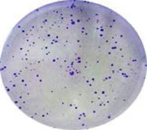
8                                                                                                             Journal of Oncology

by the increased expression of p21 and p27. In addition, Id4     18 expression and AKT2 upregulates the expression of CK18
overexpression suppressed the expression and activity of         and vimentin [35]. According to our findings, CK18 over-
PI3K, p-PI3K, AKT, and p-AKT, suggesting that Id4 induces        expression reverses the suppressive effects of Id4 on the
the suppression of CRC cell growth which may be related to       PI3K/AKT pathway and EMT. It means that CK18 may be a
the inhibition of the PI3K/AKT signaling pathway. EMT is a       target of Id4 in the PI3K/AKT pathway. Together, findings in
process which involves in the shifting between two alter-        the present study indicate Id4 can inhibit the growth and
native states, mesenchymal or epithelial. Aberrant EMT           invasion of CRC cells through CK18-related inhibition of the
activation may trigger the dissociation of cancer cells from     AKT pathway and EMT.
primary carcinomas, and then, they subsequently migrate
and disseminate to distant organs. During the EMT, the           5. Conclusion
expression of cell adhesion molecules, especially E-cadherin,
is concomitantly repressed, but that of other molecules (such    In summary, our findings indicate that Id4 expression re-
as snail, zeb, and twist) is upregulated [21]. Numerous          duces in the CRC cells, and Id4 overexpression inhibits the
studies have indicated that Id protein is one of factors in-     growth of CRC cells as well as suppresses the migration,
volved in the EMT. In lung cancer cells, the inhibition of Id1   invasion, and metastasis of CRC cells in vitro and in vivo.
expression reduces the expression of EMT-related markers         Furthermore, Id4 mediates the migration and invasion of
(vimentin, intergrin-β, and snail). Even in the cancer tissues   CRC cells through interacting with CK18 to suppress the
from NSCLS patients, Id1 seems to be significantly related to     PI3K/AKT pathway and EMT. Collectively, these findings
the vimentin and other EMT-related proteins [22]. Id1            implicate that Id4 may serve as a tumor suppressor and has
opposed twist activity, thus promoting cell metastasis to the    the promise to become a target in the clinical treatments of
lung via EMT [23]. It has been indicated that Id4 can inhibit    CRC.
lung cancer metastasis and mesenchymal-epithelial transi-
tion (MET) by binding to slug and promoting E-cadherin           Data Availability
expression [24]. MMPs are also important markers of EMT
and play a major role in the biological behaviours of cells      The data used to support the findings of this study are
such as cell proliferation, migration, and differentiation.       available from the corresponding author upon request.
MMP2 and MMP9 are gelatinases capable of degrading type
IV collagen and are the most abundant components in the          Conflicts of Interest
basement membrane. The degradation of the basement               The authors declare that there are no conflicts of interest.
membrane is an essential step for the metastasis of most
cancers [25]. MMP7 is also known as matrilysin which can         Authors’ Contributions
digest components of the extracellular matrix. The active
MMP7 can also cleave the pro-MMP2 and pro-MMP9 to                Hui-Jing Chen and Yue Yu contributed equally to this work.
facilitate tumor invasion [26]. Id4 is involved in suppressing
the MMP2-mediated cell invasion in glioblastoma [27]. Our        Acknowledgments
results showed that Id4 overexpression downregulated the
expression of MMP2, MMP7, β-catenin, twist, slug, and            This study was supported by the National Natural Science
snail, but upregulated that of TIMP1 and TIMP2, indicating       Foundation of China (81472720).
that Id4 is involved in reversing the progression of EMT.
    Id proteins dimerize with bHLH protein, which inhibits       References
their DNA binding activity [28]. In the lung cancer, Id4
binds to slug, which interferes with its interaction with         [1] F. Bray, J. Ferlay, I. Soerjomataram, R. L. Siegel, L. A. Torre,
                                                                      and A. Jemal, “Global cancer statistics 2018: GLOBOCAN
E-box promoter, and then suppresses the metastasis of
                                                                      estimates of incidence and mortality worldwide for 36 cancers
cancer cells [24]. Id4 interacts with MDC1 and other DNA              in 185 countries,” CA: A Cancer Journal for Clinicians, vol. 68,
repair proteins which govern the DNA damage response                  no. 6, pp. 394–424, 2018.
[29]. Our results showed that Id4 could interact with CK18.       [2] R. Siegel, C. Desantis, and A. Jemal, “Colorectal cancer sta-
CK18 gene encodes the type I intermediated filament                    tistics, 2014,” CA: A Cancer Journal for Clinicians, vol. 64,
protein, which is primarily found in many types of single-            no. 2, pp. 104–117, 2014.
layered epithelial tissues. CK18 protein localizes in the cy-     [3] R. Benezra, R. L. Davis, D. Lockshon, D. L. Turner, and
toplasm and perinuclear region [30]. CK18 is important for            H. Weintraub, “The protein Id: a negative regulator of helix-
some cell processes including apoptosis [31], mitosis [32],           loop-helix DNA binding proteins,” Cell, vol. 61, no. 1,
cell cycle progression [33], and cell signaling [34]. Prior           pp. 49–59, 1990.
studies have noted that CK18 is an epithelial marker in the       [4] V. Riechmann, I. van Crüchten, and F. Sablitzky, “The ex-
                                                                      pression pattern ofId4, a novel dominant negative helix-loop-
diagnosis of cancers. Galarneau et al. [34] proposed that
                                                                      helix protein, is distinct fromId1,1d2 and Id3,” Nucleic Acids
CK18 was involved in the signalling pathways related to the           Research, vol. 22, no. 5, pp. 749–755, 1994.
regulation of cell growth, death, and motility. The PI3K/AKT      [5] J. P. Carey, A. J. Asirvatham, O. Galm, T. A. Ghogomu, and
pathway plays a pivotal role in these mechanisms in which             J. Chaudhary, “Inhibitor of differentiation 4 (Id4) is a po-
CK18 involves. There is a close relationship between CK18             tential tumor suppressor in prostate cancer,” BMC Cancer,
and AKT because AKT1 overexpression increases the CK8/                vol. 9, no. 1, p. 173, 2009.
Id4 Suppresses the Growth and Invasion of Colorectal Cancer HCT116 Cells through CK18-Related Inhibition of AKT and EMT Signaling - Hindawi.com
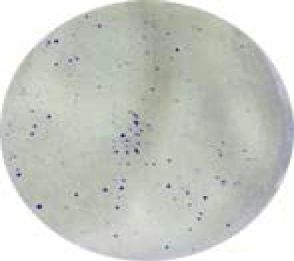
Journal of Oncology                                                                                                                           9

 [6] G. Fontemaggi, S. Dell’Orso, D. Trisciuoglio et al., “The ex-              mesenchymal-to-epithelial transition,” Cell Reports, vol. 5,
     ecution of the transcriptional axis mutant p53, E2F1 and ID4               no. 5, pp. 1228–1242, 2013.
     promotes tumor neo-angiogenesis,” Nature Structural &               [24]   C.-C. Wang, Y.-L. Hsu, C.-J. Chang, C.-J. Wang, T.-H. Hsiao,
     Molecular Biology, vol. 16, no. 10, pp. 1086–1093, 2009.                   and S.-H. Pan, “Inhibitor of DNA-binding protein 4 sup-
 [7] Y. Zhang, L.-X. Zhang, X.-Q. Liu et al., “Id4 promotes cell                presses cancer metastasis through the regulation of epithelial
     proliferation in hepatocellular carcinoma,” Chinese Journal of             mesenchymal transition in lung adenocarcinoma,” Cancers,
     Cancer, vol. 36, no. 1, p. 19, 2017.                                       vol. 11, no. 12, p. 2021, 2019.
 [8] A. S. W. Chan, W. Y. Tsui, X. Chen et al., “Downregulation of       [25]   O. R. F. Mook, W. M. Frederiks, and C. J. F. Van Noorden,
     ID4 by promoter hypermethylation in gastric adenocarci-                    “The role of gelatinases in colorectal cancer progression and
     noma,” Oncogene, vol. 22, no. 44, pp. 6946–6953, 2003.                     metastasis,” Biochimica et Biophysica Acta (BBA) - Reviews on
 [9] M. B. Ruzinova and R. Benezra, “Id proteins in development,                Cancer, vol. 1705, no. 2, pp. 69–89, 2004.
     cell cycle and cancer,” Trends in Cell Biology, vol. 13, no. 8,     [26]   K. Edman, M. Furber, P. Hemsley et al., “The discovery of
     pp. 410–418, 2003.                                                         MMP7 inhibitors exploiting a novel selectivity trigger,”
[10] Y. Yokota and S. Mori, “Role of Id family proteins in growth               ChemMedChem, vol. 6, no. 5, pp. 769–773, 2011.
     control,” Journal of Cellular Physiology, vol. 190, no. 1,          [27]   G. J. Rahme and M. A. Israel, “Id4 suppresses MMP2-me-
     pp. 21–28, 2002.                                                           diated invasion of glioblastoma-derived cells by direct inac-
[11] S. Fong, R. J. Debs, and P.-Y. Desprez, “Id genes and proteins             tivation of Twist1 function,” Oncogene, vol. 34, no. 1,
     as promising targets in cancer therapy,” Trends in Molecular               pp. 53–62, 2015.
                                                                         [28]   E. C. Roberts, R. W. Deed, T. Inoue, J. D. Norton, and
     Medicine, vol. 10, no. 8, pp. 387–392, 2004.
                                                                                A. D. Sharrocks, “Id helix-loop-helix proteins antagonize pax
[12] A. Lasorella, T. Uo, and A. Iavarone, “Id proteins at the cross-
                                                                                transcription factor activity by inhibiting DNA binding,”
     road of development and cancer,” Oncogene, vol. 20, no. 58,
                                                                                Molecular and Cellular Biology, vol. 21, no. 2, pp. 524–533,
     pp. 8326–8333, 2001.
                                                                                2001.
[13] Y. Sun, X. Lai, Y. Yu et al., “Inhibitor of DNA binding 1 (Id1)
                                                                         [29]   L. A. Baker, H. Holliday, D. Roden et al., “Proteogenomic
     mediates stemness of colorectal cancer cells through the Id1-
                                                                                analysis of Inhibitor of Differentiation 4 (ID4) in basal-like
     c-Myc-PLAC8 axis via the Wnt/β-catenin and Shh signaling                   breast cancer,” Breast Cancer Research, vol. 22, no. 1, p. 63,
     pathways,” Cancer Management and Research, vol. 11,                        2020.
     pp. 6855–6869, 2019.                                                [30]   R. Moll, W. W. Franke, D. L. Schiller, B. Geiger, and
[14] X. Lai, J. Liao, W. Lin et al., “Inhibitor of DNA-binding                  R. Krepler, “The catalog of human cytokeratins: patterns of
     protein 1 knockdown arrests the growth of colorectal cancer                expression in normal epithelia, tumors and cultured cells,”
     cells and suppresses hepatic metastasis in vivo,” Oncology                 Cell, vol. 31, no. 1, pp. 11–24, 1982.
     Reports, vol. 32, no. 1, pp. 79–88, 2014.                           [31]   S. Gilbert, A. Loranger, N. Daigle, and N. Marceau, “Simple
[15] Z. Zebedee and E. Hara, “Id proteins in cell cycle control and             epithelium keratins 8 and 18 provide resistance to Fas-me-
     cellular senescence,” Oncogene, vol. 20, no. 58, pp. 8317–8325,            diated apoptosis. The protection occurs through a receptor-
     2001.                                                                      targeting modulation,” Journal of Cell Biology, vol. 154, no. 4,
[16] W. Zeng, E. J. Rushing, D. P. Hartmann, and N. Azumi,                      pp. 763–774, 2001.
     “Increased inhibitor of differentiation 4 (id4) expression in        [32]   N.-O. Ku, S. Michie, E. Z. Resurreccion, R. L. Broome, and
     glioblastoma: a tissue microarray study,” Journal of Cancer,               M. B. Omary, “Keratin binding to 14-3-3 proteins modulates
     vol. 1, pp. 1–5, 2010.                                                     keratin filaments and hepatocyte mitotic progression,” Pro-
[17] S. K. Komaragiri, D. H. Bostanthirige, D. J. Morton et al., “ID4           ceedings of the National Academy of Sciences, vol. 99, no. 7,
     promotes AR expression and blocks tumorigenicity of PC3                    pp. 4373–4378, 2002.
     prostate cancer cells,” Biochemical and Biophysical Research        [33]   L. Galarneau, A. Loranger, S. Gilbert, and N. Marceau,
     Communications, vol. 478, no. 1, pp. 60–66, 2016.                          “Keratins modulate hepatic cell adhesion, size and G1/S
[18] K. Ruchusatsawat, J. Wongpiyabovorn, P. Protjaroen et al.,                 transition,” Experimental Cell Research, vol. 313, no. 1,
     “Parakeratosis in skin is associated with loss of inhibitor of             pp. 179–194, 2007.
     differentiation 4 via promoter methylation,” Human Pa-               [34]   P. A. Coulombe and M. B. Omary, ““Hard” and “soft”
     thology, vol. 42, no. 12, pp. 1878–1887, 2011.                             principles defining the structure, function and regulation of
[19] T. Gomez Del Pulgar, F. Valdes-Mora, E. Bandres et al.,                    keratin intermediate filaments,” Current Opinion in Cell Bi-
     “Cdc42 is highly expressed in colorectal adenocarcinoma and                ology, vol. 14, no. 1, pp. 110–122, 2002.
     downregulates ID4 through an epigenetic mechanism,” In-             [35]   A.-M. Fortier, C. Van Themsche, É. Asselin, and M. Cadrin,
     ternational Journal of Oncology, vol. 33, no. 1, pp. 185–193,              “Akt isoforms regulate intermediate filament protein levels in
     2008.                                                                      epithelial carcinoma cells,” FEBS Letters, vol. 584, no. 5,
[20] K. Qi, Y. Li, X. Li et al., “Id4 promotes cisplatin resistance in          pp. 984–988, 2010.
     lung cancer through the p38 MAPK pathway,” Anti-cancer
     Drugs, vol. 27, no. 10, pp. 970–978, 2016.
[21] M. A. Nieto, R. Y.-J. Huang, R. A. Jackson, and J. P. Thiery,
     “Emt: 2016,” Cell, vol. 166, no. 1, pp. 21–45, 2016.
[22] E. Castañón, A. Soltermann, I. López et al., “The inhibitor of
     differentiation-1 ( Id1 ) enables lung cancer liver colonization
     through activation of an EMT program in tumor cells and
     establishment of the pre-metastatic niche,” Cancer Letters,
     vol. 402, pp. 43–51, 2017.
[23] M. Stankic, S. Pavlovic, Y. Chin et al., “TGF-β-Id1 signaling
     opposes Twist1 and promotes metastatic colonization via a
Id4 Suppresses the Growth and Invasion of Colorectal Cancer HCT116 Cells through CK18-Related Inhibition of AKT and EMT Signaling - Hindawi.com
You can also read