Impact of Coenzyme Q10 Administration on Lead Acetate-Induced Testicular Damage in Rats
←
→
Page content transcription
If your browser does not render page correctly, please read the page content below
Hindawi
Oxidative Medicine and Cellular Longevity
Volume 2020, Article ID 4981386, 12 pages
https://doi.org/10.1155/2020/4981386

Research Article
Impact of Coenzyme Q10 Administration on Lead Acetate-
Induced Testicular Damage in Rats

 Manal El-khadragy ,1,2 Wafa A. Al-Megrin,1 Norah A. AlSadhan,3 Dina M. Metwally ,4,5
 Rehab E. El-Hennamy,2 Fatma Elzahraa H. Salem,2,6 Rami B. Kassab ,2,7
 and Ahmed E. Abdel Moneim 2
 1
 Biology Department, Faculty of Science, Princess Nourah Bint Abdulrahman University, Riyadh 84428, Saudi Arabia
 2
 Zoology and Entomology Department, Faculty of Science, Helwan University, Cairo 11795, Egypt
 3
 College of Medicine, AlMaarefa University, 11597 Riyadh, Saudi Arabia
 4
 Zoology Department, Faculty of Science, King Saud University, Riyadh 11451, Saudi Arabia
 5
 Faculty of Veterinary Medicine, Zagazig University, Zagazig, Egypt
 6
 Biology Department, Faculty of Science, Taibah University, Medina 42353, Saudi Arabia
 7
 Biology Department, Faculty of Science and Arts, Al Baha University, Almakhwah Branch, Saudi Arabia

 Correspondence should be addressed to Manal El-khadragy; manalelkhadragy@yahoo.com

 Received 23 March 2020; Revised 28 April 2020; Accepted 12 May 2020; Published 3 June 2020

 Guest Editor: German Gil

 Copyright © 2020 Manal El-khadragy et al. This is an open access article distributed under the Creative Commons Attribution
 License, which permits unrestricted use, distribution, and reproduction in any medium, provided the original work is
 properly cited.

 Exposure to lead (Pb) causes multiorgan dysfunction including reproductive impairments. Here, we examined the protective effects
 of coenzyme Q10 (CoQ10) administration on testicular injury induced by lead acetate (PbAc) exposure in rats. This study
 employed four experimental groups (n = 7) that underwent seven days of treatment as follows: control group intraperitoneally
 (i.p.) treated with 0.1 ml of 0.9% NaCl containing 1% Tween 80 (v : v), CoQ10 group that was i.p. injected with 10 mg/kg
 CoQ10, PbAc group that was i.p. treated with PbAc (20 mg/kg), and PbAc+CoQ10 group that was i.p. injected with CoQ10 2 h
 after PbAc. PbAc injection resulted in increasing residual Pb levels in the testis and reducing testosterone, luteinizing hormone,
 and follicle-stimulating hormone levels. Additionally, PbAc exposure resulted in significant oxidative damage to the tissues on
 the testes. PbAc raised the levels of prooxidants (malondialdehyde and nitric oxide) and reduced the amount of endogenous
 antioxidative proteins (glutathione and its derivative enzymes, catalase, and superoxide dismutase) available in the cell.
 Moreover, PbAc induced the inflammatory response as evidenced by the upregulation of inflammatory mediators (tumor
 necrosis factor-alpha and interleukin-1 beta). Further, PbAc treatment induced apoptosis in the testicular cells, as indicated by
 an increase in Bax and caspase 3 expression, and reduced Bcl2 expression. CoQ10 supplementation improved testicular function
 by inhibiting Pb accumulation, oxidative stress, inflammation, cell death, and histopathological changes following PbAc
 exposure. Our findings suggest that CoQ10 can act as a natural therapeutic agent to protect against the reproductive
 impairments associated with PbAc exposure.

1. Introduction [3], and cardiovascular system [4]. Recently, lead exposure
 has been highlighted as an important cause of testicular
Lead is an important environmental pollutant, which can malfunction and male infertility [5, 6]. Excess exposure to
cause serious illness if it is not controlled and monitored. It lead products reduces semen quality and reproductive
is widely used in industry, cosmetics, and medicine [1], and capacity causing male infertility [6]. It also promotes apopto-
accumulation of lead in the environment can exert a toxic sis and DNA damage in the spermatozoa [1] and activates
effect on humans and animals and causes damage to several Bax and caspase-3 in spermatogenic cells, which induces
important organs including the nervous system [2], skeleton apoptosis [5]. It also impairs sperm function by reducing
2 Oxidative Medicine and Cellular Longevity

cyclic adenosine monophosphate and calcium levels of sperm Ethics Committee for animal care and use at Helwan Univer-
and decreasing tyrosine phosphorylation of sperm proteins sity (approval number: HU/Z/010-18).
[7]. Pb exposure affects also the hormonal secretion as it
suppresses the anterior pituitary secretion of LH and FSH 2.2. Experimental Design. In order to explore the application
[8]. Lead’s toxic effect in the testes is facilitated by its ability of CoQ10 as a protective against lead acetate- (PbAc-)
to produce reactive oxygen species. It was reported that lead induced testicular toxicity, the rats were divided randomly
exposure causes an increase in membrane lipid peroxidation into four groups (seven rats/group):
as well as reductions in the endogenous antioxidant [9]. It
reduces the levels of glutathione, superoxide dismutase, and (i) Control group: rats were intraperitoneally (i.p.)
other enzymatic antioxidants [9]. treated with 0.1 ml of 0.9% NaCl containing 1%
 Coenzyme Q10 (CoQ10) is concentrated in the mito- Tween 80 (v : v)
chondria of eukaryotic cells, where it functions as a key ele- (ii) CoQ10 group: CoQ10 was i.p. injected at 10 mg/kg
ment in the electron transport chain and thus an important according to the protocol described in a previous
factor in energy production. CoQ10 supplementation has report [17]
been shown to have benefits in the treatment and prevention
of a number of diseases including heart disease and cancer (iii) PbAc group: rats were i.p. treated with PbAc
[10]. These benefits are associated with CoQ10’s documented (20 mg/kg) based on the protocol developed by
antioxidant and anti-inflammatory characteristics [11]. In Moneim [18]
addition, CoQ10 is able to prevent lipid peroxidation and
 (iv) PbAc+CoQ10 group: CoQ10 was i.p. injected 2 h
adjust cytoplasmic redox potentials [12]. CoQ10 is found
 after PbAc. All the treated groups were injected
naturally in the seminal fluid and plays a central role in
 intraperitoneally for seven days
enhancing several key features of semen. Deficiency in
CoQ10 has been linked to impaired sperm parameters [13]. As reprotoxicity mediated by lead is well characterized,
Therefore, it has been used in several studies as a treatment and several acute exposures at a much higher dosage as this
for infertile men. It improves sperm density, sperm motility, have already been reported [19], we have applied this treat-
sperm morphology, and sperm count [14, 15]. It also reduces ment regimen of seven days to mimic acute toxicity. CoQ10
the levels of follicle-stimulating hormone and luteinizing and PbAc doses were selected based on the earlier published
hormone [14, 15]. A recent study on men with idiopathic researches [17, 18].
oligoasthenoteratozoospermia has proven its ability to
increase sperm concentration and motility that is associated 2.3. Sampling and Tissue Preparations. At 24 h after the last
with the increase in the antioxidant capacity and enzymatic injection, rats were sacrificed by using an overdose of isoflur-
antioxidant activity as well [16]. Hence, this study was ane. Under anaesthesia and before sacrificing, blood was
designed to evaluate the protective effect of CoQ10 supple- collected by syringe puncture of the abdominal aorta, and
mentation following PbAc exposure in the testes of rats. the serum was separated. The testes were removed. One of
The study will explore the antioxidant, antiapoptotic, and the testes from each rat was used for molecular and histo-
anti-inflammatory properties of CoQ10, with a specific pathological analyses, while the other was washed, weighed,
emphasis on the Nfe212/Hmox1 pathway. and homogenized in ice-cold medium containing 50 mM
 Tris–HCl (pH 7.4). The homogenates were spun at 3000 × g
2. Materials and Methods at 4°C for 10 min; then, the supernatant was stored at –20°C
 for biochemical assay.
2.1. Chemicals and Experimental Animals. Lead acetate trihy- To evaluate the inflammatory and apoptotic markers
drate and CoQ10 were sourced from Sigma-Aldrich (St. using ELISA, testicular tissue was weighed and homogenized
Louis, MO, USA). The other reagents and chemicals were with ice-cold extracting buffer (0.1 ml), containing 0.1%
all analytical grade. Double-distilled water was used as the nondenaturing detergent (Sigma, St. Louis, MO, USA) in
solvent. CoQ10 was dissolved in saline solution (0.9% NaCl) phosphate-buffered saline with protease inhibitor cocktail
containing 1% Tween 80 (v : v) by stirring overnight at 25°C. (catalog number P8340, Sigma, St. Louis, MO, USA) in a
 Male Wistar albino rats, aged ten weeks and weighing 5 : 1 (v : w). After homogenization, the mixture was incu-
150–170 g, were purchased from the animal facility of the bated for 10 min. The mixture was spun at 15,000 × g at 4°C
Holding Company for Biological Products and Vaccines (10 min). The developed supernatant was applied for the
(Cairo, Egypt) and maintained in wire polypropylene cages estimation of TNF-α, IL-1β, Bax, caspase 3, and Bcl-2.
under standard conditions (25 ± 2° C; 12 h light-dark cycle).
The rats had access to water and standard rodent’s diet ad 2.4. Measurement of Lead Concentration in the Testis. The
libitum. Prior to the initialization of the experiment, rats were protocol established by Szkoda and Zmudzki [20] was used
given one week for acclimatization. The experimental proto- to estimate lead deposition in the left testis. Testicular speci-
cols were designed using the European Community Directive mens were dehydrated at 60°C and then dried at 150°C for
(86/609/EEC), and the national rules on animal care and all 24 h in the oven. After which, the samples were dissolved in
experiments were carried out in accordance with the NIH a hot solution of 1 M HNO3 then made up to 50 ml with
Guidelines for the Care and Use of Laboratory Animals 8th deionized water. The samples were measured using a flame
edition. This study was approved by the Institutional Animal atomic absorption spectrophotometer (Perkin-Elmer, 3100)
Oxidative Medicine and Cellular Longevity 3

at 283.3 nm. Accumulated lead was expressed as μg/g wet oxidizes NADPH to NADP+ which can be measured at
tissue weight. 340 nm.
2.5. Testicular Weight Assessment. The testicular absolute 2.9. Inflammation Marker Assays. Tumor necrosis factor-α
weight was measured using a sensitive weighing balance (TNF-α) and interleukin-1β (IL-1β) levels were evaluated
(Radwag, Model AS220/C/2, Clarkson Laboratory and Sup- in the testicular homogenate using ELISA kits purchased
ply Inc., Chula Vista, CA, USA), whereas the relative testicu- from Thermo Fisher Scientific. The kits were used as per
lar weight was calculated using the following formula: the manufacturer’s instructions.

 Left testis ðLTÞ 2.10. Apoptotic Marker Assays. Testicular Bax, caspase 3, and
 Relative testis weight = × 100 ð1Þ
 Body weight Bcl-2 expressions were evaluated using commercial ELISA
 kits obtained from BioVision, Inc., Sigma-Aldrich, and
 Cusabio, respectively. These assays were completed as per
2.6. Measurements of Sex Hormone Concentrations in the
 the manufacturer s’ instructions.
Blood. The estimation of testosterone, luteinizing hormone
(LH), and follicle-stimulating hormone (FSH) levels was
 2.11. Quantitative Real Time PCR. The RNeasy Plus Minikit
performed using specific commercial enzyme-linked immu-
 (Qiagen, Valencia, CA) was used to purify the total RNA
nosorbent assay (ELISA) kits (MyBioSource, Inc., San Diego,
 from the testicular tissues. cDNA was prepared using the
CA, USA) according to the manufacturer’s instructions.
 RevertAid™ H Minus Reverse Transcriptase kit (Fermentas,
2.7. Determination of Redox Status in the Testis. Testicular Thermo Fisher Scientific Inc., Canada). For real-time PCR
nitric oxide (NO) content was determined using the opti- analysis, samples of cDNA were run in triplicate. The
mized acid reduction method in the presence of nitrite. In reaction was performed using the Power SYBR® Green
this reaction, nitrous acid diazotized sulfanilamide couples Mastermix (Life Technologies, CA) on an Applied Biosys-
with N-(1–naphthyl) ethylenediamine, resulting in the tems 7500 system. The PCR cycling thermal conditions were
production of a bright reddish purple azo dye which can be established as follows: preliminary denaturation at 95°C for
quantified at 540 nm [21]. LPO in the testicular tissues 12 min, then by 45 cycles of denaturation at 94°C for 60 s
was assessed using the malondialdehyde (MDA) method and annealing at 55°C for 60s, extension at 72°C for 90 s,
described by Ohkawa et al. [22]. Briefly, 100 mg of the tes- and afterwards held for a final extension at 72°C for 10 min.
ticular homogenate (pH 7.4) was blended with 100 μl of Gene expression values were normalized against Actb. The
sodium thioglycolate (1%), 100 μl 100% trichloroacetic acid primer sequences and accession numbers for each of these
(TCA), and 250 μl 1 N HCl. This mixture was then incu- genes are provided in Table 1.
bated for 20 min at 100°C and then centrifuged for 10 min
(4000 rpm). The color was then used to estimate thiobarbitu- 2.12. Histological Examinations. The testes were fixed in 10%
ric acid reactive substance (TBARS) content and was evalu- neutral buffered formalin for 24 h at room temperature. The
ated at 532 nm. Additionally, the glutathione (GSH) levels tissues were dehydrated using an ascending series of alcohol
in the testes was assessed via the reduction of 5,5 ′ -dithiobis concentrations, cleared in xylene, and embedded in paraffin
(2-nitrobenzoic acid) (Ellman’s reagent) which turns yellow wax. Paraffin blocks were sectioned at 5 μm thickness then
following reduction. The proportion of the reduced chromo- hydrated and stained with hematoxylin and eosin according
gen is directly proportional to the GSH concentration, and its to the Drury and Wallington [28] method. Sections were
absorbance can be evaluated at 405 nm [23]. examined using a Nikon microscope (Eclipse E200-LED,
 Tokyo, Japan). Images represent the 400x magnification.
2.8. Measurement of the Antioxidant Status of the Testes. The
superoxide dismutase (SOD) activity was determined by 2.13. Immunohistochemical Investigations. After the deparaf-
measuring its ability to inhibit phenazine methosulfate- finization and hydration of the testis sections, the peroxidase
mediated reduction of nitroblue tetrazolium (NBT) [24]. activity was blocked by H2O2 (3%). The sections were rinsed
Catalase (CAT) was estimated by adding 50 μl of the testicu- twice with distilled water, followed by phosphate buffer for
lar homogenate to 30 mM H2O2 in 50 mM potassium 5 min. After the washing procedure, the sections were incu-
phosphate buffer (pH 8.0). H2O2 decomposition was then bated with a blocking solution, the bovine serum albumin
measured as a change in absorbance at 340 nm over 120 s at (5%), for 60 min. Testis sections were incubated with primary
20 s intervals [25]. The activity of glutathione reductase antibodies for caspase 3 (Santa Cruz, CA, USA) at 4°C over-
(GR) was assessed indirectly by assaying its catalysis of the night. Washing the sections with PBS was applied three times
glutathione reduction reaction in the presence of NADPH. (5 min) the next day. Then, the sections were incubated with
NADPH oxidation to NADP+ was measured as a reduction the secondary antibodies for 30 min. Another wash with PBS
in absorbance at 340 nm [26]. Finally, the activity of gluta- for three times (5 min) was performed before the incubation
thione peroxidase (GPx) was estimated using the Paglia with diaminobenzidine for 5 min. Finally, sections were
and Valentine [27] method. This assay measures the activ- washed with distilled water (5 min) then stained with
ity of GPx indirectly. The oxidized glutathione, resulting hematoxylin. Sections were investigated with the Eclipse
from the reduction of organic peroxide by GPx, is E200-LED microscope (Nikon, Kawasaki, Japan) at the
returned to its reduced state by glutathione reductase. This magnification of 400x.
4 Oxidative Medicine and Cellular Longevity

 Table 1: Primer sequences of genes analyzed by real time PCR.

Name Sense (5 ′ —3 ′ ) Antisense (5 ′ —3 ′ )
Actb GGCATCCTGACCCTGAAGTA GGGGTGTTGAAGGTCTCAAA
Sod2 AGCTGCACCACAGCAAGCAC TCCACCACCCTTAGGGCTCA
Cat TCCGGGATCTTTTTAACGCCATTG TCGAGCACGGTAGGGACAGTTCAC
Gpx1 CGGTTTCCCGTGCAATCAGT ACACCGGGGACCAAATGATG
Gsr TGCACTTCCCGGTAGGAAAC GATCGCAACTGGGGTGAGAA
Nfe2l2 GGTTGCCCACATTCCCAAAC GGCTGGGAATATCCAGGGC
Hmox1 GCGAAACAAGCAGAACCCA GCTCAGGATGAGTACCTCCCA
Nos2 CGAAACGCTTCACTTCCAA TGAGCCTATATTGCTGTGGCT
Il1b TGCCACCTTTTGACAGTGATG TTCTTGTGACCCTGAGCGAC
Tnf-α AGAGGCACTCCCCCAAAAGA CGATCACCCCGAAGTTCAGT
Bcl2 GACAGAAGATCATGCCGTCC GGTACCAATGGCACTTCAAG
Bax CTGAGCTGACCTTGGAGC GACTCCAGCCACAAAGATG
Casp3 GAGCTTGGAACGGTACGCTA CCGTACCAGAGCGAGATGAC
Abbreviations of the genes: Actb: beta actin; Sod2: superoxide dismutase 2 mitochondrial (MnSOD); Cat: catalase; Gpx1: glutathione peroxidase 1; Gsr:
glutathione reductase; Nfe2l2: nuclear factor erythroid 2-related factor 2; Hmox1: heme oxygenase-1; Nos2: inducible nitric oxide synthase; Il-1β: interleukin
1-beta; Tnf-α: tumor necrosis factor-alpha; Bcl2: B-cell lymphoma 2; Bax: Bcl-2-associated X protein; Casp3: caspase 3.

2.14. Statistical Analysis. The results are expressed as the 4
 Pb concentration (mg/g wet tissue)
mean ± standard error of the mean (SEM). Data were ana-
lyzed by one-way analysis of variance (ANOVA) using the A⁎⁎
 3
statistical package SPSS, version 17. In order to compare
the significance between groups, Duncan’s test was used
post hoc. A probability level of p < 0:05 was considered 2
 A⁎⁎B⁎⁎
statistically significant.
 1

3. Results
 0
Lead accumulates in various body tissues including the repro- Control CoQ10 PbAc PbAc+CoQ10
ductive organs. Here, PbAc i.p. injection (20 mg/kg, seven
 Figure 1: Effects of coenzyme Q10 (CoQ10, 10 mg/kg) administration
days) significantly increased Pb concentrations (p < 0:001) in on the concentration of Pb in the testis of rats treated with lead acetate
testicular homogenates when compared to the vehicle control (PbAc, 20 mg/kg). Data was represented as mean ± SEM (n = 7).
group. On the other hand, rats posttreated with CoQ10 A,B
 Significant change at p < 0:05; ∗ ,∗∗ Significant variation at p < 0:01
showed a marked decrease in Pb levels in the testicular tissues and p < 0:001, respectively, as compared to the control and PbAc
when compared to the PbAc-treated group (Figure 1). groups, respectively. Data was analyzed by one-way ANOVA using
 Data in Figure 2 shows that Pb accumulation correlates Duncan’s post hoc test.
with the increased absolute and relative testicular weights in
the lead-treated groups when compared to the control group.
This weight gain decreased significantly (p < 0:001) following trations and increased glutathione depletion (p < 0:001)
CoQ10 treatment. observed in PbAc-treated rats (Figure 4). Moreover, we were
 In order to evaluate PbAc-induced reprotoxicity, serolog- able to demonstrate a significant decrease in the activity and
ical levels of the sex hormones were determined (Figure 3). expression of the antioxidant enzymes ((GPx1, Gpx1), (GR,
PbAc injection significantly decreased the levels of testoster- Gsr), (CAT, Cat), and (SOD, Sod2)) following PbAc injection
one (p < 0:001), LH (p < 0:05), and FSH (p < 0:01) when when compared to the control group. As expected, CoQ10
compared to the control. In contrast, CoQ10 supplementa- supplementation restored the redox status by decreasing the
tion of Pb-free rats showed a significant increase in testoster- prooxidant concentration and promoting the activity of the
one and LH levels, while FSH remained unchanged when endogenous antioxidant enzymes and their expression as
compared to the control group. Interestingly, the group demonstrated by changes in these parameters when compared
treated with both PbAc and CoQ10 showed a significant to the PbAc-treated group (Figure 5).
increase in the levels of these sex hormones when compared To understand the molecular antioxidant activity of
to the PbAc group. CoQ10, testicular expression of Nfe2l2 and Hmox1 was
 PbAc injection impaired the balance between pro- and evaluated. Nrf2 regulates the expression and activity of
antioxidants in the testicular tissues as demonstrated by numerous antioxidant and detoxifying proteins. PbAc
the increased LPO (p < 0:001) and NO (p < 0:001) concen- treatment significantly downregulated (p < 0:001) Nfe2l2
Oxidative Medicine and Cellular Longevity 5

 1.0 0.008

 Relative testicular weight (%)
 Absolute testicular weight (g)
 A
 0.8 B A⁎
 0.006
 B⁎
 0.6
 0.004
 0.4
 0.002
 0.2

 0.0 0.000
 Control

 CoQ10

 PbAc

 PbAc+CoQ10

 Control

 CoQ10

 PbAc

 PbAc+CoQ10
Figure 2: Effects of coenzyme Q10 (CoQ10, 10 mg/kg) administration on the absolute and relative testicular weight in rats treated with lead
acetate (PbAc, 20 mg/kg). All data represented as mean ± SEM (n = 7). A,BSignificant change at p < 0:05; ∗Significant variation at p < 0:01 as
compared to the control and PbAc groups, respectively. Data was analyzed by one-way ANOVA using Duncan’s post hoc test.
 Plasma LH concentration (ng/ml)

 8 1.0 3
 A⁎

 concentration (ng/ml)
 concentration (pg/ml)

 A
 Plasma testosterone

 0.8
 6
 A⁎ AB

 Plasma FSH
 B 2
 AB⁎⁎ 0.6 A
 4
 0.4
 A⁎⁎ 1
 2 0.2

 0 0.0 0

 Control

 CoQ10

 PbAc

 PbAc+CoQ10
 Control

 CoQ10

 PbAc

 PbAc+CoQ10
 Control

 CoQ10

 PbAc

 PbAc+CoQ10

Figure 3: Effects of coenzyme Q10 (CoQ10, 10 mg/kg) administration on the plasma levels of testosterone, LH, and FSH in rats treated with
lead acetate (PbAc, 20 mg/kg). All data represented as mean ± SEM (n = 7). A,BSignificant change at p < 0:05; ∗ ,∗∗ Significant variation at
p < 0:01 and p < 0:001, respectively, as compared to the control and PbAc groups, respectively. Data was analyzed by one-way ANOVA
using Duncan’s post hoc test.
 LPO concentration (nmol/mg protein)

 Nitric oxide (mmol/mg protein)

 10 150 0.6
 A⁎⁎ A⁎
 (mmol/mg protein)
 GSH concentration

 8
 100 AB⁎⁎ AB⁎⁎
 0.4
 6 AB⁎⁎
 B⁎⁎ A⁎⁎
 4
 50 0.2
 2

 0 0 0.0
 Control

 CoQ10

 PbAc

 PbAc+CoQ10

 Control

 CoQ10

 PbAc

 PbAc+CoQ10

 Control

 CoQ10

 PbAc

 PbAc+CoQ10

Figure 4: Effects of coenzyme Q10 (CoQ10, 10 mg/kg) administration on lipid peroxidation (LPO), nitric oxide (NO), and reduced
glutathione (GSH) concentrations in the testis of rats treated with lead acetate (PbAc, 20 mg/kg). Data was represented as mean ± SEM
(n = 7). A,BSignificant change at p < 0:05; ∗ ,∗∗ Significant variation at p < 0:01 and p < 0:001, respectively, as compared to the control and
PbAc groups, respectively. Data was analyzed by one-way ANOVA using Duncan’s post hoc test.
6 Oxidative Medicine and Cellular Longevity

 2000 2.5
 A

 SOD activity (U/mg protein)
 B ⁎⁎

 Sod2 mRNA expression
 2.0
 1500

 (fold change)
 A⁎⁎ 1.5
 1000
 1.0 B ⁎⁎
 500 A⁎⁎
 0.5

 0 0.0

 CoQ10

 PbAc+CoQ10
 PbAc
 Control
 CoQ10

 PbAc+CoQ10
 PbAc
 Control

 1.0 2.5
 CAT activity (U/mg protein)

 0.8 B⁎ 2.0

 CatmRNA expression
 A⁎⁎

 (fold change)
 0.6 1.5

 0.4 1.0 B ⁎⁎
 A⁎⁎
 0.2 0.5

 0.0 0.0
 CoQ10

 CoQ10
 PbAc+CoQ10

 PbAc+CoQ10
 PbAc

 PbAc
 Control

 Control

 6 3
 GPxactivity ( mol/mg protein)

 Gpx1 mRNA expression

 AB ⁎
 4
 (fold change)

 2
 A⁎⁎

 2 1 B⁎
 A⁎

 0 0
 CoQ10

 PbAc+CoQ10
 PbAc

 CoQ10
 Control

 PbAc+CoQ10
 PbAc
 Control
 Gsr mRNA expression (fold change)

 10 2.5
 GR activity ( mol/mg protein)

 8 2.0
 AB ⁎⁎
 6 A⁎⁎ 1.5

 4 1.0 A⁎B⁎

 2 0.5 A ⁎⁎

 0 0.0

Figure 5: Effects of coenzyme Q10 (CoQ10, 10 mg/kg) administration on the activity of superoxide dismutase (SOD), catalase (CAT),
glutathione peroxidase (GPx), glutathione reductase (GR), and their corresponding mRNA expressions in the testis of rats exposed to lead
acetate (PbAc, 20 mg/kg). Data of the antioxidant enzyme activity was represented as mean ± SEM (n = 7), while the values of mRNA
expressions (mean ± SEM of triplicate assays) were normalized to the Actb mRNA levels and are expressed as the fold change relative to
the control group. A,BSignificant change at p < 0:05; ∗ ,∗∗ Significant variation at p < 0:01 and p < 0:001, respectively, as compared to the
control and PbAc groups, respectively. Data was analyzed by one-way ANOVA using Duncan’s post hoc test.
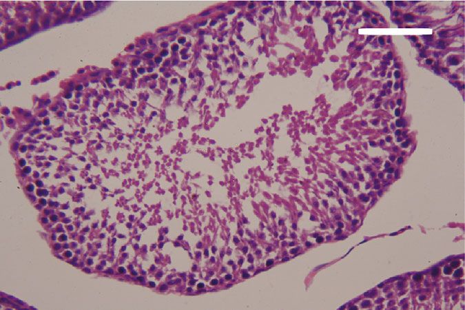
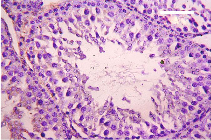
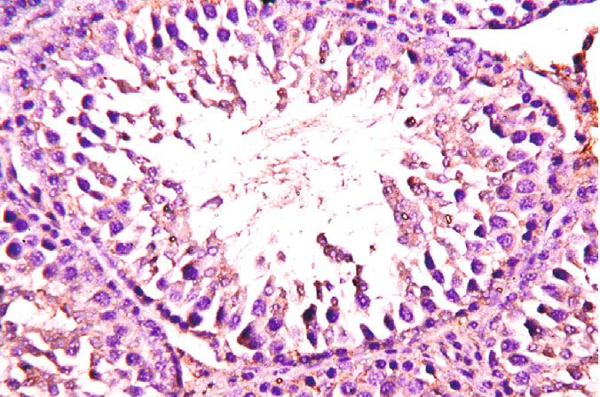
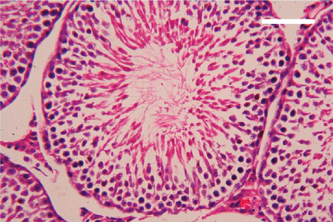
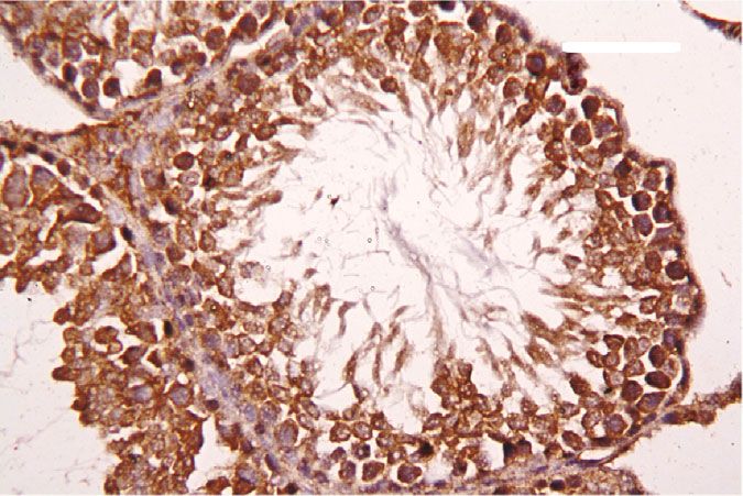
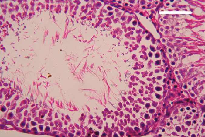
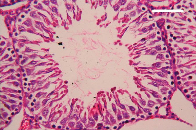
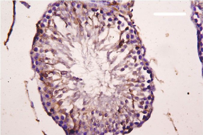
Oxidative Medicine and Cellular Longevity 7

 3 6

 Hmox1 mRNA expression
 Nfe2l2 mRNA expression
 A⁎⁎B⁎

 (fold change)
 A⁎⁎

 (fold change)
 2 4

 1 A⁎B⁎ 2
 A⁎⁎

 0 0
 Control

 CoQ10

 PbAc

 PbAc+CoQ10

 Control

 CoQ10

 PbAc

 PbAc+CoQ10
Figure 6: Effects of coenzyme Q10 (CoQ10, 10 mg/kg) administration on the mRNA levels of Nfe212 and Hmox1 in the testes of rats exposed
to lead acetate (PbAc, 20 mg/kg). Data was represented as mean ± SEM of triplicate assays. Data was first normalized to the Actb mRNA levels
then was expressed as a fold change relative to the control group. A,BSignificant change at p < 0:05; ∗ ,∗∗ Significant variation at p < 0:01 and
p < 0:001, respectively, as compared to the control and PbAc groups, respectively.

 150 80
 TNF- (pg/mg protein)

 IL-1 (pg/mgprotein)

 A A⁎⁎
 60
 100 AB
 40 A⁎⁎B⁎⁎

 50
 20

 0 0
 Control

 CoQ10

 PbAc

 PbAc+CoQ10

 Control

 CoQ10

 PbAc

 PbAc+CoQ10
Figure 7: Effects of coenzyme Q10 (CoQ10, 10 mg/kg) administration on the levels of tumor necrosis factor-α (TNF-α) and interleukin-1β
(IL-1β) in the testis of rats treated with lead acetate (PbAc, 20 mg/kg). Data was represented as mean ± SEM (n = 7). A,BSignificant change at
p < 0:05; ∗ ,∗∗ Significant variation at p < 0:01 and p < 0:001, respectively, as compared to the control and PbAc groups, respectively.

expression, while upregulated (p < 0:001) Hmox1 expression while CoQ10 injection after PbAc exposure was found to
when compared to the control group. When CoQ10 supple- protect the testicular tissues by enhancing Bcl2 expression
mentation was added after PbAc treatment, both Nfe2l2 and and inhibiting Bax and Casp3 expression (Figure 8). Constant
Hmox1 expressions were upregulated when compared to with the gene expression level of caspase-3, immunohisto-
PbAc treatment (Figure 6). chemical analysis obviously showed that PbAc enhances the
 Inflammation has been suggested as one of the pathways apoptotic cascade in the testicular tissue as presented by
that affect reproductive function following heavy metal elevating the immunostaining intensity for the proapoptotic
intoxication. Rats injected with PbAc showed increased protein, caspase-3. However, postadministration of CoQ10
inflammation in the testicular tissues as demonstrated by markedly decreased the number of the positively stained
increases in TNF-α (p < 0:05) and IL-1β (p < 0:001) expres- spermatogenic epithelial cells for caspase-3 immunoreac-
sion when compared to the control group, while CoQ10 post- tivity (Figure 9).
treatment attenuated this response following PbAc exposure Histological examination of testicular sections stained
(Figure 7). with H&E was represented in Figure 10. Control and
 To evaluate cell death and the apoptotic cascade in the CoQ10-treated alone rats revealed normal testicular mor-
testicular tissue, the proapoptotic markers (Bax and caspase phology with typical and functional seminiferous tubules
3) and the antiapoptotic marker (Bcl2) were evaluated in with well-arranged spermatogenic cells displaying all stages
each of the various groups. Rats treated with PbAc exhibited and the interstitial cells filling the space between the sem-
testicular cell loss as evidenced by the increased expression of iniferous tubules (Figures 10(a) and 10(b), respectively).
Bax (p < 0:001) and Casp3 (p < 0:001) and decreased Bcl2 On the other hand, PbAc-injected rats exhibited deleteri-
(p < 0:001) expression when compared to the control group, ous histopathological alterations verified by degeneration
8 Oxidative Medicine and Cellular Longevity

 2.0 6 5
 A⁎⁎ A⁎⁎

 Casp3 mRNA expression
 Bcl2 mRNA expression

 Bax mRNA expression
 1.5 4
 (fold change)

 (fold change)

 (fold change)
 4 A⁎⁎B⁎ A⁎⁎B⁎
 3
 1.0 A⁎B⁎
 2
 A⁎⁎ 2
 0.5
 1

 0.0 0 0
 Control

 CoQ10

 PbAc

 PbAc+CoQ10

 Control

 CoQ10

 PbAc

 PbAc+CoQ10

 Control

 CoQ10

 PbAc

 PbAc+CoQ10
Figure 8: Effects of coenzyme Q10 (CoQ10, 10 mg/kg) administration on the mRNA expression of Bcl2, Bax, and Casp3 in the testes of rats
exposed to lead acetate (PbAc, 20 mg/kg). Data was represented as mean ± SEM of triplicate assays. Data was first normalized to the Actb
mRNA levels then was expressed as the fold change relative to the control group. A,BSignificant change at p < 0:05; ∗,∗∗ Significant variation
at p < 0:01 and p < 0:001, respectively, as compared to the control and PbAc groups, respectively. Data was analyzed by one-way ANOVA
using Duncan’s post hoc test.

 100 m 100 m

 (a) (b)

 100 m 100 m

 (c) (d)

Figure 9: Effects of coenzyme Q10 (CoQ10, 10 mg/kg) administration on the caspase-3 expression in the testis tissue after lead acetate (PbAc,
20 mg/kg) exposure. (a) Control group, (b) CoQ10-administered group, (c) PbAc-exposed group, and (d) PbAc+CoQ10-treated group.
Scale bar = 100 μm.

of spermatogenic cells, detachment of the spermatogenic Doumouchtsis et al. [30] and Soleimanzadeh et al. [31].
epithelial cells from the basement membrane, and appear- Hormonal reduction resulting from Pb exposure is attributed
ance of a vacuolated area within the seminiferous tubules to impairment of the hypothalamus-pituitary-gonadal axis
(Figure 10(c)). Interestingly, CoQ10 posttreatment repaired [8, 30, 32]. Pb exposure causes degeneration of the pituitary
these abnormalities and largely preserved the testicular gland gonadotrophic cells [33] and induces apoptotic signals
structures (Figure 10(d)). in the Leydig cells [7]. In addition, Pb inhibits steroidogenic
 enzyme production in Leydig cells resulting in reduced
4. Discussion testosterone secretion [34].
 The high levels of polyunsaturated fatty acids in the tes-
Pb toxicity primarily affects the testes [5, 29]. Testicular ticular cells make them vulnerable to oxidative stress [35].
toxicity induced by PbAc treatment was characterized by Consequently, Pb toxicity-induced testicular malfunction
diminished plasma testosterone and LH and FSH concentra- may result from its ability to evoke membrane lipid peroxida-
tions, which was in agreement with the observations of tion promoting oxidative stress and apoptosis [5, 36, 37].
Oxidative Medicine and Cellular Longevity 9

 100 m 100 m

 (a) (b)

 100 m 100 m

 (c) (d)

Figure 10: Effects of coenzyme Q10 (CoQ10, 10 mg/kg) administration on the histopathological changes in the testis tissue after lead acetate
(PbAc, 20 mg/kg) exposure. (a) Control group, (b) CoQ10-administered group, (c) PbAc-exposed group, and (d) PbAc+CoQ10-treated
group. Scale bar = 100 μm.

Likewise, the prooxidant parameters, LPO and NO, increased are exposed to magnetic fields [43], ischemia/reperfusion
after Pb injection. On the other hand, the antioxidant param- injuries [44], and sodium arsenite toxicity [45]. Here, we
eters were diminished with Pb treatment. The gene expres- focused on its effect after PbAc exposure and we found that
sion of the antioxidant enzymes, Cat mRNA, Gpx1 mRNA, it could improve testicular function even in the presence of
Gsr mRNA, and Sod mRNA, and its corresponding proteins Pb. The absolute and relative testicular weight was restored
(CAT, GPX, GR, and SOD) were downregulated after Pb following CoQ10 supplementation, while the testosterone,
exposure. The GSH level and the activity of the antioxidant LH, and FSH levels were improved; signs of oxidative stress
enzymes (GPx, GR, CAT, and SOD) were also all reduced. were decreased as evidenced by the reduction in LPO and
This depletion may be caused by Pb binding of the metal NO concentrations. In addition, CoQ10 enhanced expression
cofactors of these enzymes or from the interaction of Pb and level of glutathione and the antioxidant enzymes, CAT,
and their SH group, reducing overall function [38]. SOD, GPx, and GR. Fouad et al. [45] revealed that CoQ10
 Moreover, the apoptotic pathway was activated by Pb could suppress oxidative stress in the testis by inhibiting lipid
administration which is demonstrated by the proapoptotic peroxidation and enhancing antioxidant enzyme activity.
genes (Bax and caspase-3) upregulation, as well as the down- This in turn can counteract oxidative damage and sustain
regulation of the antiapoptotic gene, Bcl2. Similarly, previous the function of Leydig cells protecting testosterone secretion
studies revealed a testicular damage and apoptosis in the [46]. The key application of CoQ10 in the testis is to increase
spermatogenic cells with Pb exposure [5, 7]. the levels of CoQ10 and its reduced form, ubiquinol, in the
 The inflammatory responses were elevated in the testes semen [13]. Ubiquinol is a potent fat-soluble antioxidant that
after Pb exposure, which was evidenced by the rise in the pro- can regenerate other antioxidants including vitamins E and C
inflammatory cytokines, TNF-α and IL1-β. Salama et al. [39] [47]. It also eliminates peroxyl radicals resulting from the
also reported an elevation in the proinflammatory cytokine lipid peroxidation process [48].
levels in the kidneys of rats after Pb exposure. Furthermore, The antiapoptotic effect of CoQ10 has been described by
PbAc administration resulted in the downregulation of several studies [40, 49], and it was confirmed in this study
Nfe212 gene expression. Suppression of Nfe212 induces apo- when CoQ10 treatment upregulated the expression of the
ptosis, activates the inflammatory response, and decreases antiapoptotic gene Bcl2 and downregulated proapoptotic
expression of the antioxidant enzymes [40]. Hmox1 gene genes Casp3 and Bax. Papucci et al. [50] attributed the anti-
expression was upregulated. The activation of the Hmox1 apoptotic effect of CoQ10 to the inhibition of DNA frag-
pathway may act to counter the elevation in the ROS and mentation and mitochondrial depolarization as well as
the inflammatory response [41]. increasing ATP levels. Additionally, CoQ10 inhibits nuclear
 Coenzyme Q10 is a natural antioxidant that plays a fun- translocation of apoptosis-inducing factors and prevents
damental role in the electron transport chain [42]. It has been cell death via the inhibition of mitochondrial complex I
shown to exhibit a protective effect on the testes when they activity [51].
10 Oxidative Medicine and Cellular Longevity

 CoQ10 is implicated in the prevention of inflammation [3] M. J. Zuscik, L. Ma, T. Buckley et al., “Lead induces chondro-
in the liver [52] and kidneys [40]. This anti-inflammatory genesis and alters transforming growth factor-beta and bone
effect can now be extended to the testes, as CoQ10 admin- morphogenetic protein signaling in mesenchymal cell popula-
istration following PbAc reduced the concentrations of pro- tions,” Environmental Health Perspectives, vol. 115, no. 9,
inflammatory cytokines, TNF-α and IL-1β, consequently pp. 1276–1282, 2007.
attenuating inflammation. Meanwhile, it activated the [4] D. Nash, L. Magder, M. Lustberg et al., “Blood lead, blood
Nfe212/Hmox1 pathway by upregulating both Nfe212 and pressure, and hypertension in perimenopausal and postmeno-
Hmox1 gene expressions. Previous studies indicated that pausal women,” JAMA, vol. 289, no. 12, pp. 1523–1532, 2003.
CoQ10 exerts its cytoprotective role and antioxidant activity [5] E. Hassan, M. El-Neweshy, M. Hassan, and A. Noreldin,
by activating the Nfe212/Hmox1 pathway [53]. Nfe212/H- “Thymoquinone attenuates testicular and spermotoxicity fol-
 lowing subchronic lead exposure in male rats: possible mecha-
mox1 signaling induces the transcription of the cytoprotec-
 nisms are involved,” Life Sciences, vol. 230, pp. 132–140, 2019.
tive and antioxidant enzymes [54] and protects cells from
 [6] C. Li, K. Zhao, H. Zhang et al., “Lead exposure reduces sperm
damage and death [55]. Nfe212, the transcription factor,
 quality and DNA integrity in mice,” Environmental Toxicol-
translocates from cytosol into the nucleus and bind to the
 ogy, vol. 33, no. 5, pp. 594–602, 2018.
antioxidant response element so upregulating the genes
 [7] Y. He, Q. Zou, H. Chen, S. Weng, T. Luo, and X. Zeng, “Lead
encoding antioxidant enzymes [56]. In addition, Nfe212
 inhibits human sperm functions by reducing the levels of
activation resulted in the inhibition of NF-κB and in turn intracellular calcium, cAMP, and tyrosine phosphorylation,”
reduces the proinflammatory cytokines [57]. Increased The Tohoku Journal of Experimental Medicine, vol. 238,
Hmox1 activity generates two important metabolites, biliru- no. 4, pp. 295–303, 2016.
bin and CO. Bilirubin acts as an antioxidant that inhibits [8] Y. S. El-Sayed and M. S. El-Neweshy, “Impact of lead toxicity
lipid peroxidation [58], while CO protects the cell during on male rat reproduction at “hormonal and histopathological
oxidative stress [59] thus inhibiting apoptosis in various cells levels”,” Toxicological & Environmental Chemistry, vol. 92,
[53], and attenuates the production of proinflammatory no. 4, pp. 765–774, 2010.
cytokine [57]. [9] A. E. A. Moniem, M. A. Dkhil, and S. Al-Quraishy, “Protective
 role of flaxseed oil against lead acetate induced oxidative stress
5. Conclusion in testes of adult rats,” African Journal of Biotechnology, vol. 9,
 no. 42, pp. 7216–7223, 2010.
Taken together, we can conclude that CoQ10 posttreatment [10] A. Sarmiento, J. Diaz-Castro, M. Pulido-Moran, N. Kajarabille,
ameliorates Pb-induced testicular toxicity by reducing oxida- R. Guisado, and J. J. Ochoa, “Coenzyme Q10 supplementation
tive stress, apoptosis, and inflammation. These observations and exercise in healthy humans: a systematic review,” Current
may be the result of Nfe212/Hmox1 pathway activation, but Drug Metabolism, vol. 17, no. 4, pp. 345–358, 2016.
this will need further study for confirmation. [11] S. A. Mirmalek, A. Gholamrezaei Boushehrinejad, H. Yavari
 et al., “Antioxidant and Anti-Inflammatory Effects of Coen-
Data Availability zyme Q10 on L-Arginine- Induced Acute Pancreatitis in
 Rat,” Oxidative Medicine and Cellular Longevity, vol. 2016, 8
All relevant data are within the paper. pages, 2016.
 [12] J. J. Challem, “Toward a new definition of essential nutrients: is
Conflicts of Interest it now time for a third 'vitamin' paradigm?,” Medical Hypoth-
 eses, vol. 52, no. 5, pp. 417–422, 1999.
The authors declare no conflict of interest. [13] G. Balercia, “Coenzyme q10 supplementation in infertile men
 with idiopathic asthenozoospermia: an open, uncontrolled
Authors’ Contributions pilot study,” Fertility and Sterility, vol. 81, no. 1, pp. 93–98,
 2004.
All the authors contributed equally to this work. [14] M. R. Safarinejad, “Efficacy of coenzyme Q10 on semen
 parameters, sperm function and reproductive hormones in
Acknowledgments infertile men,” The Journal of Urology, vol. 182, no. 1,
 pp. 237–248, 2009.
This research was funded by Deanship of Scientific Research [15] A. Mancini and G. Balercia, “Coenzyme Q10 in male infertil-
at Princess Nourah Bint Abdulrahman University through ity: physiopathology and therapy,” BioFactors, vol. 37, no. 5,
the Fast-Track research funding program. pp. 374–380, 2011.
 [16] A. T. Alahmar, “The impact of two doses of coenzyme Q10 on
References semen parameters and antioxidant status in men with idio-
 pathic oligoasthenoteratozoospermia,” Clinical and Experimen-
 [1] S. R. Kumar and A. S. Devi, “Lead toxicity on male reproduc- tal Reproductive Medicine, vol. 46, no. 3, pp. 112–118, 2019.
 tive system and its mechanism: a review,” Research Journal of [17] A. A. Fouad and I. Jresat, “Hepatoprotective effect of coen-
 Pharmacy and Technology, vol. 11, no. 3, p. 1228, 2018. zyme Q10 in rats with acetaminophen toxicity,” Environmen-
 [2] A. Rossi-George, M. B. Virgolini, D. Weston, M. Thiruchelvam, tal Toxicology and Pharmacology, vol. 33, no. 2, pp. 158–167,
 and D. A. Cory-Slechta, “Interactions of lifetime lead exposure 2012.
 and stress: behavioral, neurochemical and HPA axis effects,” [18] A. E. Abdel Moneim, “Flaxseed oil as a neuroprotective
 Neurotoxicology, vol. 32, no. 1, pp. 83–99, 2011. agent on lead acetate-induced monoamineric alterations and
Oxidative Medicine and Cellular Longevity 11

 neurotoxicity in rats,” Biological Trace Element Research, [34] B.-M. Huang and M.-Y. Liu, “Inhibitory actions of lead on ste-
 vol. 148, no. 3, pp. 363–370, 2012. roidogenesis in MA-10 mouse Leydig tumor cells,” Archives of
[19] N. Li, Y. H. Hou, D. D. Ma, W. X. Jing, H. U. Dahms, and Andrology, vol. 50, no. 1, pp. 5–9, 2009.
 L. Wang, “Lead accumulation, oxidative damage and histo- [35] N. E. Furland, S. R. Zanetti, G. M. Oresti, E. N. Maldonado,
 pathological alteration in testes and accessory glands of fresh- and M. I. Aveldaño, “Ceramides and sphingomyelins with
 water crab, Sinopotamon henanense, induced by acute lead high proportions of very long-chain polyunsaturated fatty
 exposure,” Ecotoxicology and Environmental Safety, vol. 117, acids in mammalian germ cells,” Journal of Biological Chemis-
 pp. 20–27, 2015. try, vol. 282, no. 25, pp. 18141–18150, 2007.
[20] J. Szkoda and J. Zmudzki, “Determination of lead and cad- [36] D. D. Thomas, L. A. Ridnour, J. S. Isenberg et al., “The chem-
 mium in biological material by graphite furnace atomic ical biology of nitric oxide: implications in cellular signaling,”
 absorption spectrometry method,” Bulletin of the Veterinary Free Radical Biology and Medicine, vol. 45, no. 1, pp. 18–31,
 Institute in Pulawy, vol. 49, pp. 89–92, 2005. 2008.
[21] L. C. Green, D. A. Wagner, J. Glogowski, P. L. Skipper, J. S. [37] M. A. Dkhil, M. S. Al-Khalifa, S. Al-Quraishy, R. Zrieq, and
 Wishnok, and S. R. Tannenbaum, “Analysis of nitrate, nitrite, A. E. Abdel Moneim, “Indigofera oblongifolia mitigates lead-
 and [15N] nitrate in biological fluids,” Analytical Biochemistry, acetate-induced kidney damage and apoptosis in a rat model,”
 vol. 126, no. 1, pp. 131–138, 1982. Drug Design, Development and Therapy, vol. 10, pp. 1847–
[22] H. Ohkawa, N. Ohishi, and K. Yagi, “Assay for lipid peroxides 1856, 2016.
 in animal tissues by thiobarbituric acid reaction,” Analytical [38] R. C. Patra, A. K. Rautray, and D. Swarup, “Oxidative Stress in
 Biochemistry, vol. 95, no. 2, pp. 351–358, 1979. Lead and Cadmium Toxicity and Its Amelioration,” Veteri-
[23] G. L. Ellman, “Tissue sulfhydryl groups,” Archives of Biochem- nary Medicine International, vol. 2011, 9 pages, 2011.
 istry and Biophysics, vol. 82, no. 1, pp. 70–77, 1959. [39] S. A. Salama, H. H. Arab, I. A. Maghrabi, M. H. Hassan, and
[24] Y. Sun, L. W. Oberley, and Y. Li, “A simple method for clinical M. S. AlSaeed, “Gamma-glutamyl cysteine attenuates tissue
 assay of superoxide dismutase,” Clinical Chemistry, vol. 34, damage and enhances tissue regeneration in a rat model of
 no. 3, pp. 497–500, 1988. lead-induced nephrotoxicity,” Biological Trace Element
[25] H. Lück, “Catalase,” in Methods of Enzymatic Analysis, H. U. Research, vol. 173, no. 1, pp. 96–107, 2016.
 Bergmeyer, Ed., pp. 855–888, Academic Press, New York, [40] A. M. Kabel and A. A. Elkhoely, “Ameliorative effect of coen-
 1965. zyme Q10 and/or candesartan on carboplatin-induced neph-
[26] V. M. Factor, A. Kiss, J. T. Woitach, P. J. Wirth, and S. S. Thor- rotoxicity: roles of apoptosis, transforming growth factor-Β1,
 geirsson, “Disruption of redox homeostasis in the transform- nuclear factor kappa-B and the Nrf2/HO-1 pathway,” Asian
 ing growth factor-alpha/c-myc transgenic mouse model of Pacific journal of cancer prevention: APJCP, vol. 18, no. 6,
 accelerated hepatocarcinogenesis,” The Journal of Biological pp. 1629–1636, 2017.
 Chemistry, vol. 273, no. 25, pp. 15846–15853, 1998. [41] E. M. Al Olayan, A. S. Aloufi, O. D. AlAmri, O. H. El-Habit,
[27] D. E. Paglia and W. N. Valentine, “Studies on the quantitative and A. E. Abdel Moneim, “Protocatechuic acid mitigates
 and qualitative characterization of erythrocyte glutathione cadmium-induced neurotoxicity in rats: role of oxidative
 peroxidase,” The Journal of Laboratory and Clinical Medicine, stress, inflammation and apoptosis,” Science of The Total Envi-
 vol. 70, no. 1, pp. 158–169, 1967. ronment, vol. 723, p. 137969, 2020.
[28] R. Drury and E. Wallington, Carleton’s histological techniques, [42] H. Nohl, L. Gille, and A. V. Kozlov, “Critical aspects of the
 Oxford press, London, New York, 1980. antioxidant function of coenzyme Q in biomembranes,” Bio-
[29] P. J. Landrigan, P. Boffetta, and P. Apostoli, “The reproductive Factors, vol. 9, no. 2-4, pp. 155–161, 1999.
 toxicity and carcinogenicity of lead: a critical review,” Ameri- [43] L. A. Ramadan, A. R. Abd-Allah, H. A. Aly, and A. A. Saad-El-
 can Journal of Industrial Medicine, vol. 38, no. 3, pp. 231– Din, “Testicular toxicity effects of magnetic field exposure and
 243, 2000. prophylactic role of coenzyme Q10 and L-carnitine in mice,”
[30] K. K. Doumouchtsis, S. K. Doumouchtsis, E. K. Doumoucht- Pharmacological Research, vol. 46, no. 4, pp. 363–370, 2002.
 sis, and D. N. Perrea, “The effect of lead intoxication on endo- [44] B. Erol, M. Bozlu, V. Hanci, H. Tokgoz, S. Bektas, and
 crine functions,” Journal of Endocrinological Investigation, G. Mungan, “Coenzyme Q10 treatment reduces lipid peroxi-
 vol. 32, no. 2, pp. 175–183, 2009. dation, inducible and endothelial nitric oxide synthases, and
[31] A. Soleimanzadeh, M. Kian, S. Moradi, and S. Mahmoudi, germ cell–specific apoptosis in a rat model of testicular ische-
 “Carob (Ceratonia siliqua L.) fruit hydro-alcoholic extract alle- mia/reperfusion injury,” Fertility and Sterility, vol. 93, no. 1,
 viates reproductive toxicity of lead in male mice: evidence on pp. 280–282, 2010.
 sperm parameters, sex hormones, oxidative stress biomarkers [45] A. A. Fouad, A. I. Al-Sultan, and M. T. Yacoubi, “Coenzyme
 and expression of Nrf2 and iNOS,” Avicenna journal of phyto- Q10 counteracts testicular injury induced by sodium arsenite
 medicine, 2019. in rats,” European Journal of Pharmacology, vol. 655, no. 1-3,
[32] J. Gandhi, R. J. Hernandez, A. Chen et al., “Impaired pp. 91–98, 2011.
 hypothalamic-pituitary-testicular axis activity, spermatogene- [46] C. M. Palmeira, D. L. Santos, R. Seiça, A. J. Moreno, and M. S.
 sis, and sperm function promote infertility in males with lead Santos, “Enhanced mitochondrial testicular antioxidant capac-
 poisoning,” Zygote, vol. 25, no. 2, pp. 103–110, 2017. ity in Goto-Kakizaki diabetic rats: role of coenzyme Q,” Amer-
[33] N. Hamadouche, N. Sadi, O. Kharoubi, M. Slimani, and ican Journal of Physiology-Cell Physiology, vol. 281, no. 3,
 A. Aoues, “The protective effect of vitamin E against genotoxi- pp. C1023–C1028, 2001.
 city of lead acetate intraperitoneal administration in male rat,” [47] M. Turunen, J. Olsson, and G. Dallner, “Metabolism and func-
 Archives of Biological Sciences, vol. 65, no. 4, pp. 1435–1445, tion of coenzyme Q,” Biochimica et Biophysica Acta (BBA)-
 2013. Biomembranes, vol. 1660, no. 1-2, pp. 171–199, 2004.
12 Oxidative Medicine and Cellular Longevity

[48] M. Potgieter, E. Pretorius, and M. S. Pepper, “Primary and
 secondary coenzyme Q10 deficiency: the role of therapeutic
 supplementation,” Nutrition Reviews, vol. 71, no. 3, pp. 180–
 188, 2013.
[49] A. A. K. El-Sheikh, M. A. Morsy, M. M. Mahmoud, R. A.
 Rifaai, and A. M. Abdelrahman, “Effect of coenzyme-Q10 on
 doxorubicin-induced nephrotoxicity in rats,” Advances in
 Pharmacological Sciences, vol. 2012, 8 pages, 2012.
[50] L. Papucci, N. Schiavone, E. Witort et al., “Coenzyme q10 pre-
 vents apoptosis by inhibiting mitochondrial depolarization
 independently of its free radical scavenging property,” Journal
 of Biological Chemistry, vol. 278, no. 30, pp. 28220–28228,
 2003.
[51] H. Li, G. Chen, W. Ma, and P.-A. Li, “Water-soluble coenzyme
 q10 inhibits nuclear translocation of apoptosis inducing factor
 and cell death caused by mitochondrial complex I inhibition,”
 International Journal of Molecular Sciences, vol. 15, no. 8,
 pp. 13388–13400, 2014.
[52] J. L. Tarry-Adkins, D. S. Fernandez-Twinn, I. P. Hargreaves
 et al., “Coenzyme Q10 prevents hepatic fibrosis, inflammation,
 and oxidative stress in a male rat model of poor maternal
 nutrition and accelerated postnatal growth1,” The American
 Journal of Clinical Nutrition, vol. 103, no. 2, pp. 579–588, 2016.
[53] A. E. Khodir, H. Atef, E. Said, H. A. ElKashef, and H. A. Salem,
 “Implication of Nrf2/HO-1 pathway in the coloprotective
 effect of coenzyme Q10 against experimentally induced ulcer-
 ative colitis,” Inflammopharmacology, vol. 25, no. 1, pp. 119–
 135, 2017.
[54] K. A. Kang and J. W. Hyun, “Oxidative stress, Nrf2, and epige-
 netic modification contribute to anticancer drug resistance,”
 Toxicological research, vol. 33, no. 1, pp. 1–5, 2017.
[55] A. Loboda, M. Damulewicz, E. Pyza, A. Jozkowicz, and
 J. Dulak, “Role of Nrf2/HO-1 system in development, oxida-
 tive stress response and diseases: an evolutionarily conserved
 mechanism,” Cellular and Molecular Life Sciences, vol. 73,
 no. 17, pp. 3221–3247, 2016.
[56] Y.-K. J. Zhang, K. C. Wu, and C. D. Klaassen, “Genetic activa-
 tion of Nrf2 protects against fasting-induced oxidative stress in
 livers of mice,” PLoS ONE, vol. 8, no. 3, p. e59122, 2013.
[57] H. So, H. J. Kim, Y. Kim et al., “Evidence that cisplatin-induced
 auditory damage is attenuated by downregulation of pro-
 inflammatory cytokines via Nrf2/HO-1,” Journal of the Associ-
 ation for Research in Otolaryngology, vol. 9, no. 3, pp. 290–306,
 2008.
[58] J. E. Clark, R. Foresti, P. Sarathchandra, H. Kaur, C. J. Green,
 and R. Motterlini, “Heme oxygenase-1-derived bilirubin
 ameliorates postischemic myocardial dysfunction,” American
 Journal of Physiology-Heart and Circulatory Physiology,
 vol. 278, no. 2, pp. H643–H651, 2000.
[59] L. E. Otterbein and A. M. K. Choi, “Heme oxygenase: colors of
 defense against cellular stress,” American Journal of
 Physiology-Lung Cellular and Molecular Physiology, vol. 279,
 no. 6, pp. L1029–L1037, 2000.
You can also read