Stress Urinary Incontinence- A Consequence of Failed Load Transfer Through the Pelvis? - Herman & Wallace Pelvic ...
←
→
Page content transcription
If your browser does not render page correctly, please read the page content below
Stress Urinary Incontinence –
           A Consequence of Failed Load Transfer Through the Pelvis?
                                        Diane Lee BSR, FCAMT, Canada
                                   Linda-Joy Lee BSc, BSc(PT), FCAMT, Canada
                                 th
             Presented at the 5 World Interdisciplinary Congress on Low Back and Pelvic Pain
                                       Melbourne, November 2004

INTRODUCTION

         The anatomical and biomechanical research in the last decade has led to a clearer understanding of how
load is transferred through the low back and pelvic girdle (Hodges & Richardson 1996, 1997, Hodges 1997, 2003,
Hodges et al 1999, 2001a,b,c, 2003b, Hungerford 2001, Mens et al 1999, Richardson et al 1999, Snijders et al
1993a,b Vleeming et al 1990a,b, Vleeming et al 1995, Vleeming et al 1996). From this research, an integrated
model of function evolved (Lee & Vleeming 1998, 2004) and clinical extrapolations based on this model were
developed (Richardson et al 1999, Hodges (Course Notes, 2004), Lee 2004, Lee & Lee 2004). Further research
based on the model continued (Hungerford et al 2004, O’Sullivan et al 2002, Pool-Goodzwaard 2003, Stuge et al
2004) and it soon became evident that two conditions of failed load transfer through the pelvis, low back/pelvic
girdle pain and stress urinary incontinence, had components in common. Recently, Pool-Goudzwaard (2003)
conducted a multi-centered study in Holland to investigate, in part, the prevalence of low back/pelvic girdle pain
and pelvic floor disorders. In this study of 66 patients, 52% reported a combination of low back and/or pelvic
girdle pain along with pelvic floor dysfunction which included voiding dysfunction, urinary incontinence, sexual
dysfunction and/or constipation. Of these 52%, 82% stated that their symptoms began with either low back or
pelvic girdle pain.

         Our journey into considering the relationship between load transfer through the musculoskeletal
                                                                          th
components of the pelvis and the organs it contains began at the 4 World Congress on Low Back and Pelvic
Girdle Pain after hearing a paper (O’Sullivan et al. 2002) which demonstrated via real-time ultrasound imaging the
impact of the active straight leg raise (ASLR) (Mens et al 1999) on the position of the bladder in pelvic girdle pain
patients. They noted that the bladder tended to descend during the ASLR and that this descent decreased when
compression was applied to the pelvis. Our question at the time was, “How much should the bladder move when
you lift your leg?” This led to a search of the literature pertaining to stress urinary incontinence and several
revelations followed regarding the parallel features of both low back/pelvic girdle pain and stress urinary
incontinence. We now recognize that the factors which must be optimal for effective force closure and stability of
the pelvic girdle and those which must be present for optimal force closure of the urethra are the same. The
intent of this paper is to briefly review the literature regarding effective force closure of the pelvic girdle, present
the anatomical and neurophysiological requirements for effective force closure of the urethra and conclude with a
case presentation which will clinically integrate the material. Some of the material in this paper comes from the
  rd
3 edition of The Pelvic Girdle (Lee 2004) and is reproduced here with permission.

THE INTEGRATED MODEL OF FUNCTION

        The integrated model of function has four components: form closure (structure), force closure (forces
produced by myofascial action), motor control (specific timing of muscle action/inaction during loading) and
emotions. The joints of the pelvic girdle are mobile (Jacob & Kissling 1995, Sturesson et al 2000) and therefore
force closure of the pelvic girdle is required if loads are to be transferred optimally. According to Panjabi
(1992a,b) stability (effective load transfer) is achieved when the passive, active and control systems work
together. Collectively, all three systems produce approximation of the joint surfaces (Snijders & Vleeming
1993a,b); essential if stability is to be insured. The amount of approximation required is variable and difficult to
quantify since it depends on an individual’s structure and the forces they need to control. The term ‘adequate’
has been used (Lee & Vleeming 1998, 2004) to describe how much approximation is necessary and reflects the
non-quantitative aspect of this measure. Essentially, it means ‘not too much’ and ‘not too little’; in other words,
‘just enough’ to suit the existing situation. Consequently, the ability to effectively transfer load through the pelvis
is dynamic and depends on:
            1. optimal function of the bones, joints and ligaments (form closure – not discussed in this paper)
     (Vleeming et al 1990a,b).
            2. optimal function of the muscles and fascia (force closure) (Hungerford 2003, O’Sullivan 2000,
     Richardson et al 1999, 2002, Vleeming et al 1995).
            3. appropriate neural function (motor control, emotional state) (Bø & Stien 1994, Hodges 1997,
     2003, Hodges et al, 2001, 2003, Holstege et al 1996, Hungerford 2003).

EFFECTIVE FORCE CLOSURE AND MOTOR CONTROL - THE PELVIC GIRDLE

         If the articular surfaces of the pelvis were constantly and completely compressed, mobility would not be
possible. However, compression during loading is variable and therefore motion is possible (Jacob & Kissling
1995, Sturesson 2000) and stabilization required. This is achieved by increasing compression across the joint
surface at the moment of loading (force closure). The amount of force closure required depends on the
individual’s form closure and the magnitude of the load. The anatomical structures responsible for force closure
are the ligaments, muscles and fascia.

          In the close-packed or self-locked (self-braced) position, the joints of the pelvic girdle are under significant
compression and the ability to resist shear forces is enhanced by the tension of the passive structures and
increased friction between the articular surfaces (Vleeming et al 1990b, Snijders et al 1993a,b). For the sacroiliac
joints, this position is full nutation of the sacrum or posterior rotation of the innominate (Vleeming et al 1990a,b,
van Wingerden et al 1993). Studies have shown (Egund et al 1978, Hungerford 2004, Lavignolle et al 1983,
Sturesson et al 2000) that nutation of the sacrum occurs bilaterally whenever the lumbopelvic spine is loaded.
The amount of sacral nutation varies with the magnitude of the load. Full sacral nutation (self-locking or close
packing) occurs during forward and backward bending of the trunk (Sturesson et al 2000). However, function
would be significantly compromised if joints could only be stable in the close-packed position. In the neutral
spinal position, an osteoligamentous spine (T1 to sacrum) will buckle under approximately 20N (about 4.4 lb) of
compression load (Lucas & Bresler, 1961, Panjabi 1992a,b). Consequently, stability for load transfer is required
throughout the entire range of motion and this is provided by the active, or neuromyofascial, system.

           In 1989, Bergmark proposed that muscles could be classified into two systems – a local and a global
system (Bergmark 1989). The local system pertains to those muscles essential for segmental or intrapelvic
stabilization while the global system appears to be more responsible for regional stabilization (between the thorax
and pelvis or pelvis and legs) and motion (Bergmark 1989, Comerford & Mottram 2001, Richardson et al 1999).
The function of the lumbopelvic local system is to stabilize the joints of the spine and pelvic girdle in preparation
for (or in response to) the addition of external loads. This is achieved through several mechanisms some of which
include:
                           •       increasing the intra-abdominal pressure (Cresswell, 1993, Hodges & Gandevia
                  2000a,b, Hodges et al 2001a, 2003b, McGill & Norman 1987)
                           •       increasing the tension of the thoracodorsal fascia (Cresswell 1993, Hodges 2003,
                  Hodges et al 2003b, Vleeming et al 1995a, Willard 1997) and/or
                           •        increasing the articular stiffness (Hodges et al 1997, Hodges 2003, Richardson
                  et al 2002).

         Research has shown (Barbic et al 2003, Bø & Stien 1994, Constantinou & Govan 1982, Hodges 1997,
Hodges & Gandevia 2000a,b, Hodges 2003, Hungerford 2004, Moseley et al 2002, 2003, Sapsford et al 2001)
that when the central nervous system can predict the timing of the load, the local system is anticipatory when
functioning optimally. In other words, these muscles should work at low levels at all times and increase their
action before any further loading or motion occurs. When the local system is functioning optimally, it provides
anticipatory intersegmental stiffness of the joints of the lumbar spine (Hodges et al 2003b) and pelvis (Richardson
et al 2002). This external force augments the form closure and helps to prevent excessive shearing at the time of
loading. This stiffness/compression occurs prior to the onset of any movement and prepares the low back and
pelvis for additional loading from the global system.
The research is still lacking which enables classification of all muscles according to this system and
clinically it appears that parts of some muscles may belong to both systems. With respect to the lumbopelvic
region, the following muscles fit the criteria for classification as local stabilizers - the muscles of the pelvic floor
(Barbic et al 2003, Bø et al 1994, Constantinou & Govan 1982, Deindl et al 1993, Hodges 2003, Sapsford et al
2001), the transversus abdominis (Hodges & Richardson 1997a,b, Hodges 2003), the diaphragm (Hodges &
Gandevia 2000a,b, Hodges 2003) and the deep fibres of multifidus (Hides et al 1994, 1996, Moseley et al 2002,
2003). As research continues, more muscles will likely be added to this list. The deep (medial) fibres of psoas
(Gibbons et al 2002), the medial fibres of quadratus lumborum (Bergmark 1989, McGill 2002), the lumbar parts of
the lumbar iliocostalis and longissimus (Bergmark 1989) and the posterior fibres of the internal oblique (Bergmark
1989, O’Sullivan 2000) are some likely candidates.
Motor control pertains to patterning of muscle activation (Barbic et al 2003, Comerford & Mottram 2001,
Danneels et al 2001, Deindl et al 1993, Hodges 1997, 2003, Hungerford et al 2003, Moseley et al 2002,
O’Sullivan et al 1997, O’Sullivan 2000, Richardson et al 1999), in other words, the timing of specific muscle action
and inaction. Efficient movement as well as effective load transfer through the pelvic girdle requires coordinated
muscle action, such that stability is ensured while motion is controlled and not restrained (Hodges et al 2001b,
Hodges 2003). With respect to the lumbopelvic region, it is the coordinated action between the local and global
systems that ensures stability without rigidity of posture and without episodes of collapse.
EFFECTIVE FORCE CLOSURE AND MOTOR CONTROL - THE URETHRA
Urinary incontinence is defined as the involuntary leakage of urine which is objectively demonstrable.
Stress urinary incontinence (leakage which occurs during physical exertion) is the most common type. According
to DeLancey (1994): ‘During a cough urethral closure pressure is known to rise simultaneously with abdominal
pressure to keep the urethra closed in spite of great increases in intravesical pressure.’ Essentially, when one
cannot effectively force close the urethra (maintain urethral closure pressure) stress urinary incontinence can
result. How common is this? The prevalence of this condition varies according to age, study design and
definition. Ashton-Miller et al (2001) state that 8.5% - 38% of women experience stress urinary incontinence
(SUI). Nygaard et al (1994) note that this condition is not limited to women bearing children and that in a study of
144 nulliparous female athletes ages 18 to 21 years, 28% suffered from SUI. Bø & Borgen (2001) found that 41%
of elite female athletes experience SUI. Fantl et al (1996) states that incontinence affects four out of ten women,
about one out of ten men, and about 17% of children below the age of fifteen.
Clearly, this is a significant problem but is it a different problem than a loss of effective force closure of the
musculoskeletal elements of the pelvis? It is common to hear women complain of both low back and pelvic girdle
pain as well as urinary incontinence and therapists commonly note that treating one component often impacts the
other. Minimal research has been done on the correlation between the two functional impairments. In reviewing
the literature, it appears that Panjabi’s stability model (1992a,b) can be applied to the urethra as well as to the
musculoskeletal system.
Urinary continence
In an excellent review article, Ashton-Miller, Howard and DeLancey (2001) clearly explain the mechanism
by which continence is achieved during physical exertion. Essentially, continence relies on optimal function of two
systems; the urethral support system and the sphincteric closure system.Urethral Support System
The structures which provide support for the urethral include (Fig. 1):
• the passive system – this includes
the endopelvic fascia which is anchored to a
thick fascial band called the arcus tendineus
fasciae which arises from the pubic bone
anteriorly and inserts into the ischial spine
• the active system for this fascial
hammock or sling – this includes the levator ani
muscle which contains primarily type 1 fibres
and exhibits constant tone.
• the control system – this includes
the pudendal nerve which innervates the levator
ani as well as the central control of reflex
function between the detrusor muscle and the
pelvic floor.
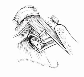
Figure 1. The urethral support system
Together, the passive and active systems form a
hammock of support for the urethra (Fig. 2) and the
integrity/function of these tissues is essential if force closure of
the urethra is to be effective. If this system gives way easily, it
cannot provide a backstop against which the urethra can be
compressed. A useful analogy (Ashton-Miller et al 2001) is to
imagine a garden hose (urethra), with water running through it
(urine), lying on a trampoline bed (the pelvic floor). Stepping on
the hose will block the flow of water if the bed is very stiff and
provides an equal and opposite counterforce (functional pelvic
floor). If however, the bed is very flexible (i.e. loss of myofascial
support), the downward pressure on the hose will cause the bed
to stretch and allow the hose to indent the bed. The flow of water
Figure 2. The hammock of support for the urethra.
will continue uninterrupted.
Sphincteric Closure System
In addition, the urethra is closed by a system of both intrinsic and extrinsic muscles. The striated muscles
within the wall of the urethra (intrinsic) contract prior to any pressure received by the bladder and these muscles
are also comprised of type 1 fibres; well-suited to maintain constant tone.
Constantinou & Govan (1982) measured the intra-urethral and intra-bladder pressures in healthy
continent women during the valsalva maneuver and coughing and found that during a cough the pressure in the
urethra increased approximately 250 ms before any pressure increase was detected in the bladder. This did not
occur during a valsalva (bearing down or straining). This suggests that an anticipatory reflex exists between thepelvic floor and the urethra (the pelvic floor is active during a cough and inactive during a valsalva). This
anticipatory closure of the urethra was confirmed in a subsequent study by Thind et al (1991). They also noted
that the urethral pressure remained elevated for a short time after the pressure normalized in the bladder.
Bø & Stien (1994) used needle EMG to measure activity in the urethral wall during a cough and valsalva
as well as during activation of the hip adductors, abdominals and gluteal muscles. They found that the urethral
wall contracted syngergistically with the pelvic floor, hip adductors and gluteals and also during a cough. They
conclude that strengthening the pelvic floor will also strengthen the urethral wall – but will it restore the
anticipatory reflex mechanism?
Deindl et al (1993) investigated via needle EMG the activity pattern of pubococcygeus in nulliparous,
continent women during a voluntary “squeeze” and during an attempt to urinate. The voluntary “squeeze” led to a
concomitant increase in activation of the targeted motor units bilaterally (this response was sustained) whereas
during the attempt to urinate a marked decrease in the ongoing tonic motor unit activity occurred.
Sapsford et al. (2001) investigated the co-activation pattern of the pelvic floor and abdominals via fine
wire EMG for the abdominal wall and surface EMG for the pelvic floor and found that the abdominals contract in
response to a pelvic floor contraction command and that the pelvic floor contracts in both a ‘hollowing’ and
‘bracing’ abdominal command. They also found that a submaximal command of pubococcygeus elicited the
greatest response in transversus abdominis. The results from this research suggest that the pelvic floor can be
facilitated by co-activating the deep abdominals and visa versa. However; it is wrong to assume that all patients
will be able to contract the muscles of the pelvic floor through verbal commands alone, either through the
abdomen or the pelvic floor. Bump et al (1991) found that only 50% of women could actually perform a pelvic floor
muscle contraction with just a verbal instruction. We have found that careful analysis is required to ensure that the
reflex connection between the transversus abdominis and the pelvic floor is intact before this strategy is used.
In conclusion, the evidence suggests that the pelvic floor plays a significant role in effective force closure
of the urethra and that these muscles fit the criteria for classification as local system muscles. Optimally the pelvic
floor muscles function constantly at low levels of tone and increase their activation in anticipation of load. They
should co-activate with transversus abdominis for lumbopelvic stabilization and play a role in increasing the intra-
urethral pressure, in pre-tensing the endopelvic fascia and in facilitating a co-activation of the striated urethral
sphincter muscles.
Stress urinary incontinence
Stress urinary incontinence can result when there is loss of the anatomical integrity or neurophysiological
function of the pelvic floor (muscles and fascia) secondary to a single major trauma or repetitive minor trauma.
Inefficient load transfer strategies through the low back and pelvis, particularly those which excessively increase
the intra-abdominal pressure and result in the bladder and pelvic organs being repetitively compressed inferiorly,
can lead to incontinence via repetitive microtrauma to the fascial supports or via altering optimal recruitment of the
pelvic floor muscles (Sapsford 2004). Also, a history of straining during bowel movements is common in women
with uterovaginal prolapses and stress urinary incontinence (Spence-Jones et al 1994). These altered motor
control strategies often lead to changes in lumbopelvic posture. Nguyen et al (2000) found that women with
uterovaginal prolapse had significantly less lumbar lordosis and a less vertically oriented pelvic inlet than groups
without prolapse. This postural change is often seen in patients with stress urinary incontinence as well as in
patients with lumbopelvic dysfunction (clinical observation). When the bladder is observed with real-time
ultrasound imaging, it can be seen that these strategies cause the bladder to descend (Fig. 3).Figure 3. Descent of the bladder imaged via real-time
ultrasound during load transfer strategies which result
in an excessive increase in the intra-abdominal
pressure.
Using real-time ultrasound imaging in a group of patients with pelvic girdle pain, O’Sullivan et al (2002)
noted that the bladder descended during an active straight leg raise test. When compression was applied to the
pelvic girdle, this descent was minimized. How much descent of the bladder is optimal, or normal, during
functional activities? Peschers et al (2001) measured the mobility of the bladder neck via perineal ultrasound
during coughing and valsalva in 39 healthy, nulliparous women. They found that the bladder neck descended a
variable amount (2 – 32mm) in both a cough and valsalva and questioned the long held view that stress urinary
incontinence was associated with urethral mobility. Like the sacroiliac joint, there appears to be a wide variation in
the amount of motion possible (Buyruk et al 1995a,b, Damen et al 2001) and continence (effective force closure
of the urethra) relies more on control and urethral closure rather than amplitude of motion.
Howard et al (2000) investigated the amplitude of descent of the bladder neck during a cough and
valsalva in three groups of women, nulliparous continent (17 subjects), primiparous continent (18 subjects) and
primiparous incontinent (23 subjects). There was no statistical difference in the amount of bladder neck mobility
between the groups, again suggesting that movement of the urethra is not what determines one’s continence
status. When they compared the amplitude of bladder neck movement during a cough and valsalva, they noted
that the two continent groups exhibited less movement during a cough. Conversely, the incontinent group
demonstrated no difference in the amount of movement during a cough or valsalva. Clearly, something was
happening during a cough in the continent women that was not happening in the incontinent group. All three
groups generated the same amount of cough pressures; however, the stiffness value (pressure change divided by
bladder neck mobility) was the greatest in the nulliparous continent group, next highest in the primiparous
continent group and lowest in the primiparous incontinent group. They hypothesize that these differences depend
on the function of the pelvic floor; assumed to be optimal in the continent women. Thind et al (1991) noted that the
amplitude of the anticipatory pressure rise in the urethra was less in women with stress urinary incontinence and
suggest that this is due to weakness of the pelvic floor. Indeed, Bø et al (1990) demonstrated in a randomized
clinical trial that retraining the function of the pelvic floor (awareness training coupled with strength and endurance
training) is effective for some women (60%) in the treatment of stress urinary incontinence.
Allen et al (1990) investigated 96 nulliparous women both pre and postnatally to determine if childbirth
caused damage to the pelvic floor muscles and/or their nerve supply. They showed that a vaginal delivery
impaired the strength of the pelvic floor and noted that recovery had not occurred at two months postpartum.
They also demonstrated via needle EMG that vaginal delivery caused a partial denervation of the pelvic floor in
80% of these women. Women who had a long, active second stage of labour showed the most EMG evidence of
denervation. Ashton-Miller et al (2001) feel that if the nerve to the levator ani is damaged, the denervated
muscles will atrophy thus placing more stress on the passive supporting structures (endopelvic fascia) which over
time will stretch and result in organ prolapse. Alternately, a paravaginal defect can occur which causes a
separation in the endopelvic fascia. This effectively reduces the stiffness of the fascial layer which supports the
urethra (Fig. 2) and can occur unilaterally or bilaterally. When this occurs, the pelvic floor must take over to
support the organ position and provide active closure to the urethra. However, they note that ‘If the muscle is
completely detached from the fascial tissues, then it may be able to contract; but that contraction may not be
effective in elevating the urethra or stabilizing its position’ (Ashton-Miller et al 2001).
Deindl et al (1994) compared the activity pattern of pubococcygeus in both nulliparous, continent and
parous stress urinary incontinent women. They found two differences in the incontinent group:
1. a voluntary “squeeze” of the pubococcygeus showed an endurance deficit (shorter holding times)
2. an asymmetrical and uncoordinated pattern of activation (left vs. right) commonly occurred.
Sometimes the response was only unilateral.Barbic et al (2003) also investigated the pattern and timing of muscle activation of the levator ani in both
continent and incontinent women. They noted that both the left and right levator ani contracted prior to any
pressure increase in the bladder and that the timing of this activation was delayed in the incontinent group. They
conclude that an important aspect of a “stable bladder neck is the timely activation of the levator ani muscle. The
activation, which precedes the contraction of other muscles…, might enable a pretension of the endopelvic fascia
tissue, which becomes less compliant for stretching by downward forces of increased abdominal pressure.”
Therefore, continence of urine (effective force closure of the urethra) during the transference of loads
through the pelvic girdle requires optimal bladder position control and this depends, in part, on the individual’s
ability to effectively contract and sustain a tonic co-contraction of the local muscle system in a properly timed
manner.
We believe that orthopaedic manual therapists who focus on restoring function to the local system of the
low back and pelvic girdle (transversus abdominis, multifidus, diaphragm, and the pelvic floor) and therapists who
specialize in pelvic floor dysfunction are treating the same condition – failed load transfer through the lumbopelvic
region, manifested either through a loss of effective force closure of the joints of the low back and pelvis, or loss
of effective force closure of the urethra. The research clearly supports that we are merging to a common
understanding of both function and dysfunction of the whole pelvis and not just its parts. Treatment of the
impaired lumbopelvic-hip region must focus on an integrated approach – one which considers the restoration of
form closure, force closure and motor control of all the structures contained within the region. Ultimately, function
requires stability with mobility (not rigidity) of the joints and organs, for any endeavor the individual chooses to do.
The subsequent section of this paper will outline the principles for evidence-based treatment of either
1. failed load transfer through the musculoskeletal components of the pelvic girdle and/or
2. failed load transfer through the organs of the pelvic girdle.
GENERAL PRINCIPLES - TREATMENT ACCORDING TO THE INTEGRATED MODEL OF FUNCTION
rd
The following material is taken from the 3 edition of The Pelvic Girdle (Lee 2004; Ch. 10 Lee & Lee) and it’s
companion DVD (Lee & Lee 2004) and is reproduced with permission (Churchill Livingstone and Lee & Lee
2004).
Treatment for the impaired lumbopelvic-hip region with or without stress urinary incontinence must be
prescriptive since every individual has a unique clinical presentation. Rarely will only one dysfunction be present
(one stiff joint or one poorly controlled joint); more commonly, multiple problems coexist such that the most effective
treatment consists of a unique combination of techniques and exercises specific for each patient. However, there
are some principles for treatment which help guide the therapist who is inexperienced in working with this model.
The first step is to analyse the findings from the assessment. Does the individual appear to be:
• primarily under too much compression from stiff joints (form closure) or hypertonicity of the
global system (force closure/motor control),
• primarily under too little compression due to loose joints (form closure), a poorly controlled
neutral zone of motion, and/or insufficient recruitment and timing of the local system (force closure/motor
control), or
• a combination of both too much and too little compression in different areas of the
lumbopelvic-hip complex.
In the first instance, the therapist may decide to use manual techniques and exercises which decompress
the joints (increase mobility) and follow this with an exercise plan that re-establishes a more optimal stabilization
strategy which emphasizes stability with mobility. In the second instance, the therapist may decide to start a
program which emphasizes retraining of the local system right away (increase stability) and then add decompression
techniques/exercises (increase mobility) later as necessary. The most common scenario is the third where a
combination of decompression and stabilization is required. Continual assessment of form closure (mobility/stability
of the joints) and force closure/motor control helps direct the therapeutic plan from treatment to treatment.
The effective management of lumbopelvic-hip pain and dysfunction requires attention to all four
components – form closure, force closure, motor control and emotions. Ultimately, the goal is to teach the patienta healthier way to live and move such that sustained compression and/or tensile forces on any one structure are
avoided.
If the clinical findings suggest that
Treatment Principles - Lumbar Spine Pelvis and Hip decompression is necessary, the treatment
principles are:
1. restore the zygapophyseal,
Restore Joint Mobility
Lumbar Zygapophyseal
sacroiliac and/or hip joint mobility (form
Sacroiliac, Hip Joints closure – mobility),
2. correct the osseous
alignment within and between the
Correct the Osseous Alignment lumbar spine, pelvic girdle and femur,
Intersegmental Lumbar Spine 3. restore optimal force closure
Intrapelvic
and control of the neutral zone through
training of the local system (force
closure/motor control),
Restore Optimal Force
4. retrain integration of the
Closure and Motor local and global systems, including
Control functional movements (rehearse
activities of daily living, work or sport
specific movement patterns - functional
integration).
If the clinical findings suggest that more compression is necessary, the treatment principles are:
1. correct the osseous alignment within and between the lumbar spine, pelvic girdle and femur
2. restore optimal force closure and motor control through training of the local system (force
closure/motor control),
3. provide an external support (not always necessary) to augment the training being taught (SI
belt, taping),
4. restore articular mobility/stability to extrinsic joints (knee, foot, thorax) since their dysfunction
may be contributing to compensatory patterns that put excessive stress on the joints of the lumbopelvic-hip
region (form closure – mobility).
Restoring joint mobility
The fibrotic stiff joint
The passive structures surrounding a joint (capsule and ligaments) can stiffen following either a traumatic
joint sprain or as a consequence of the joint being held in a compressed position for prolonged periods of time
(weeks). Passive articular mobilization techniques are the most effective for restoring mobility to stiff joints. The
technique is graded according to the irritability of the articular tissues and longstanding fibrosis requires a sustained
grade 4+ passive mobilization.
The myofascially compressed joint/region
When joints are compressed due to overactivation of muscles they feel ‘stiff’ when moved passively;
however their motion is not limited by passive restraints but rather the motion is resisted by myofascial forces. In this
situation, there are many neuromuscular techniques which effectively decrease muscular hypertonicity and therefore
restore mobility. The muscles commonly affected are those of the global system. These techniques include:
1. active mobilization or muscle energy techniques (Mitchell 2001, Schamberger 2002),
2. functional (strain, counterstrain) or craniosacral techniques,
3. trigger point techniques,
4. intramuscular stimulation (IMS - dry needling) (Gunn 1996),
5. using imagery during a combination of active mobilization, trigger point release and ‘exercise’
(Franklin 1996, Lee 2001, Lee 2003, 2004, Lee & Lee 2004),6. techniques to restore optimal breathing (Chaitow et al 2002, Lee 2003, Lee 2003),
7. exercises which encourage ‘movement with awareness’ (Feldenkrais, Hanna Somatics,
Pilates), finding neutral spine (Lee 2003) and the optimal lumbopelvic pyramid (postural re-education)
(Lee 2001).
The fixated joint
Joints can become fixated in an abnormal position when a force exceeds the resistance ability of the
passive restraints. This is not common in the spine nor the pelvic girdle, however when present, a passive articular
manipulation technique (Hartman 1997, Lee 2004) is necessary to restore the joint position and mobility before
motor control retraining can be prescribed.
Correcting alignment
Loads are transferred more effectively through joints which are properly aligned such that the compression
and tension forces induced are shared amongst all structures. Malalignment can create excessive stress on
individual structures (tension or compression) which ultimately leads to tissue breakdown (inflammation and pain).
Therefore, techniques which correct alignment and restore the path of the instantaneous centre of rotation for joint
movement (PICR) (Hall & Brody 1999, Sahrmann 2001) are necessary in most treatment plans. They include:
1. active mobilization/alignment techniques (muscle energy) (Mitchell 2001, Schamberger 2002),
2. movement with awareness exercises – finding neutral spine (Lee 2003, Lee & Lee 2004)) and
the optimal lumbopelvic pyramid (postural re-education) (Lee 2001).
Restoring force closure and motor control
Recent research has increased our understanding of muscle and joint function and consequently changed
the way exercises for back pain, dysfunction and stress urinary incontinence are prescribed (Barbic et al 2003,
Bergmark 1989, Bullock-Saxton et al 1993, Danneels et al 2000, Hides et al 1994, 1996, Hodges 2000, Hodges
1997, 1999, 2003, Jull & Richardson 2000, Moseley et al 2002, O’Sullivan at al 1997, Richardson et al 1999). New
concepts of how joints are stabilized and how load is transferred through the body (including its organs) highlight the
importance of proprioception, automatic muscle activity, balance of forces, and motor control for regaining optimal
movement after injury. It is clear from this body of evidence that successful rehabilitation of back pain, dysfunction
and stress urinary incontinence requires exercises that differ from those used for conditioning and training the
healthy, non-painful, non-injured population.
When planning rehabilitation, exercises should be
prescribed as part of an integrated treatment plan (Lee & Lee
2004), not as a stand alone treatment. If exercise is
Exercise
Joint prescribed without first restoring joint mobility (form closure),
Mobilization
or correcting skeletal alignment, the patient’s pain and
dysfunction often gets worse. This may lead to the conclusion
that certain exercises are ‘bad’ or ‘unsuccessful’, when it may
Treatment
merely be a problem of inappropriately timed exercise
intervention.
Active Myofascial
Education
Release
Similarly, the type of exercise prescribed is of utmost
Intramuscular
importance. For back and pelvic pain as well as stress urinary
Stimulation (IMS) incontinence, the evidence supports correcting deficits in
Modalities
motor control rather than focusing on strength and power of
individual muscles (Hodges 2000, 2003, Jull & Richardson
2000, Richardson et al 1999). Patients who go mindlessly through a routine of exercises will have limited success in
retraining motor patterns and may get worse with exercise if poor patterns and control are reinforced, resulting in
irritation of joint structures and symptom exacerbation. The problem may not be which exercise was prescribed, but
how the exercise was performed. Three people performing a squat can do so with three different movement
strategies, with three different combinations of muscle recruitment and timing. Therefore, when planning exercise
intervention clinicians must remember that ‘exercise A’ does not guarantee the use of ‘muscle A.’ It is up to theclinician to observe, assess, and decide if ‘exercise A’ is reaching the goal of training ‘muscle A’ (with appropriate
recruitment, timing, endurance, etc.) for each patient. The key to correcting dysfunctional patterns of muscle
activation is teaching awareness of movement; this requires mindfulness on the part of both the therapist and the
patient.
The goal of restoring force closure and motor
control for the lumbopelvic-hip region is to restore
Downtrain stabilization strategies and movement patterns such
Re-educate
Dysfunctional Neutral Spine
Global that load transfer is optimized through all joints of the
Position
Patterns kinetic chain. Optimal load transfer occurs when
there is precise modulation of force, coordination, and
timing in the local and global systems, ensuring
Exercise control of the neutral zone for each joint (segmental
Train Co-
Isolated Rehabilitation contraction & control), the orientation of the spine (spinal
Recruitment of
Local System
Program endurance of
Local system
curvatures, thorax on pelvic girdle, pelvis in relation to
the lower extremity), and the control of postural
equilibrium with respect to the environment (Hodges
2003). The result, and our goal for our patients, is
Integrate Target Specific
Coordination & Global Muscle
stability with mobility, where there is stability without
Timing of Local &
Functional
Weakness/ Restore rigidity of posture, without episodes of collapse, and
Global systems Muscle Length
Integration of with fluidity of movement. In addition, the strategy
Local & Global used for stabilization should not induce excessive
Systems
bladder descent (O’Sullivan et al 2002, 2003). The
exercises are prescribed in the context of this goal;
the focus is to balance compression and tension forces by using manual cues, imagery, and movement to address
alterations in the motor control system.
Optimal coordination of the local and global systems will produce optimal stabilization strategies. These
patients will have:
• the ability to find and maintain control of neutral spinal alignment both in the lumbopelvic region
and in relationship to the thorax and hip.
• the ability to consciously recruit and maintain a tonic, isolated contraction of the local stabilizers
of the lumbopelvis to ensure segmental control (control of the neutral zone) (Hodges 2003, 2004,
Richardson et al 1999, O’Sullivan 1997) and to control the position of the bladder and effectively keep the
urethra closed.
• the ability to move in and out of neutral spine (flex, extend, laterally bend, rotate) without
segmental or regional collapse.
• the ability to maintain all the above in coordination with the thorax and the extremities in
functional, work specific, and sport specific postures and movements.
This paper will conclude with a case presentation which demonstrates how we have integrated the related research
into an ‘apparently’ effective protocol for the management of patients with both lumbopelvic pain and stress urinary
incontinence. The next step is to investigate the efficacy of this integrated approach with a well designed clinical
trial.
REFERENCES
Ashton-Miller J A, Howard D, DeLancey J O L 2001 The functional anatomy of the female pelvic floor and stress
continence control system. Scandinavian Journal of Urology and Nephrololgy Supplement 207
Allen R E, Hosker G L, Smith A R B, Warrell D W 1990 Pelvic floor damage and childbirth: a neurophysiological study.
British Journal of Obstetrics and Gynaecology 97:770
Barbic M, Kralj B, Cor A 2003 Compliance of the bladder neck supporting structures: importance of activity pattern of
levator ani Muscle and content of elastic fibers of endopelvic fascia. Neurourology and Urodynamics 22:269
Bergmark A 1989 Stability of the lumbar spine. A study in mechanical engineering. Acta Orthopedica Scandinavica
230(60):20Bø K, Borgen J S 2001 Prevalence of stress and urge urinary incontinence in elite athletes and controls. Medical Science
Sports Exercise 33(11):1797
Bø K, Stein R 1994 Needle EMG registration of striated urethral wall and pelvic floor muscle activity patterns during
cough, Valsalva, abdominal, hip adductor, and gluteal muscles contractions in nulliparous healthy females.
Neurourology and Urodynamics 13:35
Bø K, Hagen R H, Dvarstein B, Jorgensen J, Larsen S 1990 Pelvic floor muscle exercise for the treatment of female
stress urinary incontinence: III Effects of two different degrees of pelvic floor muscle exercises. Neurourology and
Urodynamics 9:489
Bullock-Saxton J E, Janda V, Bullock M I 1993 Reflex activation of gluteal muscles in walking: an approach to restoration
of muscle function for patients with low back pain. Spine 18(6):704
Bump R C, Hurt G W, Fantl J A, Wyman J F 1991 Assessment of Kegal pelvic muscle exercise performance after brief
verbal instruction. American Journal of Obstetrics and Gynecology 165:322
Buyruk H M, Stam H J, Snijders C J, Vleeming A, Laméris J S, Holland W P J 1995a The use of colour Doppler imaging
for the assessment of sacroiliac joint stiffness: a study on embalmed human pelvises. European Journal of
Radiology 21:112
Buyruk H M, Snijders C J, Vleeming A, Laméris J S, Holland W P J, Stam H J 1995b The measurements of sacroiliac joint
stiffness with colour Doppler imaging: a study on healthy subjects. European Journal of Radiology 21:117
Chaitow L, Bradley D, Gilbert C 2002 Multidisciplinary approaches to breathing pattern disorders. Churchill Livingstone,
Edinburgh
Comerford M J, Mottram S L 2001 Movement and stability dysfunction – contemporary developments. Manual Therapy
6(1):15
Constantinou C E, Govan D E 1982 Spatial distribution and timing of transmitted and reflexly generated urethral pressures
in healthy women. Journal of Urology 127:964
Cresswell A 1993 Responses of intra-abdominal pressure and abdominal muscle activity during dynamic loading in man.
European Journal of Applied Physiology 66:315
Damen L, Buyruk H M, Guler-Uysal F, Snijders C J, Lotgering F K, Stam H J 2001 Pelvic pain during pregnancy is
associated with asymmetric laxity of the sacroiliac joints. Acta Obstetrica Gynecologica Scandinavica 80:1019
Danneels L A, Vanderstraeten G G, Cambier D C, Witvrouw E E, De Cuyper H J 2000 CT imaging of trunk muscles in
chronic low back pain patients and healthy control subjects. European Spine 9(4):266
Deindl F M, Vodusek D B, Hesse U, Schussler B 1993 Activity patterns of pubococcygeal muscles in nulliparous continent
women. British Journal of urology 72:46
Deindl F M, Vodusek D B, Hesse U, Schussler B 1994 Pelvic floor activity patterns: comparison of nulliparous continent
and parous urinary stress incontinent women. A kinesiological EMG study. British Journal of Urology 73:413
DeLancey J O L 1994 Structural support of the urethra as it relates to stress urinary incontinence: the hammock
hypothesis. American Journal of Obstetrics and Gynecology 170(6):1713
Egund N, Olsson T H, Schmid H 1978 Movements in the sacro-iliac joints demonstrated with Roentgen
stereophotogrammetry. Acta Radiologica 19:833
Fantl J A, Newman D K, Colling J et al 1996 Managing acute and chronic urinary incontinence, clinical practice guideline,
no. 2. Rockville MD, US Department of Health and Human Services
Franklin E 1996 Dynamic alignment through imagery. Human Kinetics, Illinois
Gibbons S, Comerford M, Emerson P 2002 Rehabilitation of the stability function of psoas major. Orthopaedic Division
Review Jan/Feb:9
Gunn C C 1996 The Gunn approach to the treatment of chronic pain. Intramuscular stimulation for myofascial pain of
radicolopathic origin. Churchill Livingstone, New York
Hall C M, Brody L T 1999 Therapeutic exercise – moving toward function. Lippincott/Williams & Wilkins, Philadelphia
Hartman L 1997 Handbook of osteopathic technique, 3rd edn. Chapman & Hall, London
Hides J A, Stokes M J, Saide M, Jull G A, Cooper D H 1994 Evidence of lumbar multifidus muscles wasting ipsilateral to
symptoms in patients with acute/subacute low back pain. Spine 19(2):165
Hides J A, Richardson C A, Jull G A 1996 Multifidus recovery is not automatic following resolution of acute first episode
low back pain. Spine 21(23):2763
Hodges P W 1997 Feedforward contraction of transversus abdominis is not influenced by the direction of arm movement.
Experimental Brain Research 114:362
Hodges P W 2000 The role of the motor system in spinal pain: implications for rehabilitation of the athlete following lower
back pain. Journal of Science and Medicine in Sport 3:242
Hodges P W 2001 Changes in motor planning of feedforward postural responses of the trunk muscles in low back pain.
Experimental Brain Research 141:261Hodges P W 2003 Neuromechanical control of the spine. PhD thesis. Karolinska Institutet, Stockholm, Sweden
Hodges P W 2003 Core stability exercise in chronic low back pain. Orthopaedic clinics of North America 34:245
Hodges P W 2004 – Course Notes
Hodges P W, Richardson C A 1996 Inefficient muscular stabilization of the lumbar spine associated with low back pain: a
motor control evaluation of transversus abdominis. Spine 21(22):2640
Hodges P W, Gandevia S C 2000a Changes in intra-abdominal pressure during postural and respiratory activation of the
human diaphragm. Journal of Applied Physiology 89:967
Hodges P W, Gandevia S C 2000b Activation of the human diaphragm during a repetitive postural task. Journal of
Physiology 522(1):165
Hodges P W, Moseley G L 2003 Pain and motor control of the lumbopelvic region: effect and possible mechanisms.
Journal of Electromyography and Kinesiology 13:361
Hodges P W, Richardson C A 1997 Contraction of the abdominal muscles associated with movement of the lower limb.
Physical Therapy 77:132
Hodges P W, Cresswell A G, Thorstensson A 1999 Preparatory trunk motion accompanies rapid upper limb movement.
Experimental Brain Research 124:69
Hodges P W, Cresswell A G, Daggfeldt K, Thorstensson A 2001a In vivo measurement of the effect of intra-abdominal
pressure on the human spine. Journal of Biomechanics 34:347
Hodges P W, Cresswell A G, Thorstensson A 2001b Perturbed upper limb movements cause short-latency postural
responses in trunk muscles. Experimental Brain Research 138:243
Hodges P W, Heinjnen I, Gandevia S C 2001c Postural activity of the diaphragm is reduced in humans when respiratory
demand increases. Journal of Physiology 537(3):999
Hodges P W, Kaigle Holm A, Holm S et al 2003b Intervertebral stiffness of the spine is increased by evoked contraction of
transversus abdominis and the diaphragm: in vivo porcine studies. Spine 28(23):2594
Holstege G, Bandler R, Saper C B 1996 The emotional motor system. Elsevier Science
Howard D, Miller J M, DeLancey J O L, Aston-Miller J A 2000 Differential effects of cough, Valsalva, and continence
status on vesical neck movement. Obstetrics and Gynecology 95(4):535
Hungerford B A 2003 Evidence of altered lumbopelvic muscle recruitment in the presence of sacroiliac joint pain. Spine
28(14):1593
Hungerford B, Gilleard W, Lee D 2004 Altered patterns of pelvic bone motion determined in subjects with posterior pelvic
pain using skin markers. Clinical Biomechanics (accepted).
Jacob H A C, Kissling R O 1995 The mobility of the sacroiliac joints in healthy volunteers between 20 and 50 years of age.
Clinical Biomechanics 10(7):352
Jull G A, Richardson C A 2000 Motor control problems in patients with spinal pain: a new direction for therapeutic
exercise. Journal of Manipulative and Physiological Therapeutics 23(2):115
Lavignolle B, Vital J M, Senegas J et al 1983 An approach to the functional anatomy of the sacroiliac joints in vivo.
Anatomica Clinica 5:169
Lee D G 2001 Imagery for core stabilization. Diane G. Lee Physiotherapist Corporation, Surrey, Canada. Available online
at www.dianelee.ca
Lee D G 2002 The compressor. Available online at: www.optp.com
Lee D G 2003 The thorax – an integrated approach. Diane G. Lee Physiotherapist Corporation, Surrey, Canada. Available
online at: www.dianelee.ca
Lee D G 2004 The pelvic girdle, 3rd edn. Churchill Livingstone, Edinburgh
Lee D G, Lee L J 2004 An Integrated Approach to the Assessment and Treatment of the Lumbopelvic-hip region.
Available online at www.dianelee.ca
Lee D G, Vleeming A 1998 Impaired load transfer through the pelvic girdle – a new model of altered neutral zone function.
In: Proceedings from the 3rd interdisciplinary world congress on low back and pelvic pain. Vienna, Austria
Lee D G, Vleeming A 2004 The management of pelvic joint pain and dysfunction. In: Jull G (ed) Grieve’s modern manual
therapy of the vertebral column, 3rd edn. Elsevier Science (in press)
Lee L J 2003 Restoring force closure/motor control of the thorax. Chapter 7 in: The thorax – an integrated approach.
Diane G. Lee Physiotherapist Corporation, Surrey, Canada. Available online at www.dianelee.ca
Lucas D, Bresler B 1961 Stability of the ligamentous spine. In: Technical report no. 40. Biomechanics Laboratory,
University of California, San Francisco
McGill S, Norman R W 1987 Effects of an anatomically detailed erector spinae model on L4/L5 disc compression and
shear. Journal of Biomechanics 20(6):591
McGill S 2002 Low back disorders – evidence-based prevention and rehabilitation. Human Kinetics, CanadaMens J M A, Vleeming A, Snijders C J, Stam H J, Ginai A Z 1999 The active straight leg raising test and mobility of the
pelvic joints. European Spine 8:468
Mitchell F L, Mitchell P K G 2001 The muscle energy manual – evaluation and treatment of the pelvis and sacrum. 2nd
edn. MET press, East Lansing, Michigan
Moseley G L, Hodges P W, Gandevia S C 2002 Deep and superficial fibers of the lumbar multifidus muscle are
differentially active during voluntary arm movements. Spine 27(2):E29
Moseley G L, Hodges P W, Gandevia S C 2003 External perturbation of the trunk in standing humans differentially
activates components of the medial back muscles. Journal of Physiology 547(2):581
Nguyen J K, Lind L R, Choe J Y, McKindsey F, Sinow R, Bhatia N N 2000 Lumbosacral spine and pelvic inlet changes
associated with pelvic organ prolapse. Obstet Gynecol 95(3):332-6
Nygaard I E, Thompson F L, Svengalis S L, Albright J P 1994 Urinary incontinence in elite nulliparous athletes. Obstetrics
and Gynecology 84(2):183
O’Sullivan P 2000 Lumbar segmental “instability”: clinical presentation and specific stabilizing exercise management.
Manual Therapy 5(1):2
O’Sullivan P, Twomey L, Allison G 1997. Evaluation of specific stabilising exercise in the treatment of chronic low back
pain with radiological diagnosis of spondylolysis and spondylolisthesis. Spine 15(24):2959
O’Sullivan P B, Beales D, Beetham J A et al 2002 Altered motor control strategies in subjects with sacroiliac joint pain
during the active straight leg raise test. Spine 27(1):E1
O’Sullivan P, Bryniolfsson G, Cawthorne A, Karakasidou P, Pederson P, Waters N 2003 Investigation of a clinical test and
transabdominal ultrasound during pelvic floor muscle contraction in subjects with and without lumbosacral pain.
In: 14th international WCPT congress proceedings. Barcelona, Spain, CD ROM abstracts
Panjabi M M 1992a The stabilizing system of the spine. Part I: function, dysfunction, adaptation, and enhancement.
Journal of Spinal Disorders 5(4):383
Panjabi M M 1992b the stabilizing system of the spine. Part II. Neutral zone and instability hypothesis. Journal of Spinal
Disorders 5(4):390
Peschers U M, Ganger G, Schaer G N et al 2001 Bladder neck mobility in continent nulliparous women. British Journal of
Obstetrics and Gynaecology 108:320
Pool-Goudzwaard A 2003 Biomechanics of the sacroiliac joints and the pelvic floor PhD thesis. Chapter 8: relation
between low back and pelvic pain, pelvic floor activity and pelvic floor disorders.
Richardson C A, Jull G A, Hodges P W, Hides J A 1999 Therapeutic exercise for spinal segmental stabilization in low
back pain – scientific basis and clinical approach. Churchill Livingstone, Edinburgh
Richardson C A, Snijders C J, Hides J A, Damen L, Pas M S, Storm J 2002 The relationship between the transversely
oriented abdominal muscles, sacroiliac joint mechanics and low back pain. Spine 27(4):399
Sahrmann S 2001 Diagnosis and treatment of movement impaired syndromes. Mosby, St Louis
Sapsford R R, Hodges P W, Richardson C A, Cooper D H, Markwell S J, Jull G A 2001 Co-activation of the abdominal
and pelvic floor muscles during voluntary exercises. Neurourology and Urodynamics 20:31
Sapsford R R 2004 Rehabilitation of pelvic floor muscles utilizing trunk stabilization. Manual Therapy 9(1):3-12
Schamberger W 2002 The malalignment syndrome. Implications for medicine and sport. Churchill Livingstone, Edinburgh
Snijders C J, Vleeming A, Stoeckart R 1993a Transfer of lumbosacral load to iliac bones and legs. 1: Biomechanics of
self-bracing of the sacroiliac joints and its significance for treatment and exercise. Clinical Biomechanics 8:285
Snijders C J, Vleeming A, Stoeckart R 1993b Transfer of lumbosacral load to iliac bones and legs. 2: Loading of the
sacroiliac joints when lifting in a stooped posture. Clinical Biomechanics 8:295
Spence-Jones C, Kamm M A, Henry M M, Hudson C N 1995 Bowel dysfunction: a pathogenic factor in uterovaginal
prolapse and urinary stress incontinence. Obstetrics and Gynecology 85:220-4.
Stuge B, Lærum E, Kirkesola G, Vøllestad N 2004 The efficacy of a treatment program focusing on specific stabilizing
exercises for pelvic girdle pain after pregnancy. Spine 29(4):351
Sturesson B, Uden A, Vleeming A 2000 A radiosteriometric analysis of movements of the sacroiliac joints during the
standing hip flexion test. Spine 25(3):364
Thind P, Lose G, Jorgensen L, Colstrup H 1991 Urethral pressure increment preceding and following bladder pressure
elevation during stress episode in healthy and stress incontinent women. Neurourology and Urodynamics 10:177
Van Wingerden J P , Vleeming A, Snijders C J, Stoeckart R 1993 A functional-anatomical approach to the spine-pelvis
mechanism: interaction between the biceps femoris muscle and the sacrotuberous ligament. European Spine
Journal 2:140
Vleeming A, Stoeckart R, Volkers A C W, Snijders C J 1990a Relation between form and function in the sacroiliac joint. 1:
Clinical anatomical aspects. Spine 15(2):130Vleeming A, Volkers A C W, Snijders C J, Stoeckart R 1990b Relation between form and function in the sacroiliac joint. 2:
Biomechanical aspects. Spine 15(2):133
Vleeming A, Snijders C J, Stoeckart R, Mens J M A 1995 A new light on low back pain. In: Proceedings from the 2nd
interdisciplinary world congress on low back pain. San Diego, California
Vleeming A, Pool-Goudzwaard A L, Hammudoghlu D, Stoeckart R, Snijders C J, Mens J M A 1996 The function of the
long dorsal sacroiliac ligament: its implication for understanding low back pain. Spine 21(5):556
Willard F H 1997 The muscular, ligamentous and neural structure of the low back and its relation to back pain. In:
Vleeming A, Mooney V, Dorman T, Snijders C, Stoeckart R (eds) Movement, stability and low back pain. Churchill
Livingstone, Edinburgh, p 3You can also read