Inflammatory epithelial cytokines after in vitro respiratory syncytial viral infection are associated with reduced lung function
←
→
Page content transcription
If your browser does not render page correctly, please read the page content below
ERJ OPEN RESEARCH
                                                                                                                      ORIGINAL RESEARCH ARTICLE
                                                                                                                                  W. DUAN ET AL.

Inflammatory epithelial cytokines after in vitro respiratory
syncytial viral infection are associated with reduced lung
function
Wenming Duan1, Yuchen Cen1,2, Cindy Lin1, Hong Ouyang1, Kai Du3, Anushree Kumar1, Borui Wang1,
Julie Avolio1, Hartmut Grasemann 1,4 and Theo J. Moraes 1,2,4

1
 Program in Translational Medicine, Hospital for Sick Children, Toronto, ON, Canada. 2Dept of Laboratory Medicine and Pathobiology,
University of Toronto, Toronto, ON, Canada. 3Program in Molecular Medicine, Hospital for Sick Children, Toronto, ON, Canada.
4
 Division of Respiratory Medicine, Dept of Pediatrics, Hospital for Sick Children, Toronto, ON, Canada.

                                                                               Corresponding author: Theo J. Moraes (theo.moraes@sickkids.ca)

                                        Shareable abstract (@ERSpublications)
                                        This work demonstrates an association between epithelial inflammatory cytokines after in vitro
                                        viral infection and lung function in cystic fibrosis, and reinforces the importance of studying innate
                                        immune epithelial cell function in cystic fibrosis https://bit.ly/3gDNwwo

                                        Cite this article as: Duan W, Cen Y, Lin C, et al. Inflammatory epithelial cytokines after in vitro
                                        respiratory syncytial viral infection are associated with reduced lung function. ERJ Open Res 2021; 7:
                                        00365-2021 [DOI: 10.1183/23120541.00365-2021].

                                        Abstract
    Copyright ©The authors 2021         Respiratory syncytial virus (RSV) infections in early life predispose children with cystic fibrosis (CF) to
                                        more severe lung function decline in later life. The mechanisms explaining the associations between RSV
    This version is distributed under
    the terms of the Creative
                                        and progression of CF lung disease are not clear.
    Commons Attribution Non-               In this study, a human bronchial epithelial cell line and primary human nasal epithelial cells (PNECs)
    Commercial Licence 4.0. For         from individuals with CF and healthy control donors were infected with RSV. Real-time PCR, plaque
    commercial reproduction rights      assay, cytokine detection, immunofluorescence and Western blot analyses were performed.
    and permissions contact
                                           RSV is replicated to a higher degree in CF epithelial cells as compared to control cells; however, no
    permissions@ersnet.org
                                        defects in innate immune pathways were identified in CF cells. Rather, primary p.Phe508del cystic fibrosis
    This article has supplementary      transmembrane conductance regulator PNECs produced more cytokines after RSV infection than control
    material available from             cells. Moreover, interleukin-8 and tumour necrosis factor-α production post RSV negatively correlated with
    openres.ersjournals.com             lung function (% predicted forced expiratory volume in 1 s) in the individuals who donated the cells.
    Received: 2 June 2021
                                           These data suggest that CF epithelium has a dysfunctional response to RSV allowing for enhanced viral
    Accepted: 11 June 2021              replication and an exaggerated inflammatory response that ultimately may predispose to greater airway
                                        inflammation and reduced lung function.

                                        Introduction
                                        Respiratory syncytial virus (RSV) is a leading cause of upper and lower respiratory tract infections among
                                        infants throughout the world resulting in significant morbidity and mortality [1–3]. While RSV readily
                                        infects healthy infants with no obvious risk factors, there are also a number of conditions that place infants
                                        at higher risk for more severe disease [4, 5]. Cystic fibrosis (CF) is one such condition, where some
                                        evidence suggests that infants with CF may fare worse with RSV than infants without CF [2, 6–8]. There
                                        are also some data to suggest that infants with CF who have a severe RSV infection are at risk for more
                                        significant subsequent lung disease [6, 7, 9].

                                        It is not clear why severe RSV infection is associated with progression of CF lung disease, but one
                                        hypothesis is that inflammation associated with any infection may lead to lung damage. Consistent with
                                        this suggestion are reports from the AREST CF group that early life lung inflammation (at 3 months of
                                        age) in infants with CF is associated with the development of bronchiectasis at age 3 years [10]. In that
                                        work, neutrophil elastase levels at 3 months of age, a marker of lung inflammation, were a predictive factor

https://doi.org/10.1183/23120541.00365-2021                                                                          ERJ Open Res 2021; 7: 00365-2021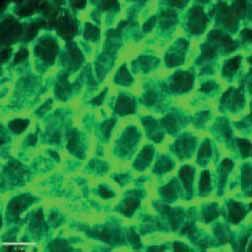
ERJ OPEN RESEARCH                                                                                  ORIGINAL RESEARCH ARTICLE | W. DUAN ET AL.

                                   for subsequent bronchiectasis. It is known that neutrophils, the primary source of elastase in the airway, are
                                   recruited to the airway in acute RSV infection [11].

                                   The respiratory epithelial cell is the primary target for RSV infection, and thus we were interested in
                                   examining the epithelial response to RSV in the setting of CF to better understand the link between RSV
                                   infection and subsequent progression of CF lung disease.

                                   Materials and methods
                                   For all sections, further details are available in the online supplementary material.

                                   Participants
                                   All participants were recruited and studied at SickKids in Toronto under local Research Ethics Board
                                   approved protocols. Participants or their guardians signed informed consent prior to participating. Twelve
                                   patients homozygous for p.Phe508del cystic fibrosis transmembrane conductance regulator (CFTR) and 12
                                   non-CF healthy controls were recruited and donated nasal cells for primary cell culture (REB
                                   1000044783). The age range of the homozygous p.Phe508del patients was between 12 and 16 years, and
                                   50% of them were males. The age range of the healthy controls was between 28 and 63 years, and 25% of
                                   them were males.

                                   Nasal brushing and air–liquid interface culture
                                   Nasal brushing and air–liquid interface (ALI) culture were performed as previously described [12–18].

                                   Human bronchial epithelial cell culture
                                   Human bronchial epithelial (HBE) cells CF-HBE (CFF-16HBEge CFTR p.Phe508del V470) were
                                   obtained from Cystic Fibrosis Foundation Therapeutics (Lexington, MA, USA) and wild-type HBE
                                   (WT-HBE=16HBE14o) were obtained from Drs D. Gruenert and B. Illek (University of California,
                                   San Francisco, CA, USA).

                                   RSV propagation and purification
                                   RSV-A2 strain production was performed as previously described [15, 19, 20]. The recombinant strain of
                                   RSV expressing green fluorescent protein rgRSV224 (RSV-GFP) was a gift from Dr M.E. Peeples
                                   (Children’s Research Institute, Columbus, OH, USA) and Dr P.L. Collins (National Institutes of Health,
                                   Bethesda, MD, USA) [21].

                                   RSV infection and transepithelial resistance measurement
                                   At day 21 of ALI culture, nasal epithelial cells and HBE cells were infected with RSV-A2 and RSV-GFP.
                                   Nasal epithelial cells were infected apically with 100 µL RSV in PBS at 0.5 MOI in duplicates for each
                                   insert. Mock infection was performed with PBS addition to cells. Transepithelial resistance (TER) was
                                   measured with an ohmmeter (World Precision Instruments, Sarasota, FL, USA) following the
                                   manufacturer’s instructions. HBE cells were treated with RSV-GFP in the same way as nasal cells.

                                   Quantitative real-time PCR
                                   Buffer RLT was added onto the nasal cells and incubated at room temperature for 5 min. Then cells were
                                   scraped and stored at −80°C. mRNA purification was conducted using Qiagen RNeasy Mini Kit (Qiagen,
                                   Hilden, Germany) following the manufacturer’s protocols.

                                   Plaque assay
                                   PBS washes of the apical surface of cells were diluted and added onto HEp-2 cells in 6-well plates.
                                   DMEM-F12/agarose was overlaid onto the cells and incubated for 6 days at 37°C as previously described
                                   [19, 20, 22].

                                   Immunofluorescence
                                   Immunofluorescence was conducted as described previously [16, 17, 23].

                                   Western blots
                                   Primary nasal epithelial cells (PNECs) were lysed in 150 µL radioimmunoprecipitation assay (RIPA)
                                   buffer (50 mM Tris–HCl PH 7.4, 150 mM NaCl, 1 mM EDTA, pH 8.0, 0.2% SDS, and 0.1% Triton
                                   X-100, Roche complete protease inhibitor cocktail) on ice for 10 min. The cells were then scraped from
                                   the membrane and transferred to Eppendorf tubes. The cell lysates were spun down at 8000 g for 15 min
                                   and then the supernatant was collected and stored at −80°C.

https://doi.org/10.1183/23120541.00365-2021                                                                                                    2
ERJ OPEN RESEARCH                                                                                                                     ORIGINAL RESEARCH ARTICLE | W. DUAN ET AL.

                                              Cytokine measurements
                                              Basolateral medium was collected 72 h post infection (hpi) and cytokine expression was measured by
                                              Luminex bead assay (Bio-Rad Laboratories, Hercules, CA, USA).

                                              Statistical analysis
                                              Statistical analyses were performed with SAS Version 9.4 (SAS Institute, Cary, NC, USA) and GraphPad
                                              Prism Version 7.0 (GraphPad Software, La Jolla, CA, USA). Mann–Whitney, matched Wilcoxon and
                                              two-way ANOVA tests were applied to analyse the data. Spearman test was used to analyse the correlation.
                                              The values below the limit of detection (LOD) for cytokine measurements were calculated using LOD/
                                              SQRT(2). All data were represented as mean±SEM. p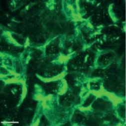
ERJ OPEN RESEARCH ORIGINAL RESEARCH ARTICLE | W. DUAN ET AL.
p.Phe508del Control
Mock RSV Mock RSV
E-Tubulin
E-Cadherin
E-Catenin
ZO-1
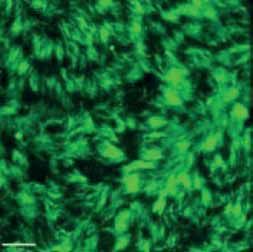
FIGURE 2 Morphology of epithelial cells post respiratory syncytial virus (RSV) infection in cystic fibrosis and
control cells. Homozygous p.Phe508del cystic fibrosis transmembrane conductance regulator (CFTR) and
control primary human nasal epithelial cells (PNECs) in mock and RSV infection were stained at 72 h post
infection. Representative immunofluorescence images demonstrate β-tubulin staining in epithelial cilia,
E-cadherin and β-catenin staining in cell–cell adherens junctions and ZO-1 staining in tight junctions.
Scale bar=10 µm.
The gene expression of pattern recognition receptors in PNECs
Owing to the difference seen in the viral load between p.Phe508del CFTR cells and control cells, we
wondered if there was a defect in innate immune pathways. We measured pattern recognition receptors’
(PRRs) mRNA expressions in the cells at 72 hpi (figure 4). Toll-like receptor-2 (TLR2), TLR3, TLR4,
TLR7 and TLR9 levels were not significantly different between p.Phe508del CFTR cells and control cells,
whereas RIG-I and MDA-5 levels were higher in p.Phe508del CFTR cells than in control cells post RSV
infection. TLR8 was undetectable in PNECs. In addition, ISG56 mRNA expression was higher in p.
Phe508del CFTR cells than in control cells post RSV infection.
Cytokine measurements
The basolateral medium from the primary human epithelial cells grown at ALI was collected at 72 hpi, and
36 cytokines were measured (figure 5 and supplementary figure S1). Interleukin (IL)-2, IL-5, IL-13, IL-22,
IL-26, granulocyte–macrophage colony-stimulating factor (GM-CSF), basic fibroblast growth factor and
macrophage inflammatory protein-1α were not detected. The cells from p.Phe508del CFTR patients
produced significantly more IL-8, IL-9, IL-10, IL-12p70, IL-15, IL-17A, tumour necrosis factor-α
(TNF-α), vascular endothelial growth factor and granulocyte colony-stimulating factor (G-CSF) compared
to cells from healthy controls at 72 h post RSV infection. At baseline, p.Phe508del CFTR cells produced
more IL-12p70 and IL-15 than control cells (figure 5). Importantly, we did not see reduced production of
innate antiviral cytokines (IL-28A, IL-29) in p.Phe508del cells (figure 6).
https://doi.org/10.1183/23120541.00365-2021 4
ERJ OPEN RESEARCH ORIGINAL RESEARCH ARTICLE | W. DUAN ET AL.
Mock RSV
NS
a) p.Phe508del Control b) 2.5 NS
(normalised to total protein)
M R M R M R M R M R M R
2.0
E-Tubulin
E-Tubulin
1.5
E-Cadherin 1.0
0.5
E-Catenin
0.0
p.Phe508del Control
NS
ZO-1 2.5 NS
(normalised to total protein)
2.0
CNX
E-Cadherin
1.5
1.0
Total
protein 0.5
0.0
p.Phe508del Control
1.5 NS
NS
(normalised to total protein)
*
c) 1200 p.Phe508del_RSV (n=10)
Control_RSV (n=10) 1.0
E-Catenin
1000
0.5
Resistance :cm2
NS NS NS
800
0.0
p.Phe508del Control
600 5 NS
NS
(normalised to total CNX)
4
400
0 24 48 72 3
ZO-1
Hours post RSV infection
2
1
0
p.Phe508del Control
FIGURE 3 Protein expression and transepithelial resistance (TER) post respiratory syncytial virus (RSV) infection
in cystic fibrosis and control cells. Protein was harvested from homozygous p.Phe508del cystic fibrosis
transmembrane conductance regulator (CFTR) and control primary human nasal epithelial cells in mock and
RSV infection at 72 h post infection. Western blots were performed. a) Epithelial cilia marker β-tubulin, cell–cell
adherens markers E-cadherin and β-catenin and tight junctions marker ZO-1. b) Quantification of Western blots
demonstrates that expressions of the proteins are not significant between p.Phe508del CFTR and control cells
(n=5–6), by Mann–Whitney test. c) The TER between homozygous p.Phe508del CFTR and control cells is not
significant at 24, 48 and 72 hpi. The data were analysed with two-way ANOVA analysis followed by post hoc
Bonferroni multiple comparisons. *p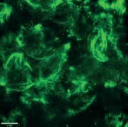
ERJ OPEN RESEARCH                                                                                                       ORIGINAL RESEARCH ARTICLE | W. DUAN ET AL.

                                                        Mock
                                                        RSV
                      8       **         **                                          6        **        ***                                  4       **        NS
                                                ***                                                             ***                                                   ***

                                                               MDA-5 relative mRNA

                                                                                                                        TLR2 relative mRNA
RIG-I relative mRNA

                      6                                                                                                                      3
                                                                                     4
                      4                                                                                                                      2
                                                                                     2
                      2                                                                                                                      1

                      0                                                              0                                                       0
                          p.Phe508del         Control                                     p.Phe508del         Control                            p.Phe508del        Control

                                                                                              **        NS
                      4       ***        NS                                          6                          ***                          4      ***        NS
                                                ***                                                                                                                   **
                                                               TLR4 relative mRNA
TLR3 relative mRNA

                                                                                                                        TLR7 relative mRNA
                      3                                                                                                                      3
                                                                                     4
                      2                                                                                                                      2
                                                                                     2
                      1                                                                                                                      1

                      0                                                              0                                                       0
                          p.Phe508del         Control                                     p.Phe508del         Control                            p.Phe508del        Control

                      3                  NS                                          10       **        ***
                                                                                                                ***
                                                               ISG56 relative mRNA
TLR9 relative mRNA

                                                                                     8
                      2
                                                                                     6

                                                                                     4
                      1
                                                                                     2

                      0                                                              0
                          p.Phe508del         Control                                     p.Phe508del         Control

            FIGURE 4 Pattern recognition receptors gene expression is increased in cystic fibrosis (CF) and control cells post respiratory syncytial virus (RSV).
            RSV increases RIG-I, MDA-5, TLR2, TLR3, TLR4 and TLR7 mRNA expression. TLR9 mRNA is unchanged and TLR8 mRNA is undetectable using Qiagen
            SBH0265788-200 TLR8 primers. The relative mRNA levels of RIG-I and MDA-5 are significantly higher in homozygous p.Phe508del cystic fibrosis
            transmembrane conductance regulator (CFTR) cells compared to control cells post RSV infection. No significant differences are seen in TLR genes
            post RSV between CF and control cells. Interferon-stimulated gene ISG56 shows more mRNA expression in homozygous p.Phe508del CFTR cells.
            Non-paired data were analysed by Mann–Whitney test, and paired data were analysed by matched Wilcoxon test (n=11–12). **p
ERJ OPEN RESEARCH                                                                                                    ORIGINAL RESEARCH ARTICLE | W. DUAN ET AL.

                                                        Mock
                                                        RSV
                                **        *                                       50        **        **                               10                  **
                   85 000                        **                                                          **
                   65 000                                                         40                                                     8
                   45 000

                                                                                                                      IL-10 pg·mL–1
                                                                  IL-9 pg·mL–1
IL-8 pg·mL−1

                   25 000                                                         30                                                    6
                    5000
                    5000
                    4000                                                          20                                                     4
                    3000
                    2000                                                          10                                                     2
                    1000
                        0                                                          0                                                    0
                            p.Phe508del       Control                                   p.Phe508del        Control                           p.Phe508del        Control

                     150        **        *                                       1.5       **        **                               30        **        **
                                      *          **                                               *                                                               **
IL-12p70 pg·mL−1

                                                                                                                      IL-17A pg·mL–1
                                                               IL-15 pg·mL–1

                     100                                                          1.0                                                  20

                      50                                                          0.5                                                  10

                       0                                                          0.0                                                    0
                            p.Phe508del       Control                                   p.Phe508del        Control                           p.Phe508del        Control

                      20        **        *                                      5000       *         **                               400       **        *
                                                 **                                                          **                                                   **
                                                                                 4000
TNF-D pg·mL−1

                      15                                                                                                               300

                                                                                                                      G-CSF pg·mL–1
                                                               VEGF pg·mL–1

                                                                                 3000
                      10                                                                                                               200
                                                                                 2000
                       5                                                                                                               100
                                                                                 1000

                       0                                                            0                                                   0
                            p.Phe508del       Control                                   p.Phe508del        Control                           p.Phe508del        Control

      FIGURE 5 Cystic fibrosis cells produce higher amounts of cytokines post respiratory syncytial virus (RSV) infection as compared to control cells.
      The production of cytokines was measured by Luminex in basal media at 72 hpi in primary human nasal epithelial cells. Among the 36 cytokines
      measured, the levels of interleukin (IL)-8, IL-9, IL-10, IL-12p70, IL-15, IL-17A, tumour necrosis factor-α (TNF-α), vascular endothelial growth factor
      (VEGF) and granulocyte colony-stimulating factor (G-CSF) are significantly higher in homozygous p.Phe508del cystic fibrosis transmembrane
      conductance regulator (CFTR) cells than in control cells. Non-paired data were analysed by Mann–Whitney test, and paired data were analysed by
      matched Wilcoxon test (n=10). *p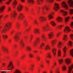
ERJ OPEN RESEARCH                                                                                                                      ORIGINAL RESEARCH ARTICLE | W. DUAN ET AL.

                                                                                                          Mock
                                                                                                          RSV
                                                                       **                         **                                 4000         **                        **
                                                     700 000
                                                     500 000
                                                                                                                                     3000

                                    IL-28A pg·mL−1

                                                                                                                   IL-29 pg·mL–1
                                                     300 000
                                                     100 000                                                                         2000
                                                     100 000
                                                      80 000
                                                      60 000                                                                         1000
                                                      40 000
                                                      20 000
                                                           0                                                                              0
                                                                   p.Phe508del               Control                                          p.Phe508del                 Control

                                       FIGURE 6 The production of Type III interferons (IFNs) is not reduced in cystic fibrosis cells. The production of
                                       Type III IFNs interleukin (IL)-28A and IL-29 were measured. IL-28A and IL-29 are not different between
                                       respiratory syncytial virus (RSV)-infected p.Phe508del cystic fibrosis transmembrane conductance regulator cells
                                       and control cells. The production of IL-28A and IL-29 in mock cells were below the limit of detection.
                                       Non-paired data were analysed by Mann–Whitney test, and paired data were analysed by matched Wilcoxon
                                       test (n=9–10). **p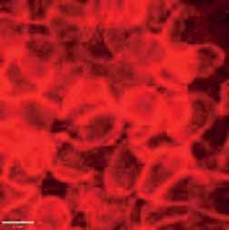
ERJ OPEN RESEARCH                                                                                   ORIGINAL RESEARCH ARTICLE | W. DUAN ET AL.

                                   neutrophil recruitment with more elastase production and then subsequent lung damage and dysfunction.
                                   This work is consistent with data from the AREST CF group where bronchoalveolar lavage elastase levels
                                   at age 3 months were linked with bronchiectasis at age 3 years [10].

                                   One caveat to the line of reasoning suggesting a causal link between primary epithelial IL-8 production
                                   and secondary reduced lung function is that it is also possible that the reverse relationship exists. Thus, it is
                                   feasible that elevated IL-8 levels seen in CF cells are explained by epigenetic changes within those cells.
                                   Individuals with reduced lung function likely have been exposed to a different infectious and inflammatory
                                   milieu over their life course when compared to individuals with higher lung function. This different milieu
                                   may result in an altered epigenetic profile of epithelial cells which in turn results in a different IL-8
                                   (inflammatory) response to RSV when studied in vitro. Outside of epithelial cells, there are data to suggest
                                   that neutrophilic inflammation predisposes the lung to more severe RSV infection [51]. Thus, it is
                                   conceivable that inflammation leads to worse infection as opposed to the reverse. It is also possible that a
                                   causal association exists in both directions leading to an auto-amplification loop (i.e. virus induces
                                   inflammation à inflammation alters cells to be pro-inflammatory post infection à virus induces
                                   inflammation). These possibilities remain a focus of future investigations.

                                   Fourth, we observed a number of cytokines (including IL-9, IL-10, IL-12, IL-15, IL-17A and TNF-α)
                                   produced at higher levels in CF cells post RSV infection consistent with the idea of an exaggerated
                                   inflammatory response. In the past, it has been reported that infants with RSV bronchiolitis have greater
                                   IL-9 mRNA levels than control infants; elevated IL-9 production was thought to increase the inflammatory
                                   response and lung disease severity [52]. IL-10 is a known anti-inflammatory cytokine; however, its impact
                                   on the antiviral immune response is complex. Thus, broadly in the setting of viral infection, IL-10 can
                                   inhibit viral replication but has also been reported to promote viral persistence [53, 54]. This is a pattern
                                   that has also been reported for IL-12p70 and IL-15 where both pro-inflammatory or anti-inflammatory viral
                                   responses have been reported likely reflecting differences in model systems studied and when in the course
                                   of the infection the analyses were performed (i.e. early versus late in infections) [55–57]. IL-17A is
                                   thought to promote RSV pathogenesis as in a mouse model, anti-IL-17A treatment reduced both
                                   inflammation and viral load significantly [58]. TNF-α is a pro-inflammatory cytokine and is known to be
                                   elevated in infants post RSV infection [59]. Both TNF-α and IL-17A are known to induce G-CSF, an
                                   important mediator of neutrophil function [60]. In our study, IL-17A, TNF-α and G-CSF all showed higher
                                   levels in CF cells.

                                   Finally, our work highlights the importance of examining more than just current CFTR as an in vitroCF
                                   epithelial outcome measure. Clearly, CF epithelial cells have a number of deficits that can be evaluated and
                                   may predispose to or be associated with lung disease. Viral clearance and cytokine production are such
                                   outcomes, and changes should be considered at baseline and post candidate interventions/therapies. With
                                   this in mind, a future direction of this work will be to examine the impact of current and novel CFTR
                                   modulator agents on in vitro viral clearance and in vitro cytokine production.

                                   In summary we present data supporting an association between in vitro IL-8 production and lung function
                                   and also the notion that innate immune function is an important epithelial phenotype that should be studied
                                   in CF cells as we look towards improving clinical outcomes.

                                   Limitations
                                   There are a few limitations to this study. Only 12 CF patients and 12 controls donated nasal cells for the
                                   culture work; it is possible that a larger sample size may unveil significantly different findings. In addition,
                                   CF and control samples were not age or sex matched. Lastly, sputum was not collected in this study to
                                   analyse elastase activity. Future studies could address these limitations.

                                   Acknowledgements: CF-HBEs were obtained from the Cystic Fibrosis Foundation Therapeutics and wild-type HBEs
                                   were obtained from D. Gruenert and B. Illek at UCSF. The recombinant strain of RSV expressing GFP rgRSV224 was
                                   obtained from M.E. Peeples at the Children’s Research Institute in Columbus and P.L. Collins at the National
                                   Institutes of Health. We would like to thank all members of CFIT for supporting this work.

                                   Provenance: Submitted article, peer reviewed.

                                   Author contributions: W. Duan conceived of, performed and planned all experiments, analysed the data, and wrote
                                   the paper. Y. Chen, C. Lin, H. Ouyang, K. Du, A. Kumar and B. Wang performed experiments and edited the
                                   manuscript. J. Avolio performed the nasal brushing and edited the manuscript. H. Grasemann provided guidance

https://doi.org/10.1183/23120541.00365-2021                                                                                                       9
ERJ OPEN RESEARCH ORIGINAL RESEARCH ARTICLE | W. DUAN ET AL.
and edited the paper. T.J. Moraes conceived of experiments, analysed data, wrote and edited the paper,
supervised, and provided support for the project.
Conflict of interest: None declared.
Support statement: T.J. Moraes was supported by CF Canada–SickKids Program for Individualised CF Therapy and
Emily’s Entourage.
References
1 Leader S, Kohlhase K. Recent trends in severe respiratory syncytial virus (RSV) among US infants, 1997 to
2000. J Pediatr 2003; 143: Suppl. 5, S127–S132.
2 Pelletier AJ, Mansbach JM, Camargo CA, Jr. Direct medical costs of bronchiolitis hospitalizations in the
United States. Pediatrics 2006; 118: 2418–2423.
3 Samson L. Prevention of respiratory syncytial virus infection. Paediatr Child Health 2009; 14: 521–532.
4 Robinson JL, Le Saux N. Preventing hospitalizations for respiratory syncytial virus infection. Paediatr Child
Health 2015; 20: 321–333.
5 Byington CL, Wilkes J, Korgenski K, et al. Respiratory syncytial virus-associated mortality in hospitalized
infants and young children. Pediatrics 2015; 135: e24–e31.
6 Abman SH, Ogle JW, Butler-Simon N, et al. Role of respiratory syncytial virus in early hospitalizations for
respiratory distress of young infants with cystic fibrosis. J Pediatr 1988; 113: 826–830.
7 Armstrong D, Grimwood K, Carlin JB, et al. Severe viral respiratory infections in infants with cystic fibrosis.
Pediatr Pulmonol 1998; 26: 371–379.
8 Somayaji R, Goss CH, Khan U, et al. Cystic fibrosis pulmonary exacerbations attributable to respiratory
syncytial virus and influenza: a population-based study. Clin Infect Dis 2017; 64: 1760–1767.
9 Hiatt PW, Grace SC, Kozinetz CA, et al. Effects of viral lower respiratory tract infection on lung function in
infants with cystic fibrosis. Pediatrics 1999; 103: 619–626.
10 Sly PD, Gangell CL, Chen L, et al. Risk factors for bronchiectasis in children with cystic fibrosis. N Engl J Med
2013; 368: 1963–1970.
11 Kirsebom FCM, Kausar F, Nuriev R, et al. Neutrophil recruitment and activation are differentially dependent
on MyD88/TRIF and MAVS signaling during RSV infection. Mucosal Immunol 2019; 12: 1244–1255.
12 Cao H, Ouyang H, Ip W, et al. Testing gene therapy vectors in human primary nasal epithelial cultures. Mol
Ther Methods Clin Dev 2015; 2: 15034.
13 Eckford PDW, McCormack J, Munsie L, et al. The CF Canada-Sick Kids Program in individual CF therapy: a
resource for the advancement of personalized medicine in CF. J Cyst Fibros 2018; 18: 35–43.
14 Wu YS, Jiang J, Ahmadi S, et al. ORKAMBI-mediated rescue of mucociliary clearance in cystic fibrosis primary
respiratory cultures is enhanced by arginine uptake, arginase inhibition, and promotion of nitric oxide
signaling to the cystic fibrosis transmembrane conductance regulator channel. Mol Pharmacol 2019; 96:
515–525.
15 Norris MJ, Malhi M, Duan W, et al. Targeting intracellular ion homeostasis for the control of respiratory
syncytial virus. Am J Respir Cell Mol Biol 2018; 59: 733–744.
16 Cao H, Ouyang H, Grasemann H, et al. Transducing airway basal cells with a helper-dependent adenoviral
vector for lung gene therapy. Hum Gene Ther 2018; 29: 643–652.
17 Molinski SV, Ahmadi S, Ip W, et al. Orkambi(R) and amplifier co-therapy improves function from a rare CFTR
mutation in gene-edited cells and patient tissue. EMBO Mol Med 2017; 9: 1224–1243.
18 Ahmadi S, Bozoky Z, Di Paola M, et al. Phenotypic profiling of CFTR modulators in patient-derived respiratory
epithelia. NPJ Genom Med 2017; 2: 12.
19 Tayyari F, Marchant D, Moraes TJ, et al. Identification of nucleolin as a cellular receptor for human
respiratory syncytial virus. Nat Med 2011; 17: 1132–1135.
20 Norris MJ, Duan W, Cen Y, et al. Agonistic 4-1BB antibody fails to reduce disease burden during acute
respiratory syncytial virus (RSV) infection. Antiviral Res 2016; 125: 46–50.
21 Hallak LK, Spillmann D, Collins PL, et al. Glycosaminoglycan sulfation requirements for respiratory syncytial
virus infection. J Virol 2000; 74: 10508–10513.
22 Marchant DJ, Bellac CL, Moraes TJ, et al. A new transcriptional role for matrix metalloproteinase-12 in
antiviral immunity. Nat Med 2014; 20: 493–502.
23 Du K, Karp PH, Ackerley C, et al. Aggregates of mutant CFTR fragments in airway epithelial cells of CF lungs:
new pathologic observations. J Cyst Fibros 2015; 14: 182–193.
24 Houben ML, Coenjaerts FE, Rossen JW, et al. Disease severity and viral load are correlated in infants with
primary respiratory syncytial virus infection in the community. J Med Virol 2010; 82: 1266–1271.
25 Hijano DR, Brazelton de Cardenas J, Maron G, et al. Clinical correlation of influenza and respiratory syncytial
virus load measured by digital PCR. PLoS ONE 2019; 14: e0220908.
https://doi.org/10.1183/23120541.00365-2021 10ERJ OPEN RESEARCH ORIGINAL RESEARCH ARTICLE | W. DUAN ET AL.
26 Bhowmick R, Gappa-Fahlenkamp H. Cells and culture systems used to model the small airway epithelium.
Lung 2016; 194: 419–428.
27 Villenave R, Thavagnanam S, Sarlang S, et al. In vitro modeling of respiratory syncytial virus infection of
pediatric bronchial epithelium, the primary target of infection in vivo. Proc Natl Acad Sci USA 2012; 109:
5040–5045.
28 Guo-Parke H, Canning P, Douglas I, et al. Relative respiratory syncytial virus cytopathogenesis in upper and
lower respiratory tract epithelium. Am J Respir Crit Care Med 2013; 188: 842–851.
29 Lay MK, González PA, León MA, et al. Advances in understanding respiratory syncytial virus infection in airway
epithelial cells and consequential effects on the immune response. Microbes Infect 2013; 15: 230–242.
30 Colasurdo GN, Fullmer JJ, Elidemir O, et al. Respiratory syncytial virus infection in a murine model of cystic
fibrosis. J Med Virol 2006; 78: 651–658.
31 Chattoraj SS, Ganesan S, Faris A, et al. Pseudomonas aeruginosa suppresses interferon response to
rhinovirus infection in cystic fibrosis but not in normal bronchial epithelial cells. Infect Immun 2011; 79:
4131–4145.
32 Kieninger E, Singer F, Tapparel C, et al. High rhinovirus burden in lower airways of children with cystic
fibrosis. Chest 2013; 143: 782–790.
33 Parker D, Cohen TS, Alhede M, et al. Induction of type I interferon signaling by Pseudomonas aeruginosa is
diminished in cystic fibrosis epithelial cells. Am J Respir Cell Mol Biol 2012; 46: 6–13.
34 Zheng S, De BP, Choudhary S, et al. Impaired innate host defense causes susceptibility to respiratory virus
infections in cystic fibrosis. Immunity 2003; 18: 619–630.
35 Schögler A, Stokes AB, Casaulta C, et al. Interferon response of the cystic fibrosis bronchial epithelium to
major and minor group rhinovirus infection. J Cyst Fibros 2016; 15: 332–339.
36 Vareille M, Kieninger E, Alves MP, et al. Impaired type I and type III interferon induction and rhinovirus
control in human cystic fibrosis airway epithelial cells. Thorax 2012; 67: 517–525.
37 Majzoub K, Wrensch F, Baumert TF. The innate antiviral response in animals: an evolutionary perspective
from flagellates to humans. Viruses 2019; 11: 758.
38 Kim TH, Lee HK. Innate immune recognition of respiratory syncytial virus infection. BMB Rep 2014; 47:
184–191.
39 Brisse, M, Ly H. Comparative structure and function analysis of the RIG-I-like receptors: RIG-I and MDA5. Front
Immunol 2019; 10: 1586.
40 Okabayashi T, Kojima T, Masaki T, et al. Type-III interferon, not type-I, is the predominant interferon induced
by respiratory viruses in nasal epithelial cells. Virus Res 2011; 160: 360–366.
41 Glaser L, Coulter PJ, Shields M, et al. Airway epithelial derived cytokines and chemokines and their role in
the immune response to respiratory syncytial virus infection. Pathogens 2019; 8: 106.
42 Standiford TJ, Kunkel SL, Basha MA, et al. Interleukin-8 gene expression by a pulmonary epithelial cell line. A
model for cytokine networks in the lung. J Clin Invest 1990; 86: 1945–1953.
43 Hillian AD, Londono D, Dunn JM, et al. Modulation of cystic fibrosis lung disease by variants in interleukin-8.
Genes Immun 2008; 9: 501–508.
44 Polineni D, Dang H, Gallins PJ, et al. Airway mucosal host defense is key to genomic regulation of cystic
fibrosis lung disease severity. Am J Respir Crit Care Med 2018; 197: 79–93.
45 Cordoba-Lanus E, Baz-Dávila R, Espinoza-Jiménez A, et al. IL-8 gene variants are associated with lung
function decline and multidimensional BODE index in COPD patients but not with disease susceptibility: a
validation study. COPD 2015; 12: 55–61.
46 Esposito S, Iererdi V, Daleno C, et al. Genetic polymorphisms and risk of recurrent wheezing in pediatric age.
BMC Pulm Med 2014; 14: 162.
47 Charrad R, Kaabachi W, Rafrafi A, et al. IL-8 gene variants and expression in childhood asthma. Lung 2017;
195: 749–757.
48 Wang SZ, Smith PK, Lovejoy M, et al. Shedding of L-selectin and PECAM-1 and upregulation of Mac-1 and
ICAM-1 on neutrophils in RSV bronchiolitis. Am J Physiol 1998; 275: L983–L989.
49 Smith PK, Wang SZ, Dowling KD, et al. Leucocyte populations in respiratory syncytial virus-induced
bronchiolitis. J Paediatr Child Health 2001; 37: 146–151.
50 McNamara PS, Ritson P, Selby A, et al. Bronchoalveolar lavage cellularity in infants with severe respiratory
syncytial virus bronchiolitis. Arch Dis Child 2003; 88: 922–926.
51 Habibi MS, Thwaites RS, Chang M, et al. Neutrophilic inflammation in the respiratory mucosa predisposes to
RSV infection. Science 2020; 370: eaba9301.
52 McNamara PS, Flanagan BF, Baldwin LM, et al. Interleukin 9 production in the lungs of infants with severe
respiratory syncytial virus bronchiolitis. Lancet 2004; 363: 1031–1037.
53 Rojas JM, Avia M, Martín V, et al. IL-10: a multifunctional cytokine in viral infections. J Immunol Res 2017;
2017: 6104054.
54 Naicker DD, Werner L, Kormuth E, et al. Interleukin-10 promoter polymorphisms influence HIV-1 susceptibility
and primary HIV-1 pathogenesis. J Infect Dis 2009; 200: 448–452.
https://doi.org/10.1183/23120541.00365-2021 11ERJ OPEN RESEARCH ORIGINAL RESEARCH ARTICLE | W. DUAN ET AL.
55 Chang HD, Radbruch A. The pro- and anti-inflammatory potential of interleukin-12. Ann NY Acad Sci 2007;
1109: 40–46.
56 Bolger G, Lapeyre N, Dansereau N, et al. Primary infection of mice with high titer inoculum respiratory
syncytial virus: characterization and response to antiviral therapy. Can J Physiol Pharmacol 2005; 83: 198–213.
57 Perera PY, Lichy JH, Waldmann TA, et al. The role of interleukin-15 in inflammation and immune responses to
infection: implications for its therapeutic use. Microbes Infect 2012; 14: 247–261.
58 Mukherjee S, Lindell DM, Berlin AA, et al. IL-17-induced pulmonary pathogenesis during respiratory viral
infection and exacerbation of allergic disease. Am J Pathol 2011; 179: 248–258.
59 Dou Y, Zhao Y, Zhang ZY, et al. Respiratory syncytial virus infection induces higher Toll-like receptor-3
expression and TNF-α production than human metapneumovirus infection. PLoS ONE 2013; 8: e73488.
60 Kim YM, Kim H, Lee S, et al. Airway G-CSF identifies neutrophilic inflammation and contributes to asthma
progression. Eur Respir J 2020; 55: 1900827.
https://doi.org/10.1183/23120541.00365-2021 12You can also read