Ingestion of microplastics by free-living marine nematodes, especially Enoplolaimus spp., in Mallipo Beach, South Korea
←
→
Page content transcription
If your browser does not render page correctly, please read the page content below
Plankton Benthos Res 16(2): 109–117, 2021
                                                                                                              Plankton & Benthos
                                                                                                                   Research
                                                                                                              © The Japanese Association of Benthology

Ingestion of microplastics by free-living marine
nematodes, especially Enoplolaimus spp., in
Mallipo Beach, South Korea
Teawook Kang1, Dongsung Kim2,* & Je Hyeok Oh2
1
    Marine Research Center, National Park Research Institute, Korea National Park Service Yeosu 59723, Republic of Korea
2
    Marine Ecosystem Research Center, KIOST, 385 Haeyang-ro, Yeongdo-Gu, Busan 49111, Republic of Korea
Received 8 April 2020; Accepted 28 October 2020 Responsible Editor: Motohiro Shimanaga
doi: 10.3800/pbr.16.109

            Abstract: Many plastics cause pollution in the marine environment, with microplastics (0.1 µm–5 mm) representing
            a key research focus. The number of microplastics in sediments may increase rapidly, affecting organisms inhabiting
            marine sediments. The aim of this study was to determine how microplastics affect nematodes in intertidal sand. We
            assessed: (1) intake of microplastic particles (10 µm, 5 µm, 1 µm, or 0.5 µm) by Enoplolaimus spp. over 48 h; (2) mi-
            croplastic intake by nematodes depending on feeding type (selective deposit feeders, non-selective deposit feeders, epi-
            strate feeders, or predators/omnivores) over 48 h; and (3) microplastic egestion by Enoplolaimus spp. The proportion
            of Enoplolaimus spp. individuals containing microplastics was significantly less in the 10-µm microplastic treatment
            than in the treatments where Enoplolaimus spp. were exposed to microplastic particles of smaller sizes (5 µm, 1 µm,
            or 0.5 µm). The ingestion rates of microplastics by predators/omnivores, non-selective deposit feeders, and selective
            deposit feeders increased as the size of the microplastic decreased. After transferring Enoplolaimus spp. to filtered
            seawater following microplastic ingestion, the proportion of Enoplolaimus spp. individuals containing the smallest size
            microplastic (0.5 µm) decreased by 15% of the ingested amount in 3 days. In conclusion, there was a significant differ-
            ence among microplastic-size treatments, but not among feeding types or in the interaction between microplastic size
            and feeding type. The size of microplastics, rather than feeding type of nematodes, impacted ingestion rates. It is pos-
            sible that microplastics in the sediment are ingested by nematodes living in marine benthic ecosystems.

            Key words: egestion, ingestion, meiofauna, microplastic, nematodes

                                                                           Additionally, approximately 18% of the plastic waste found
                           Introduction
                                                                           in the ocean environment is associated with the fishing
   Plastic is an important and widely used material, from                  industry, with aquaculture also being a significant con-
the production of basic goods (such as clothes and cosmet-                 tributor to plastic debris in the oceans (Hinojosa & Thiel
ics) to complicated and high-tech products (such as aircraft               2009).
and rocket nozzles). In the medical industry, plastic goods                   Many plastics are transported to sediments in the ma-
are crucial as aseptic and disposable materials (Hamid et                  rine environment (van Cauwenberghe et al. 2015b). Marine
al. 2018). Plastics are excellent packaging materials be-                  plastic debris represents one of the most serious environ-
cause of their low cost, excellent oxygen/moisture barrier                 mental issues worldwide, with microplastics being the pri-
properties, bio-inertness, and light weight (Andrady 2011).                mary focus of many studies (Thompson et al. 2004, Eer-
Annual global plastic production is continuously rising and                kes-Medrano et al. 2015, Rochman et al. 2016, Galloway
reaching 348 million tons in 2017 (PlasticsEurope 2018),                   et al. 2017). Microplastics are generally defined as being
and, as of 2010, 5–13% of this annual plastic production                   0.1 µm–5 mm in diameter (Thompson et al. 2004, Moore
ended up in the marine environment (Jambeck et al. 2015).                  2008) and in the marine environment, can be divided into
                                                                           two components: (1) primary microplastics that are manu-
                                                                           factured directly for various consumer and industrial ap-
* Corresponding author: Dongsung Kim; E-mail, dskim@kiost.ac.kr            plications, and (2) secondary microplastics from the break-
110                                                      T. Kang et al.

down of larger plastic items through decomposition, such           ameter
Ingestion and egestion of MPs by marine nematodes                                   111

                                                                  in the dark. After 48 h, seawater containing Enoplolaimus
Microplastics
                                                                  spp. in a petri dish was placed in a sieve with a 38 µm
   We experimented with fluorescent polystyrene micro-             mesh size. To remove the microplastics from the surface of
plastics to identify microplastics in the nematode body           Enoplolaimus spp., they were washed using a water spray
under a fluorescence microscope. Fluorescent polysty-              containing fresh water. Then, the remaining Enoplolaimus
rene microplastics (FLUOR Polystyrene; MAGSPHERE,                 spp. in the sieve were transferred to a petri dish and fixed
Pasadena, CA, USA), used in all experiments, are com-             with formalin. Enoplolaimus spp. were mounted on micro-
mercially available and were made with fluorescent                 scope slides to check and identify ingested microplastics
dye (yellow-green color). Four diameters of microplas-            by using fluorescence microscopy.
tics were used: 10 µm (4.57×10⁷ particles ml−1), 5 µm             Experiment 2-Microplastic intake of nematodes,
(3.65×10⁸ particles ml−1), 1 µm (4.57×10¹⁰ particles ml−1),       depending on feeding type
and 0.5 µm (3.65×10¹¹ particles mL−1). The specific gravity           In the second experiment, the feeding type of nematodes
of the microplastics used in this study was 1.05. Therefore,      was evaluated in relation to the microplastic ingestion rate.
the microplastics used in the experiment sink by specific          Twelve acrylic cores were collected for this analysis. All
gravity, because the density of the microplastic is greater       nematodes from one acrylic core were separated and trans-
than that of seawater.                                            ferred to a petri dish (i.e., there were 12 petri dishes each
                                                                  containing the nematodes from one sediment sample).
Experimental design
                                                                     The seawater and living nematodes used in Experiment
Experiment 1-Microplastic intake by Enoplolaimus spp.             2 were collected and extracted in the same way as in Ex-
   In the first experiment, Enoplolaimus spp. were used            periment 1. The nematodes selected for Experiment 2 were
to investigate how different sizes of microplastics affected        transferred to filtered seawater to confirm movement. The
ingestion rates. Before the experiment, we analyzed sedi-         nematodes were then transferred to a petri dish contain-
ment in the intertidal zone of Mallipo Beach to identify          ing 5 mL of seawater. In each petri dish, approximately
the dominant nematode. Enoplolaimus was the dominant              100 nematodes sorted from one sediment sample were con-
nematode genus at the sampling site and could be distin-          tained. One microliter of microplastic (10 µm, 5 µm, 1 µm,
guished under a dissecting microscope owing to its large          and 0.5 µm) was added to the petri dishes (three replicates
size. In addition, the feeding type of Enoplolaimus spp.          for each microplastic size). The petri dishes were stored at
is carnivorous/predator, and it has a large mouth through         20°C in the dark.
which 10 µm sized microplastics can enter.                           After 48 h, all nematodes were removed using a fine
   Enoplolaimus spp. were separated from the collected            pin under a stereoscopic microscope, transferred from for-
sediments. We put the collected sediments containing liv-         malin to 3% glycerin, and then mounted on stereoscopic
ing meiofauna in 500-ml bottles. To anesthetize living            microscope slides in anhydrous glycerine for identifica-
meiofauna, seawater containing 5% MgCl2 was added to              tion. Nematodes were identified to the genus level by us-
the bottles containing sediments. The bottles were then           ing the pictorial keys of Platt & Warwick (1983, 1988)
left for approximately 15 min to anesthetize the meiofauna.       and Warwick et al. (1998), with the aid of a microscope
The bottle was stirred with a spoon so that the meiofauna         (Olympus BX51, Tokyo, Japan). The nematodes that had
would float to the surface, and the supernatant containing         ingested microplastics were identified using fluorescence
the meiofauna was poured into a 63 µm sieve. The nema-            microscopy. The mounted nematodes were classified ac-
todes in the sieve were transferred to a petri dish. using a      cording to the original groupings of Wieser (1953) into
spray with seawater containing 5% MgCl2. Next, the nem-           four feeding groups: (1A) selective deposit feeders, (1B)
atodes were distinguished from other meiofauna under a            non-selective deposit feeders, (2A) epistrate feeders, and
dissecting microscope by using a hook or O-type loop, and         (2B) predators/omnivores.
they were confirmed to have been anesthetized (i.e., there         Experiment 3-Microplastic egestion of Enoplolaimus spp.
was no movement).                                                    In Experiment 3, we investigated whether the micro-
   Enoplolaimus spp. were then separated from other               plastics ingested by Enoplolaimus spp. were egested. We
nematodes under a microscope by using a hook-or O-type            allowed the Enoplolaimus spp. to ingest microplastics, then
loop. The selected Enoplolaimus spp. were transferred to          moved them to filtered seawater for incubation for 24 and
a petri dish containing clean filtered seawater to ensure          72 h. We then measured the amount of microplastics in
that they were alive and moving. As Enoplolaimus spp.             the nematodes immediately after removal from the water
emerged from anesthesia, they began to move. Twenty live          containing microplastics and after 24 h and 72 h of incuba-
Enoplolaimus spp. were placed on each of 16 petri dish-           tion in the filtered seawater. A total of 24 petri dishes were
es containing 5 mL of filtered seawater. One microliter of         prepared, and 10 Enoplolaimus spp. in each petri dish were
each size of microplastic (10 µm, 5 µm, 1 µm, and 0.5 µm)         placed in 5 mL of seawater containing microplastics of
was added to each of four petri dishes containing live            each size (10, 5, 1, and 0.5 µm). There were six petri dishes
Enoplolaimus spp. (i.e., there were four replicates for each      for each microplastic size. The petri dishes were stored
microplastic size). The petri dishes were stored at 20°C          at 20°C in the dark. After 48 h of microplastic ingestion,
112 T. Kang et al.
Fig. 3. Microplastic intake rate (%) of Enoplolaimus spp. for
Fig. 2. Images of Enoplolaimus spp. ingesting microplastics of different microplastic particle-size treatments. Error bars represent
different sizes. standard deviation.
the ingestion rate of Enoplolaimus spp. in two petri dishes sis, PIngestion and egestion of MPs by marine nematodes 113
Table 1. The abundance of nematodes that ingested microplastics by genus in different microplastic-size treatments.
Nematodes Feeding 10 µm 5 µm 1 µm 0.5 µm
(spp.) type Avg. Ind. MP Ingest. Avg. Ind. MP Ingest. Avg. Ind. MP Ingest. Avg. Ind. MP Ingest.
Metachromodora 2B 37.0 0.0 50.0 9.3 71.0 65.3 74.0 73.0
Enoplolaimus 2B 48.7 10.0 32.7 12.3 14.0 13.3 11.7 11.7
Daptonema 1B 17.0 0.3 3.3 1.7 1.0 0.7 0.3 0.3
Enoploides 2B 11.3 1.3 5.0 2.7 3.7 3.3 1.3 1.3
Enoplus 2B 2.7 0.0 3.0 1.3 3.3 3.3 0.3 0.3
Oncholaimus 2B 2.3 0.0 0.7 0.3 1.0 1.0
Araeolaimus 1A 1.0 0.7 0.7 0.7
Halalaimus 1A 1.0 0.0 0.3 0.3
Anoplostoma 1B 0.7 0.0 0.3 0.3
Theristus 1B 0.7 0.0
Polysigma 2A 0.7 0.0
Total 121.0 11.7 95.7 27.7 95.7 88.3 88.3 87.3
Percentage (%) 9.6 28.9 92.3 98.9
Avg. Ind.: Average number of individuals (average for three replicates). MP Ingest.: Average number of individuals that ingested microplas-
tics. 1A, selective deposit feeders; 1B, non-selective deposit feeders; 2A, epistrate feeders; and 2B, predators/omnivores.
Fig. 4. Microplastic intake rate (%) of nematodes depending on
microplastic size and feeding type. Error bars represent standard
deviation. Fig. 5. Proportion of Enoplolaimus spp. containing microplastic
particles in different microplastic particle-size treatments and after
transfer to filtered seawater.
other nematodes is shown in Table 1.
found tended to decrease regardless of particle size. We
Experiment 3-Excretion of microplastics by Enoplolaimus also identified the location of microplastics in the body of
spp. Enoplolaimus spp. The microplastics were found close to
After transferring Enoplolaimus spp. that had ingested the head of Enoplolaimus spp. after 48 h exposure to the
microplastics to filtered seawater, the proportion of indi- microplastic environment; 72 h after being transferred to
viduals in which microplastics were found decreased (Fig. filtered seawater, the microplastics were closer to the tail
5, Table 2). This decrease was attributed to the egestion of (Fig. 6).
microplastics. The proportion of Enoplolaimus spp. with
microplastics was 52.7% at 10 µm, 64.3% at 5 µm, 73.2%
Discussion
at 1 µm, and 88.2% at 0.5 µm after 48 h in a petri dish with
microplastics. After transfer to and incubation in filtered In this study, we investigated the extent to which mi-
seawater for 24 h, the proportion of Enoplolaimus spp. croplastics are ingested by nematodes (in particular
with microplastics was 43.7% at 10 µm, 50.0% at 5 µm, Enoplolaimus spp.) collected from Mallipo Beach in South
74.1% at 1 µm, and 79.2% at 0.5 µm. After transfer to and Korea. We showed that microplastics were ingested by
incubation in filtered seawater for 72 h, the proportion of free-living nematodes collected in the marine environment
Enoplolaimus spp. with microplastics was 48.3% at 10 µm, by using laboratory microcosm experiments. Few stud-
58.3% at 5 µm, 66.7% at 1 µm, and 75.0% at 0.5 µm. The ies have demonstrated that free-living marine nematodes
proportion of individuals in which microplastics were consume microplastics. However, there have been several114 T. Kang et al.
Table 2. The percentage (%) of Enoplolaimus spp. that have microplastic in different microplastic-size treatments and after transfer to
filtered seawater.
Size (µm) Replication Microplastic ingestion rate (48 h) 24 h in filtered seawater 72 h in filtered seawater
a 42.9 42.9 16.7
10 b 62.5 44.4 80.0
Avg. 52.7 43.7 48.3
a 57.1 25.0 50.0
5 b 71.4 75.0 66.7
Avg. 64.3 50.0 58.3
a 71.4 62.5 66.7
1 b 75.0 85.7 66.7
Avg. 73.2 74.1 66.7
a 88.9 75.0 80.0
0.5 b 87.5 83.3 70.0
Avg. 88.2 79.2 75.0
lation of microplastics through food chains in the marine
environment. However, lower trophic organisms, such as
invertebrates, likely ingest and accumulate microplastics,
introducing them to the food chains of the marine environ-
ment (Wright et al. 2013a).
In Experiment 2, we showed that the size of microplas-
tics, rather than feeding type of nematodes, affected inges-
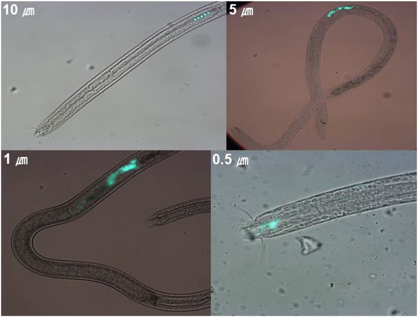
Fig. 6. Image of microplastics in the intestines of Enoplolaimus tion rates. The size and shape of microplastics, exposure
spp. A, exposure to plastic environment; B, 72 h after transfer to concentration and time, morphological features of nema-
filtered seawater. todes (e.g., buccal cavity size and intestine dimensions),
and species-specific feeding habits probably constrain the
recent studies investigating microplastic ingestion by one uptake and ingestion of microplastics by nematodes (Gray
freshwater nematode species, C. elegans (Zhao et al. 2017, & Weinstein 2017, Lehtiniemi et al. 2018, Fueser et al.
Lei et al. 2018, Fueser et al. 2019). C. elegans worms ac- 2019). Most nematodes collected in natural sediments for
tively accumulate 0.5 and 1 µm fluorescent polystyrene mi- this experiment were predators/omnivores. Additionally,
crospheres in the presence and absence of bacterial food, selective deposit feeders and non-selective deposit feeders
whereas microspheres of 3 µm are rarely appeared with low abundance. However, few nematodes
accumulated (Kiyama et al. 2012). were of the epistrate feeding type. For this reason, the
In the current study, Experiment 1 showed that smaller results of non-predator/omnivore feeding-type nematodes
microplastics were more often detected in Enoplolaimus may not have been reflected well. In contrast, the size of
spp. bodies than larger microplastics. The reason for this microplastics had a great influence on the intake rate. The
result could be that given the smaller the size of the mi- small size of microplastics is an important factor affect-
croplastics and the higher the number of microplastics ing bioavailability, particularly for lower trophic organisms
particles added to the petri dishes, these small microplas- (Wright et al. 2013a). The ingestion of microplastics by
tics have a higher probability of entering the mouth of nematodes is predominantly determined by their feeding
nematodes than large microplastics. Previous studies have habits and can be predicted by the morphology of the buc-
reported that the size of microplastics influences the likeli- cal cavity. Fueser et al. (2019) showed that feeding habit
hood of ingestion. For example, a review paper summariz- and the buccal cavity of nematodes have larger effects than
ing microplastic studies conducted on various organisms other factors (such as species-specific feeding differences,
showed that ingestion depends on the size of microplastic exposure time, and exposure concentration) on the inges-
particles, among other factors (Phuong et al. 2016). Lehti- tion of microplastics and could be important for control-
niemi et al. (2018) showed that the size of microplastic ling the quantity of microplastic uptake.
particles, rather than shape, strongly influences the amount In Experiment 3, we investigated whether the microplas-
of microplastics ingested in an experiment using fish and tic particles were retained in the bodies of Enoplolaimus
mysid shrimps. Few studies have evaluated the bioaccumu- spp. or egested. The excretion of microplastics by macroor-Ingestion and egestion of MPs by marine nematodes 115
ganisms has been investigated by measuring the ingestion of microplastics by macroorganisms. For example, Gra-
of microplastic particles and quantifying the microplastics ham et al. (2019) showed that pacific oysters have effi-
in fecal debris. For example, Graham & Thompson (2009) cient egestion rates of microplastics (84.6±2%) and Van
quantified the excretion of microplastics by four species Cauwenberghe et al. (2015a), who analyzed the fecal casts
of sea cucumber. However, it is more difficult to quantify of Polychaeta, showed that they excrete some microplastic
the excretion of microplastics by microorganisms such as particles.
marine nematodes. Therefore, in this study, we first deter- Nematodes are widely distributed globally, including in
mined the proportion of nematodes that ingested micro- extreme environments (such as in polar, deep-sea, and hy-
plastics and then determined the proportion of nematodes drothermal areas). Microplastics are also widely distrib-
that still contained microplastics after being transferred to uted in the marine environment; thus, they are available
and incubated in filtered seawater for 24 h or 72 h. to nematodes. Our study showed that sunk microplastics
The proportion of Enoplolaimus spp. with 10 µm micro- are ingested by nematodes living in marine benthic eco-
plastics in the body decreased by 4.4 percentage points, systems. In these experiments, conducted on Enplolaimus
that with 5 µm microplastics decreased by 6.0 percent- spp., which have a large mouth, the microplastics of the
age points, that with 1 µm microplastics decreased by 6.5 sizes used in this study could be sufficiently ingested. As a
percentage points, and that with 0.5 µm microplastics de- result, microplastics of all sizes were identified in the body
creased by 13.2 percentage points after 72 hours. When the of nematodes. In addition, since the number of nematodes
size of the microplastic decreased, it was expected that mi- used in Experiment 2 by feeding type is small, the dif-
croplastics would easily escape from the body along the di- ference in the ingestion rate of the nematode, depending
gestive tract; however, this effect cannot be identified sig- on the feeding type is not clear. However, the intake rate
nificantly differently due to the lack of sufficient samples to can be expected to be more affected by the size of the mi-
achieve relevant statistical power. Also, in Experiment 3, croplastics rather than the difference in feeding types. In
we transferred the nematodes to the filtered seawater and Experiment 3, contrary to expectations, the discharge rate
checked the discharge of microplastics for just three days. of microplastics into the body of nematodes according to
After three days of experiments in filtered sea water, we the size of microplastics could not be detected. After trans-
observed that the transferred nematodes slowed down and ferring Enoplolaimus spp. to filtered seawater, the propor-
their health condition deteriorated, therefore, we no longer tion of nematodes containing the smallest size microplas-
proceeded with the experiments. However, we expected tic (0.5 µm) decreased by 15% of the ingested amount in
that the egestion rates of microplastics would increase fur- 3 days. As microplastics sink through the water column
ther if the nematode is provided conditions to maintain a when they become covered with biofilm, eventually set-
healthy state in the petri dish for more than three days. Be- tling onto sediments (Wright et al. 2013a), the microplas-
cause the intestinal tract of nematodes is a simple, hollow, tics ingested by nematodes will likely be transferred to
straight tube consisting of a single layer of epithelial cells, animals that feed on them. Future studies should evaluate
it is not difficult to move any sized microplastic from the how microplastics are transmitted and how they accumu-
mouth to the anus (Basyoni & Rizk 2016). Recently, Fue- late within the benthic food web.
ser et al. (2020) showed that the two nematode species, C.
elegans and P. pacificus, rapidly ingest and egest PS beads
Acknowledgements
(0.5 and 1.0 µm) along with bacteria. These two nematodes
are not free-living marine nematodes, but the results are This research was a part of the project titled “Under-
very important in relation to the bioconcentration of mi- standing the deep-sea biosphere on seafloor hydrothermal
croplastics. vents in the Indian Ridge” (No. 20170411), funded by the
Finally, we checked the location of microplastics in the Ministry of Oceans and Fisheries, Korea.
body of Enoplolaimus spp. In Experiment 1, the location of
microplastics in the body of Enoplolaimus spp. was mostly
References
close to the head. However, in Experiment 3, after trans-
ferring the Enoplolaimus spp. that ingested microplastics Andrady AL (2011) Microplastics in the marine environment.
into filtered seawater, microplastics in the body of the Mar Pollut Bull 62: 1596–1605.
Enoplolaimus spp. were mainly located close to the tail. As Auta HS, Emenike CU, Fauziah SH (2017) Distribution and im-
a result, the egestion of microplastics in the nematode body portance of microplastics in the marine environment: a review
is expected to proceed slowly. of the sources, fate, effects, and potential solutions. Environ
If organisms that ingest microplastics do not egest mi- Int 102: 165–176.
croplastics, then the plastics may accumulate in animals Basyoni MM, Rizk EM (2016) Nematodes ultrastructure: com-
higher in the food chain. Conversely, if microorganisms plex systems and processes. J Parasit Dis 40, 1130–1140.
egest microplastics, the adverse effects of plastics on ani- Bonanno G, Orlando-bonaca M (2018) Perspectives on using ma-
mals higher in the food chain may be limited (Wright et al. rine species as bioindicators of plastic pollution. Mar Pollut
Bull 137: 209–221.
2013a). Some studies have been conducted on the egestion116 T. Kang et al. Browne MA, Crump P, Niven SJ, Teuten E, Tonkin A, Galloway na. Washington, DC: Smithsonian Institution Press. 11–13 pp. T, Thompson R (2011) Accumulation of microplastic on shore- Hinojosa I, Thiel M (2009) Floating marine debris in fjords, lines worldwide: sources and sinks. Environ Sci Technol 45: gulfs and channels of southern Chile. Mar Pollut Bull 58: 341– 9175–9179. 350. Browne MA, Dissanayake A, Galloway TS, Lowe DM, Thomp- Jambeck, JR, Andrady A, Geyer R, Narayan R, Perryman M, son RC (2008) Ingested microscopic plastic translocates to the Siegler T, Wicox C, Lavender Law K (2015) Plastic waste in- circulatory system of the mussel, Mytilus edulis (L). Env Sci puts from land into the ocean. Science 347: 768–771. Technol 42: 5026–5031. Kaiser D, Kowalski N, Waniek JJ (2017) Effects of biofouling on Browne MA, Galloway T, Thompson R (2007) Microplastic-an the sinking behavior of microplastics. Environ Res Lett 12: emerging contaminant of potential concern? Integr Environ 124003. Asses 3: 559–566. Karlsson TM, Vethaak AD, Almroth BC, Ariese F, van Velzen Du Y, Gao S, Warwick RM, Hua E (2014) Ecological functioning M, Hassellöv M, Leslie HA (2017) Screening for microplas- of free-living marine nematodes in coastal wetlands: an over- tics in sediment, water, marine invertebrates and fish: method view. Chinese Sci Bull 59: 4692–4704. development and microplastic accumulation. Mar Pollut Bull Duis K, Coors A (2016) Microplastics in the aquatic and terres- 122: 403–408. trial environment: sources (with a specific focus on personal Kim SW, Kim D, Jeong SW, An YJ (2020) Size-dependent ef- care products), fate and effects. Environ Sci Eur 28: 1–25. fects of polystyrene plastic particles on the nematode Eerkes-Medrano D, Thompson RC, Aldridge DC (2015) Mi- Caenorhabditis elegans as related to soil physicochemical croplastics in freshwater systems: a review of the emerging properties. Environ Pollut, 258: 113740. threats, identification of knowledge gaps and prioritisation of Kiyama Y, Miyahara K, Ohshima Y (2012) Active uptake of arti- research needs. Water Res 75: 63–82. ficial particles in the nematode Caenorhabditis elegans. J Exp Eo S, Hong SH, Song YK, Han GM, Shim WJ (2019) Spatio- Biol 215: 1178–1183. temporal distribution and annual load of microplastics in the Lehtiniemi M, Hartikainen S, Näkki P, Engström-Öst J, Koistin- Nakdong River, South Korea. Water Res 160: 228–237. en A, Setälä O (2018) Size matters more than shape: ingestion Fueser H, Mueller MT, Weiss L, Höss S, Traunspurger W (2019) of primary and secondary microplastics by small predators. Ingestion of microplastics by nematodes depends on feeding Food Webs 17: e00097. strategy and buccal cavity size. Environ Pollut 255: 113227. Lei L, Wu S, Lu S, Liu M, Song Y, Fu Z, Shi H, Raley-Susman Galloway TS, Cole M, Lewis C (2017) Interactions of microplas- KM, He D (2018) Microplastic particles cause intestinal dam- tic debris throughout the marine ecosystem. Nat Ecol Evol 1: age and other adverse effects in zebrafish Danio rerio and 1–8. nematode Caenorhabditis elegans. Sci Tot Envt 619: 1–8. Graham ER, Thompson JT (2009) Deposit-and suspension-feed- Maes T, Van der Meulen M, Devriese LI, Leslie HA, Huvet A, ing sea cucumbers (Echinodermata) ingest plastic fragments. J Frère L, Robbens J, Vethaak AD (2017) Microplastics baseline Exp Mar Biol Ecol 368: 22–29. surveys at the water surface and in sediments of the North- Graham P, Palazzo L, de Lucia GA, Telfer TC, Baroli M, Car- East Atlantic. Front Mar Sci 4: 135. boni S (2019) Microplastics uptake and egestion dynamics in Mahmoudi E, Essid N, Beyrem H, Hedfi A, Boufahja F, Vitiello Pacific oysters, Magallana gigas (Thunberg, 1793), under con- P, Aissa P (2005) Effects of hydrocarbon contamination on a trolled conditions. Environ Pollut 252: 742–748. free-living marine nematode community: results from micro- Gray AD, Weinstein JE (2017) Size-and shape-dependent effects cosm experiments. Mar Pollut Bull 50: 1197–1204. of microplastic particles on adult daggerblade grass shrimp McIntyre AD (1969) Ecology of marine meiobenthos. Biol Rev (Palaemonetes pugio). Environ Toxicol Chem 36: 3074–3080. 44: 245–290. Gyedu-Ababio TK, Baird D (2006) Response of meiofauna and Moore CJ (2008) Synthetic polymers in the marine environment: nematode communities to increased levels of contaminants in a rapidly increasing, long-term threat. Environ Res 108: 131– a laboratory microcosm experiment. Ecotoxicol Environ Saf 139. 63: 443–450. Mueller MT, Fueser H, Höss S, Traunspurger W (2020) Spe- Haegerbaeumer A, Mueller MT, Fueser H, Traunspurger W cies-specific effects of long-term microplastic exposure on the (2019) Impacts of micro-and nano-sized plastic particles on population growth of nematodes, with a focus on microplastic benthic invertebrates: a literature review and gap analysis. ingestion. Ecol Indic 118: 106698. Front Environ Sci 7: 17. Naji A, Nuri M, Vethaak AD (2018) Microplastics contamination Harrison JP, Hoellein TJ, Sapp M, Tagg AS, Ju-Nam Y, Ojeda in molluscs from the northern part of the Persian Gulf. Envi- JJ (2018) Microplastic-associated biofilms: a comparison of ron Pollut 235: 113–120. freshwater and marine environments. In Freshwater micro- Phuong NN, Zalouk-Vergnoux A, Poirier L, Kamari A, Châtel plastics (eds Wagner M, Lambert S). Springer, Cham. 181– A, Mouneyrac C, Lagarde F (2016) Is there any consistency 201 pp. between the microplastics found in the field and those used in Hermi M, Mahmoudi E, Beyrem H, Aissa P, Essid N (2009) laboratory experiments? Environ Pollut 211: 111–123. Responses of a free-living marine nematode community to PlasticsEurope (2018) Plastics-the Facts 2018. An analy- mercury contamination: results from microcosm experiments. sis of European plastics production, demand and waste Arch Environ Contam Toxicol 56: 426–433. data. Available at: https://www.plasticseurope.org/application/ Higgins RP, Thiel H (1988) Introduction to the study of meiofau- files/6315/4510/9658/Plastics_the_facts_2018_AF_web.pdf
Ingestion and egestion of MPs by marine nematodes                                     117

  (accessed on 23 Sept 2019)                                           natural habitats. Environ Pollut 199: 10–17.
Platt HM, Warwick RM (1983) A Synopsis of the Free-living            Van Cauwenberghe L, Devriese L, Galgani F, Robbens J, Jans-
  Marine Nematodes. Part I. British Enoplids. Cambridge Uni-           sen CR (2015b) Microplastics in sediments: a review of tech-
  versity Press, Cambridge, 35–85 pp.                                  niques, occurrence and effects. Mar Environ Res 111: 5–17.
Platt HM, Warwick RM (1988) A synopsis of the free-living            Wang J, Wang M, Ru S, Liu X (2019) High levels of microplastic
  marine nematodes. Part II. British Chromadorids. Cambridge           pollution in the sediments and benthic organisms of the South
  University Press, Cambridge, 35–85 pp.                               Yellow Sea, China. Sci Total Environ 651: 1661–1669.
Rochman CM, Browne MA, Underwood AJ, VanFraneker JA,                 Wright SL, Thompson RC, Galloway TS (2013a) The physical
  Thompson RC, Amaral-Zettler LA (2016) The ecological im-             impacts of microplastics on marine organisms. Environ Pollut
  pact of marine debris: unraveling the demonstrated evidence          178: 438–492.
  from what is perceived. Ecology 97: 302–312.                       Wright SL, Rowe D, Thompson RC, Galloway TS (2013b) Micro-
Shahul Hamid F, Bhatti MS, Anuar N, Anuar N, Mohan P, Peria-           plastic ingestion decreases energy reserves in marine worms.
  thamby A (2018) Worldwide distribution and abundance of              Curr Biol 23: 1031–1033.
  microplastic: how dire is the situation? Waste Manag Res 36:       Warwick RM, Platt HM, Somerfield PJ (1998) A Synopsis of the
  873–897.                                                             Free-Living Marine Nematodes. Part III. Monhysterids. Field
Shang X, Lu J, Feng C, Ying Y, He Y, Fang S, Lin Y, Dahlgren           Studies Council, Shrewsbury, 35–85 pp.
  R, Ju J (2020) Microplastic (1 and 5 µm) exposure disturbs         Wieser W (1953) Die beziehung zwischen Mundhohlengestalt,
  lifespan and intestine function in the nematode Caenorhabditis       Ernahrungsweise und Vorkommen bei freilebenden marinen
  elegans. Sci Total Environ 705: 135837.                              Nematoden. Eine okologisch-morphologische Studie. Ark
Thompson RC, Olson Y, Mitchell RP, Davis A, Rowland SJ, John           Zool 4: 439–484 (in German).
  AW, McGonigle D, Russell AE (2004) Lost at sea: where is all       Zhao L, Qu M, Wong G, Wang D (2017) Transgenerational tox-
  the plastic? Science (Washington) 304: 838.                          icity of nanopolystyrene particles in the range of µg L−1 in
Van Cauwenberghe L, Claessens M, Vandegehuchte MB,                     the nematode Caenorhabditis elegans. Environ Sci-Nano 4:
  Janssen CR (2015a) Microplastics are taken up by mussels             2356–2366.
  (Mytilus edulis) and lugworms (Arenicola marina) living in
You can also read