Intrauterine Ultrasound-Guided Transcervical Radiofrequency Ablation - Systematic Review - LBI-HTA
←
→
Page content transcription
If your browser does not render page correctly, please read the page content below

Intrauterine
Ultrasound-Guided
Transcervical
Radiofrequency
Ablation
Systematic Review

Decision Support Document No. 120
ISSN online: 1998-0469


Intrauterine
Ultrasound-Guided
Transcervical
Radiofrequency
Ablation
Systematic Review

Vienna, March 2020
Project Team
Authors: Robyn Lambert, BSc, MPh.
         Christoph Strohmaier, Bakk.rer.soc.oec.

Project Support
Systematic literature searches: Tarquin Mittermayr, BA
External Review: Prof. Dr. Heinrich Husslein; Universitätsklinik für Frauenheilkunde, Klinische Abteilung für
                 Allgemeine Gynäkologie und gynäkologische Onkologie/Stv. Leiter Endometriosezentrum Wien;
                 Medizinische Universität Wien
Internal Review: PD. Dr. Claudia Wild

Correspondence
Christoph Strohmaier, christoph.strohmaier@aihta.at

This report should be referenced as follows:
Lambert R. and Strohmaier C. Intrauterine Guided Transcervical Radiofrequency Ablation – Systematic Review.
Decision Support Document No. 120; 2020. Vienna: Ludwig Boltzmann Institute for Health Technology Assessment.

Conflict of interest
All authors and the reviewers involved in the production of this report have declared they have no conflicts of interest
in relation to the technology assessed according to the Uniform Requirements of Manuscripts Statement of Medical
Journal Editors (www.icmje.org).

Disclaimer
The external reviewers did not co-author the scientific report and do not necessarily all agree with its content. Only the
LBI-HTA is responsible for errors or omissions that could persist. The final version and the policy recommendations are
under the full responsibility of the LBI-HTA.

The HTA Core Model®, developed within EUnetHTA (www.eunethta.eu), has been utilised when producing the
contents and/or structure of this work. The following version of the Model was used: [HTA Core Model®, Version 4.2]
Use of the HTA Core Model® does not guarantee the accuracy, completeness, quality or usefulness of any information
or service produced or provided by using the Model.

Commissioned by the Austrian Ministry of Health, this report systematically assessed the intervention described herein
as decision support for the inclusion in the catalogue of benefits.

CONTENT INFORMATION
Publisher:
Ludwig Boltzmann Gesellschaft GmbH
Nußdorferstr. 64, 6 Stock, A-1090 Wien
https://hta.lbg.ac.at/page/imprint

Responsible for content:
Ludwig Boltzmann Institute for Health Technology Assessment (LBI-HTA)
Garnisongasse 7/20, A-1090 Vienna
https://hta.lbg.ac.at/
Decision support documents of the LBI-HTA do not appear on a regular basis and serve to publicize
the research results of the Ludwig Boltzmann Institute of Health Technology Assessments.
Decision support documents of the LBI-HTA are only available to the public via the Internet at
http://eprints.hta.lbg.ac.at
Decision Support Document No.: 120
ISSN-online: 1998-0469
© 2020 LBI-HTA – All rights reserved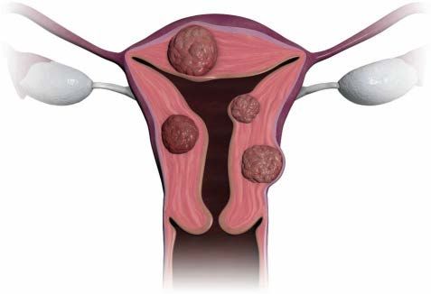
Content

     Executive Summary ............................................................................................................................................. 7
     Zusammenfassung ............................................................................................................................................. 10
1    Scope .................................................................................................................................................................... 15
     1.1      PICO question ............................................................................................................................................ 15
     1.2      Inclusion criteria ........................................................................................................................................ 15

2    Methods ............................................................................................................................................................... 17
     2.1      Research questions ..................................................................................................................................... 17
     2.2      Sources......................................................................................................................................................... 18
     2.3      Systematic literature search ....................................................................................................................... 19
     2.4      Flow chart of study selection ..................................................................................................................... 19
     2.5      Analysis ....................................................................................................................................................... 20
     2.6      Synthesis...................................................................................................................................................... 21

3    Description and technical characteristics of technology ............................................................................. 23
4    Health Problem and Current Use.................................................................................................................... 27
5    Clinical effectiveness ......................................................................................................................................... 35
     5.1      Outcomes ..................................................................................................................................................... 35
     5.2      Included studies.......................................................................................................................................... 37
     5.3      Results ......................................................................................................................................................... 40

6    Safety .................................................................................................................................................................... 45
     6.1      Outcomes ..................................................................................................................................................... 45
     6.2      Included Studies ......................................................................................................................................... 45
     6.3      Results ......................................................................................................................................................... 46

7    Quality of evidence ............................................................................................................................................ 49
8    Discussion ........................................................................................................................................................... 53
9    Recommendation ............................................................................................................................................... 59
10 References............................................................................................................................................................ 61
     Appendix ............................................................................................................................................................. 65
              Evidence tables of individual studies included for clinical effectiveness and safety ............................ 65
              Risk of bias tables and GRADE evidence profile .................................................................................... 77
              Applicability table ...................................................................................................................................... 84
              List of ongoing randomised controlled trials ........................................................................................... 84
              Literature search strategies ........................................................................................................................ 85
                    Search strategy for Cochrane .................................................................................................................... 85
                    Search strategy for CRD ........................................................................................................................... 85
                    Search strategy for Embase ....................................................................................................................... 86
                    Search strategy for Medline ...................................................................................................................... 87
                    Search strategy for ClinicalTrials.gov ..................................................................................................... 88
                    Search strategy for WHO-ICTRP ............................................................................................................ 88
                    Search strategy for EU Clinical Trials (EUdraCT) ............................................................................... 88

LBI-HTA | 2020                                                                                                                                                                   3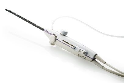
Intrauterine Ultrasound-Guided Transcervical Radiofrequency Ablation

List of Figures
Figure 2-1: Flow chart of study selection (PRISMA Flow Diagram) ................................................................... 20
Figure 3-1: Sonata® device: handpiece and console (images supplied by Gynesonics, Inc.) .............................. 23
Figure 4-1: Treatable fibroid types (image supplied by Gynesonics Inc.) ........................................................... 28
Figure 4-2: Management of uterine leiomyoma ..................................................................................................... 31

List of tables
Table 1-1:      Inclusion criteria ................................................................................................................................... 15
Table 3-1:      Requirements associated with comparators to TFA .......................................................................... 26
Table 4-1:      Classification of uterine fibroids ......................................................................................................... 27
Table 4-2:      Treatment reported by women with a diagnosis of uterine leiomyoma ........................................... 33
Table 5-1:      Comparison of eligibility criteria across studies of TFA ................................................................... 39
Table 5-2:      Percentage reduction in menstrual bleeding scores at 3, 6 and 12 months post TFA ..................... 41
Table 5-3:      Improvement in symptoms (UFS-QOL SSS score) at 3, 6 and 12 months post TFA ...................... 41
Table 7-1:      Summary of findings table of transcervical radiofrequency ablation ............................................... 51
Table 9-1:      Evidence based recommendations ....................................................................................................... 59
Table A-1: Transcervical radiofrequency ablation results from observational studies (part 1) ........................ 65
Table A-1: Transcervical radiofrequency ablation results from observational studies (part 2) ........................ 72
Table A-2: Risk of bias – study level (case series),
           using the Institute for Health Economics appraisal tool ................................................................... 77
Table A-3: Evidence profile: efficacy and safety of TFA ...................................................................................... 79
Table A-4: Summary table characterising the applicability of a body of studies ............................................... 84

List of abbreviations
AdHopHTA ............. Adopting Hospital Based Health Technology Assessment
AE ............................. Adverse event
CI .............................. Confidence interval
CRD ......................... Centre for Reviews and Dissemination
CTR .......................... Clinical trial registry
DARE ....................... Database of Abstracts of Reviews of Effects
DVT.......................... Deep vein thrombosis
EMAS ....................... European Menopause and Andropause Society
EU ............................ European Union
FDA.......................... Food and Drug Administration
FIGO ........................ International Federation of Gynecology and Obstetrics
GA ............................ General anaesthesia
GRADE .................... Grading of Recommendations Assessment, Development and Evaluation
HIFU ........................ High Intensity Focused Ultrasound ablation
ICD .......................... International Statistical Classification of Diseases
Lap-RFA .................. Laparoscopic radiofrequency volumetric thermal ablation
MIQ .......................... Menorrhagia Impact Questionnaire

4                                                                                                                                                 LBI-HTA | 2020
Content
MP ............................ Menstrual pictogram
MRgFUS .................. Magnetic resonance-guided focused ultrasound
MRI .......................... Magnetic resonance imaging
NA ............................ Not applicable
NHS-EED ................ National Health Service Economic Evaluation Database
NPV .......................... Non-perfused volume
NR ............................ Not reported
PBAC ....................... Pictorial blood loss assessment chart
POP .......................... Planned and Ongoing Projects
PRISMA ................... Preferred Reporting Items for Systematic Reviews and Meta-Analyses
PTS ........................... Patients
RCT .......................... Randomised Controlled Trial
RF ............................. Radiofrequency
RFA .......................... Radiofrequency ablation
RFVTA ..................... Radiofrequency volumetric thermal ablation
ROB .......................... Risk of bias
SD ............................. Standard deviation
SOGC ....................... Society of Obstetricians and Gynaecologists of Canada
TFA .......................... Transcervical fibroid ablation
UAE .......................... Uterine artery embolization
UAO ......................... Uterine artery occlusion
UFS-QOL-HRQL ... Uterine Fibroid Symptom and Health-Related Quality of Life Questionnaire
UFS-QOL-SSS ........ Uterine Fibroid Symptom and Health-Related Quality of Life Questionnaire
Symptom Severity Scale
USD.......................... United States Dollar
USgFUS ................... Ultrasound-guided focused ultrasound
USgHIFU ................ Ultrasound-guided high frequency focused ultrasound
UTI ........................... Urinary tract infection
VAS .......................... Visual analogue scale
WHO-ICTRP .......... World Health Organisation International Clinical Trials Registry Portal
LBI-HTA | 2020 5Executive Summary
Introduction
Health Problem
This systematic review is focused on women with symptomatic uterine fibro- uterine fibroids are
ids who wish to preserve their uterus. Uterine fibroids, also called myomas benign tumours that
or leimyomas, are benign tumours that can cause significant symptoms in- can cause significant
cluding pain, heavy menstrual bleeding and pelvic pressure. Definitive treat- symptoms including
ment is hysterectomy, however, less invasive uterine-preserving interventions pain, heavy menstrual
may be favoured by many women. bleeding and pelvic
pressure
Description of Technology
Intrauterine ultrasound-guided transcervical radiofrequency ablation (often TFA by RF energy
referred to as transcervical fibroid ablation [TFA]) is a method of delivering (high frequency energy)
radiofrequency (RF) energy to uterine leiomyomas to cause coagulative ne- is a uterus preserving
crosis of the tissue. There is one marketed TFA device, the Sonata® Sonog- method for the
raphy-Guided Transcervical Fibroid Ablation System (Gynesonics, Inc.). The treatment of myomas
technology aims to reduce symptoms through reducing the fibroid volume
without requiring a surgical incision. This intervention allows women to re-
tain their uterus.
Other uterus preserving interventions which aim to reduce the volume of optional interventions:
leiomyomas or remove them, include open, laparoscopic or hysteroscopic myomectomy, UAE,
myomectomy, uterine artery embolization (UAE), uterine artery occlusion UAO, MRgFUS,
(UAO), magnetic resonance-guided focused ultrasound (MRgFUS), ultra- USgFUS/USgHIFU,
sound-guided focused ultrasound (USgFUS/USgHIFU), and laparoscopic RFVTA
radiofrequency volumetric thermal ablation (laparoscopic RFVTA).
Methods
The research question was investigated through a systematic review of the question: is TFA by
current literature on TFA. The EUnetHTA Core Model® for Rapid Assess- RF energy vs. other
ment of Relative Effectiveness was the main source for selecting relevant as- interventions more
sessment elements. The question was whether TFA is more effective and safe effective and safer?
or equally effective, but safer with respect to the specified crucial outcomes
in the effectiveness and safety domain.
The search was executed in four biomedical databases (Medline, Embase, search in 4 databases;
the Cochrane Library, the University of York Centre for Reviews and Dis- 304 hits in total;
semination) on the 4th to 6th of December 2019. Overall, 303 citations were 225 hits after
identified from the database searches and one publication was identified deduplication
through handsearching (overall 304). After removing duplications, 225 cita-
tions were identified.
Ongoing and unpublished studies were searched in three clinical trials re- search in CTRs
gistries (CTRs: ClinicalTrials.gov; World Health Organisation Internation- for ongoing
al Clinical Trials Registry Portal [WHO-ICTRP]; EU Clinical Trials) on the trials, 20 hits
28th of January 2020 resulting in 20 potential hits (0 relevant). The manufac- (0 relevant);
turer from the most common product (Sonata®, Gynesonics, Inc.) was con- no new publications
tacted and provided 23 publications, but no new citations were identified. from the manufacturer
Study selection, data extraction, and quality appraisal was conducted by two
authors (RL, CS).
LBI-HTA | 2020 7Intrauterine Ultrasound-Guided Transcervical Radiofrequency Ablation
Domain effectiveness
effectiveness The outcomes used as evidence to derive a recommendation on the effec-
outcomes to derive a tiveness of TFA included reduction in symptom severity, reduction in men-
recommendation strual bleeding, rates of surgical reintervention, reduction in fibroid volume,
improvement in quality of life, patient satisfaction and fertility following the
procedure.
Domain safety
safety outcomes The outcomes used as evidence to derive a recommendation on the safety of
to derive a TFA included major adverse events and all adverse events.
recommendation
Results
Available evidence
no comparative studies; No comparative studies were identified by the search strategy and therefore
3 single-arm case series the evidence base evaluating TFA comprises of prospective single arm stud-
studies (FAST-EU, IDE ies. Some outcomes reported were analysed retrospectively. A total of three
and OPEN Trial) single-arm case series studies (FAST-EU, IDE and OPEN Trial) published
published in in seven publications [2-8] reporting on three unique cohorts (234 patients)
7 publications (234 Pts.) were identified for inclusion.
all 3 case-series: All three studies described above were sponsored by Gynesonics, Inc. Patients
sponsored, were followed up for a maximum of 12 months (FAST-EU Trial, IDE Trial)
max. FU ranged from and minimum of 6 weeks (OPEN Trial). Losses to follow-up were reported
6 weeks to 12 months in each of the studies. In the FAST-EU Trial after 12 months two patients
and 2 to 4 pts exited the study and two patients were lost-to follow up in the OPEN Trial.
Four patients missed the 12 months visit or were withdrawn before the 12
month visit in the IDE trial.
Clinical effectiveness
mean bleeding score re- Compared to pre-procedure measures TFA resulted in statistically signifi-
duction (improvement) cant reductions in menstrual bleeding three months after the procedure and
at 12 months: persisting at 12 months. Two studies reported this outcome and mean reduc-
53.8% (FAST-EU) and tions in bleeding scores at 12 months were 53.8% in the FAST-EU trial [3]
51.1% (IDE); and 51.1% in the IDE trial [2]. In both studies 64% of patients achieved at
64% of patients least a 50% reduction in bleeding from baseline at 12 months.
achieved a >50%
Statistically significant improvements in symptoms were also reported follow-
reduction in bleeding
ing TFA that were observed by three months post-procedure and remained
from baseline at
at 12 months. Two studies reported this outcome and mean improvement in
12 months
symptoms at 12 months were 35.3 [3] and 32.1 points [2] with respect to the
Uterine Fibroid Symptom and Health-Related Quality of Life Questionnaire
mean improvement in Symptom Severity Scale score (UFS-QOL SSS score). Improvement in health
symptoms at 12 months: related quality of life at 12 months was documented in two studies with a
UFS-QOL SSS, mean increase in Uterine Fibroid Symptom and Health-Related Quality of
35.3 (FAST-EU) and Life Questionnaire (UFS-QOL HRQOL) score of 45.7 and 43.7 points in the
32.1 (IDE) points; FAST-EU [3] and IDE [2] trial respectively. When measured using the EQ-
UFS-QOL HRQOL, 5D-3L health utility was statistically significantly improved for the cohort as
45.7 (FAST-EU) and a whole at 12 months compared to baseline. One study reported patient sat-
43.7 (IDE) points isfaction with 70.4% of patients being very satisfied [2].
8 LBI-HTA | 2020Executive Summary Reintervention was reported with a range of
Intrauterine Ultrasound-Guided Transcervical Radiofrequency Ablation

                               Zusammenfassung

                               Einleitung
                               Indikation und therapeutisches Ziel
    uterine Myome sind         Myome der Gebärmutter sind die häufigsten gutartigen Neubildungen im
  gutartige Tumore, die        weiblichen Becken und stellen auch die Hauptindikation für eine Gebärmut-
  Symptome wie starke          terentfernung (Hysterektomie) dar. Die vorliegende systematische Übersicht
Menstruationsblutungen,        befasst sich mit symptomatischen Gebärmuttermyomen: Uterusmyome, auch
  Schmerzen und Druck          Fibroide oder Leiomyoma genannt, sind gutartige Tumore, die erhebliche
      im Beckenbereich         Symptome wie Schmerzen, starke Menstruationsblutungen und Druck im Be-
    verursachen können         ckenbereich verursachen können. Eine Behandlungsoption ist die Hysterek-
                               tomie, jedoch bevorzugen viele Frauen weniger invasive, uteruserhaltende
                               Eingriffe.
      genaue Aussage über      Eine genaue Aussage über die Anzahl der betroffenen Frauen ist schwierig,
       die Prävalenz in der    da die Schätzungen über die Prävalenz in der publizierten Literatur eine gro-
           Zielbevölkerung     ße Bandbreite aufweisen (4,5 % bis 68,6 %) [9]. Diese Bandbreite ergibt sich
               ist schwierig   durch die unterschiedliche Methodik der untersuchten Population und der
                               Methode zur Diagnoseerhebung in den verschiedenen Studien. Zusätzlich
ca. 15-30 % der Myome          wird berichtet, dass die Myome bei 15-30 % der Frauen symptomatisch sind
können symptomatisch           [10], und es wird angenommen, dass die Symptome mit einer endometrialen
                    sein;      und vaskulären Dysfunktion assoziiert sind. Viele der Prävalenzdaten basie-
Symptome beeinflussen          ren auf Selbsteinschätzungen der Patientinnen zu Symptomen und/oder der
     das körperliche und       Diagnose und können daher unzuverlässig sein. Die Symptome können die
             emotionale        Arbeitsfähigkeit und die Ausübung von körperlichen und sozialen Aktivitä-
 Wohlbefinden negativ          ten negativ beeinflussen und zu Beeinträchtigung des körperlichen und emo-
                               tionalen Wohlbefindens führen [11].
         insgesamt 5.112       In Österreich belief sich die Anzahl der Krankenhausaufenthalte (stationär)
Krankenhausaufenthalte         von Frauen mit der Diagnose Leiomyom der Gebärmutter im Jahr 2018 auf
        mit der Diagnose       insgesamt 5.112 [12]. Davon wurden 537 (10,5 %) tagesklinisch behandelt.
 Leiomyom in AT, davon         Für die Altersgruppe 15 bis 44 Jahre beliefen sich die Krankenhausaufent-
 537 tagesklinisch (2018)      halte auf 2.035 Fälle, für die Altersgruppe 45 bis 64 Jahre auf 2.914 und für
                               die Altersgruppe 65+ wurden 163 Fälle dokumentiert. Die durchschnittliche
                               Aufenthaltsdauer betrug 4,4 Tage [12].

                               Beschreibung der Technologie
   TFA durch RF-Energie        Die intrauterine ultraschallgesteuerte transzervikale Radiofrequenzablation
 (Hochfrequenzenergie)         (oft als transzervikale Myomablation [TFA] bezeichnet) ist eine uteruserhal-
ist eine uteruserhaltende      tende Methode, bei der unter Verwendung von Radiofrequenzenergie (RF)
         Methode für die       eine koagulative Nekrose des Gewebes verursacht wird, um somit das Volu-
         Behandlung von        men von uterinen Leiomyomen minimal-invasiv zu reduzieren. In der Fach-
                Myomen         literatur wird TFA als ein ambulanter Eingriff beschrieben, der von einer/m
                               Gynäkologen/in entweder unter Vollnarkose, Sedierung unter Bewusstsein
                               oder unter regionaler Anästhesie durchgeführt wird. Der Eingriff kann von
                               ÄrztInnen, die eine entsprechende Ausbildung absolviert haben, durchgeführt
                               werden [2].

 10                                                                                               LBI-HTA | 2020
Zusammenfassung Ein Gerät zur ultraschallgesteuerten transzervikalen Myomablation mit dem Sonata®-System hat Namen Sonata® wird von dem Hersteller Gynesonics, Inc. vertrieben. Das seit Februar 2019 eine System hat seit Februar 2019 eine CE-Kennzeichnung [8] und wurde 2018 CE-Kennzeichnung und von der Food and Drug Administration (FDA) für die "diagnostische intra- ist seit 2018 durch die uterine Bildgebung und transzervikale Behandlung von symptomatischen FDA zugelassen Uterusmyomen, einschließlich solcher, die mit schweren Menstruationsblu- (FDA Cleared) tungen einhergehen" [13], zugelassen (FDA Cleared). Das Sonata®-System besteht aus einem RF-Generator (Signalgenerator), ei- Komponenten: nem Ultraschallsystem mit Führungssoftware und einem Behandlungsgerät RF-Generator, (d. h. ein Handstück zur Radiofrequenzablation [RFA] kombiniert mit einer Ultraschallsystem Ultraschallsonde). Das RFA-Handstück ist für den einmaligen Gebrauch be- inkl. Software, stimmt, weshalb für jeden Eingriff ein neues Handstück erforderlich ist [2, 3]. Behandlungsgerät Die Technologie zielt darauf ab, die Symptome durch eine Verringerung des optionale Eingriffe: Myomvolumens zu reduzieren, ohne dass eine chirurgische Inzision erfor- Myomektomie, VAE, derlich ist. Dieser Eingriff ermöglicht es, Frauen, ihre Gebärmutter zu erhal- VAO, MRgFUS, ten. Optionale uteruserhaltende Eingriffe, die darauf abzielen das Volumen USgFUS/USgHIFU, von Leiomyomen zu reduzieren, sind die Myomektomie, die Embolisation der RFVTA Gebärmutterarterie (UAE), der Verschluss der Gebärmutterarterie (UAO), der magnetresonanzgesteuerte fokussierte Ultraschall (MRgFUS), der ultraschall- gesteuerte fokussierte Ultraschall (USgFUS/USgHIFU) und die laparosko- pische volumetrische Thermoablation mit Radiofrequenz (laparoskopische RFVTA). Methoden Das Ziel dieser systematischen Übersichtsarbeit war es, die Frage zu beant- Forschungsfrage: worten, ob TFA wirksamer und sicherer oder ebenso wirksam, aber sicherer Ist die TFA durch RF- in Bezug auf Patientinnen-relevante Endpunkte ist. Die Forschungsfrage Energie im Vergleich zu wurde durch eine systematische Auswertung der rezenten Literatur zu TFA anderen Interventionen untersucht. Dabei wurde das EUnetHTA-Core-Modell® für Rapid Assess- wirksamer und sicherer? ment of Relative Effectiveness herangezogen. Die systematische Suche wurde vom 4. bis 6. Dezember 2019 in vier Daten- Suche in banken (Medline via Ovid, Embase, The Cochrane Library, CRD [DARE, 4 Datenbanken; NHS-EED, HTA]) durchgeführt. Die Literatursuche beschränkte sich nicht insgesamt 304 Treffer; auf ein Publikationsjahr oder Studiendesign, jedoch gab es sprachliche Ein- 225 Treffer nach schränkungen auf englische und deutsche Artikel. Insgesamt wurden 303 Zi- Deduplizierung tate aus den Datenbankrecherchen und eine Publikation durch Handsuche identifiziert (insgesamt 304). Nach der Deduplikation wurden 225 Zitate identifiziert. Drei klinische Studienregister (ClinicalTrials.gov; World Health Organisa- Suche in CTRs tion International Clinical Trials Registry Portal [WHO-ICTRP]; EU Clini- nach laufenden Studien, cal Trials) wurden am 28. Januar 2020 nach laufenden oder unveröffentlich- 20 Treffer (0 relevant); ten Studien durchsucht. Die Suche ergab 20 potenzielle Treffer von denen 0 keine neuen Studien als relevant eingestuft wurden. Der Hersteller des gängigsten Produkts (So- durch den Hersteller nata®, Gynesonics, Inc.) wurde kontaktiert und stellte 23 Publikationen zur Verfügung, wobei keine neuen Publikationen identifiziert werden konnten. Die Studienauswahl, Datenextraktion und Qualitätsbewertung wurde von zwei AutorInnen (RL, CS) durchgeführt. LBI-HTA | 2020 11
Intrauterine Ultrasound-Guided Transcervical Radiofrequency Ablation

                               Klinische Wirksamkeit
        Endpunkte für          Zur Ableitung einer Empfehlung zur Wirksamkeit von TFA wurden folgen-
Empfehlung hinsichtlich        de Endpunkte herangezogen: die Verringerung des Schweregrads der Symp-
      der Wirksamkeit          tome, die Verringerung der Menstruationsblutungen, die Anzahl der chirur-
                               gischen Reinterventionen, die Verringerung des Myomvolumens, die Verbes-
                               serung der Lebensqualität, die Zufriedenheit der Patientinnen und die Fer-
                               tilität nach dem Eingriff.

                               Sicherheit
        Endpunkte für          Die Endpunkte für die Ableitung einer Empfehlung zur Sicherheit von TFA
Empfehlung hinsichtlich        umfassten schwerwiegende bzw. wesentliche unerwünschte Ereignisse (SAE)
         der Sicherheit        und alle unerwünschten Ereignisse bzw. Nebenwirkungen (AE).

                               Ergebnisse
                               Verfügbare Evidenz
   keine vergleichenden        Im Rahmen der Suchstrategie wurden keine vergleichenden Studien iden-
                 Studien;      tifiziert, und daher besteht die Evidenzbasis zur Bewertung der TFA aus
    3 ein-armige Studien       prospektiven einarmigen Studien. Insgesamt wurden drei einarmige Studien
    (FAST-EU-, IDE- und        (FAST-EU-, IDE- und OPEN-Trial), die in sieben Publikationen [2-8] veröf-
          OPEN-Trial), in      fentlicht wurden und über drei Kohorten (234 Patienten) berichteten, iden-
7 Publikationen (234 Pts.)     tifiziert.
      alle 3 Studien wurden    Alle drei oben beschriebenen Studien wurden durch den Hersteller Gyneso-
                  gesponsert   nics, Inc. finanziert. Die Follow-Up-Zeiten der Patientinnen beliefen sich auf
                               maximal 12 Monate (FAST-EU-Studie, IDE-Trial) und 6 Wochen (OPEN-
          min./max. FU         Trial). In jeder der Studien wurde über das Loss-to-Follow-Up berichtet. In
6 Wochen bis 12 Monate         der FAST-EU-Studie beendeten zwei Patientinnen nach 12 Monaten die Stu-
  Loss-to-FU 2 bis 4 Pts.      die und zwei Patientinnen gingen zur Nachbeobachtung verloren (OPEN-
                               Trial). Vier Patientinnen verpassten den 12-monatigen Besuch oder schieden
                               vor dem 12-monatigen Besuch aus (IDE-Trial).

                               Klinische Wirksamkeit
          Reduktion der        Zwei Studien berichteten über den Endpunkt Reduktion der Menstruations-
        Blutungen nach         blutungen. Es kam im Vergleich zu den Ausgangswerten vor der Behandlung
           12 Monaten:         durch die Intervention zu einer statistisch signifikanten Verringerung nach
  53,8 % (FAST-EU) und         drei Monaten, die auch nach 12 Monaten noch beobachtet werden konnte.
           51,1 % (IDE);       Die durchschnittliche Verringerung der Blutungswerte nach 12 Monaten be-
         64 % Pts. nach        trug 53,8 % in der FAST-EU-Studie [3] und 51,1 % in der IDE-Studie [2].
 12 Monaten: >50 %-ige         In beiden Studien erreichten 64 % der Patientinnen nach 12 Monaten eine
Reduktion der Blutungen        mindestens 50 %-ige Reduktion der Blutungen gegenüber dem Ausgangs-
                               wert.
         Verbesserung der      Zwei Studien berichteten über die Veränderung im Schweregrad der Symp-
                Symptome       tome. Es wurden statistisch signifikante Verbesserungen nach drei und 12
         nach 12 Monaten:      Monaten beobachtet. Die durchschnittliche Verbesserung der Symptome wur-
             UFS-QOL SSS,      den anhand des validierten UFS-QOL-SSS-Fragebogens (Uterine Fibroid
       35,3 (FAST-EU) und      Symptom and Quality of Life Symptom Severity Score) erhoben und betru-
         32,1 (IDE) Punkte     gen nach 12 Monaten 35,3 (FAST-EU [3]) bzw. 32,1 Punkte (IDE [2]).

 12                                                                                               LBI-HTA | 2020
Zusammenfassung Zwei Studien berichteten die gesundheitsbezogene Lebensqualität: Es wurde Verbesserung nach 12 Monaten ein mittlerer Anstieg des Scores von 45,7 bzw. 43,7 Punk- der HRQOL nach ten in der FAST-EU [3] und IDE [2] Studie dokumentiert. Hierbei wurde 12 Monaten: der Fragebogen zu den uterinen Fibroid-Symptomen und der gesundheits- UFS-QOL, 45,7 bezogenen Lebensqualität (UFS-QOL HRQOL) herangezogen. Auch bei der (FAST-EU) und Messung mit dem EQ-5D-3L hat sich der Nutzen hinsichtlich der Gesundheit 43,7 (IDE) Punkte für die gesamte Kohorte nach 12 Monaten im Vergleich zum Ausgangswert statistisch signifikant verbessert. Eine Studie berichtete über die Patientin- nenzufriedenheit, wobei 70,4% der Patientinnen sehr zufrieden waren [2]. Zwei Studien berichteten über die Reinterventionsrate. Sie bewegte sich in Reinterventionsrate im einer Bandbreite von 12 Monate) typischerweise bei weniger als 10 % der Patientinnen auf). Über die langfristige Sicherheit von TFA (über 12 Monate hinaus) wurde in keiner prospektiven Studie berichtet. Laufende Studien Es wurden keine laufenden relevanten randomisierten kontrollierten Studien es wurden keine (RCTs) zur Bewertung der Sicherheit oder Wirksamkeit von TFA identifi- laufenden relevanten ziert. Studien identifiziert Kostenerstattung TFA wird in Österreich derzeit nicht für die Behandlung von symptomati- bisher keine schen Gebärmuttermyomen erstattet. Kostenerstattung in AT LBI-HTA | 2020 13
Intrauterine Ultrasound-Guided Transcervical Radiofrequency Ablation

                            Diskussion
      Risk of Bias (RoB):   Von den eingeschlossenen sieben Publikationen wurden drei mit einem mitt-
     3 Publikationen mit    leren Biasrisiko [2-4] und vier mit einem hohen Biasrisiko [5-8] bewertet.
        mittlerem Risiko,   Die Evidenzstärke (Qualität der Evidenz) für die Wirksamkeit und Sicher-
                            heit wurde für alle relevanten Endpunkte als niedrig oder sehr niedrig ein-
     4 Publikationen mit    gestuft. Zum einen war dies auf die inhärenten Einschränkungen von einar-
        hohem Biasrisiko    migen Studien zurückzuführen, d. h. auf das Versäumnis, Kointerventionen/
                            Störfaktoren zu beschreiben, und zum anderen auf die Verwendung von un-
                            verblindeten, von Patientinnen berichteten Endpunkten.
         Evidenzstärke      Obwohl die Stärke der Evidenz niedrig bis sehr niedrig war, berichteten die
 (Qualität der Evidenz)     Studien durchwegs über eine Verbesserung der Symptome und eine Verrin-
        ist sehr niedrig    gerung der Menstruationsblutungen nach der Intervention. Frauen mit symp-
             bis niedrig    tomatischen Uterusmyomen haben jedoch derzeit Zugang zu uteruserhalten-
                            den Alternativen oder Hysterektomie, die ebenfalls die Symptome und die
                            Menstruationsblutungen reduzieren.
                            Vergleichende Ergebnisse zur Wirksamkeit und Sicherheit der intrauterinen
                            ultraschallgesteuerten transzervikalen Radiofrequenzablation (TFA) bei symp-
                            tomatischen Myomen zu anderen optionalen Interventionen liegen nicht vor.

                            Empfehlung
       Aufnahme in den      Die derzeitige Evidenz reicht nicht aus, um zu zeigen, dass die intrauterine
  Leistungskatalog wird     ultraschallgesteuerte transzervikale Radiofrequenzablation (TFA) wirksamer
derzeit nicht empfohlen     und ebenso sicher oder genauso wirksam ist, aber sicherer als optionale ute-
                            ruserhaltende Interventionen bei symptomatischen Myomen. Daher wird die
                            Aufnahme in den Leistungskatalog derzeit nicht empfohlen.

14                                                                                             LBI-HTA | 2020
1 Scope
1.1 PICO question
In patients with symptomatic uterine leiomyoma, is intrauterine ultrasound- PIKO-Frage
guided transcervical radiofrequency ablation more effective and safe than al-
ternative uterine-preserving interventions, concerning reduction in leiomyo-
ma volume, menstrual blood loss, quality of life, need for surgical reinterven-
tion, fertility outcomes or adverse events?
1.2 Inclusion criteria
Inclusion criteria for relevant studies are summarized in Table 1-1. Einschlusskriterien
für relevante Studien
Table 1-1: Inclusion criteria
Population Women with symptomatic uterine leiomyomas
International classification of diseases (ICD)-10-CM code: D25 Leiomyoma of uterus
Contraindications/exclusions: Active pelvic infection; cervical dysplasia; endometrial
hyperplasia; uterine malignancy.
MeSH Terms: Leiomyoma [C04.557.450.590.450], Myoma [C04.557.450.590.540]
Rationale: Recent international guidelines on the place in therapy of transcervical
radiofrequency ablation of leiomyomas is not available.Therefore, the population has been
defined based on the inclusion criteria for patients treated in clinical trials [3-8, 14].
Intervention Intrauterine ultrasound-guided transcervical radiofrequency ablation of uterine leiomyoma
Product names: Sonata®, VizAblate® (previous version of the Sonata® system)
MeSH Term: Radiofrequency Ablation [E02.808.750], Ultrasonography [E01.370.350.850]
The radiofrequency ablation device the Acessa™ was excluded as it requires laparoscopic
ultrasound guidance
Control Uterine-preserving interventions for uterine leiomyoma including, but not limited to:
myomectomy, uterine artery embolization (UAE), uterine artery occlusion (UAO), magnetic
resonance-guided focused ultrasound (MRgFU), and radiofrequency volumentric thermal
ablation (RFVTA)
Rationale: Hysterectomy is a definitive procedure for relief of symptoms and prevention of
recurrent leiomyoma-related problems [15]; however, many women would prefer a less
invasive treatment and/or wish to preserve their uterus. Comparators to the intervention
are presumed to include all alternative uterine-preserving options.
Outcomes
Efficacy Clinical endpoints include:
Clincial endpoints (Crucial):
Reduction in menstrual blood loss (pre vs post procedure), measured by but not limited to:
Menorrhagia Impact Questionnaire (MIQ)
Menstrual pictogram (MP) scores
Pictorial blood loss assessment chart (PBAC)
Self report (e.g. symptom free, better, worse)
Quantitative measures
Improvement in quality of life measures (surrogate measure of pelvic pain and/or bulk
related symptoms) including but not limited to the Uterine Fibroid Symptom-Quality of
Life (UFS-QOL) questionnaire including the Symptom Severity Score (SSS) subscale
Rates of surgical reintervention (measure of treatment failure) at any time point
LBI-HTA | 2020 15Intrauterine Ultrasound-Guided Transcervical Radiofrequency Ablation
Efficacy Surrogate endpoints (Important):
(continuation) Reduction in leiomyoma volume
Recurrence of uterine leiomyoma at any time point
Fertility outcomes following the procedure
Pregnancy
Outcome of pregnancy (live birth, miscarriage, complications)
Rationale: Appropriate clinical outcomes have been informed by clinical studies using the
Sonata® device [3, 5, 14] as well as a systematic review protocol addressing interventions
for uterine leiomyoma [16].
Safety All adverse events reported, including but not limited to:
Mortality
Abnormal uterine bleeding
Pain
Urinary tract infection
Other infection
Re-admission
Rationale: Appropriate clinical outcomes have been informed by clinical studies
using the Sonata® device [3, 5, 14] and the EUnetHTA guidelines [17].
Study design
Efficacy Systematic reviews and meta-analyses
Randomised controlled trials
Prospective non-randomised controlled trials
In the absence of comparative evidence, prospective case series
with ≥ 10 participants will be included
Excluded: conference abstracts, narrative reviews, letter to the editor, author response,
case reports.
Safety Systematic reviews and meta-analyses
Randomised controlled trials
Prospective non-randomised controlled trials
Prospective case-series with ≥ 10 participants
Excluded: conference abstracts, narrative reviews, letter to the editor, author response,
case reports, retrospective case series.
Abbreviations: ICD – International Statistical Classification of Diseases; MIQ – Menorrhagia Impact Questionnaire;
MP – Menstrual pictogram; MRgFU – Magnetic resonance-guided focused ultrasound; PBAC – Pictorial Blood Loss
Assessment Chart; RFVTA – Radiofrequency volumetric thermal ablation; SSS – Symptom Severity Score;
UAE – Uterine artery embolization; UAO – Uterine artery occlusion; UFS-QOL – Uterine Fibroid Symptom-Quality
of Life; VAS – Visual analogue scale.
16 LBI-HTA | 20202 Methods
2.1 Research questions
Description of the technology
Element ID Research question
B0001 What is intrauterine ultrasound-guided transcervical radiofrequency ablation
and the comparator(s)?
A0020 For which indications has intrauterine ultrasound-guided transcervical radiofrequency
ablation received marketing authorisation or CE marking?
B0002 What is the claimed benefit of the technology in relation to the comparators?
B0003 What is the phase of development and implementation of intrauterine ultrasound-guided
transcervical radiofrequency ablation and the comparator(s)?
B0004 Who administers intrauterine ultrasound-guided transcervical radiofrequency ablation
and the comparators and in what context and level of care are they provided?
B0008 What kind of special premises are needed to use intrauterine ultrasound-guided transcervical
radiofrequency ablation and the comparator(s)?
B0009 What supplies are needed to use intrauterine ultrasound-guided transcervical radiofrequency
ablation and the comparator(s)?
A0021 What is the reimbursement status of intrauterine ultrasound-guided transcervical
radiofrequency ablation?
Health problem and Current Use
Element ID Research question
A0001 For which health conditions, and for what purposes is intrauterine ultrasound-guided
transcervical radiofrequency ablation used?
A0002 What is the disease or health condition in the scope of this assessment?
A0003 What are the known risk factors for the disease or health condition?
A0004 What is the natural course of the disease or health condition?
A0005 What is the burden of disease for the patients with the disease or health condition?
A0006 What are the consequences of the disease or health condition for the society?
A0024 How is the disease or health condition currently diagnosed according to published guidelines
and in practice?
A0025 How is the disease or health condition currently managed according to published guidelines
and in practice?
A0007 What is the target population in this assessment?
A0023 How many people belong to the target population?
A0011 How much are the technologies utilised?
Clinical Effectiveness
Element ID Research question
D0005 How does intrauterine ultrasound-guided transcervical radiofrequency ablation affect
symptoms and outcomes of the disease or health condition?
D0006 How does intrauterine ultrasound-guided transcervical radiofrequency ablation affect
progression (or recurrence) of the disease or health condition?
D0011 What is the effect of intrauterine ultrasound-guided transcervical radiofrequency ablation
on fertility?
LBI-HTA | 2020 17Intrauterine Ultrasound-Guided Transcervical Radiofrequency Ablation
Clinical Effectiveness
Element ID Research question
D0012 What is the effect of intrauterine ultrasound-guided transcervical radiofrequency ablation
on generic health-related quality of life?
D0013 What is the effect of intrauterine ultrasound-guided transcervical radiofrequency ablation
on disease-specific quality of life?
D0017 Was the use of intrauterine ultrasound-guided transcervical radiofrequency ablation
worthwhile?
Safety
Element ID Research question
C0008 How safe is intrauterine ultrasound-guided transcervical radiofrequency ablation
in comparison to the comparator(s)?
C0002 Are the harms related to dosage or frequency of applying intrauterine ultrasound-guided
transcervical radiofrequency ablation?
C0004 How does the frequency or severity of harms change over time or in different settings?
C0005 What are the susceptible patient groups that are more likely to be harmed through the use
of intrauterine ultrasound-guided transcervical radiofrequency ablation?
C0007 Are intrauterine ultrasound-guided transcervical radiofrequency ablation and comparator(s)
associated with user-dependent harms?
B0010 What kind of data/records and/or registry is needed to monitor the use of intrauterine
ultrasound-guided transcervical radiofrequency ablation and the comparator?
2.2 Sources
verschiedene Quellen A range of sources were used to identify relevant literature to answer the re-
herangezogen: search questions relating to the description of the technology, health prob-
lem and current use, including:
Description of the technology
systematische Suche, Handsearch in the Planned and Ongoing Projects (POP), Adopting
Handsuche Hospital Based Health Technology Assessment (AdHopHTA) and
sowie Centre for Reviews and Dissemination (CRD) databases for Health
Informationen des Technology Assessments
einreichenden Background publications identified in database search: see Section 2.3
Krankenhauses und
des Herstellers
Documentation provided by the manufacturers
Questionnaire completed by the submitting hospitals
Health problem and Current Use
Handsearch in the POP, AdHopHTA and CRD databases for Health
Technology Assessments
Background publications identified in database search: see Section 2.3
Documentation provided by the manufacturers
Questionnaire completed by the submitting hospitals
18 LBI-HTA | 2020Methods
2.3 Systematic literature search
The systematic literature search was conducted on the systematische
4th to 6th of December 2019 in the following databases: Literatursuche in
Medline via Ovid 4 Datenbanken
Embase
The Cochrane Library
CRD (Database of Abstracts of Reviews of Effects [DARE], National
Health Service Economic Evaluation Database [NHS-EED], HTA)
The systematic search was not limited by publication year or to study de-
sign. Only articles published in English or German were eligible for inclu-
sion. The specific search strategy employed can be found in the Appendix.
Furthermore, to identify ongoing and unpublished studies, a search in three Suche nach laufenden
clinical trials registries (ClinicalTrials.gov; World Health Organisation Inter- Studien in 3 klinischen
national Clinical Trials Registry Portal [WHO-ICTRP]; EU Clinical Trials) Studienregistern und
was conducted on the 28th of January 2020. Searches of the clinical trial reg- Nachfrage beim
istries identified 20 potential relevant hits. However, no relevant ongoing ran- Hersteller
domised controlled trials (RCTs) evaluating the safety or effectiveness of TFA
were identified. The manufacturer from the most common product (Sonata®,
Gynesonics, Inc.) was contacted. They submitted 23 publications and no new
citations were identified.
2.4 Flow chart of study selection
The search results were screened by two independent researchers (RL, CS), Literaturauswahl
and in case of disagreement a third researcher was involved to resolve the
differences. The selection process is displayed in Figure 2-1.
Overall, 303 citations were identified from the database searches and one pub- insgesamt wurden
lication was identified through handsearching (overall 304). After removing 304 Publikationen
duplications, 225 citations were identified. identifiziert
One recent systematic review [18] that aimed to assess the clinical perfor- rezente systematische
mance of radiofrequency ablation of uterine fibroids was identified. This re- Übersichtsarbeit wurde
view included literature on transcervical radiofrequency ablation (TFA) but ausgeschlossen wegen
was excluded from this report because the efficacy analysis had significant methodischen Mängeln
methodological flaws (i.e., pooling across different study designs) and safety
outcomes were not reported.
A total of seven publications reporting on three case series studies were iden- 7 Publikationen mit
tified for inclusion. Multiple studies reporting on the same patient cohort 3 distinkten Kohorten
were included only if they contributed new information. wurden eingeschlossen
LBI-HTA | 2020 19Intrauterine Ultrasound-Guided Transcervical Radiofrequency Ablation
Records identified through Additional records identified
Identification
database searching through other sources
(n=303) (n=1)
Records after duplicates
removed
(n=225)
Screening
Records screened
(n=225)
Records excluded
(n=182)
Full-text articles Full-text articles excluded,
Eligibility
assessed for eligibility with reasons
(n=43) (n=36)
Other study design (n=20)
Other intervention (n=10)
Wrong population* (n=1)
Background literature (n=1)
Not English/German (n=1)
Studies included in Poor quality systematic review
Included
qualitative synthesis (n=1)
(n=7) Not a comprehensive review (n=1)
Case series** (n=3) Duplicate patient population (n=1)
* This study was identified by handsearching; ** Published in 7 Publications
Figure 2-1: Flow chart of study selection (PRISMA Flow Diagram)
2.5 Analysis
Datenextraktion und Relevant data from the included studies were systematically extracted into
Validierung data extraction tables by one reviewer (RL) (See Appendix Table A-1) based
on study design and research question. The extracted data tables were vali-
dated for accuracy by a second reviewer (CS). Due to the paucity of available
evidence and limited study designs, the safety and effectiveness results are
reported narratively.
Bewertung des Two independent researchers (RL, CS) conducted quality appraisal, including
Bias-Risikos gemäß risk of bias assessment, with differences settled via consensus. Quality ap-
IHE-Checkliste praisal was conducted using the Institute of Health Economics (IHE) check-
list for single arm studies [19].
20 LBI-HTA | 2020Methods 2.6 Synthesis The research questions based on the EUnetHTA Core Model® for Rapid As- Evidenzsynthese sessment of Relative Effectiveness were answered in plain text format with mittels GRADE reference to GRADE (Grading of Recommendations Assessment, Develop- ment and Evaluation) evidence tables that are included in the Appendix [20]. The GRADE results are summarised in Table 7-1. No quantitative synthesis for any outcome could be performed due to the limitations of the primary ev- idence. The outcomes included in the GRADE summary of findings table (Table 7-1) wesentliche include the outcomes designated as crucial for decision-making, and repre- Endpunkte zur sent outcomes that are most relevant to patients and decision makers. Sever- Entscheidungsfindung al of these outcomes were reported at 3, 6 and 12-month time points. For ease wurden in of interpretation, only 12-month outcomes are included in the GRADE tables. Tabellen dargestellt This was chosen as the most informative time point as 3-month outcomes do not inform an assessment of effect durability and 6-month outcomes were in- consistently reported. The 3 and 6-month results (if available) are presented in Table A-1 and are discussed narratively in the results section. The rating (quality) associated with the 12-month results represented in the GRADE tables is directly ap- plicable to the 3 and 6-month results for the respective outcomes. LBI-HTA | 2020 21
3 Description and technical
characteristics of technology
Features of the technology and comparators
B0001 – What is intrauterine ultrasound-guided transcervical
radiofrequency ablation and the comparator(s)?
Intrauterine ultrasound-guided transcervical radiofrequency ablation
Intrauterine ultrasound-guided transcervical radiofrequency ablation, referred TFA mittels
to hereafter as transcervical fibroid ablation (TFA) is a method of delivering Radiofrequenz:
radiofrequency (RF) energy to uterine leiomyomas, also called myomas or uteruserhaltende
fibroids1, to cause coagulative necrosis of the tissue. The technology aims to Methode zur
reduce symptoms through reductions in fibroid volume without requiring Behandlung von
surgical incisions. This intervention allows women to retain their uterus. Uterusmyomen
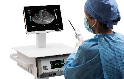
There is one marketed TFA device, the Sonata® Sonography-Guided Trans- Sonata®-TFA-System
cervical Fibroid Ablation System (Gynesonics, Inc.). This device was previ- umfasst ein Handgerät
ously marketed as the VizAblate® System. The system is comprised of a sin- mit einer
gle-use RFA handpiece attached to a reusable intrauterine ultrasound probe. Ultraschallsonde
The ultrasound probe provides imaging that can be used for guidance during
the procedure and for diagnostic purposes (Figure 3-1). The system also in-
cludes software which is used for targeting and planning of ablation zones [4].
The system delivers up to 150 W of RF energy for a duration of 2-7 minutes
as dictated by the required ablation size. The probe is capable of producing
an ablation zone of 4.0 cm x 5.0 cm [4]. Multiple ablations per leiomyoma may
be required.
Figure 3-1: Sonata® device: handpiece and console (images supplied by Gynesonics, Inc.)
Comparators
There are many treatment options for symptomatic uterine leiomyoma that Behandlungsoption ist
include pharmacological, ablative and minimally invasive therapies, and sur- abhängig von mehreren
gical therapies. The choice of treatment is guided by patient preference, re- Faktoren
productive intentions and history, symptom burden, and the number, size and
location of leiomyoma [21].
1 Note that the terms leiomyoma, myoma and fibroid are used interchangeably within
this report.
LBI-HTA | 2020 23Intrauterine Ultrasound-Guided Transcervical Radiofrequency Ablation
(uteruserhaltende) The definitive treatment for uterine leiomyoma is hysterectomy. TFA of leio-
Interventionen bei myoma is intended to provide a uterine-preserving alternative to hysterecto-
einem symptomatischen my for women who wish to preserve their uterus. Other interventions which
Uterusmyom: aim to remove or reduce the volume of leiomyomas, whilst preserving the
uterus, include:
Myomektomie Myomectomy: surgical removal of leiomyomas and can be performed
via an open, laparoscopic, hysteroscopic or vaginal approach. Robot
assisted myomectomy has also been described [21].
Uterusmyom Uterine artery embolisation (UAE): occlusion of vessels supplying fibro-
embolisation (UAE) ids using microspheres or polyvinyl alcohol particles. The procedure
is percutaneous with access via the femoral artery [22].
Verschluss der Uterine artery occlusion (UAO or L [laparoscopic] UAO): a laparoscopic
Gebärmutterarterie surgical procedure in which the uterine arteries are occluded using
(UAO) surgical techniques (e.g., ligation, coagulation etc.) [22].
Magnetresonanz- Magnetic resonance-guided focused ultrasound (MRgFUS): the non-inva-
gesteuerter fokussierter sive use of ultrasound energy to cause thermal destruction of tissue.
Ultraschall (MRgFUS) Patients undergo the procedure in a magnetic resonance imaging (MRI)
Ultraschall-gesteuerter machine, with ultrasound delivered via an external transducer [23].
fokussierter Ultraschall Ultrasound-guided high intensity focused ultrasound ablation (USgFUS/
(USgFUS/USgHIFU) USgHIFU): the non-invasive use of ultrasound energy to cause ther-
laparoskopische mal destruction of tissue under ultrasound guidance [24].
volumetrische Radiofrequency volumetric thermal ablation (Lap-RFA or RFVTA): radio-
Thermoablation mit frequency ablation of fibroids under ultrasound guidance performed
Radiofrequenz using a laparoscopic approach [21].
(laparoskopische
RFVTA) A0020 – For which indications has transcervical radiofrequency
ablation received marketing authorisation or CE marking?
Sonata®-TFA-System The Sonata® Sonography-Guided Transcervical Fibroid Ablation System was
durch FDA zugelassen developed to provide a uterus-conserving, transcervical (i.e., incisionless) treat-
(2018) und ment for International Federation of Gynecology and Obstetrics (FIGO) uter-
CE-Zertifizierung (2019) ine myoma types 1,2, 3, 4, 5, 6 and 2–5 [8]. Types 1 and 2 refer to myomas
located in the lining of the uterine cavity whilst type 3,4 and 5 refer to myo-
mas located within the uterine wall [25]. The system is CE Marked [8] since
February 2019 and was approved (FDA cleared) by the Food and Drug Ad-
ministration (FDA) in 2018 for “diagnostic intrauterine imaging and trans-
cervical treatment of symptomatic uterine fibroids, including those associat-
ed with heavy menstrual bleeding” [13].
B0002 – What is the claimed benefit of transcervical
radiofrequency ablation in relation to the comparators?
TFA ist als weitere TFA is purported to reduce leiomyoma volume and associated symptoms in
minimalinvasive und women with uterine leiomyoma whilst avoiding or delaying hysterectomy [18].
inzisionsfreie Compared to other volume-reducing interventions, with the exception of hys-
Therapiemöglichkeit teroscopic myomectomy, MRgFUS, and USgFUS/USgHIFU the technology
bei gleichzeitigem may be considered less invasive as it requires no incisions. Consequently the
Erhalt des Uterus technology may be associated with a reduced rate of complications (specifi-
und reduzierter cally infection, seroma, haematoma, adhesions) relative to open or laparoscop-
Komplikationsrate ic interventions [3]. Further, it has been claimed that the procedure requires
beabsichtigt a smaller volume of hypotonic fluid (used for acoustic coupling) and lower in-
trauterine pressures than hysteroscopic procedures, and may therefore have
a reduced risk of fluid intravasion and consequent fluid overload [3].
24 LBI-HTA | 2020You can also read