Intravascular ultrasound, performed during resuscitative endovascular balloon occlusion of the aorta (REBOA), confirms correct balloon deployment ...
←
→
Page content transcription
If your browser does not render page correctly, please read the page content below
12 Journal of the Royal Naval Medical Service 2018; 104(1)
Intravascular ultrasound, performed during
resuscitative endovascular balloon occlusion of the
aorta (REBOA), confirms correct balloon deployment
and haemostasis - a potential solution for remote,
austere and military settings
P S C Rees, A M Buckley, S A Watts, E Kirkman
Abstract
Introduction
Resuscitative endovascular balloon occlusion of the aorta (REBOA) is rapidly evolving as an emergency haemorrhage control
technique. It has wide potential applicability in remote and austere settings, and following military trauma where prolonged
field care might be required. However, rapid confirmation of balloon delivery is a challenge which relies on estimates derived
from anatomical measurements or trans-abdominal ultrasound. In addition, confirmation of adequate balloon expansion is
difficult. Intravascular ultrasound (IVUS) offers a solution to these two issues, making REBOA a deliverable therapy in the
pre-hospital and early hospital settings.
Methods
In an animal model of severe ballistic trauma, following characterisation of the technique, an IVUS-REBOA device was con-
figured, combining a peripheral angioplasty balloon and a digital coronary IVUS catheter. This was introduced via a sheath into
the femoral vessel over a conventional angioplasty guide wire.
Results
Real time IVUS imaging allowed confirmation of delivery of the balloon to the aorta, and also demonstrated full apposition
once deployed. Furthermore, using ChromaFlo imaging, the device confirmed loss of pulsatile flow in the aorta after deploy-
ment, correlating with loss of transduced femoral pressure traces. Post-mortem examination confirmed correct anatomical
balloon placement.
Summary
For the first time, in a porcine pilot study, we have demonstrated that IVUS-REBOA is feasible and confirms both correct bal-
loon placement and haemostasis. It has potential to offer advantages to REBOA operators especially during the pre-hospital and
retrieval phases, and in the early phase of hospital delivered damage control resuscitation at remote locations.
Rees P S C, Buckley A M, Watts S A, et al. J R Nav Med Serv 2018;104(1):12–17
Introduction easily be remedied by early medical intervention with pres-
sure dressings, combat tourniquets or haemostatic trauma
Haemorrhage is the primary cause of death following injuries
dressings.2 The only effective therapies for shocked patients
sustained in conflict. Effective medical systems which deliver
in this group are early blood component transfusion, whilst
early resuscitation and control of bleeding are vital to preserve
preparing for emergency laparotomy or thoracotomy in or-
life. An analysis of 4,596 United States military deaths during
der to gain proximal aortic haemorrhage control, followed by
kinetic operations in Iraq and Afghanistan demonstrated that
damage control surgery. If the patient presents in the peri-ar-
87.3% of deaths occurred early, prior to arrival at a medical
rest phase, outwith a facility capable of providing immedi-
treatment facility. 24.3% of deaths were assessed as potential-
ate surgery, death is likely to ensue. Whilst the presence of
ly survivable had swifter intervention been possible.1
a full military trauma system proved successful during the
Non-compressible torso haemorrhage carries a high mortali- recent counter-insurgency operations in Afghanistan,3 this
ty, and presents a particular therapeutic challenge as it cannot multi-professional system is difficult to mirror in less mature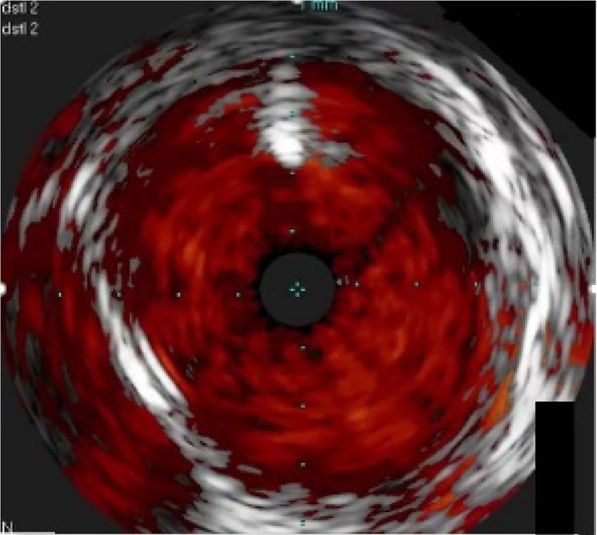
Original articles 13
c ontingency operations. Often assembled at relatively short deployed into the target vessel over a guidewire, it allows
notice, casualty evacuation may be hampered by an inability for real time two-dimensional imaging to be performed. The
to allow air, boat or ground transport assets to be dedicated images obtained can be analysed to assess vessel diameter or
solely for medical transport. Similarly, casualty evacuation the burden of atheroma, and to guide interventional therapies
timelines may be long due to geographical distance, unfavour- such as stents. IVUS devices are now small-calibre, simple to
able meteorological variables or tactical issues. Accordingly, use and interpret, and are approved by international regulatory
a haemorrhage control solution that could be deployed into a bodies for use in humans. In interventional cardiology, data
prolonged field care or early in-hospital setting is an attractive strongly support IVUS use especially in complex cases, and
prospect with life-saving potential due to the ability to buy international guidelines recommend their use. 12 ChromaFlo™
time for surgical haemorrhage control. is an imaging modality which colourises movement in the
IVUS image pixels as red, presenting a form of 2-dimensional
Resuscitative endovascular balloon occlusion of the aorta Doppler. Pixel movement is thus used as a surrogate for flow,
(REBOA) is a rapidly evolving solution to haemorrhage con- and this feature can be used to clearly delineate the vessel lu-
trol. It involves occlusion of the descending aorta by delivery men. This study also aimed to evaluate its use in confirming
of a balloon catheter device via an introducer sheath placed in loss of aortic flow following REBOA balloon deployment, as
the common femoral artery – effectively acting as an endovas- a potential tool for evaluating REBOA effect in vivo.
cular aortic cross-clamp. Three anatomically-defined aortic
landing zones are described: zone I – thoracic aorta between Methods
the left subclavian and coeliac artery; zone 2 – between the
coeliac artery and renal arteries; zone 3 – between the renal The study was ethically reviewed, approved, and conducted
arteries and aortic bifurcation. Only zones I and III are usu- in accordance with the Animals (Scientific Procedures) Act
ally used as landing zones for emergency haemorrhage con- 1986. Two large white swine, weighing 35kg, were termi-
trol. Commonly, balloon placement is performed under direct nally anaesthetised using isoflurane followed by alfaxalone.
fluoroscopic guidance in the emergency department or in a They were instrumented for central and cardiac blood pressure
hybrid interventional radiology or vascular surgical theatre. A measurement, cardiac output measurement and the removal of
small number of cases have been performed in the pre-hospital blood and administration of medications and fluids. Following
setting by clinicians of London’s Air Ambulance, which de- the completion of an experimental protocol simulating severe
ploys a helicopter-delivered, physician-led advanced trauma haemorrhagic shock followed by damage control resuscita-
team to the scene of serious incidents.4 tion, the following pilot studies were performed:
In multiple animal studies, REBOA has been shown to offer Run 1 – IVUS assessment
an effective endovascular solution for severe torso and pelvic To confirm the ability to gain access to and visualise the aorta,
haemorrhage, resulting in rapid control of bleeding and im- a 9 French gauge (F) sheath with haemostatic valve and a side
proved survival, most likely mediated by increased proximal arm was placed in the right common femoral artery via a lim-
blood pressure, brain oxygen delivery and carotid blood flow, ited cut down to expose the vessel, using a modified Seldinger
compared with standard therapy.5-7 In human cohorts, vascu- technique under direct vision. Through this, a 175cm, 0.035”
lar surgeons have reported the use of balloon occlusion as an J-tipped guidewire was passed, over which a 10MHz, 8.5F
effective means of gaining rapid haemorrhage control in the compatible digital IVUS catheter (Visions PV, Philips Volca-
setting of ruptured aortic aneurysm prior to endovascular re- no™) was introduced. The position of the aortic bifurcation
pair,8,9 and a number of case series have been reported on the was visualised under direct IVUS guidance, and the diameter
use of REBOA in the setting of major traumatic haemorrhage. of the distal aorta was noted to aid REBOA balloon selection
To date, the medical literature contains data on 1355 cases for further runs. The IVUS catheter was then removed.
treated with REBOA, including reports of non-trauma indica-
tions such as gastrointestinal and post-partum bleeding.10
Outside a resuscitation room equipped with fluoroscopy, with
which X-ray screening can be used to track the passage of the
balloon, confirmation of balloon delivery to the intended aor-
tic landing zone presents a significant limitation to REBOA.
A blind approach is possible, though the potential risk of de-
vice failure or unintentional balloon deployment in a smaller
branch vessel exists. Trans-abdominal ultrasound techniques
have been reported, using contrast-enhanced ultrasonography
to confirm REBOA placement, but these require additional re-
sources and training.11
Figure 1: Right femoral access site. Limited cut-down, with 12F
Intravascular ultrasound (IVUS) is an endovascular imaging sheath in femoral artery. REBOA balloon catheter shaft (right of
technique in common use in interventional cardiology and picture) and IVUS catheter passing through haemostatic valve. A
radiology. Using a miniaturised, solid-state imaging device venous sheath is also present.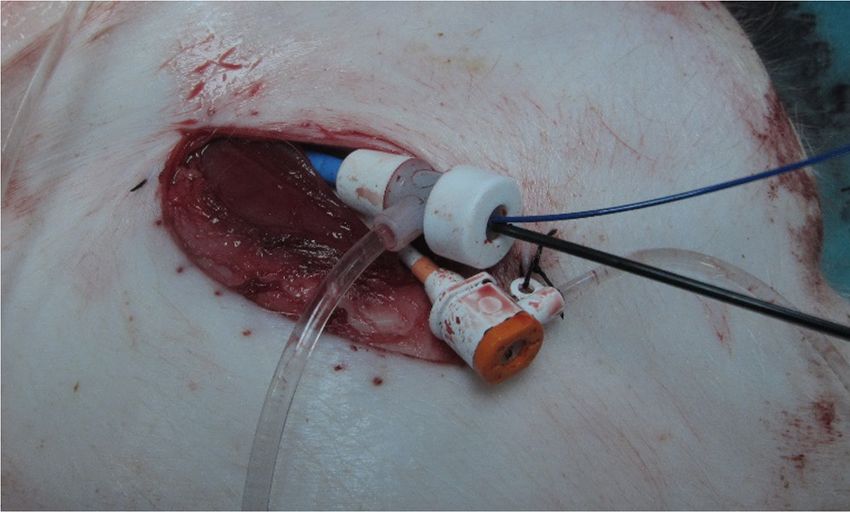
14 Journal of the Royal Naval Medical Service 2018; 104(1)
Run 2 – ChromaFlo
Through the femoral sheath, a 0.014” angioplasty guidewire
(Balance Middle Weight, Abbott™) was introduced into the
aorta. A hydrophilic-coated 20MHz, 5F compatible digital
Figure 3: Representative aortic IVUS image. ChromaFlo is activat-
ed, delineating areas of blood flow. A 0.035” guidewire is noted at
the top of the image as a bright signal, and the inferior vena cava is
also captured to the left of the aortic signal.
the contralateral femoral arterial sheath (LabChart, ADI
Figure 2: – Tip of combined IVUS-REBOA balloon catheter, with
Instruments™).
balloon deflated. Inset: close-up of IVUS catheter attached to bal-
loon catheter shaft (Eagle Eye Platinum, Philips Volcano™). Run 4 - Combined IVUS-REBOA device
A bespoke IVUS-REBOA device was fashioned by combin-
ing the angioplasty balloon and the IVUS probe. Multiple
IVUS catheter was passed over this (Eagle Eye Platinum,
REBOA runs were performed, delivering the device through
Philips Volcano™). Once delivered, ChromaFlo imaging was
the right femoral artery sheath to the distal aorta with Chroma-
commenced and the image was interpreted.
Flo active. Once in position, the balloon was deployed, and the
clinical effect was assessed using both ChromaFlo and obser-
Run 3 – ChromaFlo guided REBOA vation of the transduced pressure trace from the contralateral
With the IVUS catheter in situ in the distal aorta, a 9.0*40mm femoral arterial sheath.
angioplasty balloon (Mustang, Boston Scientific™) was
passed over a 0.035” guidewire. The balloon was deployed, The position of the balloon from the final run was confirmed
and the aortic flow was monitored using both ChromaF- by direct visualisation of the opened aorta at post-mortem ex-
lo and observation of the transduced pressure trace from amination.
Inflation Deflation
70
60
50
40
30
20
10
5:35:52 5:36:02 5:36:12 5:36:22 5:36:32
Figure 4: Transduced femoral arterial trace during IVUS-REBOA deployment and deflation. A marked reduction in downstream pressure is
visible during balloon deployment, which resolves on balloon deflation.
Original articles 15
Length markers
P-tipTM
Compliant A A
balloon
Balloon
lumen
Arterial line
port
Arterial line
lumen
Radiopaque (pressure
marker bands monitoring)
Peel-away
sheath
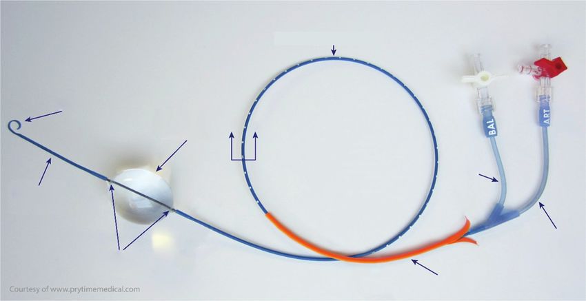
Figure 5: Commercially available REBOA device (ER-REBOA™ catheter, image courtesy of Prytime Medical) for comparison, with balloon
deployed.
Results ChromaFlo signal and transduced distal arterial pressure trace.
The mean time taken from balloon insertion into the femoral
Two pigs were studied in this initial technical evaluation pilot.
sheath to balloon deployment was 21.25 seconds (n=10 runs,
The results of each component are reported. Femoral access
range 18.1-24.0 seconds).
was gained bilaterally without incident in both subjects.
Post-mortem examination confirmed correct delivery of the
Run 1 – delivery of the 0.035” guidewire and larger Vision
IVUS-REBOA device to the distal aortic landing zone in both
catheter was successful at first pass in both cases, and internal
subjects. No vascular injuries related to either vascular access
diameter of the target aortic area was noted as ~10mm.
or REBOA were found.
Run 2 – delivery of the angioplasty guidewire and Eagle Eye
Discussion
catheter was successful at first pass in both cases. Chroma-
Flow imaging clearly delineated the aortic wall, and confirmed This series of studies demonstrates, for the first time in a por-
pulsatile flow in time with the cardiac cycle and corresponding cine model, that a commercially available IVUS catheter can
to the transduced arterial pressure traces. Even in the setting of be used to confirm delivery of the REBOA balloon to the cor-
cardiac arrest with chest compressions underway (according rect anatomical landing area. It also allows real-time obser-
to study protocol) by a mechanical cardiopulmonary resusci- vation of balloon deployment and confirms apposition of the
tation device (LUCAS 2, Physio Control™), ChromaFlo was balloon to the vessel wall. The addition of ChromaFlo imaging
able to detect the presence of aortic flow. allowed confirmation of loss of pulsatile aortic flow distal to
the deployed balloon, confirming clinical effect. Deployment
Run 3 – delivery of the REBOA guidewire and balloon was suc- of the device was rapid, safe and did not require fluoroscopy.
cessful at first pass, and easily visualised using live IVUS imag-
ing, with both guidewire and balloon shafts visible as they passed Limitations
the IVUS catheter. In addition, on deployment of the REBOA This was a proof-of-concept pilot study, hence the number of
balloon, ChromaFlo pulsation was lost simultaneously with a animal subjects was small. An expanded study using a nov-
dramatic reduction in transduced distal arterial pressure trace. el pressurised human cadaveric model is planned. The fem-
oral access devices used were relatively large (9-12F) due to
Run 4 – delivery of the IVUS-REBOA device was success- the calibre required to introduce the IVUS catheter as a pig-
ful at first pass in all cases (total 10 runs). IVUS imaging gy-backed device coupled to the REBOA balloon. If a com-
confirmed correct deployment of the device above the iliac bined device were to be manufactured, with a lower cross-sec-
bifurcation, in aortic zone 3. ChromaFlo imaging confirmed tional profile, a smaller femoral sheath could be used. A recent
full apposition of the REBOA balloon to the aortic wall, and multicentre registry study confirms that the use of smaller
loss of pulsatile signal confirmed aortic occlusion, again cor- sheaths is associated with fewer access site complications,
responding with loss of transduced distal arterial pressure such as thrombus and distal ischaemia.13 The use of smaller
trace. On deflating the REBOA balloon, forward flow rapidly sheaths (e.g. 7F) is likely to broaden REBOA delivery among
re-established, confirmed by restoration of a visible pulsatile physicians who are not regular endovascular operators. In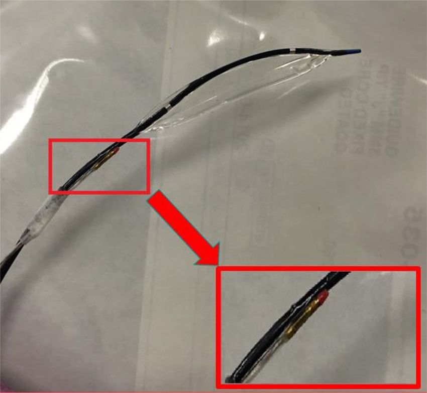
16 Journal of the Royal Naval Medical Service 2018; 104(1)
a ddition, the use of vascular access sites other than the femoral another undergoes damage control surgery. This would include
area is attractive, especially following trauma where the groin the Royal Navy Role 2 Afloat, and Commando Forces Surgical
might be anatomically disrupted and involved in the primary Group Role 2 Light Manoeuvre units.
injury. Interventional cardiology procedures, using catheters
most commonly 6F, but ranging from 5-7.5F, predominantly IVUS-REBOA could also help to mitigate downstream tissue
use the radial artery as the primary access site of choice, due ischaemia. With the REBOA balloon totally inflated, down-
to lower procedural access site related complications. REBOA stream haemorrhage stops. Unfortunately, this results in an ac-
from non-femoral sites has been reported, but confirmation cumulation of metabolites in downstream tissues. Long periods
of delivery to the intended target landing zone would usual- of balloon inflation have been shown to result in a rise in lac-
ly mandate fluoroscopy. Forward-looking, three-dimensional tate, interleukin-6, an increased vasopressor requirement and
IVUS, currently an investigative tool, might offer a solution respiratory distress syndrome.17 Partial REBOA (pREBOA),
to this, allowing the operator to steer the aortic balloon from with subtotal aortic occlusion which allows a limited amount
the subclavian system into the descending aorta, and on to the of forward flow, can be used to extend the therapeutic window
intended landing zone. Staying away from the femoral site as demonstrated in recent animal studies. pREBOA results
would also reduce procedural complications such as pseu- in reduced levels of serum lactate, with reduced splanchnic
doaneurysm formation, the development of arteriovenous fis- injury, when compared with complete REBOA (cREBOA),
tulae and post-procedural bleeding. Care of the radial access and also avoids the supra-normal pressure surge seen in the
site is also much easier, using direct pressure with a currently vascular tree above the inflated REBOA balloon.18 pREBOA
available compression band device (e.g. TR band, Terumo™). thus offers a physiologically and haemodynamically tolerable
option for endovascular haemorrhage control, which might
IVUS imaging requires connection of the endovascular part of extend the golden hour after traumatic injury.19 Pragmatically,
the catheter to an external processing unit via a connector ca- an initial cREBOA inflation, to rapidly achieve control is ad-
ble. This is currently built solely for in-hospital use in the inter- vocated. Following this, pREBOA could be considered, sup-
ventional suite, and is a wheeled tower containing a processing ported by registry data which further suggests that pREBOA
unit and a display screen. Whilst the current unit could be suit- is associated with better outcomes in human subjects.13 Clin-
able for emergency department-delivered IVUS-REBOA, it ically, there is no easy way to confirm whether cREBOA or
would require extensive miniaturisation to enable its use in the pREBOA has been achieved. IVUS imaging offers a solution
pre-hospital setting. Ultrasound devices using wireless probes to this, allowing direct visualisation of balloon apposition to
such as the Accuson Freestyle™ have already been used by the vessel wall, allowing the operator to see whether complete
military units in austere settings,14 and the development of a or partial occlusion has been performed. ChromaFlo imaging
combined IVUS-REBOA device which connects wirelessly to would allow the operator to visualise a degree of forward flow
a handheld tablet-sized screen could easily be achieved and should pREBOA be desired. Further research is required to
make this a therapy which is truly suited to pre-hospital and assess the optimum degree of forward flow, and it is likely that
prolonged field care situations. Most critical care clinicians this will vary between patients. An endovascular variable aor-
are used to interpreting ultrasound images, and little additional tic control (EVAC) system, regulating the degree of REBOA
training uplift would be required to interpret IVUS images. based on changes in proximal pressure has been trialled in an
animal study,20 and IVUS-REBOA technology could have a
REBOA has the potential to save lives in a military trauma co- role in developing future variants of such an EVAC catheter,
hort. Retrospective analysis of a UK military cohort identified able to automatically perform these functions.
174 combat deaths due to exsanguinating trauma, with a mean
pre-hospital time of 61 minutes.7 66 deaths occurred en route to IVUS could be part of an advanced endovascular resuscita-
hospital, and 29 shortly after arrival at the receiving facility. From tion toolkit. As part of an advanced endovascular resuscitation
injury analysis, 20% of patients had a focus of haemorrhage in strategy, REBOA offers a bridge to damage control surgery. If
the abdomen or around the abdomino-pelvic junction that could traumatic cardiac arrest ensues, access to the aorta gives the
have been anatomically controlled with REBOA. It is likely that added advantage of allowing future therapies such as selective
a significant number of the remainder could also have benefitted aortic arch perfusion using either oxygenated blood or novel
from the physiological stabilisation offered by REBOA.7 A pop- resuscitation fluids.21 If initial efforts still fail, the addition of a
ulation-based gap analysis of civilian trauma patients in England large-bore venous cannula would make full cardiopulmonary
and Wales revealed similar data and suggested a role for REBOA bypass possible following damage control surgery. This com-
mostly within high volume major trauma centres.15 REBOA is bination of techniques – emergency preservation and resusci-
now well established as a potential therapy for exsanguinating tation (EPR) and resuscitation for cardiac arrest from trauma
trauma, which could significantly improve outcomes in critically is currently undergoing a feasibility and safety study in the
ill patients for whom there are no viable options. REBOA, as United States (EPR-Cat study; ClinicalTrials.gov identifier
part of an armoury of advanced endovascular techniques, may NCT01042015). It is likely, as data emerges, that REBOA,
be ready for exploitation when planning deployed damage con- SAAP and EPR will overlap to form a hybrid advanced endo-
trol resuscitation and surgery facilities for military operations.16 vascular toolkit for the management of haemorrhagic shock.22
Potential UK military uses might include Role 2 units, especially Developing a robust REBOA capability is the first step to en-
where surgical capacity is limited to one operating table, and a abling deployed damage control resuscitation teams to be able
potential requirement might exist to stabilise one patient whilst to exploit these techniques as the data emerges in future.Original articles 17
References
1. Eastridge BJ, Mabry RL, Seguin P. Death on the battlefield (2001–2011): implications for the future of combat casualty care. J Trau-
ma Acute Care Surg 2012;73:431-7.
2. Morrison JJ, America TER. Noncompressible torso hemorrhage: a review with contemporary definitions and management strategies. Surg
Clin North Am 2012;92:843-58.
3. Penn-Barwell JG, Roberts, SAG, Midwinter, MJ. Improved survival in UK combat casualties from Iraq and Afghanistan: 2003–2012. J
Trauma Acute Care Surg 2015;78:1014-20.
4. Sadek S, Lockey DJ, Lendrum RA, Perkins Z, Price J, Davies GE. Resuscitative endovascular balloon occlusion of the aorta (REBOA) in
the pre-hospital setting: an additional resuscitation option for uncontrolled catastrophic haemorrhage. Resuscitation 2016 Oct 31;107:135-8.
5. Avaro JP, Mardelle V, Roch A. Forty-minute endovascular aortic occlusion increases survival in an experimental model of uncontrolled
hemorrhagic shock caused by abdominal trauma. J Trauma Acute Care Surg 2011;71:720-6.
6. Morrison JJ, Percival TJ, Markov NP. Aortic balloon occlusion is effective in controlling pelvic hemorrhage. J Surg Res 2012;177:341-7.
7. Morrison JJ, Ross JD, Houston RIV, Watson JDB. Use of resuscitative endovascular balloon occlusion of the aorta in a highly lethal model
of noncompressible torso hemorrhage. Shock 2014;41:130-7.
8. Malina M, Veith F, Ivancev, K. Balloon occlusion of the aorta during endovascular repair of ruptured abdominal aortic aneurysm. J Endo-
vasc Ther 2005;12:556-9.
9. Matsuda H, Tanaka Y, Hino Y et al. Transbrachial arterial insertion of aortic occlusion balloon catheter in patients with shock from ruptured
abdominal aortic aneurysm. J Vasc Surg 2003;38:1293-6.
10. Gamberini E, Coccolini F. Resuscitative Endovascular Balloon Occlusion of the Aorta in trauma: a systematic review of the literature.
World J Emerg Surg 2017;12.
11. Chaudery M, Clark J, Morrison JJ, Wilson MH, Bew D, Darzi A. Can contrast-enhanced ultrasonography improve Zone III REBOA place-
ment for prehospital care? J Trauma Acute Care Surg 2016;80:89-94.
12. Bavishi C, Sardar P, Chatterjee S et al. Intravascular ultrasound--guided vs angiography-guided drug-eluting stent implantation in complex
coronary lesions: Meta-analysis of randomized trials. Am Heart J 2017;185:26-34.
13. Matsumura Y, Matsumoto J, Kondo H, Idoguchi JK. Fewer REBOA complications with smaller devices and partial occlusion: evidence
from a multicentre registry in Japan. Emerg Med J 2017;34:793-9.
14. Rees PSC, Lamb LEM, Nicholson-Roberts TC et al. Safety and feasibility of a strategy of early central venous catheter insertion in a de-
ployed UK military Ebola virus disease treatment unit. Intensive Care Med 2015;41:735-43.
15. Barnard EBG, Morrison JJ, Madureira RM. Resuscitative endovascular balloon occlusion of the aorta (REBOA): a population based gap
analysis of trauma patients in England and Wales. Emerg Med J 2015;32:926-32.
16. Tisherman SA, Brenner ML. Taking advanced endovascular techniques out of the hospital: Ready for prime time. Resuscitation
2016;107:A3-A4.
17. Markov NP, Percival TJ, Morrison JJ, Ross JD, Scott DJ. Physiologic tolerance of descending thoracic aortic balloon occlusion in a swine
model of hemorrhagic shock. Shock 2013;153:848-56.
18. Russo RM, Williams TK, Grayson JK. Extending the golden hour: partial resuscitative endovascular balloon occlusion of the aorta in a
highly lethal swine liver injury model. J Trauma Acute Care Surg 2016;80:372-80.
19. Russo RM, Neff LP, Lamb CM. Partial resuscitative endovascular balloon occlusion of the aorta in swine model of hemorrhagic shock. J
Am Coll Surg 2016;223:359-68.
20. Williams TK, Neff LP, Johnson MA et al. Extending REBOA: endovascular variable aortic control (EVAC) in a lethal model of hemorrhagic
shock. J Trauma Acute Care Surg 2016;81:294.
21. Manning JE, Murphy CA, Hertz CM. Selective aortic arch perfusion during cardiac arrest: a new resuscitation technique. Ann Emerg Med
1992;21:1058-65.
22. Kutcher ME, Forsythe RM, Tisherman SA. Emergency preservation and resuscitation for cardiac arrest from trauma. Int J Surg
2016;33:209-12.
Conflict of interest statement
The authors report no conflicts of interest.
Authors
Surgeon Commander PSC Rees Royal Navy
Consultant in Interventional Cardiology, Acute and Pre-hospital Medicine
Reader in Military Medicine
Chair, Defence Resuscitation Committee
cardiacexpert@nhs.net
Academic Department of Military Medicine and University of St Andrews School of Medicine
Major AM Buckley RAMC
Academic Department of Military Medicine
Dr S A Watts, DSTL Porton Down
Dr E Kirkman, DSTL Porton DownYou can also read