SingHealth Radiology Archives pictorial essay Part 1: cardiovascular, respiratory and neurological cases
←
→
Page content transcription
If your browser does not render page correctly, please read the page content below
Singapore Med J 2020; 61(12): 633-640
Pictorial Essay
                                                                                                                        https://doi.org/10.11622/smedj.2020177

CMEArticle
SingHealth Radiology Archives pictorial essay
Part 1: cardiovascular, respiratory and neurological cases
                            Mark Bangwei Tan1, MRCS, FRCR, Kim Ping Tan1, FRCR, FAMS, Joey Chan Yiing Beh2, MMed, FRCR,
         Eugenie Yi Kar Chan3, MBBS, Kenneth Fu Wen Chin3, MD, Zong Yi Chin3, MBChB, MRCS, Wei Ming Chua3, MMed, FRCR,
                             Aaron Wei-Loong Chong3, MB BCh BAO, Gary Tianyu Gu3, MBBS, MRCS, Wenlu Hou3, MMed, FRCR,
                            Anna Chooi Yan Lai1, MMed, FRCR, Rebekah Zhuyi Lee3, MBBS, Perry Jia Ren Liew4, MMed, FRCR,
                   May Yi Shan Lim3, MRCS, FRCR, Joshua Li Liang Lim3, MBBS, Zehao Tan3, MMed, FRCR, Eelin Tan3, BMed, MD,
                                     Grace Siew Lim Tan3, MBBS, Timothy Shao Ern Tan3, MBChB, Eu Jin Tan1, MMed, FRCR,
                     Alexander Sheng Ming Tan1, MRCS, FRCR, Yet Yen Yan4, MMed, FRCR, Winston Eng Hoe Lim1, FRCR, FAMS

    The Singapore Health Services cluster (SingHealth) radiology film archives are a valuable repository of local radiological
    cases dating back to the 1950s. Some of the cases in the archives are of historical medical interest, i.e. cerebral angiography
    in the workup of patients with hemiplegia. Other cases are of historical social interest, being conditions seen during
    earlier stages of Singapore’s development, i.e. bound feet. The archives form a unique portal into the development of
    local radiology as well as the national development of Singapore. A selection from the archives is published in 2020 in
    commemoration of the 20th anniversary of the formation of SingHealth, the 55th National Day of Singapore, and the
    125th anniversary of the International Day of Radiology. This pictorial essay comprises cardiovascular, respiratory and
    neurological cases from the archives.

Keywords: archives, historical, national development, pictorial essay, SingHealth

INTRODUCTION                                                                        A radiologist roster from November 1976 is shown in Fig. 1.
The Singapore Health Services (SingHealth) radiology film archives                  Today, the radiological services of these same hospitals have
comprise radiography case records dating back to the 1950s. The                     expanded to include ultrasonography, computed tomography
archives are located within Singapore General Hospital (SGH)                        (CT), magnetic resonance (MR) imaging and nuclear medicine
and comprise cases of historical medical interest as well as of                     investigations, provided by a staff of at least 150 radiologists
historical social interest. The archives form a unique portal into the              and trainees.
development of local radiology as well as the national development
of Singapore. Additional commentary and anecdotes on these cases                    STANFORD TYPE A AORTIC DISSECTION
were obtained via oral interview with Dr Tan Kim Ping, a prominent                  Fig. 2 shows transfemoral catheter aortography that was
local radiologist, former head of department of the SGH Department                  performed for a patient with chest pain and unequal pulses.
of Radiology and key proponent of the archives. A selection from                    In the thoracic aorta, a dissection flap arising near the aortic
the archives was published in 2020 in commemoration of the 20th                     root is visualised (black arrow, Fig. 2a). The false lumen is less
anniversary of the formation of SingHealth, the 55th National Day                   dense (white arrows in Fig. 2). There is also non-opacification
of Singapore, and the 125th anniversary of the International Day of                 of the left subclavian artery, indicating its origin from the false
Radiology. This pictorial essay comprises cardiovascular, respiratory               lumen. As the dissection involves the ascending aorta proximal
and neurological cases from the archives.                                           to the left subclavian artery, this is classified as a Stanford Type
                                                                                    A aortic dissection. In the abdominal aorta, the presence of a
R A D I O LO G I S T S TA F F I N G I N N OV E M B E R                              right nephrogram (black arrow, Fig. 2b) without opacification of
1976                                                                                the left kidney and renal artery suggests that the latter structures
In the 1970s, nine radiologists provided fluoroscopy, interventional                arise from the false lumen.
radiology and plain radiography reporting services to SGH and                           Catheter aortography was previously the gold standard
three satellite (outstation) hospitals – Thomson Road General                       for suspected aortic dissection. One of its pitfalls, however,
Hospital (precursor of the current Changi General Hospital),                        is a false negative due to thrombosis of the false lumen.(1)
Alexandra Hospital and Kandang Kerbau Maternity Hospital                            CT has since replaced this procedure due to its high image
(renamed KK Women’s and Children’s Hospital in 1997). A single                      resolution and amenability to post-processing reconstructions,
doctor would provide on-call services for these four hospitals.                     as displayed in a three-dimensional reconstruction and

1
 Department of Diagnostic Radiology, Singapore General Hospital, 2Department of Radiology, Ng Teng Fong General Hospital, 3Singhealth Diagnostic Radiology Residency
Programme, 4Department of Radiology, Changi General Hospital, Singapore
Correspondence: Dr Mark Tan Bangwei, Associate Consultant, Department of Diagnostic Radiology, Singapore General Hospital, Outram Road, Singapore 169608.
marktanbangwei@gmail.com

633
Pictorial Essay

		                         Fig. 1 Radiologist roster in November 1976.

                            2a                                                   2b

Fig. 2 Transfemoral catheter aortogram shows the (a) thoracic aorta and (b) abdominal aorta in the setting of a Stanford Type A aortic dissection.

maximum intensity projection sagittal image (Fig. 3), whereby                such as mitral valve disease, as well as in congenital conditions
the dissection flap (arrows) and the subtle contrast differences             such as a ventricular septal defect or patent ductus arteriosus.
between the true lumen (asterisks) and false lumen can be                    Patients with left atrial enlargement may present with dysphagia to
appreciated in exquisite detail. Nonetheless, the recognition                solids due to extrinsic compression of the oesophagus (dysphagia
of this appearance of aortic dissection on angiography                       megalatriensis), as well as hoarseness of voice due to compression
remains relevant to this day, as it can occasionally be seen                 of the recurrent laryngeal nerve (Ortner syndrome). The barium
during modern procedures involving cannulation of the                        swallow study was a commonly used surrogate method to
aorta (e.g. cardiac catheterisation, visceral embolisation).                 diagnose left atrial enlargement before echocardiography (Fig. 5)
Notably, this study was performed at Tan Tock Seng Hospital                  became more widely available.
(TTSH), which was previously the designated hospital for
cardiothoracic surgery in Singapore.                                         EISENMENGER SYNDROME SECONDARY
                                                                             TO ATRIAL SEPTAL DEFECT
LEFT ATRIAL ENLARGEMENT ON BARIUM                                            Chest radiography was performed in a woman with chronic
SWALLOW                                                                      shortness of breath (Fig. 6). There is enlargement of the
A middle-aged woman presented with hoarseness of voice. A                    pulmonary trunk and pulmonary arteries with decreased size
barium swallow study performed in the lateral projection (Fig. 4)            of the peripheral vessels (i.e. ‘pruning’). This is suggestive of
demonstrated posterior displacement of the distal oesophagus                 the presence of an atrial septal defect with imaging features
(arrows), which is a classic radiological finding of left atrial             of resultant pulmonary arterial hypertension. Some patients
enlargement.(2) The left atrium is the most posteriorly located              may develop shunt reversal from a left-to-right to a right-
cardiac chamber. It can become enlarged secondary to increased               to-left direction as a result of chronic pulmonary arterial
intra-atrial pressures, which may be seen in acquired conditions             hypertension, leading to right ventricular hypertrophy and

                                                                                                                                                     634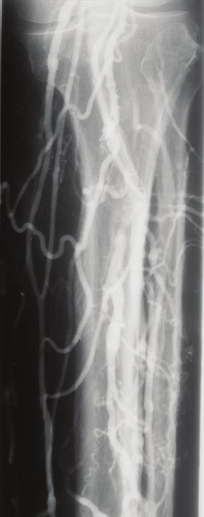
Pictorial Essay

                                 3a                                               3b

Fig. 3 (a) Three-dimensional reconstruction and (b) maximum intensity projection sagittal CT images show the dissection flap (arrows) and the subtle
contrast differences between the true lumen (asterisks) and false lumen in the setting of a Stanford Type A aortic dissection.

Fig. 4 Lateral projection radiograph shows posterior displacement of the          Fig. 6 Chest radiograph of a woman with chronic shortness of breath shows
distal oesophagus (arrows) in the setting of left atrial enlargement.             enlargement of the pulmonary trunk and pulmonary arteries with decreased
                                                                                  size of the peripheral vessels. Findings are suggestive of the presence of
                                                                                  an atrial septal defect with resultant pulmonary arterial hypertension. Film
                                                                                  degradation due to ageing is noted.

                                                                                  deoxygenated blood entering the systemic circulation causing
                                                                                  cyanosis, termed Eisenmenger syndrome. In Singapore today,
                                                                                  this condition can be suspected at the prenatal stage by fetal
                                       LV
                                                                                  echocardiography, followed up in the postnatal period, and
                        RV                                                        treated with percutaneous or surgical closure if indicated,
                                                                                  decreasing morbidity and mortality.
                                                                                       The upper half of the film (Fig. 6) is degraded due to ageing.
                                                                                  X-ray film consists of silver halide emulsion, which undergoes
                                                                                  oxidation to produce a silver ion and an electron when exposed
                                                                                  to light. Over time, the protective layer of gelatin over the film
                                                                                  may be damaged by this oxidative process, as seen in this case.
                        RA
                                                                                  This is no longer seen in the current day due to the advent of
                                        LA                                        digital radiography and the presence of computerised picture
                                                                                  archiving and communication systems.
Fig. 5 Transthoracic two-dimensional echocardiography was performed
on a middle-aged man (different patient from the earlier patient                  CONJOINED TWINS
who underwent a barium swallow procedure) with a history of atrial
fibrillation. Echocardiogram in four-chamber view shows both cardiac atria        A pair of term dicephalus parapagus conjoined male twins born
enlarged relative to the ventricles. Note that the appearance of apparent         in the 1980s underwent contrast-enhanced aortography for
communication between the cardiac atria was due to technical factors
during image acquisition, and the patient has no atrial septal defect. LA: left
                                                                                  pre-surgical separation evaluation, which was performed using
atrium; LV: left ventricle; RA: right atrium; RV: right ventricle                 catheter-guided contrast instillation via the shared umbilical

635
Pictorial Essay
DEEP VEIN THROMBOSIS OF THE LEFT
FEMORAL VEIN
Ascending phlebography was described in the 1970s and was
previously the gold standard for the diagnosis of deep venous
thrombosis (DVT). It involves cannulation of the dorsal foot
veins, and assessment of the profunda vein is done with the
Valsalva manoeuvre. Pitfalls of interpretation include false
positive examination due to under-filling, dilution or admixture
of contrast.(4) In Fig. 8, ascending phlebography shows filling
defects and distension of the left femoral vein to the distal femur,
in keeping with DVT. The ‘tram-track’ sign (white arrows) of
contrast outlining the incompletely occlusive thrombus is seen.
The abrupt transition of the interface between the opacified
and non-opacified portion of the femoral vein suggests acute
thrombosis (black arrow). The left profunda femoris vein also
shows similar evidence of thrombosis (white dashed arrow).
Ultrasonography Doppler assessment has since replaced
phlebography for diagnosis of DVT. Nonetheless, this appearance
of DVT on venography can still be seen during venous
thrombolysis, which is performed to reduce post-thrombotic
syndrome.
CHRONIC VENOUS INSUFFICIENCY
Invasive catheter venography was the gold standard test for
Fig. 7 A pair of term dicephalus parapagus conjoined male underwent
catheter-guided contrast instillation via the shared umbilical artery. venous disease before the introduction of duplex ultrasonography
Contrast-enhanced aortogram shows a single cardiac silhouette in in 1989.(5) Plain radiographs of the calf (Fig. 9) show diffuse
the right hemithorax with a shared common ventricle. A single united
thoracoabdominal aorta in the midline provides arterial supply to each twin periosteal reaction along the tibia and fibula, soft tissue swelling
via the innominate arteries. Cardiovascular anatomy – A: aorta (shared); and phleboliths. Ascending catheter venography of the same limb
H: heart; (R) CCA: right common carotid artery of right twin; (R) IA: right
innominate artery (shared); (R) SCA: right subclavian artery in right twin; shows prominent superficial calf veins that are consistent with
(L) CCA: left common carotid artery of left twin; (L) IA: left innominate chronic venous insufficiency (CVI). The deep veins are patent.
artery (shared); (L) SCA: left subclavian artery in left twin; SVC: superior
vena cava, likely shared; UAC: umbilical arterial catheter, tip likely in the left
In the context of suspected CVI, ascending venography
hepatic lobe at the level of L2–3. Other structures – L: liver, shared between evaluates perforator competency and deep venous patency.
both twins, bridging the midline; LS: stomach of the left twin, which shows
Contrast is injected into a dorsal foot vein under fluoroscopy
organoaxial rotation and has herniated through the left hemidiaphragm into
the left hemithorax; RS: stomach of right twin; SB: small bowel loops of the with the patient in a 30°–45° foot down position and a tourniquet
left twin, with some loops herniated into the left hemithorax at the ankle. The tourniquet occludes the superficial veins
so that contrast is forced into the deep venous system. Any
artery. The study outlines a single cardiac silhouette in the right retrograde flow into perforating veins is diagnostic of perforator
hemithorax with a shared common ventricle (Fig. 7). A single incompetence.(5) Descending venography via contrast injection
united thoracoabdominal aorta in the midline provides arterial into the common femoral vein grades the anatomic extent of the
supply to each twin via the innominate arteries. Conjoined twins reflux.(6) Catheter venography remains relevant today if venous
present a unique challenge due to complex anatomy involving reconstruction is contemplated.
multiple body systems. Preoperative planning is aided by
gleaning pertinent information from a combination of imaging SILICOSIS
modalities, depending on the site(s) of fusion.(3) In cases of thoracic Silicosis was the predominant occupational lung disease in
conjunction, conventional catheter angiography was traditionally Singapore in the 1970s.(7) This was largely attributed to the
performed to define cardiac malformations and aberrant vascular presence of granite mines such as those in Bukit Timah, Bukit
connections. This has now been completely replaced by MR Batok and Pulau Ubin, where workers were exposed to free
imaging or CT and digital subtraction angiography. silica dust from detonating and processing the granite stones.
Unfortunately, the depicted case was not amenable A chest radiograph (Fig. 10) shows the presence of opacities
to surgical separation given the shared heart. The surgical with a hyperdense rim in the bilateral hilar regions (arrows)
considerations in the separation of conjoined twins were fraught consistent with calcified lymph nodes. The calcification outlines
with ethical considerations and decisions depending on the the peripheries of the hilar nodes and are termed eggshell
anatomical configuration encountered, such as considering calcifications. This appearance is suggestive for silicosis given the
which twin should receive the pair of lower limbs or the set of appropriate setting of occupational exposure, with the differential
sexual organs. diagnosis of sarcoidosis (post radiation).
636Pictorial Essay
Fig. 8 Ascending phlebogram shows filling defects and distension of the left femoral vein, in keeping with deep venous thrombosis.
9a 9b 9c
Fig. 10 Chest radiograph shows imaging features of bilateral hilar
adenopathy demonstrating an eggshell calcification pattern (arrows), seen
in the setting of silicosis.
Fig. 9 (a) Plain radiograph of the calf shows diffuse periosteal reaction, soft
tissue swelling and phleboliths along the tibia and fibula. (b & c) Venograms
of the same limb show prominent superficial veins. Overall features are ASBESTOS LUNG DISEASE
consistent with chronic venous insufficiency.
From the 1950s to 1980s, raw asbestos was imported and mixed
with cement in a factory to produce roof tiles, ceiling boards
Measures were taken to decrease the prevalence of and pipes. It was also used as insulating material in shipyard
silicosis. Mines were asked to employ dust control measures. industries and buildings as well as friction material for heavy
The government necessitated dust exposure monitoring, vehicle components. The chest radiograph of an asymptomatic
encouraged use of breathing devices and instituted medical factory worker (Fig. 12) showed curvilinear calcifications along
surveillance. In the late 1990s, granite mining licensing was both hemidiaphragms and irregular calcifications in the bilateral
reduced and eventually stopped. Other industries associated lower zones (arrows), representing pleural calcifications. The
with development of silicosis in its workers included the rubber multiplicity, bilaterality and diaphragmatic involvement are
industry, kaolin mines, foundries and brickworks. In the current pathognomonic for asbestos plaques rather than other differentials
age, as seen in Fig. 11, CT shows superior characterisation of such as pleural masses, rib fractures, prior empyema, tuberculosis
mediastinal lymph node calcifications compared to radiography and haemothorax.
(asterisk). CT also better demonstrates the presence of During the 1980s, as the association between asbestos and
pulmonary nodules in the upper lobes forming conglomerate lung disease became apparent, its use was curtailed. Asbestos
focal masses, some sausage-shaped, with irregular margins was completely banned for use in 2001. Occupational asbestos
and adjacent emphysematous changes (arrows), compatible exposure is, however, still seen today, mainly during demolition
with a diagnosis of complicated pneumoconiosis secondary or renovation of buildings constructed before 1989.(8) Under
to silicosis. the Workplace Safety and Health Regulations 2011, chest
637Pictorial Essay
Fig. 11 CT image of a patient with complicated pneumoconiosis secondary
to silicosis. Note the superior characterisation of mediastinal lymph node
calcifications on CT compared to radiography (asterisk).
Fig. 13 A patient with a history of oesophageal carcinoma presented with
acute paraplegia. Emergent fluoroscopy-guided myelogram shows a filling
defect within the anterior aspect of the column of contrast within the spinal
canal (arrow) secondary to local invasion by the tumour.
Fig. 12 Chest radiograph of an asymptomatic factory worker shows
curvilinear calcifications along both hemidiaphragms (arrows) representing
pleural calcifications. Asbestos lung disease must be considered in the
imaging differential.
radiography is to be performed every 36 months for workers with Fig. 14 Catheter cerebral angiogram of the right internal carotid artery
asbestos exposure. shows leftward deviation of the branches of the anterior and middle
cerebral arteries across the midline of the skull with a clear crescentic
avascular space under the skull (arrowheads). These findings are worrisome
EMERGENCY MYELOGRAM IN ACUTE for the presence of a subdural haematoma; this diagnosis was subsequently
confirmed on right craniectomy.
PARAPLEGIA
A patient with a history of oesophageal carcinoma presented with
acute paraplegia, and emergent myelography was performed. The advent of MR imaging in the late 1980s. At present, myelography
fluoroscopy-guided myelogram (Fig. 13) demonstrates a filling is still performed in patients for whom MR imaging is not possible
defect within the anterior aspect of the column of contrast within for safety reasons(9) (e.g. non-MR-compatible pacemakers), and
the spinal canal (arrow) secondary to local invasion by the tumour. in patients with metal spinal implants that would result in severe
Myelography was first described in 1921. This procedure degradation of MR image quality due to artefacts.
involved intrathecal instillation of contrast under fluoroscopic
guidance by a radiologist, followed by the acquisition of SUBDURAL HAEMATOMA
fluoroscopic and, in later years, CT images of the spinal canal. Catheter cerebral angiography of the right internal carotid artery
The first contrast medium used was oil-based Myodil, which was performed in a patient with left hemiplegia (Fig. 14). The
was used for years and later found to cause arachnoiditis. It was angiogram demonstrates leftward deviation of the branches of
replaced when non-ionic water soluble contrast media became the anterior and middle cerebral arteries across the midline of
available in the 1980s. the skull with a clear crescentic avascular space under the skull.
For decades, myelography was the only diagnostic method These findings are consistent with an extra-axial mass lesion
available to investigate pathology within the spinal canal until the adjacent to the right cerebral hemisphere, likely representing
638Pictorial Essay
hemiparesis (Fig. 15). No vascular occlusion was detected.
However, there was a prominent artery arising from the cavernous
segment of the left internal carotid artery, running posteriorly and
terminating at a localised collection of capillaries with adjacent
contrast staining (arrow). The location, course and size of this
vessel is in keeping with a prominent meningohypophyseal
artery(11) leading to a region of abnormal appearing blood vessels
which show a characteristic ‘tumour blush’ angiographic pattern.
The normal meningohypophyseal artery is often too small to be
visualised on angiography. When it is seen to be prominent, a
pathological process should be considered, for example, the
presence of a tumour at the tentorium cerebelli, which is the
region this vessel supplies. The patient underwent surgery and a
tentorium cerebelli meningioma was found intraoperatively and
excised. CT and MR imaging brain studies have since superseded
catheter angiography for the diagnosis and localisation of
Fig. 15 Angiogram of the left internal carotid artery shows an abnormally
prominent meningohypophyseal artery arising from the cavernous segment intracranial lesions.
of the left internal carotid artery and leading to a region of abnormal-
seeming blood vessels, which show a characteristic ‘tumour blush’ (arrow)
angiographic pattern. Surgery was performed, which demonstrated ACKNOWLEDGEMENTS
tentorium cerebelli meningioma. The authors would like to acknowledge the staff of the Department
of Diagnostic and Interventional Imaging, KK Women’s and
a subdural haematoma (arrowheads). The diagnosis was Children’s Hospital, Singapore, for their assistance in digitising
subsequently confirmed on right craniectomy in which a blood the hard copy films from the SingHealth Radiology Archives;
clot was evacuated. Dr Kamalesh Anbalakan (Cardiology) for the two-dimensional
Prior to the advent of routine head CT imaging, emergent echocardiography image on the case ‘left atrial enlargement’;
catheter cerebral angiography was the default radiological and Dr David Alexander Stringer (Paediatric Radiology) for his
investigation for patients with neurological symptoms.(10) This input on the case ‘conjoined twins’.
study was performed at TTSH, which was the first hospital
designated in Singapore for specialised neurosurgical procedures. REFERENCES
1. Syme J. Transfemoral catheter aortography in dissecting aneurysm. Australas
The investigation, performed via femoral catheter or direct arterial
Radiol 1971; 15:133-9.
puncture, was used to differentiate the pathological processes of 2. Gotsman I, Mogle P, Shapira MY. An unusual cause of dysphagia. Postgrad
intracranial mass (tumour blush pattern or deviation of vascular Med J 1999; 75:629-31.
3. Kingston CA, McHugh K, Kumaradevan J, Kiely EM, Spitz L. Imaging in the
structures), infarct (absence of blood flow) and intracranial preoperative assessment of conjoined twins. Radiographics 2001; 21:1187-208.
haemorrhage (vascular blush pattern in intraparenchymal 4. Thomas ML. Phlebography. Arch Surg 1972; 104:145-51.
5. van Bemmelen PS, Bedford G, Beach K, Strandness DE. Quantitative segmental
haemorrhage, or the presence of an irregularly contoured evaluation of venous valvular reflux with duplex ultrasound scanning. J Vasc
intracranial aneurysm with or without mass effect in ruptured Surg 1989; 10:425-31.
6. Kistner RL. Diagnosis of chronic venous insufficiency. J Vasc Surg 1986; 3:185-8.
aneurysmal subarachnoid haemorrhage). Hydrocephalus may 7. Lee HS, Phoon WH, Wang SY, Tan KP. Occupational respiratory diseases in
also be suspected and confirmed by air encephalography or Singapore. Singapore Med J 1996; 37:160-4.
8. Lim JW, Koh D, Khim JSG, Le GV, Takahashi K. Preventive measures to eliminate
ventriculography.
asbestos-related diseases in Singapore. Saf Health Work 2011; 2:201-9.
9. Ozdoba C, Gralla J, Rieke A, Binggeli R, Schroth G. Myelography in the age of
MENINGIOMA WITH PROMINENT MRI: why we do it, and how we do it. Radiol Res Pract 2011; 2011:329017.
10. Lee Ligon B. Biography: history of developments in imaging techniques: Egas
MENINGOHYPOPHYSEAL ARTERY Moniz and angiography. Semin Pediatr Infect Dis 2003; 14:173-81.
Urgent catheter cerebral angiography of the left internal carotid 11. Pribram HF, Boulter TR, McCormick WF. The roentgenology of the
meningo-hypophyseal trunk. Am J Roentgenol Radium Ther Nucl Med 1966;
artery was performed for a patient who presented with right 98:583-94.
639Pictorial Essay
SINGAPORE MEDICAL COUNCIL CATEGORY 3B CME PROGRAMME
(Code SMJ 202012B)
Question 1. Regarding aortic dissection: True False
(a) A Stanford Type A dissection arises proximal to the left subclavian artery. □ □
(b) The true lumen is of lower radiographic density than the false lumen. □ □
(c) The presence of a dissection flap is a sign of aortic dissection on computed tomography (CT) □ □
angiography.
(d) CT angiography has superseded catheter aortography in the diagnosis of aortic dissection. □ □
Question 2. Regarding left atrial enlargement:
(a) A barium swallow performed in the lateral position in the setting of left atrial enlargement shows □ □
posterior displacement of the distal oesophagus.
(b) The left atrium is the most anteriorly located cardiac chamber. □ □
(c) Mitral valve disease can cause left atrial enlargement. □ □
(d) Patients with this condition can present with hoarseness of voice due to Ortner syndrome. □ □
Question 3. Classic radiological features of silicosis include:
(a) Calcified lymph nodes. □ □
(b) Pulmonary nodules. □ □
(c) Ground-glass changes. □ □
(d) Cystic lymph nodes. □ □
Question 4. Regarding Eisenmenger syndrome:
(a) It is a sequela of chronic pulmonary arterial hypertension. □ □
(b) It may be associated with an atrial septal defect. □ □
(c) It is associated with cyanosis due to a right-to-left shunt. □ □
(d) It is associated with right ventricular hypertrophy. □ □
Question 5. Regarding catheter cerebral angiography:
(a) In the presence of an intracranial mass lesion, the vascular structures will deviate towards the side □ □
of the lesion.
(b) A focal collection of capillaries with adjacent contrast staining is present in an acute infarct. □ □
(c) It remains the gold standard method for the characterisation of intracranial aneurysms. □ □
(d) Its role as the default radiological investigation for patients with neurological symptoms has since □ □
been superseded by CT and MR imaging.
Doctor’s particulars:
Name in full:___________________________________________ MCR no.:�����������������������������������������������
Specialty: ______________________________________________ Email:��������������������������������������������������
SUBMISSION INSTRUCTIONS:
Visit the SMJ website: http://www.smj.org.sg/current-issue and select the appropriate quiz. You will be redirected to the SMA login page.
For SMA member: (1) Log in with your username and password (if you do not know your password, please click on ‘Forgot your password?’). (2) Select your answers for each
quiz and click ‘Submit’.
For non-SMA member: (1) Create an SMJ CME account, or log in with your SMJ CME username and password (for returning users). (2) Make payment of SGD 21.40 (inclusive of
7% GST) via PayPal to access this month’s quizzes. (3) Select your answers for each quiz and click ‘Submit’.
RESULTS:
(1) Answers will be published online in the SMJ February 2021 issue. (2) The MCR numbers of successful candidates will be posted online at the SMJ website by 8 February 2021.
(3) Passing mark is 60%. No mark will be deducted for incorrect answers. (4) The SMJ editorial office will submit the list of successful candidates to the Singapore Medical Council.
(5) One CME point is awarded for successful candidates. (6) SMC credits CME points according to the month of publication of the CME article (i.e. points awarded for a quiz
published in the December 2020 issue will be credited for the month of December 2020, even if the deadline is in February 2021).
Deadline for submission (December 2020 SMJ 3B CME programme): 12 noon, 1 February 2021.
640You can also read