Journal of Virology Research & Reports - Scientific Research ...
←
→
Page content transcription
If your browser does not render page correctly, please read the page content below
Journal of Virology Research &
Reports

Research Article                                                                                                                             Open         Access

Enteric Viruses and Bacterial Species from the Faecal Droppings of
Bats in Selected Bat Colonies in Ondo State, Nigeria
Ajayi AO*1,2, Owaboriaye M1, Rufus J1, Arowolo HM1 and Osunla CA1

Environmental Microbiology, Antimicrobials and Biotechnology Research Unit, Department of Microbiology, Adekunle Ajasin University,
1

Akungba Akoko, Ondo state, Nigeria

Director, Center for Bio-computing and Drug Development (CBDD), Adekunle Ajasin University, Akungba-Akoko Ondo State, Nigeria
2

     ABSTRACT
     Background: Pathogenic viruses and bacteria of bat origin causes a lot of diseases that often leads to epidemics and pandemics. Fruit bats were documented
     reservoir of many viral infections including COVID-19. This study was designed to detect the presence of some enteric viruses of gastroenteritis as well as
     bacterial species from Eidolon helvum bat species in Nigeria.

     Methodology: Twenty-five samples were collected between January and March 2018, from six different locations in Ondo State, Nigeria. The samples were
     tested for the presence of Rotavirus and Adenovirus antigens using the CerTest Biotech 4th generation Quadruple Enzyme Immunoassay (EIA) kit according
     to the manufacturers’ specification. The bacterial species were isolated by pour plating and identified using standard microbiological methodsand API kits.

     Results: Throughout the period of this study, two samples (10%) were tested positive for both Norovirus and Adenovirus in bat fecal samples from Epinmin
     Akoko, Nigeria, while the isolated bacterial species from bats in Owo included Streptococcus Spp, Micrococcus Spp, Staphylococcus aureus, Enterococcus Spp.,
     Staphylococcus epidermidis, Salmonella typhi, Echerichia coli, Klebsiella oxytoca, Acinetobacter iwoffi, Citrobacter freundii, Serratia marcescens, Enterobacter
     cloacae, Proteus vulgaris, Enterobacter agglomerans and Proteus mirabillis.

     Conclusion: The isolation of these microbes are suggestive of infectious agents that can be of threat to public health and caution bush meat consumers
     from erratic consumption of unverified bush meat culinaries. The data obtained from this study is valuable for public health management as well as disease
     prevention and control.

*Corresponding author
Ajayi AO, Environmental Microbiology, Antimicrobials and Biotechnology Research Unit, Department of Microbiology; Director, Center for Bio-
computing and Drug Development (CBDD), Adekunle Ajasin University, Akungba Akoko, Ondo state, Nigeria, E-mail: olajide.ajayi@aaua.edu.ng
ORCID ID: https://orcid.org/0000-0002-5309-2653

Received: August 05, 2020; Accepted: August 11, 2020; Published: August 27, 2020

Keywords: Adenovirus, Bacteria, Fruitbats, Norovirus, Rotavirus.                      Noroviruses (NoVs) of the family Caliciviridae are one of the
                                                                                      viruses being transmitted by bats. Noroviruses are small non-
Introduction                                                                          enveloped viruses approximately 27–35 nm in diameter with
Bats (order Chiroptera) are a diverse group of mammals found                          a positive-sense, single stranded RNA genome that were first
on every continent of the world except the Polar Regions and a                        discovered in human outbreak of gastroenteritis in Norwalk,
few oceanic islands. There are 1150 species of bats in the world,                     Ohio, which gave the name Norwalk virus (NV) to the prototype
meaning that one in five (21 percent) mammal species is a bat.                        strain of NoV, in 1968. NoV infection occurs in the small intestine
Bats are distinguished from other mammals by their evolution                          although the replication may not be restricted to the enterocytes.
of true flight, as opposed to the gliding capabilities of mammals                     Epidemiological studies have repeatedly shown spread of this virus
in other orders. Bats are mammals whose forelimbs have been                           in human populations, while serological prevalence of 22.1% was
adapted to form webbed wings, making them the only mammals                            found in laboratory mice in North America. Norovirus infects a
naturally capable of true and sustained flight. The taxonomy                          wide range of animals including dairy cattle in The Netherlands,
of bats is currently being revised in the light of molecular                          veal calves in the US, In Germany and Japan as well as 4-week-
phylogenetics, as they have traditionally been split into two                         old lion cub in Pistoia zoo in Italy [10-20]. The virus is infectious
suborders – Microchiroptera and Megachiroptera, based largely                         at very low doses and transmits rapidly by the feco-oral route via
on the evolution of echo-location (ability to navigate using the                      contaminated food and water, causing a severe, sporadic or more
reflection of sound waves). Bats are the second largest order of                      than 85% of epidemic diarrhea and vomiting in all age groups,
mammals (after the rodents), with about 1,240 bat species with                        especially during the winter. In a previous hospital based study
the less specialized and largely fruit-eating megabats, or flying                     in Lagos, the prevalence of norovirus diarrhea was 37.3%, while
foxes, and the highly specialized and echolocating microbats                          reported a prevalence of 25.5% in a community based study in
which are potential reservoirs of many infectious diseases [1-9].                     Osun state, Nigeria [22, 32, 33].

J Viro Res Rep, 2020                                                                                                                                  Volume 1(1): 1-10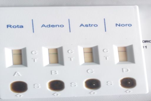
Citation: Ajayi AO, et al (2020) Enteric Viruses and Bacterial Species from the Faecal Droppings of Bats in Selected Bat Colonies in Ondo State, Nigeria. Journal of
Virology Research & Reports. SRC/JVRR-106. DOI: https://doi.org/10.47363/JVRR/2020-(1)106

Members of the Adenoviridae family are non-enveloped,                              Sample Collection
icosahedral viruses, of about 70–90nm in size, with a linear, double-              Between January to March 2013, during each visit to the bat
stranded DNA genome ranging from 26 to 46 kb. DNA sequence                         colonies, early in the morning. Bat fecal samples were collected
analysis indicates that this family is divided into five genera:                   from five different locations (Ikare, Supare, Ugbe, Epinmi and
Mastadenovirus, Aviadenovirus, Atadenovirus, Siadenovirus and                      Akure) in Ondo State. Freshly voided samples were aseptically
Ichtadenovirus. Adenoviruses (AdVs) infect a wide range of                         collected into sample bottles and preserved with ice packs/blocks
vertebrates, including mammals, birds, amphibians, reptiles and                    in a clean cooler before transportation to the laboratory for analysis.
fish. Members of the Mastadenovirus and Aviadenovirus genera
only infect mammals and birds [24,25]. Recently, great attention                   Sample Analysis
has been paid to bats given their role as reservoirs of emerging                   The fecal samples were tested for the presence of Rotavirus and
viruses, including SARS coronavirus, Ebolavirus, Hendra virus                      Adenovirus antigens using the Certest Biotech Quadriple EIA
and Nipah virus [6]. Acute infective gastroenteritis is a major                    kit according to the manufacturer’s specification (Certest, 2013).
global health problem, which manifests as three or more watery
or loose bowel evacuations with fever and vomiting in a 24-hour                    Test Principle
period that may last several days. Children under 5 years of age                   The strip consists of a nitrocellulose membrane pre coated
are particularly susceptible, and global estimates indicated a mean                with mouse monoclonal antibodies on the test line (T), in the
of between 3.5 and 7 episodes of severe diarrhea during the first 2                result window, against Rotavirus or Adenovirus and with rabbit
years of life, and the greatest burden is in the developing countries              polyclonal antibodies on the control line (C) against a specific
because of poor sanitation, lack of safe drinking water, and bad                   protein. The label/sample absorbent pad is sprayed with labelled
sanitary habits. Several studies in Nigeria showed that more than                  solution (mouse monoclonal antibodies anti-Rotavirus) conjugated
315,000 deaths of preschool age children are recorded annually as                  to red polystyrene latex and control label solution (Specific binding
a result of diarrhea disease humans include Group-A rotaviruses,                   protein) conjugated to green polystyrene latex, forming coloured
astroviruses, human caliciviruses and the enteric adenoviruses.                    conjugate complexes.
Rotaviruses are members of the family Reoviridae and genus
Rotavirus and contain 3 primary species: Rotavirus A, Rotavirus                    Test Procedures
B, and Rotavirus C. The diagnostic system is also applicable to                    Sample was mixed properly with the Extraction buffer to ensure
other viral groups [26-35].                                                        good sample dispersion and extraction of the virus antigen. The
                                                                                   Certest kit was removed from its sealed bag and four drops of the
The most widely used method for laboratory diagnosis is the                        homogenized sample was introduced into the window marked with
enzyme immunoassay (EIA or ELISA), which detects the RVA                           letter A and B. The results were taken at ten minutes. Materials
group antigen (VP6) directly on stools. The genetic methods -                      used for this study includes sample bottles, 70% of Ethanol,
microarray and real-time PCR (RT-qPCR) - are very sensitive and                    cotton wool, nose cover, hand gloves, cardboard, CerTest Biotech
capable of discriminating mixed RVA infections, and have been                      Quadruple EIA kit for the detection of adenovirus and rotavirus,
successfully developed and employed in diagnosis of RV (34). The                   cooler for the preservation of samples and kits used. Similarly, the
bacterial population of bat feacal dropping was also described by                  bacterial species were identified using standard microbiological
Wolkers et al., whose study emphasized that bats are commonly                      methods including API kits.
regarded as vectors for viruses, but paucity of information is
available about bacterial communities in bats and the possible role                Bacteriological Analysis
of bats in the transmission of foodborne diseases. The reported                    Eosine methylene blue agar, peptone water and Nutrient agar
bacterial genera predominantly recovered from fecal bacteria in                    were generally used for the bacteriological analysis. 1gram of the
bats were found to be diverse with Corynebacterium, Serratia,                      sample was weighed and was diluted in 9ml sterile peptone water
Pseudomonas, Enterococcus and Yersinia. The opportunistic                          and serial dilution was done .1ml of inoculum was taken from 10-
bacterial pathogens including Citrobacter freundii, Escherichia                    1
                                                                                     ,10-2,10-3 dilution to seed sterile plates of Eosine methylene blue
coli, Enterococcus faecalis, Serratia fonticola and Rahnella                       (EMB) agar and Nutrient agar in replicates employing pour plate
aquatilis were also obtained [8, 36]. The microbial species detected               technique An un-inoculated plate serve as the growth control for
in bat feacal droppings in this study can cause acute diarrheal                    the two culture plates. (Nutrient agar and EMB agar). The plates
disease (ADD) amongst other infectious diseases that can result to                 were swirled for even saturation of the inoculum and the media,
diseases outbreaks. Acute diarrheal disease (ADD) is a syndrome                    the plates were properly labelled, inverted and incubated at 37ᵒC
caused by different agents (bacteria, viruses and parasites), and                  for 24hrs. After 24hrs, the result colonies were carefully examined
its major manifestation is increased number of bowel movements,                    and determined for its cultural characteristics, the diversity and
with watery or loose stools in the susceptible host [5,8,12]. This                 proportion of the isolates. The Gram negative organisms were
study therefore helps to elucidate the involvement of different                    identified using API kit.
microbial groups that can be harbored by bats. This will help to
control infection from these sources.                                              Biochemical Characteristics of the Bacterial Isolates Using
                                                                                   Analytical Profile Index (Api) Kit
Materials and Methods                                                              The API-20E fast identification system combines some of
Study Site                                                                         conventional tests and allows the identification of a limited number
The study site used in Ondo State, Nigeria were based on the                       of Gram negative Enterobacteriaceae or Non-Enterobacteriaceae.
availability of Bat colonies in this area as listed here. This includes,           The test system is stored in 20 small reaction tubes or wells
Akure, Epinmi-Akoko, Ikare-Akoko, Supare-Akoko, Ugbe-Akoko                         containing dehydrated substrate to detect enzymatic activity,
as well as on a roof top of an uncompleted building at Owo,                        usually related to fermentation of carbonhydrates or catabolism of
Ondo state Nigeria for intensive enteric virus screening and                       proteins or amino acids by the inoculated organisms. A bacterial
bacteriological analysis analysis.                                                 suspension is used to rehydrate each of the wells and the strips
                                                                                   are incubated. During incubation, metabolism produces colour
                                                                                   changes that are either spontaneous or revealed by the addition
J Viro Res Rep, 2020                                                                                                                            Volume 1(1): 2-10
Citation: Ajayi AO, et al (2020) Enteric Viruses and Bacterial Species from the Faecal Droppings of Bats in Selected Bat Colonies in Ondo State, Nigeria. Journal of
Virology Research & Reports. SRC/JVRR-106. DOI: https://doi.org/10.47363/JVRR/2020-(1)106
of reagents. All positive and negative test results are compiled to the bacterial isolates to commonly used antibiotics. This test was
obtain a profile numbers in a commercial codebook or manual to carried out following the Kirby-Bauer disk diffusion methods of
determine the identification of the bacterial isolates. CLSI, (37). Inoculum from culture of bacteria isolates on nutrient
agar slants were inoculated into test tubes containing sterilized
nutrient broth and incubated at 37 for 18hours which serve as the
The Api20e Kit Contain the Following Dehydrated Substrate stock for the test.1ml from this stock was then transferred into
1. ONPG: Test for β-galactosidase enzyme by hydrolysis of the sterilized petri plate and 20ml of already prepared and sterilized
substrate o-nitrophenyl-b-D-galactopyranoside Mueller Hinton agar was inoculated. The plates were then allowed
2. ADH: Decarboxylation of the amino acid arginine by arginine to cool for about 15 minutes so as to allow the inoculated plate to
dihydrolase gel and excess surface moisture to be absorbed before applying the
3. LDC: Decarboxylations of the amino acid lysine by lysine antibiotics impregnated discs. Predetermined commercial Gram
decarboxylase negative and gram positive discs were applied to the surface of
4. ODC: Decarboxylations of the amino acid ornithine by the well labelled inoculated agar plates aseptically using sterile
ornithine decarboxylase forceps. The disks were placed firmly by slightly pressing on the
5. CIT: Utilization of citrate as only carbon source inoculated plates with the sterilized forceps to ensure complete
6. H2S: Production of hydrogen sulfide contact with the agar, the discs were applied according. After
7. URE: Test for the enzyme urease 18hours of incubation, each plate was examined. The susceptibility
8. TDA (Tryptophan deaminase): Detection of the enzyme responses of the isolates to respective antibiotics were indicated by
tryptophan deaminase: Reagent to put- Ferric Chloride. a clear zone of inhibition measured using a calibrated ruler which
9. IND: Indole Test-production of indole from tryptophan by was held on the back of the inverted petri-plate and was recorded.
the enzyme tryptophanase. Reagent- The isolates were then scored as either sensitive to the antibiotics
10. VP: The Voges-Proskauer test for the detection of acetoin or resistant depending on the diameter of the zone of inhibition.
(acetyl methylcarbinol) produced by fermentation of glucose
by bacteria utilizing the butylene glycol pathway Results
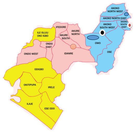
11. GEL: Test for the production of the enzyme gelatinase which Figure 1 shows the map of the study area in Ondo State, Nigeria,
liquefies gelatin from where this bat dropping samples were collected. The specific
12. GLU: Fermentation of glucose (hexose sugar) areas with bat colonies includes Owo in Owo Local Government
13. MAN: Fermentation of mannose (hexose sugar) for the bacterial species analysis, Akure, Akure South Local
14. INO: Fermentation of inositol (cyclic polyalcohol) Government Area; Epinmi in Akoko South East Local Government
15. SOR: Fermentation of sorbitol (alcohol sugar) Area, Ikare in Akoko North East Local Government Area, Supare
16. RHA: Fermentation of rhamnose (methyl pentose sugar) in Akoko South West Local Government Area and Ugbe in Akoko
17. SAC: Fermentation of sucrose (disaccharide) South East Local Government Area, Ondo State manily for the
18. MEL: Fermentation of melibiose (disaccharide) viral species analysis.
19. AMY: Fermentation of amygdalin (glycoside)
20. ARA: Fermentation of arabinose (pentose sugar)
Procedure
Suspension of each isolates were prepared in a sterile distilled
water and it was introduced into the API20E biochemical test
strip which contains dehydrated bacterial bio-chemical reagents
in 20 separate compartments accordingly with a sterile syringe.
Sterile oil was added into the Arginine dihydrolase (ADH), Lysine
decarboxylase (LDC), Ornithine decarboxylase (ODC), Hydrogen
sulphide (H2S), and Urease (URE) compartments, Some drops
of water is added in the tray and the API test strip was placed on
the tray and it was incubated for 24 hours.
Reading of Results
The colour change was observed after 24 hours, reagents were
added to some specific compartments. One drop of ferric chloride
was added to Tryptophan deaminase (TDA), one drop of Kovacs
reagent was added to indole (IND) and one drop of KOH (VP
reagent1 and reagent 2) was added to Voges-Proskauer (VP) and
the result was gotten after 10minutes. The organism was identified
by using API catalog.
Antibiotics Susceptibility Testing of the Bacteria Isolates
The test was performed to determine the phenotypic resistance of Figure 1: Map of Ondo State, showing Areas of Sample collection
J Viro Res Rep, 2020 Volume 1(1): 3-10Citation: Ajayi AO, et al (2020) Enteric Viruses and Bacterial Species from the Faecal Droppings of Bats in Selected Bat Colonies in Ondo State, Nigeria. Journal of
Virology Research & Reports. SRC/JVRR-106. DOI: https://doi.org/10.47363/JVRR/2020-(1)106
Throughout the period of this study, two samples tested positive for both Adenovirus and Norovirus in bat fecal samples from Epinmin
in Akoko South East Local Government Area, Ondo State, Nigeria (Tables 1 and 2). Between January and March, 2018, a total number
of twenty samples were collected from the five locations and tested. The fecal characteristics and result of the test are presented in
Tables 1, 2 and Fig. 2. Throughout the period of this study, two samples tested positive for both Norovirus and Adenovirus in bat
fecal samples from Epinmin in Akoko South East Local Government Area, Ondo State, Nigeria.
Table 1: Bat Droppings Collection Logistics
Specimen Location Sample code Time of Time of analysis Stool Norovirus
collection date collection characteristics
20/01/2018 Ikare A 7:00am 8:00am Solid and dry -
21/01/2018 Ikare B 8:00am 9:00am Solid and dry -
23/01/2018 Supare C 8:00am 9:00am Solid and dry -
24/01/2018 Ugbe D 9:00am 10:00am Solid and dry -
26/01/2018 Ugbe E 9:00am 10:00am Semi-solid -
31/01/2018 Epinmi F 10:00am 11:00am Semi-solid -
01/02/2018 Epinmi G 10:00am 11:00am Semi-solid +
02/02/2018 Epinmi H 10:00am 11:00am Semi-solid -
03/02/2018 Epinmi I 10:00am 11:00am Semi-solid -
04/02/2018 Epinmi J 09:00am 10:00am Semi-solid -
05/02/2018 Epinmi K 09:00am 10:00am Semi-solid -
06/02/2018 Epinmi L 09:00am 10:00am Semi-solid -
07/02/2018 Akure M 10:00am 11:00am Semi-solid -
08/02/2018 Akure N 10:00am 11:00am Semi-solid -
09/02/2018 Akure O 10:00am 11:00am Semi-solid -
10/02/2018 Akure P 10:00am 11:00am Semi-solid -
11/02/2018 Akure Q 10:00am 11:00am Semi-solid -
12/02/2018 Akure R 10:00am 11:00am Semi-solid -
15/02/2018 Akure S 10:00am 11:00am Semi-solid -
Table 2: Test Analysis of Adenovirus and Rotavirus Detection in Bat Fecal Samples from Various Locations in Ondo State
Specimen Location Sample Time of Time of Stool Adenovirus Rotavirus
collection date indicator collection analysis characteristics
0/01/2018 Ikare A 7:00am 8:00am Solid and dry - -
21/01/2018 Ikare B 8:00am 9:00am Solid and dry - -
23/01/2018 Supare C 8:00am 9:00am Solid and dry - -
24/01/2018 Ugbe D 9:00am 10:00am Solid and dry - -
26/01/2018 Ugbe E 9:00am 10:00am Semi-solid - -
31/01/2018 Epinmi F 10:00am 11:00am Semi-solid - -
01/02/2018 Epinmi G 10:00am 11:00am Semi-solid + -
02/02/2018 Epinmi H 10:00am 11:00am Semi-solid - -
03/02/2018 Epinmi I 10:00am 11:00am Semi-solid - -
04/02/2018 Epinmi J 09:00am 10:00am Semi-solid - -
05/02/2018 Epinmi K 09:00am 10:00am Semi-solid + -
06/02/2018 Epinmi L 09:00am 10:00am Semi-solid - -
07/02/2018 Akure M 10:00am 11:00am Semi-solid - -
08/02/2018 Akure N 10:00am 11:00am Semi-solid - -
09/02/2018 Akure O 10:00am 11:00am Semi-solid - -
10/02/2018 Akure P 10:00am 11:00am Semi-solid - -
11/02/2018 Akure Q 10:00am 11:00am Semi-solid - -
12/02/2018 Akure R 10:00am 11:00am Semi-solid - -
15/02/2018 Akure S 10:00am 11:00am Semi-solid - -
16/02/2018 Akure T 10:00am 11:00am Semi-solid - -
J Viro Res Rep, 2020 Volume 1(1): 4-10Citation: Ajayi AO, et al (2020) Enteric Viruses and Bacterial Species from the Faecal Droppings of Bats in Selected Bat Colonies in Ondo State, Nigeria. Journal of
Virology Research & Reports. SRC/JVRR-106. DOI: https://doi.org/10.47363/JVRR/2020-(1)106
Figure 2: Sample of Certest Kit Used For Adenovirus, Norovirus and Rotavirus Examination in Faecal Sample Collected From
Sample sources
The Gram positive bacteria recovered from fecal pellets of bat belongs to five genera namely: Streptococcus spp, Micrococcus spp,
Staphylococcus Aureus, Enterococcus spp. and Staphylococcus Epidermidis [5]. Similarly, the Gram negative bacteria recovered
from fecal pellets of bat belongs to ten genera namely: Salmonella Typhi, Echerichia Coli, Klebsiella Oxytoca, Acinetobacter
Iwoffi, Citrobacter Freundii, Serratia Marcescens, Enterobacter Cloacae, Proteus Vulgaris, Enterobacter Agglomerans and Proteus
Mirabillis (Tables 3 to 5) [10].
Table 3: Cultural and Morphological Characteristics of the Bacterial Isolates from the Fecal Pellets of Bats
Cultural characteristics. Cellular morphology
Isolates edge pigment shape Elevation Morphology Gram
M1 Pink irregular Rough Flat Cocci G +ve
M2 Milky Circular Rough Flat Rods G – ve
M3 Milky Circuler Rough Flat Tiny Cocci G + ve
M4 Milky Irregular Crenate Raised Rods G – ve
M5 Pink Circular Crenate Flat Rods G – ve
M6 Creamy Irregular Crenate Flat Cocci + Rods G – ve
M7 Pink Irregular Rough Flat Cocci G + ve
M8 Milky irregular Rough Flat Cocci G + ve
M9 Milky Circuler Crenate Flat Cocci G + ve
M10 Pink Irregular Crenate Flat Rods G – ve
M11 Milky Irregular Rough Flat Rods G – ve
M12 Creamy Irregular Rough Raised Rods G – ve
M13 Milky Circular Smooth Raised Cocci G + ve
M14 Pink Irregular Lobate Raised Rods G – ve
M15 Pink Irregular Smooth Raised Rods G – ve
M16 Pink Circular Smooth Raised Rods G – ve
M17 Milky Circular Rough Flat Tiny Cocci G + ve
M18 Creamy Circular Smooth Raised Rods G – ve
M19 Pink Circular Lobate Raised Rods G – ve
M20 Milky Irregular Rough Flat Rods G - ve
J Viro Res Rep, 2020 Volume 1(1): 5-10Citation: Ajayi AO, et al (2020) Enteric Viruses and Bacterial Species from the Faecal Droppings of Bats in Selected Bat Colonies in Ondo State, Nigeria. Journal of
Virology Research & Reports. SRC/JVRR-106. DOI: https://doi.org/10.47363/JVRR/2020-(1)106
Table 4: Biochemical characteristics and probable identity of the Gram positive Bacteria isolates
Isolate Catalase Coagulase Spore test Oxidase Haemolysin Glucose Sucrose Lactose Probable organism
test
M1 - - - - + + - + Streptococcus spp.
NG NG NG
M3 + - + - - + + Micrococcus spp
NG GP GP
M7 - - - - + + - + Streptococcus spp.
NG NG NG
M8 + + - + + + - + Staphylococcus aureus
NG GP GP
M9 - + - - - + - - Enterococcus spp.
M13 + - - - - + - + Staphylococcus
epidermidis
M17 + - - + - - + + Micrococcus spp.
NG GP GP
KEY: NG= No Gas Production, GP= Gas Production
Table 5: Biochemical characteristics and probable identity of the Gram Negative Bacteria isolates (API Kit)
Isolates API identification code Probable Organism % probability
M2 4404540 Salmonella typhi 99.96
M4 7542001 Echerichia coli 98.97
M5 5445673 Klebsiella oxytoca 99.50
M6 6306042 Acinetobacter iwoffi 95.30
M10 5215772 Klebsiella pneumonia 99.87
M11 3634552 Citrobacter freundii 94.10
M12 7532000 Echerichia coli 80.58
M14 7312153 Serratia marcescens 95.64
M15 3305572 Enterobacter cloacae 71.47
M16 2756000 Proteus vulgaris 81.06
M18 5315572 Enterobacter agglomerans 97.76
M19 2756400 Proteus mirabillis 99.43
M20 7512000 E. coli (inactive) 76.82
The bacterial isolates showed multiple antibiotic resistant to antibiotics provided, with Levofloxacin and Ampiclox having high
susceptibility effect against Gram positive bacteria. Similarly, Ofloxacin and Nalidixic acid having high susceptibility effect against
Gram negative bacteria (Table 6A and B).
Table 6A: Antibiotic susceptibility profile of the Gram positive bacterial isolate from fecal pellet of bats
Zones of inhibition by the antibiotics(MM)
ISOLATES CIP CIN AMX STR LVX ERY CHL APX
M1 12R 10R 0R 5R 10R 10R 15S 16S
M2 13R 14R 0R 4R 15S 8R 10R 17S
M3 13R 12R 6R 3R 13R 8R 18S 18S
M4 10R 10R 9R 0R 13R 0R 12R 20S
M5 13R 15S 0R 0R 15S 12R 13R 13R
M6 13R 13R 6R 0R 12R 13R 14R 14R
M7 11R 15S 4R 6R 10R 14R 15S 15S
M8 14R 10R 0R 8R 14R 15S 16S 18S
M9 15S 12R 5R 10R 13R 15S 15S 19S
M10 10R 11R 6R 12R 15S 8R 8R 18S
M11 14R 8R 0R 9R 18S 10R 6R 15S
M12 13R 9R 8R 10R 16S 13R 13R 13R
J Viro Res Rep, 2020 Volume 1(1): 6-10Citation: Ajayi AO, et al (2020) Enteric Viruses and Bacterial Species from the Faecal Droppings of Bats in Selected Bat Colonies in Ondo State, Nigeria. Journal of
Virology Research & Reports. SRC/JVRR-106. DOI: https://doi.org/10.47363/JVRR/2020-(1)106
M13 10R 12R 0R 8R 18S 6R 6R 14R
M14 12R 13R 6R 9R 15S 15S 15S 14R
M15 14R 15S 8R 11R 10R 18S 8R 15S
M16 10R 13R 9R 5R 0R 6R 16S 20S
M17 11R 12R 0R 8R 0R 12R 17S 15S
M18 13R 11R 5R 0R 16S 8R 8R 20S
M19 12R 10R 8R 0R 15S 10R 15S 20S
M20 14R 8R 0R 11R 10R 10R 15S 0R
CIP= Ciprofloxacin, CIN= cinoxacin, AMX= Amoxicillin STR= Streptomycin, LVX= levofloxacin, ERY= Erythromycin, CHL=
Chloramphenicol, APX= Ampiclox,
R-Resistant
S- Sensitive
Table 6B: Antibiotic Susceptibility Profile of the Gram Negative Bacterial Isolate from Fecal Pellet of Bats
ISOLATES Zones of inhibition by the antibiotics (MM)
OFX PEF CPX AUG CIN GEN CEP NAL SXT PEN
M1 23S 10R 0R 0R 13R 13R 10R 21S 0R 0R
M2 26S 0R 19S 0R 10R 18S 12R 22S 19S 0R
M3 30S 9R 0R 0R 0R 13R 10R 0R 15S 0R
M4 36S 16S 0R 0R 9R 13R 0R 34S 16S 7R
M5 36S 10R 0R 0R 16S 14R 10R 18S 14R 8R
M6 17S 11R 0R 0R 10R 13R 10R 22S 20S 6R
M7 30S 16S 0R 0R 11R 15S 18S 22S 20S 5R
M8 33S 38R 0R 0R 16S 14R 0R 35S 21S 0R
M9 44S 0R 24S 0R 38S 13R 0R 0R 18S 0R
M10 28R 19S 0R 0R 0R 16S 6R 22S 18S 0R
M11 42S 0R 0R 0R 17S 14R 10R 0R 10R 0R
M12 43S 9R 0R 0R 18S 14R 11R 0R 8R 0R
M13 0R 0R 0R 0R 0R 15S 16S 24S 15S 0R
M14 28S 16S 0R 0R 9R 15S 0R 18R 18S 0R
M15 42S 10R 0R 0R 0R 0R 0R 15S 15S 0R
M16 43S 11R 0R 0R 16S 15S 0R 20S 15S 0R
M17 0R 10R 0R 0R 15S 16S 0R 21S 14R 0R
M18 34S 16S 0R 0R 0R 13R 15S 22S 14R 0R
M19 0R 0R 0R 0R 10R 14R 18S 35S 14R 0R
M20 26S 14R 0R 0R 19S 15S 10R 0R 6R 0R
OFX= Ofloxacin, PEF= Pefloxacin, CPX= Ciprofloxacin, AUG= Augmentin, CIN= cinoxacin, GEN= Gentamycin, CEP=
Cephalosporine, NAL= Nalidixic acid,
SXT= Trimethopri-sulphamethoxazole PEN= Penicillin
R-Resistant
S-Sensitive
Discussion papillomavirus, parvovirus and a calicivirus (sapovirus), reported
The global diversity of viruses found in bats is still largely by [27, 41-47]. It is difficult to make specific or direct comparisons
unknown, and it is thought that a great number of virus taxa with the present study due to significant methodological variation
are yet to be discovered. Bats are often asymptomatic during among the studies including: sample type (i.e. fresh guano, urine,
infection, but the disease can be severe for a new host species when roost guano). Perhaps the most comparable study is that of, who
cross-species transmission occurs. Surveys of the bat virome and used a metagenomics approach to examine virus diversity in
monitoring of virus dynamics in bat populations can inform our African straw coloured fruit bats (Eidolon helvum) [41]. Human
fundamental understanding on how viral pathogens emerge and adenoviruses (HAdV) are major causes of clinical infections
evolve, enabling strategies for the prevention of future outbreaks including respiratory diseases, gastroenteritis, and conjunctivitis
or pandemics [38-40]. This present study shows the presence of [16]. Human Adenovirus belongs to the genus Mastadenovirus of
Adenovirus in fecal bat from Epinmi, Ondo state. Metagenomic the family Adenoviridae, The virion consists of non-enveloped,
studies in bats from other countries have identified adenovirus, linear, dsDNA genome (26–45 kb) encapsidated in an icosahedral
J Viro Res Rep, 2020 Volume 1(1): 7-10Citation: Ajayi AO, et al (2020) Enteric Viruses and Bacterial Species from the Faecal Droppings of Bats in Selected Bat Colonies in Ondo State, Nigeria. Journal of
Virology Research & Reports. SRC/JVRR-106. DOI: https://doi.org/10.47363/JVRR/2020-(1)106
protein shell. There are 52 serotypes classified into six species, A-G, the opportunity for infections [7, 53]. Like bird droppings, bat
of which species F serotypes 40 and 41 primarily affect the gut, guano can contain a potentially infectious fungus Histoplasma
contributing 5%-20% of hospitalizations for childhood diarrhea. capsulatum that causes lung infection known as histoplasmosis
The incidence of infection is nearly 3 times greater in developing [55]. Bats breed in high densities within human populations in
countries than developed ones. In recent years, bats have gained some countries, especially in tropics including Nigeria, whereby
significant notoriety for being implicated in numerous emerging humans and animals are in close contact. Hence, there is increased
infectious disease events, and their importance as reservoir hosts risk for cross species transmission. Bat populations appear to be
for viruses that can cross species barriers to infect humans and declining noticeably in response to human induced environmental
other domestic and wild mammals is increasingly recognized. factors like habitat destruction and fragmentation, disturbance to
Historically, there has been a significant body of work on the role of caves, depletion of food resources, overhunting for bush meat
bats as reservoirs of infectious agents, but relatively little available and persecution. Other factors that stress the environment such
information on members of the suborder Megachiroptera (flying as use of pesticides, infectious disease, and industrial activities
foxes and fruit bats). Although the role of bats in harbouring viruses including wind energy turbine contributes to this. The role of bat
(alphaviruses, flaviviruses, rhabdoviruses and arenaviruses) is well as reservoir of deadly etiologic agent become more pronounced
established, there is increasing global interest in evaluating the during the COVID-19 pandemic when it was documented as a
broad range of potential infectious agents that bats harbour, with probable zoonotic source of the disease. Also, bats are known
a particular focus on potential emerging pandemic threats. This to feed mostly on fruit, therefore this study recommends that
concern may be somewhat exaggerated, as bats themselves do not fruits bought from markets should be washed thoroughly before
represent the real threat to people as regards potential pathogens consumption. Nevertheless, much caution should be taken on
leading to large-scale zoonotic disease events. However, it is worth bat hibernating in domestic places as well as the humans and
investigating the infectious agents like noroviruses harboured by animals contact. The result obtained in this study will help in
bats, and integrating this information with an understanding of health management system.
the risk of transmission from bats to people or livestock, which
may serve as intermediate hosts and transmission vectors linking Acknowledgement
bats and people. In this study, Norovirus was detected in two We appreciate the support of Environmental Microbiology and
samples (2/20) representing a rate of 10% in bats of the studied Virology research unit of the Department of Microbiology,
area. This finding indicates that bats may be a potential route of Adekunle Ajasin University, Akungba-Akoko, Ondo State,
transmission of enteric norovirus infection to humans. Viral host Nigeria. This is in terms of samples collection and logistics used
switching is probably determined by the chances of interspecies for the study.
contact, as well as by the concentration and prevalence of virus
in the donor species. To judge zoonotic risks associated with bats, References
when and where these 2 variables would favor transmission must 1. IUCN (2010) IUCN Red List of Threatened Species, version,
be determined [47-52]. www.iucnredlist.org/initiatives/ mammals.
2. Koopman KF (1993) Chiroptera In D.E. Wilson and D.M.
The samples from the location mentioned in our study were Reeder, eds. Mammalian species of the world, Washington,
selected because they are regularly domiciled with bats and thus DC, Smithsonian Institution Press, pp. 137-241.
provide a certain chance of detection. Attempts to characterize 3. Wang LF, Shi Z, Zhang S, Field H, Daszak P, Eaton BT (2006)
virus dynamics in bat populations have been made earlier by using Review of bats and SARS. Emerg Infect Dis 12: 1834-1840.
the examples of lyssaviruses (rabies virus and related species), 4. Wibbelt G, Kurth A, Yasmum N (2007) Discovery of
filoviruses (Ebola and Marburg viruses) and henipaviruses herpesviruses in bats, Journal of General Virology 88: 2651-
(Hendra and Nipah viruses). However, because these viruses are 2655.
found rarely, only vague conclusions have so far been made. For 5. Wolkers JCM, Rebmann K, Bosch T, Hazeleger WC (2019)
instance, increased contact between bats and humans through bat Fecal Bacterial Communities in Insectivorous Bats from
migratory events or fruit harvesting periods have been temporally the Netherlands and Their Role as a Possible Vector for
linked with individual human cases of Ebola and Nipah virus Foodborne Diseases. Acta Chiropterologica. 20: 475-483.
infection. This present study shows the presence of Norovirus in 6. Moratelli R (2013) Evolutionary history of bats: fossils,
fecal bat from Epinmi, Ondo state. Bats are hosts to a range of molecules and morphology. Journal of Mammalogy 94:
zoonotic and potentially zoonotic pathogens. They differ from 520-521.
other disease reservoirs because of their unique and diverse 7. Hayman DTS (2016) Bats as Viral reservoirs. Annu Rev
lifestyles, including their ability to fly, often highly gregarious Virol. https://pubmed.ncbi.nlm.nih.gov/27578437, Accessed
social structures, long life spans, and low fecundity rates. They on 27th July 27, 2020.
represent a potential epidemiologic of several diseases that 8. Li J, Li L, Jiang H, Yuan L, Zhang L, et al. (2018) Fecal
can be fatal to humans, including rabies, Ebola, leptospirosis, bacteriome and mycobiome in bats with diverse diets in South
histoplasmosis, and pseudotuberculosis [7, 53]. Bats are reservoirs China. Current Microbiology 75: 1352-1361.
of several pathogens, whose spread may be related to physiological 9. Vengust M, Knapic T, Weese JS (2018) The fecal bacterial
stress associated with habitat loss or alteration [54]. On basis of microbiota of bats; Slovenia. PLoS ONE 13: e0196728.
this, our study shows that the bacterial isolates from bat feacal 10. McCracken GF, Gustin MK (1991) Nursing behavior in
droppings had multiple antibiotic resistance to antibiotics provided. Mexican free-tailed bat maternity colonies. Ethology 89:
Levofloxacin and Ampiclox have high susceptibility effect against 305-321.
Gram positive bacteria. Similarly, Ofloxacin and Nalidixic acid 11. Green KY, Chanock RM, Kapikian AZ (2001) Human
having high susceptibility effect against Gram negative bacteria caliciviruses. In: Knipe, D.M., Howley, P.M. (Eds.), Fields
(Table 6a and b). This corroborates with reports of Wibbelt et al. Virology 1: 841-874.
and Calisher, et al. who demonstrated the presence of various 12. Adler JL, Zickl R (1969) Winter vomiting disease. Journal
pathogenic organisms including bacterial species from bat sources. of Infectious Diseases 119: 668-673
Human activities that increase exposure to bats will likely increase 13. Green KY, Ando T, Balayan MS, Berke T, Clarke IN, et al.
J Viro Res Rep, 2020 Volume 1(1): 8-10Citation: Ajayi AO, et al (2020) Enteric Viruses and Bacterial Species from the Faecal Droppings of Bats in Selected Bat Colonies in Ondo State, Nigeria. Journal of
Virology Research & Reports. SRC/JVRR-106. DOI: https://doi.org/10.47363/JVRR/2020-(1)106
(2000) Taxonomy of the caliciviruses. Journal of Infectious genome analysis of snake adenovirus type 1, a representative
Diseases 181: S322-S330. of the reptilian lineage within the novel genus atadenovirus.
14. Batten CA, Clarke IN, Kempster SL, Oliver SL, Bridger JC, Virus Res 132: 132-139.
Lambden PR (2006) Characterization of a cross-reactive 33. Japhet MO, Adesina OA, Famurewa O, Svensson L, Nordgren
linear epitope in human genogroup I and bovin genogroup J (2012) Molecular Epidemiology of rotavirus and norovirus
III noroviruscapsid proteins. Virology 356: 179-187. in Ile-Ife, Nigeria: High prevalence of G12P (8) rotavirus
15. Hsu CC, Wobus CE, Steffen EK, Riley LK, Livingston strains and detection of a rare norovirus genotype. J Med
RS (2005) Development of a microsphere-based serologic Virol 84: 1489-1496.
multiplexed fluorescent immunoassay and a reverse 34. Kabayiza JC, Andersson ME, Nilsson S, Bergström T,
transcriptase PCR assay to detect murine norovirus 1 infection Muhirwa G, et al. (2014) Real-time PCR identification of
in mice. Clinical and Diagnostic Laboratory Immunology agents causing diarrhea in Rwandan children less than 5 years
12: 1145-1151. of age. Pediatr Infect Dis J 33: 1037-42.
16. Van, H.J., Ehlers, M.M., Van, Zyl, W.B. and Grabow, W.O.K. 35. Altschul SF, Gish W, Miller W, Myers EW, Lipman DJ (1990)
(2003). Incidence of adenoviruses in raw and treated water. Basic local alignment search tool. J Mol Biol 215: 403-410.
Water Re. 37: 3704 -3708. 36. Ingala MR, Simmons NB, Wultsch C, Krampis K, Speer KA,
17. Han MG, Smiley JR, Thomas C, Saif LJ (2004) Genetic et al. (2018) Comparing microbiome sampling methods in a
recombination between two genotypes of genogroup III wild mammal: fecal and intestinal samples record different
bovine noroviruses (BoNVs) and capsid sequence diversity signals of host ecology, evolution. Frontiers in Microbiology
among BoNVs and Nebraska like bovine enteric caliciviruses. 9: 803.
Journal of Clinical Microbiology 42: 5214-5224. 37. Clinical and Laboratory Standards Institute (CLSI) (2016)
18. Deng Y, Batten CA, Liu BL, Lambden PR, Elschner M, et al. Performance Standards for Antimicrobial Susceptibility
(2003) Studies of epidemiology and seroprevalence of bovine Testing. 26th ed. CLSI supplement M100S. Clinical and
norovirusesin Germany. Journal of Clinical Microbiology Laboratory Standards Institute, 950 West Valley Road, Suite
41: 2300-2305. 2500, Wayne, Pennsylvania 19087, USA.
19. Farkas T, Nakajima S, Sugieda M, Deng X, Zhong W, et al. 38. Anthony SJ, Epstein JH, Murray KA, Navarrete-Macias I,
(2005) Seroprevalence of noroviruses in swine. Journal of Zambrana-Torrelio CM, et al. (2013) A strategy to estimate
Clinical Microbiology 43: 657-661. unknown viral diversity in mammals. M Bio 4: e00598-
20. Martella V, Bányai K, Matthijnssens J, Buonavoglia C, Ciarlet e00e13.
M (2010) Zoonotic aspects of rotaviruses. Vet Microbiol 39. Chan JF-W, To KK-W, Tse H, Jin D-Y, Yuen K-Y (2013)
140: 246-55. Interspecies transmission and emergence of novel viruses:
21. Lopman BA, Brown DWG, Koopmans M (2002) Human lessons from bats and birds. Trends Microbiol 21: 544-555.
calicivirus in Europe. Journal of Clinical Virology 24: 137-60. 40. Smith I, Wang LF (2013) Bats and their virome: an important
22. Zheng M, Zhang Y, Zhu Q, Wang X, Yu H (2010) Clinical source of emerging viruses capable of infecting humans. Curr
and molecular epidemiology of rotavirus in children with Opin Virol 3: 84-91.
community acquired and hospital acquired diarrhea in 41. Baker KS, Leggett RM, Bexfield NH, Alston M, Daly G,
Shangai, China. Pediatr Infect Dis J 29: 177-80. et al. (2013) Metagenomic study of the viruses of African
23. Duizer E, Schwab KJ, Neill FH, Atmar RL, Koopmans MP, et straw-coloured fruit bats: detection of a chiropteran poxvirus
al. (2004) Laboratory efforts to cultivate noroviruses. Journal and isolation of a novel adenovirus. Virology 441: 95-106.
of General Virology 85: 79-87. 42. Drexler JF, Corman VM, Wegner T, Tateno AF, Zerbinati
24. Harrach B (2015) Random sampling of the Central European RM, et al. (2011) Amplification of emerging viruses in a bat
bat fauna reveals the existence of numerous hitherto unknown colony. Emerg Infect Dis 17: 449-456.
adenoviruses. Acta Vet Hung 63: 508-525. 43. He B, Li Z, Yang F, Zheng J, Feng Y, et al. (2013) Virome
25. Benkö M, Harrach B (2003) Molecular evolution of profiling of bats from Myanmar by metagenomic analysis
adenoviruses. Curr Top Microbiol Immunol 272: 3-35. of tissue samples reveals more novel mammalian viruses.
26. Corman, VM, Baldwin HJ, Tateno AF, Zerbinati RM, Annan PLOS ONE 8: e61950.
A, et al. (2015). Evidence for an ancestral association of 44. Li L, Victoria JG, Wang C, Jones M, Fellers GM, et al. (2010).
human coronavirus 229E with Bats. J Virol 89: 11858-11870. Bat guano virome: predominance of dietary viruses from
27. Ge XY, Li JL, Yang XL, Chmura AA, Zhu G, et al. (2013) insects and plants plus novel mammalian viruses. J Virol
Isolation and characterization of a bat SARS-like coronavirus 84, 6955–6965.
that uses the ACE2 receptor. Nature 503: 535-538. 45. Sonntag M, Mühldorfer K, Speck S, Wibbelt G, Kurth A
28. Leroy EM, Kumulungui B, Pourrut X, Rouquet P, Hassanin (2009) New adenovirus in bats, Germany. Emerg Infect Dis
A, et al. (2005) Fruit bats as reservoirs of Ebola virus. Nature 15: 2052-2055.
438: 575-576. 46. Wu Z, Ren X, Yang L, Hu Y, Yang J, et al. (2012). Virome
29. Li W, Shi Z, Yu M, Ren W, Smith C, et al. (2005) Bats are analysis for identification of novel mammalian viruses in bat
natural reservoirs of SARS-like coronaviruses. Science 310: species from Chinese provinces. J Virol. 86: 10999-11012.
676-679. 47. Tse H, Chan WM, Li KSM, Lau SKP, Woo PCY, et al.
30. World Health Organization (2009) Diarrhoeal Disease. Fact (2012) Discovery and genomic characterization of a novel
Sheet. 330 Available: http://www.who.int/mediacentre/f bat sapovirus with unusual genomic features and phylogenetic
actsheets/fs330/en/index.html. position. PLOS ONE 7: e34987.
31. Jakab F, Peterfai J, Meleg E, Banyai K, Mitchell DK 48. Sdiri-Loulizi K, Gharbikhelifi H, de Rougemont A (2008)
(2005) Comparison of clinical characteristics between Acute infant gastroenteritis associated with human enteric
astrovirusandrotavirus infections diagnosed in 1997 to 2002 viruses in Tunisia. J ClinlMicrobiol 43: 1462-1464.
in Hungary. Acta Paediatr 94: 667-71. 49. Girard MP, Steele D, Chaignat CL, Kieny MP (2006) A
32. Farkas SL, Harrach B, Benko M (2008) Completion of the review of vaccine research and development: Human enteric
J Viro Res Rep, 2020 Volume 1(1): 9-10Citation: Ajayi AO, et al (2020) Enteric Viruses and Bacterial Species from the Faecal Droppings of Bats in Selected Bat Colonies in Ondo State, Nigeria. Journal of
Virology Research & Reports. SRC/JVRR-106. DOI: https://doi.org/10.47363/JVRR/2020-(1)106
infections. Vaccine 24: 2732-50. 53. Calisher CH, Childs JE, Field HE, Holmes KV, T Schountz
50. Mohammad M, Amini E, Shirazi PT (2013) Frequency of (2006) Bats: important reservoir hosts of emerging viruses.
rotavirus and adenovirus gastroenteritis among children in Clinical Microbiology Reviews 19: 531-545.
Shiraz, Iran. Iran Red Cres Med J 15: 729-33. 54. Fenton MB, Davison M, Kunz TH, McCracken GF, Racey
51. Epstein JH, Olival KJ, Pulliam JRC, Smith C, Westrum J, et al. PA (2006) Linking bats to emerging diseases. Science 311:
(2009) Pteropus vampyrus,a hunted migratory species with a 1098-1099.
multinational home-range and a need for regionalmanagement. 55. Tuttle MD, Moreno A (2005) Cave-Dwelling Bats of
Journal of Applied Ecology 46: 991-1002. Northern Mexico: Their Value and Conservation Needs, Bat
52. Bisson I, Safi K, Holland RA (2009) Evidence for repeated Conservation International, Austin, Tex, USA.
independent evolution of migration in the largest family of
bats. Public Library of Science ONE 4: e7504.
Copyright: ©2020 Ajayi AO, et al. This is an open-access article distributed
under the terms of the Creative Commons Attribution License, which permits
unrestricted use, distribution, and reproduction in any medium, provided the
original author and source are credited.
J Viro Res Rep, 2020 Volume 1(1): 10-10You can also read