Lateral flow assays for the detection of African swine fever virus antigen are not fit for field diagnosis of wild boar carcasses
←
→
Page content transcription
If your browser does not render page correctly, please read the page content below
Posted on Authorea 2 May 2021 — The copyright holder is the author/funder. All rights reserved. No reuse without permission. — https://doi.org/10.22541/au.161994432.24828107/v1 — This a preprint and has not been peer reviewed. Data may be preliminary.
Lateral flow assays for the detection of African swine fever virus
antigen are not fit for field diagnosis of wild boar carcasses
Paul Deutschmann1 , Jutta Pikalo1 , Martin Beer1 , and Sandra Blome1
1
Friedrich-Loeffler-Institut Hauptbibliothek Bibliothek Insel Riems
May 2, 2021
Abstract
African swine fever (ASF) is one of the most important viral diseases of domestic pigs and wild boar. Apart from endemic cycles
in Africa, ASF is now continuously spreading in Europe and Asia. As ASF leads to severe but unspecific clinical signs and
high lethality, early pathogen detection is of utmost importance. Recently, “point-of-care” (POC) tests have been intensively
discussed for the use in remote areas but also in the context of on-farm epidemiological investigations and wild boar carcass
screening. Along these lines, the INGEZIM ASFV CROM Ag lateral flow assay (Eurofins Technologies Ingenasa) promises virus
antigen detection under field conditions within minutes. In the present study, we evaluated the performance of the assay with
selected high-quality reference blood samples, and also with real field samples from wild boar carcasses in different stages of
decay from the ongoing ASF outbreak in Germany. While we observed a sensitivity of roughly 77% in freeze-thawed matrices
of close to ideal quality, our approach to simulate field conditions in direct carcass testing without any modification resulted
in a drastically reduced sensitivity of only 12.5%. Freeze thawing increased the sensitivity to roughly 44% which mirrored the
overall sensitivity of 49% in the total data set of carcass samples. A diagnostic specificity of 100% was observed. However,
most of the German ASF cases in wild boar would have been missed using the lateral flow assay (LFA) alone. Therefore, the
antigen-specific LFA should not be regarded as a substitute for any OIE listed diagnostic method and has very limited use for
carcass testing at the point of care. For optimized LFA antigen tests, the sensitivity with field samples must be significantly
increased. An improved sensitivity is seen with freeze-thawed samples, which may indicate problems in the accessibility of
ASFV antigen.
Introduction
African swine fever (ASF) is caused by African swine fever virus (ASFV), a large double-stranded DNA virus,
and the sole member of the genusAsfivirus within the Asfarviridae family (Alonso et al., 2018). African
swine fever usually causes an exceptionally high lethality in domestic pigs and Eurasian wild boar and is a
notifiable disease according to the World Organization for Animal Health (OIE). Following its introduction
to Georgia in 2007, ASFV spread successively through neighboring countries in the Trans-Caucasian region
to several parts of Europe and Asia (Dixon, Stahl, Jori, Vial, & Pfeiffer, 2020). Since the virus reached
China in 2018 (Zhou et al., 2018), millions of pigs were culled and effects on the global pork market were
severe. First cases of ASF in Germany in 2020 (Sauter-Louis et al., 2020) sent another shockwave through
the industry, as trade restrictions on pork took hold even as only wild boar are affected until now. With
neither treatment nor a licensed vaccine available to date, strategies to fight the disease have to rely solely on
strict sanitary measures, an early and effective diagnosis and the culling of affected herds (Blome, Franzke,
& Beer, 2020). For the wild boar situation, fencing, adapted hunting and hunting rest practices, trapping,
incentives for carcass search and removal, as well as a general reduction of the wild boar populations have
been implemented (Busch et al., 2021; Chenais et al., 2019; EFSA et al., 2018).
The OIE lists conventional and real-time PCR assays, virus isolation and fluorescent antibody tests as proven
and reliable diagnostic methods for the detection of virus genome or antigen (OIE, 2019). However, these
1methods are rather time consuming and require laboratory conditions.
Posted on Authorea 2 May 2021 — The copyright holder is the author/funder. All rights reserved. No reuse without permission. — https://doi.org/10.22541/au.161994432.24828107/v1 — This a preprint and has not been peer reviewed. Data may be preliminary.
During its spread through Europe and Asia, ASF has affected countries with scarce infrastructure and limited
laboratory capacities. Even if routine laboratory diagnosis is established, infected wild boar could succumb in
remote forest areas, far from centralized testing facilities. With the effectiveness of disease control measures
relying on a timely implementation after an outbreak (Sanchez-Vizcaino, Mur, & Martinez-Lopez, 2012)
and laboratory analysis being rather cost-intensive, questions upon the utility point-of-care (POC) assays,
possibly even to replace laboratory testing, have arisen. Epidemiological investigations can also benefit from
the availability of an effective POC test, e.g. when culling measures take hold in an outbreak scenario and
the status of individual animals or farms is of scientific interest for back tracing of transmission factors. The
INGEZIM ASFV CROM Ag LFA (Sastre et al., 2016) commercialized by Eurofins Technologies Ingenasa is
designed for the diagnosis of ASFV antigen in blood under field conditions, possibly suiting these scenarios.
Rather promising results were obtained in previous studies under laboratory conditions and when sample
quality was ideal or close to ideal (Pikalo et al., 2021; Pikalo et al., 2020). Here, we aimed to assess the
applicability under field conditions with a total of 237 blood samples of different origins. These investigations
complement the previous studies on the lateral flow assay with a practice-orientated approach, simulating the
possible application as POC test for antigen detection in carcasses when possible effects of decomposition
have occurred. The results of the antigen LFA were compared to OIE listed qPCR diagnosis, and the
resulting sensitivity and specificity were evaluated for assessment of the practicability of the on-site test
under realistic conditions with real field samples.
Material and Methods
Sample Origin
Eighty-six EDTA blood samples were obtained from recent animal trials conducted at the Friedrich-Loeffler-
Institute (FLI) with domestic pigs, wild boar and minipigs infected with different ASFV strains of genotypes
I, II or X (see supporting table 1). The animal trials for strain characterization and reference material
generation were approved by the competent animal welfare authority under reference number 7221.3-2-
011/19. Blood was aliquoted and promptly frozen after sampling, allowing high sample quality. Additionally,
eleven blood samples of shot wild boar confirmed with ASF from the affected regions in Germany were
included in the study. These animals were sampled immediately after death, also ensuring close to ideal
matrix quality. Eighty blood samples originated from wild boar carcasses confirmed with ASF during the
German outbreak (see supporting table 2). In addition, 60 negative field samples originating from shot
wild boar of the same region were included. These field samples had been sent to the NRL for investigation
between September 2020 and April 2021. Blood from carcasses and shot wild boar were mainly taken by local
veterinary officers at the point-of-care and sent to the FLI (samples C17-C80 and negative field samples from
shot animals), or was obtained during necropsy of the wild boar cadavers directly at the high containment
facilities at the FLI (samples C1-C16, see supporting tables 2 and 3). Carcass-derived blood samples were in
various stages of decomposition and impaired by clotting and/or autolysis. Field samples C17-C80, negative
wild boar samples, and samples H81-H91, which the NRL received prior to the start of the study or in
which LFA testing could not immediately be conducted, were stored at -80° C before investigations. Samples
C1-C16 could be obtained during necropsy and were tested before, and, for comparison, after freeze-thawing
(see supporting table 3).
Rapid Tests
The Ingezim® ASFV CROM Ag (Eurofins Technologies Ingenasa) is a double antibody sandwich immu-
nochromatographic assay for the detection of ASFV antigen in blood samples. The test is based on the use
of two different colored latex microspheres: black microspheres that are coated with a specific monoclonal
antibody (MAb) against ASFV p72, and blue microspheres, which are used as test control. On the mem-
brane, two lines are printed: the test line (T) has a specific MAb against ASFV and the control line (C)
has a specific MAb against the control protein. In case of a positive sample, the virus binds to the black
beads conjugated with the anti-ASFV MAb. The immune complex then migrates through the membrane
2by capillarity and is captured again by the same anti-ASFV MAb absorbed on the test line, resulting in
Posted on Authorea 2 May 2021 — The copyright holder is the author/funder. All rights reserved. No reuse without permission. — https://doi.org/10.22541/au.161994432.24828107/v1 — This a preprint and has not been peer reviewed. Data may be preliminary.
the appearance of a black line. The presence of the control line serves as validation of the test, indicating
that the immunochromatography has been performed correctly (Sastre et al., 2016). The test procedure
was conducted according to the manufacturer’s instructions with the exception of also including previously
freeze-thawed samples in the study (see supporting tables 1 and 2), using 20 μl of blood, adding 3-4 drops of
running buffer after one minute, and results were recorded after ten minutes. The outcome was interpreted
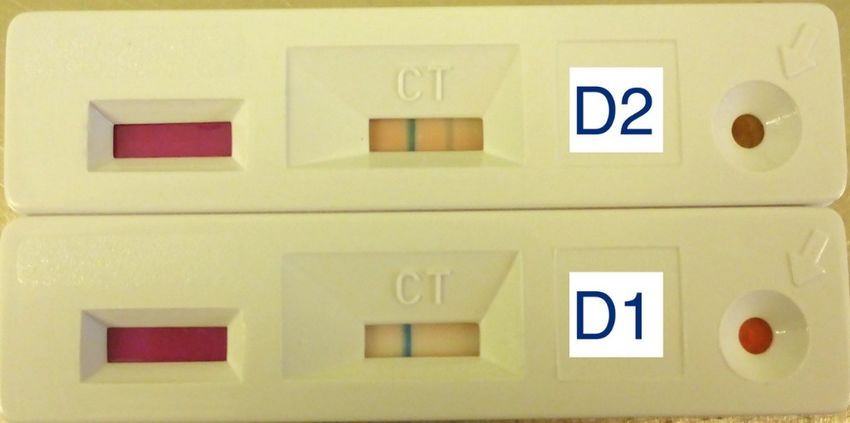
either as positive or negative (see figure 1). Only valid results were counted (appearance of the control line).
Figure 1: Exemplary antigen LFA results. D2 shows a valid positive result, D1 shows a valid negative result.
Nucleic acid extraction and real-time PCR
Viral nucleic acids were extracted using the QIAamp® RNA Viral Mini Kit (Qiagen) according to the
manufacturer’s instructions. qPCR was conducted according to the protocol published by King et al. (2003)
with slight modifications (addition of a heterologous internal control DNA), or with the commercial qPCR
kits virotype ASFV or virotype ASFV 2.0 (Indical Bioscience) according to the manufacturer’s instructions.
All qPCR runs were performed on C1000 thermal cyclers with the CFX96 Real-Time System (Biorad).
Results were recorded as quantification cycle (cq) values.
Screening for ASFV-specific antibodies
The ID Screen ASF Indirect Kit (ID.vet) was used according to the manufacturer’s instructions to screen the
field samples for ASFV-specific antibodies. The multi-antigen indirect ELISA kit detects antibodies against
ASFV p32, p62 and p72 in porcine serum, plasma or blood filter paper samples.
Statistical Analysis
Results of LFAs were evaluated in comparison to results obtained in qPCR. For this purpose, qPCR was
regarded as the standard for pathogen detection. Accordingly, the outcome of the LFA was rated true
positive (TP), true negative (TN), false positive (FP) or false negative (FN). Diagnostic sensitivity was
calculated as TP/(TP+FN)x100. Diagnostic specificity was calculated as TN/(TN+FP)x100.
Results and Discussion
Of the 97 blood samples of ideal or close to ideal quality (samples from animal trials and samples H81-
H91, see supporting table 1), 79 were positive by qPCR. The INGEZIM ASFV CROM Ag LFA detected 61
positives, resulting in a sensitivity of 77.2%. No false positives occurred, hence 100% specificity was observed
on this dataset. The performance was therefore in line with the study published by Sastre et al. (2016)
where field samples of unimpaired quality were detected with roughly 67% sensitivity when compared to
an OIE listed qPCR. Specificity was also close to 100%. Sastre et al. (2016) evaluated the LFA only with
3fresh samples, as represented by our group of samples derived from animal trials or hunted wild boar. With
Posted on Authorea 2 May 2021 — The copyright holder is the author/funder. All rights reserved. No reuse without permission. — https://doi.org/10.22541/au.161994432.24828107/v1 — This a preprint and has not been peer reviewed. Data may be preliminary.
the ongoing circulation of ASF in European wild boar, however, virus detection in carcasses has become an
important issue. Sample quality is then usually reduced due to decomposition effects, an aspect that has not
yet been elucidated for the ASFV antigen LFA. All our 80 carcass-derived blood samples were obtained from
ASF-positive wild boar and confirmed by qPCR with cq values ranging from 14 to 38 (see supporting table
2). Here, significant differences were observed between the samples that were previously frozen, and those
that were not: in native samples tested without any modifications (C1-C16, n=16, see supporting table 3),
the LFA delivered only two positive results (sensitivity of 12.5%). After freeze-thawing, testing of the same
16 samples in the LFA yielded seven positives (sensitivity of 43.75%). Surprisingly, one of the samples that
had yielded a positive result in the native context was now tested negative. The increase of overall positive
results is in accordance with the sensitivity of 48.75% we observed in all of the previously freeze-thawed
carcass-derived samples (C1-C80), where 39 positives were detected by the LFA (see supporting table 2).
Interestingly, however, we did not observe a better sensitivity after freeze-thawing in EDTA-blood samples
of high quality in a previous study by our group (Pikalo et al., 2021). The positive effect of freeze-thawing
is probably due to the fact that most of the virus in blood is associated with erythrocytes (Wardley &
Wilkinson, 1977), and therefore, the destruction of blood cells during freeze-thawing results in a higher
antigen availability for detection in the test, a process especially effective when erythrocytes are bound to
clots in samples of reduced quality. No false positive reactions occurred with any sample types.
In our study, the INGEZIM ASFV CROM Ag assay could not deliver reliable results with native blood
from carcasses. Particular samples with cq values as low as 15 (C4, see supporting table 3), indicating a
considerable virus load in the carcass, still delivered negative results in the LFA.
While we observed increased sensitivity after erythrocytolysis by freeze-thawing (12.5% vs 44% sensitivity,
samples C1 to C16, see supporting table 3), for the practical implementation of the assay in the field, of
course, freezing cannot be an option due to the technical requirements not fitting a point-of-care application.
Possible alternatives to freeze-thawing for erythrocytolysis could be the dilution of blood in aqua dest . or
lysis buffer. On a very limited dataset (n=4), hypotonic lysis seemed to improve the results (3 FN native,
1 FN after water lysis, no FN after freeze-thawing; data not further shown). While both methods could be
feasible under field conditions, the effects of this deviation from the manufacturer’s instructions on the assay
should be elucidated and could be the basis of future optimization of the assay. After all, it must be noted
that even with erythrocytolysis through freeze-thawing, we could only achieve a sensitivity of roughly 50% in
carcass-derived samples, a value not fit fur purpose. Considering the negative impact of immune-complexes,
samples were screened for the presence of antibodies. Only seven samples were positive for ASFV-specific
antibodies and three delivered doubtful results in the antibody ELISA (see supporting tables 1 and 2).
Eight of these samples were positive and two were false negative in the antigen-specific LFA (see supporting
tables 1 and 2). While the small number does not allow for evaluation of possible interference, the principal
functionality of the test in the presence of antibodies is indicated.
In general, the LFA was more reliable using samples with cq values below 30, indicating a rather high viral
load. Of those samples derived from animal trials (n=64), 56 were true positive according to the rapid test,
resulting in a sensitivity of 87.5% in that group. This goes along with observations in a previous study
performed in our group, when the LFA was most sensitive during the clinical phase of ASF, at the peak of
viral replication (Pikalo et al., 2021). In the present study, however, it was observed that the influences of
clotting and decay in the carcass-derived samples seemed be able to outweigh the effects of higher viral loads,
since here no clear correlation even with very low cq values and positive results in the LFA were observed
(see supporting table 2).
Taking into consideration the differences between the highly amplifying qPCR and native antigen detection
by LFA, the marked lower sensitivity in the later is to be expected. Still, the possibility for point-of-care
testing holds a considerable advantage and on-site assays can provide a valuable additional diagnostic tool
under certain circumstances. An acceptable sensitivity of the LFA was confirmed during the clinical phase
of the disease, when fresh samples can be obtained from live animals or immediately after death. Here, the
4application of a rapid test could be of value in domestic pig holdings, when ASF is clinically suspected and
Posted on Authorea 2 May 2021 — The copyright holder is the author/funder. All rights reserved. No reuse without permission. — https://doi.org/10.22541/au.161994432.24828107/v1 — This a preprint and has not been peer reviewed. Data may be preliminary.
live animals can be picked for sampling (given a careful interpretation of negative results in the LFA and still
immediate initiation of laboratory diagnosis). Furthermore, epidemiological investigations can benefit from
antigen assays for the on-site analysis of infected populations, when weaknesses in sensitivity are considered.
However, with a sensitivity of roughly 50%, or even well below when no erythrocytolytic procedure is applied
as proposed by the manufacturer’s instructions, our findings imply that the LFA has only very limited use
for antigen detection in blood from carcasses after extended post mortem intervals. When resources are
scarce and prioritization of diagnostic workflows is needed, the high specificity may allow for positive on-
site results in the LFA to surrogate a laboratory confirmation. Negative results, however, must always be
rated with high caution due to the low sensitivity we observed in samples of reduced quality. OIE listed
methods such as qPCR remain the only safe and proven methods for the unreserved detection of an ASFV
infection. Therefore, the on-site assay should be regarded as a complimentary option rather than a substitute
to laboratory diagnosis for carcass testing.
Author Contributions
Conceptualization, PD and SB; Data curation, PD and JP; Funding acquisition, SB and MB; Investigation,
PD; Methodology, PD, JP and SB; Visualization, PD; Writing – original draft, PD and SB; Writing – review
& editing MB. All authors have read and agreed to the published version of the manuscript.
Funding
The study received funding by the FLI-internal ASF Research Network.
Acknowledgments
The authors would like to thank Ulrike Kleinert and Robin Brandt for their excellent technical assistance.
Conflict of interest statement
The authors declare no conflict of interest.
Ethics statement
The authors confirm adherence with the journal’s ethical statements as noted in the journal’s authors guide-
line page. Blood samples listed in supporting table 1 came from animal experiments conducted at the
FLI, which were performed in accordance with EU Directive 2010/63/EC and approved by the compe-
tent authority (Landesamt fur Landwirtschaft, Lebensmittelsicherheit und Fischerei (LALLF) Mecklenburg-
Vorpommern).
Data availability statement
The data that support the findings of this study are available from the corresponding author upon reasonable
request.
References
Alonso, C., Borca, M., Dixon, L., Revilla, Y., Rodriguez, F., Escribano, J. M., & Ictv Report, C.
(2018). ICTV Virus Taxonomy Profile: Asfarviridae. The Journal of general virology, 99 (5), 613-614.
doi:10.1099/jgv.0.001049
Blome, S., Franzke, K., & Beer, M. (2020). African swine fever - A review of current knowledge. Virus
research, 287 , 198099. doi:10.1016/j.virusres.2020.198099
Busch, F., Haumont, C., Penrith, M. L., Laddomada, A., Dietze, K., Globig, A., . . . Depner, K. (2021).
Evidence-Based African Swine Fever Policies: Do We Address Virus and Host Adequately? Front Vet Sci,
8 , 637487. doi:10.3389/fvets.2021.637487
5Chenais, E., Depner, K., Guberti, V., Dietze, K., Viltrop, A., & Stahl, K. (2019). Epidemiological consider-
Posted on Authorea 2 May 2021 — The copyright holder is the author/funder. All rights reserved. No reuse without permission. — https://doi.org/10.22541/au.161994432.24828107/v1 — This a preprint and has not been peer reviewed. Data may be preliminary.
ations on African swine fever in Europe 2014-2018. Porcine Health Manag, 5 , 6. doi:10.1186/s40813-018-
0109-2
Dixon, L. K., Stahl, K., Jori, F., Vial, L., & Pfeiffer, D. U. (2020). African Swine Fever Epidemiology and
Control. Annu Rev Anim Biosci, 8 , 221-246. doi:10.1146/annurev-animal-021419-083741
EFSA, Boklund, A., Cay, B., Depner, K., Foldi, Z., Guberti, V., . . . Gortazar, C. (2018). Epidemiological
analyses of African swine fever in the European Union (November 2017 until November 2018). EFSA Journal,
16 (11), e05494. doi:https://doi.org/10.2903/j.efsa.2018.5494
King, D. P., Reid, S. M., Hutchings, G. H., Grierson, S. S., Wilkinson, P. J., Dixon, L. K., . . . Drew, T.
W. (2003). Development of a TaqMan PCR assay with internal amplification control for the detection of
African swine fever virus. Journal of virological methods, 107 (1), 53-61. doi:10.1016/s0166-0934(02)00189-1
OIE. (2019). Chapter 3.8.1 African swine fever (Infection with African swine fever virus). Manual of
Diagnostic Tests and Vaccines for Terrestrial Animals.
Pikalo, J., Deutschmann, P., Fischer, M., Roszyk, H., Beer, M., & Blome, S. (2021). African Swine Fever
Laboratory Diagnosis-Lessons Learned from Recent Animal Trials. Pathogens (Basel, Switzerland), 10 (2).
doi:10.3390/pathogens10020177
Pikalo, J., Schoder, M. E., Sehl, J., Breithaupt, A., Tignon, M., Cay, A. B., . . . Blome, S. (2020). The
African swine fever virus isolate Belgium 2018/1 shows high virulence in European wild boar.Transboundary
and emerging diseases . doi:10.1111/tbed.13503
Sanchez-Vizcaino, J. M., Mur, L., & Martinez-Lopez, B. (2012). African Swine Fever: An Epidemiological
Update. Transboundary and emerging diseases . doi:10.1111/j.1865-1682.2011.01293.x
Sastre, P., Gallardo, C., Monedero, A., Ruiz, T., Arias, M., Sanz, A., & Rueda, P. (2016). Development of
a novel lateral flow assay for detection of African swine fever in blood. BMC veterinary research, 12 , 206.
doi:10.1186/s12917-016-0831-4
Sauter-Louis, C., Forth, J. H., Probst, C., Staubach, C., Hlinak, A., Rudovsky, A., . . . Blome, S. (2020).
Joining the club: First detection of African swine fever in wild boar in Germany. Transboundary and
emerging diseases . doi:10.1111/tbed.13890
Zhou, X., Li, N., Luo, Y., Liu, Y., Miao, F., Chen, T., . . . Hu, R. (2018). Emergence of African Swine
Fever in China, 2018.Transboundary and emerging diseases, 65 (6), 1482-1484. doi:10.1111/tbed.12989
6You can also read