Lewy Body Dementia Without Alzheimer Changes

Page created by Philip Carr
 
CONTINUE READING
Lewy Body Dementia Without Alzheimer Changes
THE CANADIAN JOURNAL OF NEUROLOGICAL SCIENCES

                                Lewy Body Dementia Without
                                    Alzheimer Changes
                                  A.A.F. Sima, A.W. Clark, N.A. Sternberger, L.A. Sternberger

              ABSTRACT: Three patients with clinical Alzheimer's disease were found at postmortem examination to have
              Lewy-bodies and Lewy-like bodies in the cerebral cortex and the pigmented brainstem nuclei. Neuritic plaques were
              found in neocortical areas but no neurofibrillary tangles. The distribution of cortical neuronal inclusions correlated
              with the proposed projection of dopamine terminals. Neuronal cell loss was marked in the ventral tegmental area
              (paranigral nucleus) and the basal nucleus of Meynert, suggesting a defect in dopaminergic and cholinergic innerva-
              tion of the cerebral cortex. Immunohistochemical investigations revealed positive staining of cortical Lewy- and
              Lewy-like bodies for monoclonal antibodies to phosphorylated neurofilaments (03-44, 06-17, 04-7). Also cerebral
              neurons containing no inclusions showed positivity, suggesting an early neurofilament abnormality, preceding the
              formation of Lewy-type inclusions.

              RESUME: La demence a corps de Lewy en l'absence des changements de type Alzheimer. Des corps de Lewy et des
              structures similaires aux corps de Lewy ont ete retrouves dans le cortex cerebral et les noyaux pigmentes du tronc
              cerebral a 1'autopsie de trois patients presentant un tableau clinique de maladie d'Alzheimer. Des plaques nevritiques
              ont ete trouvees dans les regions corticales, mais pas d'enchevetrements neurofibrillaires. II y avait correlation entre
              la distribution des inclusions neuronales au niveau du cortex et la projection proposee pour les terminaisons
              dopaminergiques. La perte cellulaire neuronale etait importante au niveau de la region de la decussation ventrale
              (noyau paranigral) et du noyau basal de Meynert, suggerant un defaut de I'innervation dopaminergique et cholinergique
              du cortex cerebral. Des etudes immunohistochimiques ont montre une reaction positive des corps de Lewy et des
              structures similaires aux corps de Lewy du cortex aux anticorps monoclonaux diriges contre des neurofilaments
              phosphoryles (03-44,06-17,04-7). Des neurones cerebraux nu contenant pas d'inclusion manifestaient egalement une
              reaction positive, suggerant une anomalie neurofilamentaire precoce, precedant la formation des inclusions du type
              des corps de Lewy.
                                                                                                  Can. J. Neurol. Sri. 1986; 13:490-497

       Lewy bodies are abnormal perikaryal inclusions of the human                               counts of subcortical projection systems in three cases of Lewy
    brain and of unknown etiology. They are the histopathologic                                  body dementia unassociated with Alzheimer's disease.
    hallmark of Parkinson's disease with predilection sites in the
    substantia nigra, locus ceruleus and substantia innominata.
    They are found rarely in the cerebral cortex, but have been                                                                   CASE REPORTS
    reported in association with changes of Alzheimer's disease,'"4
    Pick's disease,2 idiopathic Parkinson's disease without demen-                                Clinical Histories
    tia,5"6 and in neuroaxonal dystrophies.7"8
       Ultrastructurally, Lewy bodies are composed of an array of                                 Case 1
    filamentous structures radiating from a dense central core.9
                                                                                                    This 72 year old male accountant began to notice problems with
    Cerebral Lewy bodies or Lewy-like bodies show a conglomer-                                   calculation at age 66. Over the next two years, he experienced forgetful-
    ate of less well organized filaments, and the latter lack the                                ness and inability to concentrate. On evaluation at age 68, an electroen-
    central dense core.10 Positive immunoreactivity of Lewy bod-                                 cephalogram showed diffuse polyrhythmic slow-wave and sharp-wave
    ies with polyclonal and monoclonal antibodies to neurofilaments                              activity most marked over the frontal and temporal areas. The diagno-
                                                                                                 sis of Alzheimer's disease was made. At the age of 69, he showed mild
    indicates the presence of neurofilament antigens in these                                    bradykinesia of gait and he was started on Levodopa. The dementia
    inclusions.'' In the present report we describe the quantitative,                            progressed over the next two years. At age 72 he was admitted in a
    morphologic and immunohistochemical findings of cortical                                     non-communicative state because of recurrent episodes of aspiration.
    Lewy bodies and Lewy-like bodies, as well as the results of cell                             He died three months later from bronchopneumonia.

    From the Section of Neuropathology, Department of Pathology, Health Sciences Centre, Winnipeg, (Dr. Sima); Department of Pathology, Foothills Hospital, Calgary,
    (Dr. Clark) and the Center for Brain Research, University of Rochester School of Medicine, Rochester, N.Y., (Drs. N.A. Sternberger and L.A. Sternberger)
    Reprint requests to: Dr. A.A.F. Sima, Health Sciences Centre, MS459A, 820 Sherbrook Street, Winnipeg, Manitoba, Canada R3A 1R9

Downloaded from https://www.cambridge.org/core. IP address: 46.4.80.155, on 10 Sep 2021 at 06:50:59, subject to the Cambridge Core terms of use, available at
    490
https://www.cambridge.org/core/terms. https://doi.org/10.1017/S0317167100037185
LE JOURNAL CANADIEN DES SCIENCES NEUROLOGIQUES

    Case 2                                                                                          Serial 16um thick sections through paraffin blocks from the
       This 72 year old female was diagnosed as having Parkinsonism in                           basal nucleus of Meynert and the paranigral nucleus were stained
    1975. Several decades earlier the diagnosis of multiple sclerosis was
    made. In 1977 she became confined to a wheelchair with weakness and                          with Nissl stain and used for neuronal cell counts. The areas of
    spasms of both legs. She was transferred to a nursing home in 1979.                          the basal nucleus of Meynert and the paranigral nucleus were
    Physical examination in July of 1980 revealed her speech to be incom-                        delineated in each section and digitized usinga Hewlett-Packard
    prehensible. She had difficulty sitting erect and showed spastic weak-                       9874A Digitizer (Hewlett-Packard Company, Fort Collins,
    ness of all limbs. Plantar responses were extensor and deep tendon                           Colorado). Only neurons with discernible nuclei were counted
    reflexes were increased. Two years later she was noted to be emotion-
    ally labile and showed episodes of anger and aggression. She showed                          in each section. The tissue volume was calculated by adding the
    bradykinesia and rigidity and was placed on Sinemet with little im-                          products of area and thickness of the serial sections of each
    provement. Over the ensuing years, she became non-communicative,                             nucleus. The cell number was then calculated as neurons per
    increasingly stiff, and immobile and was diagnosed as having Alz-                            mm. 3
    heimer's disease. She died from cardiorespiratory arrest in November
    of 1985.                                                                                        For immunocytochemical examination paraffin sections were
                                                                                                 incubated with trypsin (GIBCO) at 37°C for 10 min. (400 ug/ml)
    Case 3                                                                                       in 0.05 M Tris-HCI, pH 7.6,0.3 M sodium chloride and 0.02 M
       This 71 year old woman was first admitted in June of 1984 with                            calcium chloride. The incubated paraffin sections were stained
    decreasing appetite and forgetfulness. She displayed a child-like be-                        immunocytochemically by using monoclonal first-layer anti-
    haviour with angry outbursts. She became disoriented to time and                             bodies in a dilution of 1:1000, goat anti-mouse immunoglobulin,
    place, appeared weaker and gradually needed more help with ambula-
    tion. Neurologic examination was reported as normal. In January of                           and ClonoPAP (peroxidase-monoclonal anti-peroxidase com-
     1985, she was admitted in a dehydrated state with disorientation to time                    plex) (12). The first-layer antibodies were against phosphory-
    and place and person. A CT scan showed mild diffuse atrophy of the                           lated neurofilament epitopes (03-44, 06-17, and 04-7) (12).
    brain. Over the next 7 months, she showed a rapid deterioration of her
    mental status and was thought to have Alzheimer's disease. No clinical                       Gross Examinations
    evaluation was performed during this period. She died three months
    later from bronchopneumonia.                                                                    All three cases showed mild to moderate atrophy of the
                                                                                                 frontal and temporal areas and moderate ventricular dilatation.
                                                                                                 The brain weights were 1305,1210and 1220grams respectively,
                       NEUROPATHOLOGY EXAMINATION                                                in the three cases. Examination of the brain and spinal cord in
                                                                                                 Case 2 could not confirm the diagnosis of multiple sclerosis.
    Methods
       The brains from the 3 cases were fixed in 10% buffered                                    Lightmicroscopic Findings
    formalin for at least 10 day s. Tissue blocks for light microscopic                             Lightmicroscopic examinations of the three cases revealed
    examination were taken from various parts of the central ner-                                similar pathologic findings. The most striking finding was the
    vous system. They were embedded in paraffin and stained with                                 presence of Lewy bodies and Lewy-like bodies in the pigmented
    hematoxylin and eosin (H & E), periodic acid-Schiff (PAS),                                   brainstem nuclei and the cerebral cortex. They were found to
    Congo-red, Nissl stain, and Bielschowsky's silver stain. Small                               be most numerous in the entorhinal cortex, the anterior cingu-
    pieces of formalin fixed tissue from selected areas were fixed in                            late gyrus, the temporal and frontal cortices, in that order
    2.5% phosphate-buffered (pH 7.15) glutaraldehyde. They were                                  (Table 1). They were found to be concentrated to the 5th and
    postfixed in 1% osmium tetroxide, dehydrated and embedded                                    6th layers of the cortex in most areas, except for the anterior
    in Epon. Ultrathin sections were examined electronmicroscop-                                 cingulate gyrus and the entorhinal cortex, where they were
    ically.                                                                                      equally numerous in the 3rd cortical layer (Figure I). The

                      Table 1: Distribution of Lewy bodies and Lewy-like bodies in Case 1, 2, and 3.
                      Site                                                                                      Pathologic Change
                                                                                           Lewy Bodies                                    Lewy-like Bodies
                      Cerebral Cortex                                              I            11             Ill                  I            11             III
                               Frontal                                             +             +              +                  ++             +              +
                               Temporal                                            -            ++              +                   +           +++              +
                               Parietal                                            +              -             -                  ++            +               +
                               Insular                                             -            +               +                  ++           ++               +
                               Cingulate Ant.                                     ++            ++              +                 +++           +++              +
                               Cingulate Post.                                     +             +              +                 +++            ++              +
                               Occipital                                           +              -             -                   +              -             -
                               Entorhinal                                          -           +++              +                  ++           +++              +
                               Hippocampus                                         -              -             -                   -            ++              +
                      Substantia lnnominata                                       ++            +               +                   -            ++              +
                      Subthalamic Nucleus                                          -            +               +                   -              -             +
                      Amygdala                                                     -            +               +                   -              -             +
                      Substantia Nigra                                          +++            ++               +                 +++              +             +
                      Nucleus Paranigralis                                      ++              +               +                  +               -             +
                      Locus Ceruleus                                             +             +++              +                   -              -            +++
                      Dorsal Raphe Nucleus                                       +                -             -                   +              +             +
                          No Change; + Small Number; + + Moderate Number; + + + Large Number

   Volume 13, No. 4 (Supplement) — November 1986
Downloaded from https://www.cambridge.org/core. IP address: 46.4.80.155, on 10 Sep 2021 at 06:50:59, subject to the Cambridge Core terms of use, available at
                                                                                                                                                                      491
https://www.cambridge.org/core/terms. https://doi.org/10.1017/S0317167100037185
THE CANADIAN JOURNAL OF NEUROLOGICAL SCIENCES

                                        CASE 1

                                                                                   1a

                                                                                                    Figure I — Distribution of Lewy bodies (x) and Lewy-like bodies (*) in the
                                                                                                        temporal (A), and thefrontal (B) lobes, the cingulate gyrus (C) and the
                                                                                                        entorhinal cortex (D) in Case 1,2, and 3.

Downloaded from https://www.cambridge.org/core. IP address: 46.4.80.155, on 10 Sep 2021 at 06:50:59, subject to the Cambridge Core terms of use, available at
     492
https://www.cambridge.org/core/terms. https://doi.org/10.1017/S0317167100037185
LE JOURNAL CANADIEN DES SCIENCES NEUROLOGIQUES

     inclusions were found to be most numerous in the depth of the                                                                                                                                black with Bielschowsky's silver stain. They did not stain with
     sulci (Figure 1). In areas where Lewy bodies and Lewy-like                                                                                                                                   PAS, or Nissl stain. Lewy bodies showed a bright eosinophilic
     bodies were numerous, neuronal loss and mild astrogliosis                                                                                                                                    core with the H-E stain (Figure 3A), which also stained more
     could be demonstrated. A moderate number of senile plaques                                                                                                                                   intensely with Bielschowsky's silver stain. The light micro-
     were demonstrated throughout the neocortex in all three cases,                                                                                                                               scopic staining pattern of the inclusions is summarized in Table
     whereas no neurofibrillary tangles were found in neocortical                                                                                                                                 3.
     areas in any of the cases (Table 2). Cerebrovascular amyloid                                                                                                                                    Neuronal cell counts revealed a moderate neuronal loss in
     deposition was not identified. Lewy-like bodies stained faintly                                                                                                                              the basal nucleus of Meynert (Table 4). The paranigral nucleus
     eosinophilic with the H-E stain (Figure 2A) and dark brown to                                                                                                                                of the ventral tegmental area showed marked neuronal loss and
                                                                                                                                                                                                  gliosis (Figure 4; Table 4).
     l                                                       '                                   "

     J                                                                          _                                                                                                                                                                                      V

                                                                                                                                                                                                   f'                                 ,
                                                                                                                                                            .
     *                   #                 "
                                           7
                                                        .                   .           .        .                                                                                                      •
      •*.                    .:**                               .                       4w..•/-•
                                                                                                                                                                                                   **        •
                                                                                                                                                                                                                            ^ &
                                                                                                                                                                                                                                                         'v-4v •
                             #•                                                                      *

                                                                                                                                                                                                                 1
             '       ' *•-                     f                                             "

                                                                                                             **                                                                                             •Jp* •«&> <

                                                                                                                                                                                                                        .

                                                                                                                                                                                                        •                                                          ,
                                                                                                                                                                                                                                                        #
                                                                                                                                                                                                        .

                                                                                                                                                                                                                             •

                                                                                                                                                                                          2a                                      •

      Figure 2(A) — Lewy-like bodv in the superior temporal gyrus in Case 3,
         H.-E., Mag. 870x.                                                                                                                                                                                                                                     *           3a
                                                                                                                                                                                                   Figure 3(A) — Lewy bodies in the anterior cingulate gyrus in Case 2, H.-E.,
         v                   ••*                   »                                                 .   x
                                                                                                                                        . •'                                _/,'.                     Mag 870x.
                                                                                                              >         •'      '   .   •   "   *   '   -           *
                                                                                                                                                                              * * %
                                                                                                                                                                                  .-
                 *                     *                                            f                ^ _,                     < » " * "'                                ,' jr r : .
THE CANADIAN JOURNAL OF NEUROLOGICAL SCIENCES

                                                                                                       . *                          ;.           »r»                              . *,—
                                                                                                       **                          :• •                                                                      ''.*','

                                                                                                                                         i               ••«•                     . »        .
                                                                                                                                                                                                          1' A-'
                                                                                                                              •      *
                                                                                                                                                 >
                                                                                                                                                     *
                                                                                                                                                      4
                                                                                                                                                                .'*
                                                                                                                                                                      v. *
                                                                                                                                                                             ••     .•           •    •
                                                                                                                                                                                                              •V
                                                                                                               *.
                                                                                                                                   %* *
                                                                                                                                                     "                *                  _       -                 «

                                                                                                       T;l •
                                                                                                                                                                                                          • • ' . *

                                                                                                                           •>".:             >

                                                                                                           / .0           •                                            . >- • f                      ;•           » •
                                                                                                                                                                  : ff
                                                                                                                     t            * "»           ' - . - V :-                                             .''          - •

                                                                                                                    • •
                                                                                                               *

                                                                                                       f       - »
                                                                                                                           '£                    .••*,                #.•                '• ft'
                                                                                                                                                                                                          V- •' ••.
                                                                                                                                                                                                                        •

                                                                                                                                                      •if.".                             •;- *
                                                                                                                                                          ,V
                                                                                                                                                 VA             T.« "*»•
LE JOURNAL CANADIEN DES SCIENCES NEUROLOGIQUES

    Electronmicroscopic Findings                                                                     Lewy body dementia without concomitant histopathologic
       The cortical Lewy-like bodies consisted of both granular and                               changes of Alzheimer's disease is a seldom recognized en-
    filamentous material. Lewy bodies showed a variably increased                                 tity.13"14 In the present cases, cerebral Lewy bodies and Lewy-
    density of the central zone. The filaments measured 10-18 nm in                               like bodies showed a distributional pattern not previously
    both types of inclusions. The radial arrangement of peripheral                                described. In the neocortex the Lewy-type inclusions were
    filaments typical of brainstem Lewy bodies was less pronounced                                confined to the deeper layers 5 and 6, whereas in the anterior
    (Figure 5).                                                                                   cingulate gyrus and in the entorhinal cortex, inclusions were
    Immunohistochemical Findings                                                                  also widely distributed in the 3rd cortical layer. This distribu-
       Three monoclonal antibodies (06-17, 03-44, 04-7) directed                                  tion of Lewy bodies and Lewy-like bodies correlates well with
    against phosphorylated neurofilament epitopes were employed.                                  the proposed mesocortical dopaminergic projections to the
    Lewy-like bodies of the substantia nigra showed weak reactiv-                                 cerebral cortex.' 516 The dopaminergic fibers projecting to the
    ity (Figure 6A). Lewy bodies of the same nucleus showed a
    weakly reacting central zone and a strongly reacting peripheral
    ring (Figure 6B). A similar pattern was occasionally seen in
    cortical Lewy and Lewy-like bodies. Most commonly, however,
    Lewy-like bodies showed a strong reactivity with antibodies to
    phosphorylated epitopes of neurofilaments (Figure 2B). Corti-
    cal Lewy bodies showed strong reactivity of the central core
    and weak or no reactivity of the periphery (Figure 3B). Neur-
    ones of the 5th cortical layer containing no identifiable inclu-
    sions often revealed a diffuse positive immunostaining with
    monoclonal antibodies to phosphorylated neurofilaments (Fig-
    ure 7). The immunoreactivity of Lewy bodies and Lewy-like
    bodies for the three monoclonal antibodies is summarized in
    Table 5.                                                                                                                    » "   'i

                                       DISCUSSION

      Alzheimer type changes were found in the present 3 cases,
    consistent with the advanced age of the patients, but with a
    distribution that did not warrant the diagnosis of Alzheimer's
    disease.

                                                                                                   Figure 6(A) — Lewy-like inclusion in the substantia nigra in Case 3, showing
                                                           PC1/'* - - H • *                            no reactivity with antibody 03-44.

                                                                                                                                           2*

                                                                                                      "1/
                                                                                                                                           ^L \ -

                                                                                                                                           *        V
                                                                                                    » »                     J     f

                                                                                                                                      jjfij^
                                                                                                                      1 *                                                      4
                                                                                                                                                                           V

                               ^^t^&^sL^roT^ ^
                                                                                                                                                                %

                                       >«•
                 &&
THE CANADIAN JOURNAL OF NEUROLOGICAL SCIENCES

    cerebral cortex originate from the ventral tegmental area, mainly                               Neurons of the same cortical layers containing no inclusions
    from the paranigral nucleus and to a lesser extent from locus                                showed abnormal phosphorylated neurofilaments, not seen in
    ceruleus (Figure 8).16 They terminate most frequently in the                                 normal controls, suggesting an early post-translational phos-
    layers 5 and 6 of the frontal cortex, whereas the projections to                             phorylation of neurofilaments preceding the formation of Lewy
    the anterior cingulate and ventral entorhinal areas terminate in                             type inclusions.
    the 2nd and 3rd layers as well.15"17                                                            Based on the pathology of the present three cases, we pro-
       In the present cases the basal nucleus of Meynert showed a                                pose that Lewy body dementia without Alzheimer's disease
    moderate neuronal loss, whereas the paranigral nucleus showed                                may be a primary defect in mesocortical dopaminergic projec-
    a 90% neuronal loss in Case 1 and 2 and somewhat less in Case                                tions resulting in a transsynaptic abnormal neurofilamentous
    3, suggesting a substantial diminution of cortical dopaminergic                              reaction of the target neurons, expressed by the presence of
    input.                                                                                       Lewy-like bodies and Lewy bodies.
       It is possible that dopaminergic and cholinergic deafferenta-
    tion of the cortical neuronal population may underlie not only
    the intellectual impairment and cognitive defects observed in
    the present patients but also the histopathologic findings of
    Lewy-type inclusions.16 In other systems such as the inferior
    olivary nucleus and the substantia nigra, abnormal neurofilament
    accumulations are thought to represent a transsynaptic reac-
    tion.18"20
       The ultrastructural features and dimensions of the neurofila-
    ments constituting the inclusions were similar to those described
    in brainstem Lewy bodies.21
       Immunohistochemical investigations using monoclonal anti-
    bodies to phosphorylated neurofilaments revealed positive stain-
    ing of Lewy-like bodies and of the central core in Lewy bodies
    of the cortices. This is different from the staining pattern of
    brainstem Lewy bodies in which phosphorylated neurofilaments
    are found in the peripheral rim of Lewy bodies.21

                                                                                                 Figure 8 — Schematic illustration of proposed mesocortical dopaminergic
                                                                                                    projections. Most dopaminergic fibers originate from the paranigral
                                                                                                    nucleus and project to layers 5 and 6 of the temporal, frontal and parietal
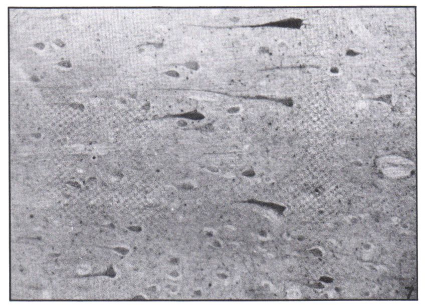
    Figure 7 — Positive immunostaining with antibody 06-17 of neurons devoid                        cortices, and to layers 5-6 and 3 in the entorhinal and anterior cingulate
       of inclusions in the 5th cortical layer of the frontal cortex. Case 2, Mag                   cortices. These projections correspond to the distribution of Lewy bodies
       104x.                                                                                        and Lewy-like bodies in the present cases (cf. Fig I).

    Table 5: Immunoreactivity to phosphorylated neurofilaments.

                                                                Lewy Bodies                                                                           Lewy-Like Bodies
                                        Substantia Nigra                               Cortex
    Antibodies                  Central Core           Periphery          Central Core                             Periphery                Substantia Nigra             Cortex
    0 6 - 17                                                   ++                         +++                           +                            -                   ++
     03-44                             +                       +++                        ++                            -                             -                  +++
     04- 7                             +                       ++                         ++                            +                             +                  ++
     - Negative; + Weak; + + Moderate; and + + + Strong Reactivity

    496
Downloaded from https://www.cambridge.org/core. IP address: 46.4.80.155, on 10 Sep 2021 at 06:50:59, subject to the Cambridge Core terms of use, available at
https://www.cambridge.org/core/terms. https://doi.org/10.1017/S0317167100037185
LE JOURNAL CANADIEN DES SCIENCES NEUROLOGIQUES

                                 ACKNOWLEDGEMENT                                                  10. Itoh T, Momma Y, Ogasawara N. An electron microscopic study
                                                                                                         on atypical presenile dementia with numerous Lewy bodies in
      The authors are indebted to Mrs. Jackie McKane for preparing the                                   the cerebral cortex. Folia Psychiatr Neurol Jpn 1982;36:99-106.
     manuscript.                                                                                  11. Goldman JE, Yen SH, Chiu F-C, Peress NS. Lewy bodies of
                                                                                                         Parkinson's disease contain neurofilament antigen. Science 1983;
                                                                                                         221: 1082-1084.
                                       REFERENCES                                                 12. Sternberger LA, Sternberger NA. Monoclonal antibodies distin-
                                                                                                         guish phosphorylated and nonphosphorylated forms of neuro-
      1. Woodard JS. Concentric hyalin inclusion body formation in mental                                filaments in situ. Proc Natl Acad Sci USA, 1983; 80: 6126-6130.
           disease. Analysis of 27 cases. J Neuropath Exp Neurol 1962; 21:                        13. Ikeda K, Hori A, Bode G. Progressive dementia with "diffuse
           442-449.                                                                                      Lewy-type inclusions" in cerebral cortex. A case report. Arch
      2. Kosaka K, Oyanagi S, Matsushita M, et al. Presenile dementia                                    Psychiatr Nervenkr 1980; 228: 243-248.
           with Alzheimer-, Pick- and Lewy body changes. Acta Neuro-                              14. Eggertson DE, Sima AAF. Dementia with cerebral Lewy bodies.
           pathol(Berl) 1976;36:221-233.                                                                 A mesocortical dopaminergic defect? Arch Neurol 1986; 43:
      3. Forno LS, Barbour PJ, Norville RL. Presenile dementia with                                      524-527.
           Lewy bodies and neurofibrillary tangles. Arch Neurol 1978; 35:                         15. Lindvall O, Bjorklund A, Divac I. Organization of mesencephalic
           818-822.                                                                                      dopamin neurons projecting to neocortex and septum. In Costa
      4. Kosaka K, Yoshimura M, Ikeda K, et al. Diffuse type of Lewy                                     E and Gessa, GL eds. Advances in Biochemical Psychopharma-
           body disease: progressive dementia with abundant cortical Lewy                                cology Vol. 16, New York, Raven Press, New York, 1977, pp
           bodies and senile changes of varying degree — a new disease?                                  39-46.
           Clin Neuropath 1984; 3: 185-192.                                                       16. Javoy-Agid F, Agid Y. Is the mesocortical dopaminergic system
      5. YagashitaS,ItohY,AmanoN,etal.Atypicalseniledementiawith                                         involved in Parkinson disease? Neurology 1980; 30: 1326-1330.
           widespread Lewy-type inclusion in the cerebral cortex. Acta                            17. Lindvall O, Bjorklund A, Moore RY, et al. Mesencephalic dopa-
           Neuropath. (Berl) 1980; 49: 187-191.                                                           mine neurons projecting to the neocortex. Brain Res 1974; 81:
      6. Yoshimura M. Cortical changes in the Parkinsonian brain: a contri-                              325-331.
           bution to the delineation of "diffuse Lewy body disease". J                            18. Horoupian DS, Wisniewski H. Neurofilamentous hyperplasia in
           Neurol 1983; 229: 17-32.                                                                       inferior olivary hypertrophy. J Neuropath Exp Neurol 1971; 30:
      7. Vuia O. Neuroaxonal dystrophy, ajuvenile adult form. Clin Neurol                                571-582.
           Neurosurg 1977; 79: 305-315.                                                           19. Forno LS. Reaction of the substantia nigra to massive basal ganglia
      8. Williamson K, Sima AAF, Curry B, Ludwin SK. Neuroaxonal                                          infarction. Acta Neuropath (Berl) 1983 62: 96-102.
           dystrophy in young adults: A clinicopathologic study of two                            20. Yagishita S, Itoh Y, Nakano T. Hypertrophy of the olivary nucleus.
           unrelated cases. Ann Neurol 1982; 11: 335-343.                                                 An ultrastructural study. Acta Neuropath (Berl) 198669: 132-138.
      9. Duffy PE, Tennyson UM. Phase and electron microscopic observa-                           21. Forno LS, Sternberger LA, Sternberger NA, et al. Reaction of
           tions of Lewy bodies and melanin granules in the substantia                                    Lewy bodies with antibodies to phosphorylated and non-phos-
           nigra and locus coeruleus in Parkinson's disease. J Neuropath                                  phorylated neurofilaments. Neuroscience Letter 1986; 64:
           Exp Neurol 1965; 24: 398-414.                                                                  253-258.

    Volume 13, No. 4 (Supplement) — November 1986                                                                                                                   497
Downloaded from https://www.cambridge.org/core. IP address: 46.4.80.155, on 10 Sep 2021 at 06:50:59, subject to the Cambridge Core terms of use, available at
https://www.cambridge.org/core/terms. https://doi.org/10.1017/S0317167100037185
You can also read