LOCAL EMERGENCE AND DECLINE OF A SARS-COV-2 VARIANT WITH MUTATIONS L452R AND N501Y IN THE SPIKE PROTEIN
←
→
Page content transcription
If your browser does not render page correctly, please read the page content below
medRxiv preprint doi: https://doi.org/10.1101/2021.04.27.21254849; this version posted April 29, 2021. The copyright holder for this preprint
 (which was not certified by peer review) is the author/funder, who has granted medRxiv a license to display the preprint in perpetuity.
                                     It is made available under a CC-BY-NC-ND 4.0 International license .

     Local emergence and decline of a SARS-CoV-2 variant with
     mutations L452R and N501Y in the spike protein

     Jan-Philipp Mallm1, Christian Bundschuh2, Heeyoung Kim3, Niklas Weidner2, Simon Steiger4,
     Isabelle Lander4, Kathleen Börner2, Katharina Bauer1, Daniel Hübschmann5,6,7, Vladimir
     Benes8, Tobias Rausch8, Nayara Trevisan Doimo de Azevedo8, Anja Telzerow8, Katharina
     Laurence Jost2, Sylvia Parthé2, Paul Schnitzler2, Michael Boutros9, Barbara Müller2, Ralf
     Bartenschlager3,10,11, Hans-Georg Kräusslich2,11* and Karsten Rippe4,*

     1
         Single Cell Open Lab, German Cancer Research Center (DKFZ) and Bioquant, Heidelberg,
     Germany
     2
         Department of Infectious Diseases, Virology, Heidelberg University, Heidelberg, Germany
     3
         Department of Infectious Diseases, Molecular Virology, Heidelberg University, Heidelberg,
     Germany
     4
         Division Chromatin Networks, German Cancer Research Center (DKFZ) and Bioquant,
     Heidelberg, Germany
     5
         Computational Oncology, Molecular Diagnostics Program, National Center for Tumor
     Diseases (NCT) Heidelberg and German Cancer Research Center (DKFZ) Heidelberg,
     Germany
     6
         Pattern Recognition and Digital Medicine, Heidelberg Institute for Stem cell Technology and
     Experimental Medicine (HI-STEM), Heidelberg, Germany
     7
         German Cancer Consortium (DKTK), Heidelberg, Germany
     8
         Genomics Core Facility, European Molecular Biology Laboratory (EMBL), Heidelberg,
     Germany
     9
         Division Signaling and Functional Genomics, German Cancer Research Center (DKFZ),
     Department for Cell and Molecular Biology, Medical Faculty Mannheim, Heidelberg University,
     Heidelberg, Germany
     10
          Division Virus-Associated Carcinogenesis, German Cancer Research Center, Heidelberg,
     Germany
     11
          Deutsches Zentrum für Infektionsforschung, partner site Heidelberg, Germany

     *Correspondence: Karsten Rippe (karsten.rippe@dkfz.de) or Hans-Georg Kräusslich (hans-
     georg.kraeusslich@med.uni-heidelberg.de)
      NOTE: This preprint reports new research that has not been certified by peer review and should not be used to guide clinical practice.

                                                                                                                                           1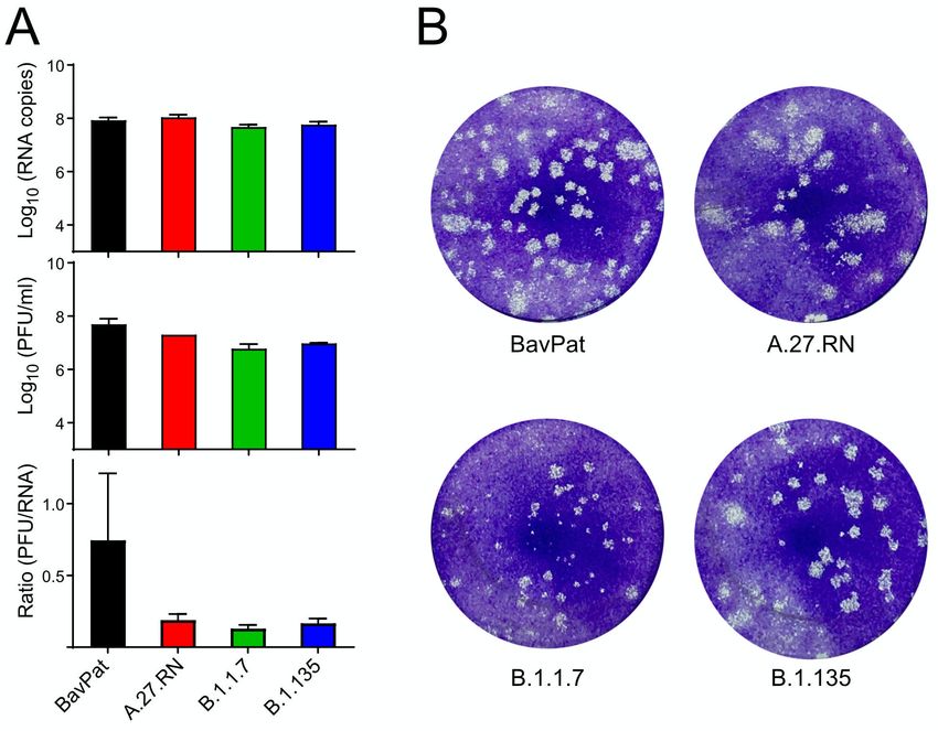
medRxiv preprint doi: https://doi.org/10.1101/2021.04.27.21254849; this version posted April 29, 2021. The copyright holder for this preprint
 (which was not certified by peer review) is the author/funder, who has granted medRxiv a license to display the preprint in perpetuity.
                                     It is made available under a CC-BY-NC-ND 4.0 International license .

     Summary
     Variants of severe acute respiratory syndrome coronavirus 2 (SARS-CoV-2) are replacing the
     initial wild-type strain, jeopardizing current efforts to contain the pandemic. Amino acid
     exchanges in the spike protein are of particular concern as they can render the virus more
     transmissible or reduce vaccine efficacy. Here, we conducted whole genome sequencing of
     SARS-CoV-2 positive samples from the Rhine-Neckar district in Germany during January-
     March 2021. We detected a total of 166 samples positive for a variant with a distinct mutational
     pattern in the spike gene comprising L18F, L452R, N501Y, A653V, H655Y, D796Y and
     G1219V with a later gain of A222V. This variant was designated A.27.RN according to its
     phylogenetic clade classification. It emerged in parallel with the B.1.1.7 variant, increased to
     >50% of all SARS-CoV-2 variants by week five. Subsequently it decreased to
medRxiv preprint doi: https://doi.org/10.1101/2021.04.27.21254849; this version posted April 29, 2021. The copyright holder for this preprint
 (which was not certified by peer review) is the author/funder, who has granted medRxiv a license to display the preprint in perpetuity.
                                     It is made available under a CC-BY-NC-ND 4.0 International license .

     Other RBD mutations that provide partial resistance to neutralizing antibodies include K417N/T
     in P.1 and B.1.351 (3, 4, 12) and L452R in B.1.427/1.429 and B.1.617 (5-8, 19). L452R is a
     marker mutation of the B.1.427/1.429 strain from California (5-7) and is found together with
     the E484Q mutation in a newly emerging B.1.617 strain from India (8, 20). Outside the RBD,
     two other S mutations have been associated with a phenotype. The D614G mutation, that
     occurred early in the pandemic, has been associated with increased transmissibility (21, 22),
     and the L18F exchange has been linked to faster spreading of SARS-CoV-2 in England (23).

     VOC B.1.1.7 has become dominant in the UK in late 2020 accounting for >90% of all infections
     in 12/2020 and has spread to other regions of Europe and beyond, where it became dominant
     as well (24). In Germany, B.1.1.7 accounted for the vast majority of all new infections in April
     2021 (9). Similarly, VOC P.1 has been responsible for a second wave of infections in Brazil
     with the epicenter in Manaus (4) and VOC B.1.351 is becoming the major cause of Covid-19
     in South Africa (3) with both viruses also spreading globally (1). A study of 2,172 samples
     collected in California reported that B.1.427/B.1.429 has emerged in late 2020 and increased
     to >50% of all infections by end of January 2021 with a calculated 18.6-24% increase in
     transmissibility relative to wild-type (6). B.1.617, another strain with the L452R mutation, has
     been linked to surge in the number of cases in April 2021 during the second COVID-19 wave
     in India (20).

     Viral genome sequencing is of crucial importance to track the emergence of mutations that
     define existing and new VOC. By applying a tiling amplicon scheme in combination with
     multiplex PCR, entire SARS CoV-2 genomes can be sequenced with sufficient coverage in a
     cost-effective manner (25-27). The comprehensive sequence information obtained by this
     strategy provides direct insight into the evolution of SARS-CoV-2 genomes and the spreading
     of variants. In addition, marker mutations identified by sequencing can be exploited to design
     melting curve-based PCR screens for the rapid detection of relevant variants (28, 29).

     Here, we have conducted whole virus genome sequencing of SARS-CoV-2 positive samples
     from the Rhine-Neckar district in Germany during the first quarter of 2021. We report on the
     local emergence of a variant designated as A.27.RN that simultaneously carries the N501Y
     and L452R mutations in the S gene, and its subsequent displacement by variant B.1.1.7.
     Variant A.27.RN was isolated from patient samples and characterized in tissue culture.
     Antibody neutralization assays with sera from BNT162b2 vaccinees showed a moderate
     decrease in neutralizing titers for A.27.RN compared to SARS-CoV-2 wild-type, but better
     neutralization than for the B.1.351 variant. Our results provide evidence for the transient
     appearance of a variant of potential concern, carrying the signature N501Y mutation. These
     observations indicate important differences regarding population spread between N501Y
     strains that are not recapitulated in infectivity assays in cell culture. Furthermore, they underpin

                                                                                                                                     3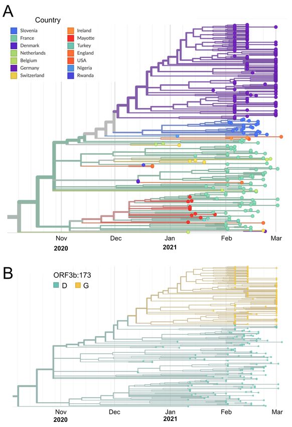
medRxiv preprint doi: https://doi.org/10.1101/2021.04.27.21254849; this version posted April 29, 2021. The copyright holder for this preprint
 (which was not certified by peer review) is the author/funder, who has granted medRxiv a license to display the preprint in perpetuity.
                                     It is made available under a CC-BY-NC-ND 4.0 International license .

     the importance of unbiased surveillance by whole genome sequencing of SARS-CoV-2
     positive samples on a population level.

     Materials and methods
     RNA purification
     For SARS-CoV-2 PCR and sequencing analysis, RNA was isolated from nasopharyngeal and
     oropharyngeal swabs applying automated magnetic bead-based nucleic acid extraction
     protocols. Either the QIASymphony, DSP Virus/Pathogen mini Kit (Qiagen) or Chemagic, Viral
     DNA/RNA 300 Kit H96 (PerkinElmer) were used, following the manufacturers’ protocols and
     using the respective devices. The sample input volume was 140 µl, spiked with 10 µl per
     sample of the internal control (TIB Molbiol) and the elution volume was set to 100 µl. To confirm
     the mutation pattern after viral passaging in tissue culture, RNA was extracted with the QIAamp
     Viral RNA body fluid kit which was carried out on the QIAcube system (Qiagen) with manual
     lysis according to the manufacturer’s protocol.

     RT-qPCR
     Samples were screened for SARS-CoV-2 RNA by commercially available dual-target RT-PCR
     assays using automated analysis for E & orf1a/b employing Cobas 6800 (Roche Diagnostics)
     or analysis for E & N Gene (TIB Molbiol) or E & S gene (Altona Diagnostics) by Roche
     LightCycler and LightCycler II. Positive samples with a Ct value ≤35 (reduced to Ct ≤32 in
     week 10 because of a high rate of inconclusive results in samples with Ct>32) were
     characterized by VirSNiP SARS-CoV-2 Spike N501Y, DelHV69/70 and K417N Assays (TIB
     MOLBIOL) using Roche LightCycler II. All commercial assays and devices were used
     according to the manufacturer’s instructions.

     SARS-CoV-2 genome sequencing
     SARS-CoV-2 genomes were sequenced with two approaches, based on the Nextera library
     generation approach (Cov-seq) and the ARTIC protocol (25) modified and developed by New
     England Biolabs (NEB, p/n E7658), respectively. For Cov-seq, cDNA was generated with
     SARS-Cov2 specific primers and a PCR handle in 384-well plates. Reverse transcription (RT)
     of 2 µl of purified RNA was conducted with the Maxima reverse transcriptase (Thermo Fisher
     Scientific) and 7.5% PEG-8000. After 50 min at 50 °C and RT inactivation, PCR was performed
     by adding KAPA PCR mix and custom forward primers and a single reverse primer
     complementary to the PCR handle. Due to overlapping primer design, the RT and PCR were
     performed in two separate reactions for each sample. cDNA was purified with SPRI beads at
     a 0.5x bead ratio and selected samples were measured with Qubit and Tapestation. cDNA

                                                                                                                                     4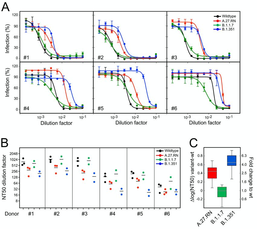
medRxiv preprint doi: https://doi.org/10.1101/2021.04.27.21254849; this version posted April 29, 2021. The copyright holder for this preprint
 (which was not certified by peer review) is the author/funder, who has granted medRxiv a license to display the preprint in perpetuity.
                                     It is made available under a CC-BY-NC-ND 4.0 International license .

     was diluted to 0.3 to 0.5 ng/µl and libraries were prepared with the Illumina Nextera kit using a
     downscaled version with the mosquito system in 384-well plates. The libraries from 384
     samples were individually indexed, pooled and purified with SPRI beads.
     The ARTIC sequencing generally followed the recommended protocol, but we could further
     expedite and scale sample processing by implementation of three modifications: (i) With the
     help of NEB's technical support team it was possible to omit the magnetic bead clean-up step
     after PCR amplification of cDNA. (ii) The clean-up of adaptor ligated fragments was conducted
     on an automated liquid-handling system. (iii) After the final PCR, libraries were pooled, and
     bead purified with SPRI beads at a 0.9x ratio.
     All libraries generated were sequenced in pools of up to 384 samples with the Illumina NextSeq
     500/550 sequencing instrument in paired-end mode with a read-length of 75 bases from each
     direction.

     Data analysis
     Sequencing data from Cov-seq were processed using the nf-core viralrecon pipeline (30, 31)
     with the following settings: (i) NC_045512.2 genome, (ii) metagenomic protocol without primer
     sequence removal, (iii) no duplicate filtering, (iv) minimal coverage of 20 for variant calling, and
     (v) maximum allele frequency of 0.9 for filtering variant calls. Primer sequences were not
     removed due to the protocol’s specific amplicon design. Variant calls and reconstructed
     consensus sequences from iVar were used (32). For the data analysis of the ARTIC
     sequencing, paired-end sequencing data of read-length 75 bases were filtered for sequencing
     adapters (trim galore) and host-read contamination (33). Remaining reads were aligned to the
     NC_045512.2 reference genome using bwa (34). Alignments were sorted and indexed using
     samtools (35) and quality-controlled using alfred (36). We then masked priming regions with
     iVar (32). Variant calling employed FreeBayes (37) and bcftools (38) for quality-filtering and
     normalization of variants. Samtools, bcftools and iVar were also employed for viral consensus
     sequence generation. Lineage and clade classification of the SARS-CoV-2 sequences was
     carried out using Pangolin (39) and Nextclade as part of the Nextstrain framework (40).

     Cells and viruses
     Vero E6 cells (ATCC#1586) were cultured in Dulbecco’s modified Eagle medium (DMEM, Life
     Technologies) containing 10% fetal bovine serum, 100 U/mL penicillin, 100 µg/mL
     streptomycin and 1% non-essential amino acids (complete medium). The original SARS-CoV-
     2 strain BavPat1/2020 was kindly provided by Christian Drosten, Institute for Virology, Charité
     Berlin, via the European Virus Archive (Ref-SKU: 026V-03883) and amplified in Vero E6 cells.
     Cells were inoculated with the original virus isolate (passage 2) at an MOI of 0.01. At 48 h post
     infection (h.p.i.), the supernatant was harvested, and cell debris was removed by centrifugation

                                                                                                                                     5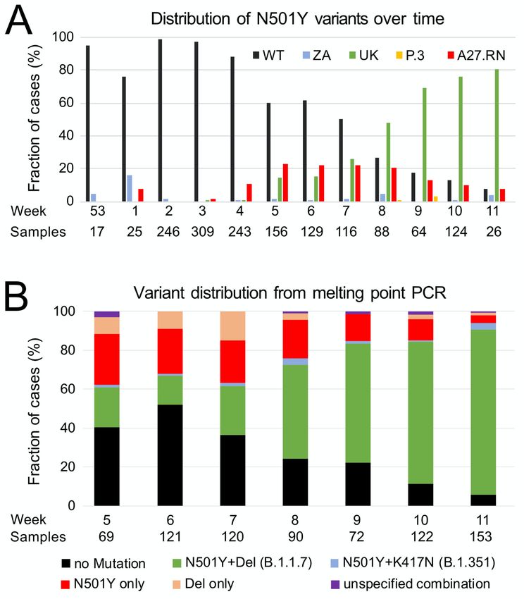
medRxiv preprint doi: https://doi.org/10.1101/2021.04.27.21254849; this version posted April 29, 2021. The copyright holder for this preprint
 (which was not certified by peer review) is the author/funder, who has granted medRxiv a license to display the preprint in perpetuity.
                                     It is made available under a CC-BY-NC-ND 4.0 International license .

     at 800xg for 10 min. Virus stocks were produced by one additional amplification in Vero E6
     (passage 4), aliquoted and stored at -80 ⁰C. Virus titers were determined by plaque assay as
     reported earlier (41). To isolate SARS-CoV-2 variants, nasopharyngeal swabs collected from
     PCR confirmed SARS-CoV-2-positive patients were resuspended and used to inoculate Vero
     E6 cells that were seeded in 24 well plates at a density of 2.5x10⁵ cells per well. 100 µl of
     clinical specimen were used to inoculate cells in the first well and five-fold serial dilutions were
     added to cells in subsequent wells. After 1 h, inoculum was removed and replaced by fresh
     complete DMEM. Cells were inspected by microscopy for the appearance of a cytopathic effect
     on a daily basis. In most cases, extensive CPE was observed at four days post-inoculation.
     Supernatant was collected (passage 0), centrifuged at 1,200 x g for 5 min and 0.35ml were
     used to inoculate 3x10⁶ Vero E6 cells, seeded into a T25 cm2 flask one day prior to inoculation,
     in a total volume of 5 ml complete DMEM. After two days, CPE was observed, supernatant
     was collected (passage 1) and used for SARS-CoV-2 genome sequencing. Virus stocks were
     produced by one further passage in Vero E6 cells (passage 2; B.1.1.7, B.1.351 and A27.RN),
     virus titer was determined, and stocks were stored at -80 ⁰C.

     Serological analyses
     Sera from healthy donors vaccinated with the BTN162b2 SARS-CoV-2 mRNA vaccine were
     collected at day 10-28 after the second vaccine dose and used immediately or stored at 4°C
     until use. IgG reactivity against SARS-CoV-2 S1RBD was analyzed by a commercial
     chemoluminescence immunoassay (Siemens sCOVG; #11207377) run on a Siemens ADVIA
     Centaur XP instrument according to the manufacturer’s instructions. Interference of sera with
     RBD-ACE2 interaction was assessed by a titration experiment using serial 1:3 dilutions of sera
     and a commercial ELISA based test system (GenScript SARS-CoV-2 Surrogate Virus
     Neutralization Test Kit; #L00847), following the manufacturer’s instructions. Based on the
     results from these analyses, six sera covering a range of relative reactivities were selected for
     virus neutralization assays (Supplementary Table S3).

     Antibody neutralization tests
     Neutralization titers were determined in titration experiments on Vero E6 cells as described
     previously (42, 43). Vaccinee sera were serially diluted and mixed with the respective SARS-
     CoV-2 variants. After 1 h at 37 °C, the mixture was added to Vero E6 cells and 24 h later, cells
     were fixed in the plates with 5% formaldehyde. Virus replication was determined by
     immunostaining for the viral nucleoprotein using an in-cell ELISA. Values were normalized to
     the non-treated control (100% infection) and non-infected cells (0% infection). Dose response
     curves were generated by non-linear regression sigmoidal dose response analysis with
     Prism 7 (GraphPad Software).

                                                                                                                                     6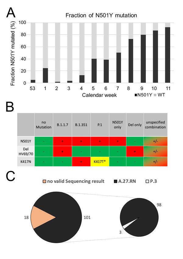
medRxiv preprint doi: https://doi.org/10.1101/2021.04.27.21254849; this version posted April 29, 2021. The copyright holder for this preprint
 (which was not certified by peer review) is the author/funder, who has granted medRxiv a license to display the preprint in perpetuity.
                                     It is made available under a CC-BY-NC-ND 4.0 International license .

     Determination of viral RNA and infectious virus
     Vero E6 cells were seeded into 24 well plates at 2.5x10⁵ cells per well. On the next day, cells
     were inoculated (MOI=0.05) for 1 h, inoculum was removed, and fresh medium was added.
     Supernatant was harvested at 48 h post infection. Viral RNA was isolated from supernatants
     using the NucleoSpin RNA extraction kit (Macherey-Nagel) according to the manufacturer’s
     specification. Viral RNA quantification was performed using the qScript XLT 1-Step RT-PCR
     Kit (Quantabio) and probe 2019-nCoV_N1 (5’-FAM-ACC CCG CAT TAC GTT TGG TGG ACC-
     BHQ1-3’) and the primer set 2019-nCoV_N1 (forward primer: 5’-GAC CCC AAA ATC AGC
     GAA AT-3’; reverse primer; 5’-TCT GGT TAC TGC CAG TTG AAT CTG-3’). To determine
     infectious virus titer, 2.5x10⁵ Vero E6 cells were seeded into each well of a 24-well plate. On
     the next day, cells were inoculated with serial dilutions of SARS-CoV-2 for 1 h. Inoculum was
     removed,         and      cells     were      overlaid       with     serum       free     media       containing        0.75%
     carboxymethylcellulose. After 72 h, cells were fixed with 5% formaldehyde for 1 h followed by
     staining with 1% crystal violet solution. Plaque forming units per ml (PFU/ml) were estimated
     by counting viral plaques.

     Data availability
     SARS-CoV-2 viral genome sequences have been uploaded to the DESH database
     (https://www.rki.de/DE/Content/InfAZ/N/Neuartiges_Coronavirus/DESH/DESH.html) of the
     Robert       Koch       Institute,     which       will    make       the     data       available      via    the     GISAID
     (https://www.gisaid.org)              (44)       and       the      European          Nucleotide          Archive         (ENA,
     https://www.ebi.ac.uk/ena/browser/home) databases. Representative sequences of A.27.RN
     with and without the A222V mutation are included as Supplementary Data.

     Ethical statement
     This study was approved by the ethics committee of the Medical Faculty at the University of
     Heidelberg for the analysis of proband samples by whole genome sequencing of the viral RNA
     (S-316/2021) as well as the virus propagation and neutralization assays and use of sera from
     vaccinated donors (S-203/2021).

     Results and discussion
     A SARS-CoV-2 variant with L452R and N501Y mutations emerged locally
     Due to the recent spreading of VOC in Europe, we implemented whole genome sequencing of
     all SARS-CoV-2 positive samples detected in the diagnostic laboratory of the Department of
     Infectious diseases at Heidelberg University Hospital in January 2021. A SARS-CoV-2 variant
     was identified in 166 out of 1,543 samples acquired from December 2020 to March 2021.

                                                                                                                                     7
medRxiv preprint doi: https://doi.org/10.1101/2021.04.27.21254849; this version posted April 29, 2021. The copyright holder for this preprint
(which was not certified by peer review) is the author/funder, who has granted medRxiv a license to display the preprint in perpetuity.
It is made available under a CC-BY-NC-ND 4.0 International license .
According to its phylogenetic lineage A.27, this variant is referred to in the following as A.27.RN
(Fig. 1). The first occurrence of A.27.RN in the Rhine-Neckar district was detected in a SARS-
CoV-2 positive sample collected in calendar week 1/2021. A.27.RN comprised S gene
mutations L18F, L452R, N501Y, A653V, H655Y, D796Y and G1219V in a D614 wild-type
background (Table 1).
Table 1. Comparison of S gene mutations in different variantsa
B.1.1.7 B.1.351 P.1 B.1.429 B.1.617 A.27.RN
20I/501Y.V1 20H/501Y.V2 20J/501Y.V3 CAL.20C (India)
(UK) (South Africa) (Brazil) (California)
S13I
(L18F) L18F L18F L18F
G142D
W152C
E154K
(A222V)
K417N K417T
L452R L452R L452R
(E484K) E484K E484K E484Q
N501Y N501Y N501Y N501Y
D614G D614G D614G D614G D614G
A653V
H655Y H655Y
A570D
P681H P681R
T716I Q1071H D796Y
S982A H1001D G1219V
D1118H
a
Mutations in A.27.RN considered to confer altered transmissibility or immune escape are shown in bold
Mutations present in addition to the defining variant genome sequence in some samples are in brackets.
Subsequently, the additional S gene mutation A222V was found in samples collected in
February and the relative fraction carrying this additional mutation increased to 10-30% in
calendar week 7. The A222V mutation has been described as a marker mutation of the
20A.EU1 strain that has been spreading in Europe in 2020 (45). The location of the mutated
residues of A.27.RN in the S protein structure and in the RBD - ACE2 interface is depicted in
Supplementary Fig. S1. Interestingly, all exchanges in the S protein of A.27.RN are located
in unstructured regions at the surface of the protein. In addition to the S gene mutations,
A.27.RN carries a D173G mutation and an 8 bp deletion in ORF3a as well as additional
8medRxiv preprint doi: https://doi.org/10.1101/2021.04.27.21254849; this version posted April 29, 2021. The copyright holder for this preprint
(which was not certified by peer review) is the author/funder, who has granted medRxiv a license to display the preprint in perpetuity.
It is made available under a CC-BY-NC-ND 4.0 International license .
mutations listed in Table S1. A comparison of A.27.RN with the three main N501Y strains
detected so far as well as the B.1.429/CAL.20C and B1.617 strains with L452R is given in
Table 1. A.27.RN lacks the E484K and the D614G mutations but carries the unusual
combination of L452R and N501Y, which has not been observed in the established VOC.
Fig. 1. Phylogenetic tree of SARS-CoV-2 variants carrying the N501Y signature mutation. An
overview of a global phylogenetic tree analysis is shown in the upper panel. It contains 4,200 genomes
sequenced from January 2020 to March 2021 and is colored by the amino acid residue at position 501
of the S protein. Green, N501; yellow, N501T; blue, N501Y. The bottom panel shows a zoom into the
A.27 clade with the samples sequenced in our study colored in purple. Six additional samples from
GISAID that also fall into the A.27.RN clade are marked in red.
9medRxiv preprint doi: https://doi.org/10.1101/2021.04.27.21254849; this version posted April 29, 2021. The copyright holder for this preprint
(which was not certified by peer review) is the author/funder, who has granted medRxiv a license to display the preprint in perpetuity.
It is made available under a CC-BY-NC-ND 4.0 International license .
A.27.RN is phylogenetically separated from other N501Y variants
The sequences obtained were placed into the established SARS-CoV-2 phylogenetic tree (40),
yielding the results depicted in Fig. 1. A.27 is a newly designated lineage in clade 19B (46).
Within this lineage, the A.27.RN sequences form a separate branch (Fig. 1) since they carry
the unique D173G mutation in ORF3a, which was absent only in 10/166 cases
(Supplementary Fig. 2B). A.27 is clearly separated from the three currently prevailing N501Y
containing strains (Fig. 1). The latter, as well as the L452R carrying B.1.427/B.1.429 and
B1.617 variants, are classified into clade 20 and lineages therein. At the end of March 2021,
162 A.27 sequences were present in the GISAID database (without the 166 sequences of our
study). The total number of A.27 sequences for calendar week 1-11 of 2021 reported to the
central database of the Robert Koch Institute for sequencing results in Germany amounts to
424 (47).
A.27 sequences in GISAID were previously reported for samples from Denmark, England,
Netherlands, Switzerland, Turkey, Slovenia, France, Belgium and Mayotte, an island between
northwestern Madagascar and northeastern Mozambique and an overseas department of
France (Supplementary Fig. S2A). Together with the 166 cases of A.27.RN reported here,
three other clustered occurrences of A.27 variants have been reported, namely 12 in Mayotte
(January 2021), 92 in France (January to February 2021), and 22 in Slovenia (February 2021),
while the remaining 36 were associated with sporadic occurrences. Thus, A.27 strains show a
repeated pattern of localized clustered occurrences across different countries during the first
quarter of 2021 with 639 cases detected in Germany until calendar week 14/2021 (9).
Rapid local spread of A.27.RN was followed by outcompetition by B.1.1.7
The first month of 2021 in the Rhine Neckar region was characterized by a rapid local spread
of variant A.27.RN. During the period of calendar weeks 53/2020-11/2021 covered in our
sample set, N501 wild-type variants were almost completely displaced by variants carrying the
N501Y mutation (Fig. 2A, Table S2). Initially, A.27.RN dominated and increased to a peak
value of ~58% of all N501Y variants in calendar weeks four and five (Fig. 2A, Table S2).
Interestingly, A.27.RN and B.1.1.7 (UK) became the prevailing N501Y variants in calendar
week seven with each of them comprising >25% of all samples sequenced (Fig. 2B).
Subsequently the occurrence of A.27.RN decayed to a 10% fraction in calendar week eleven,
while B.1.1.7 became dominant and was found in more than 85% of all samples at that time
(Table S2). A.27.RN as well as B.1.1.7 and (in a much lower number of cases) B.1.351 (ZA)
were consistently present.
10medRxiv preprint doi: https://doi.org/10.1101/2021.04.27.21254849; this version posted April 29, 2021. The copyright holder for this preprint
(which was not certified by peer review) is the author/funder, who has granted medRxiv a license to display the preprint in perpetuity.
It is made available under a CC-BY-NC-ND 4.0 International license .
Fig. 2. Development of variants in Q1/2021. (A) N501Y positive samples split up into the different
variants and wild-type virus. Both the fraction of UK B.1.1.7 and the A.27.RN variant increased relatively
fast until calendar week seven and started to diverge from week eight. While the ZA variant B.1.351 was
present in almost every calendar week, the P.3 mutant occurred very rarely. (B) SARS-CoV-2 typing by
melting curve analysis over time. Relative amounts of suspected variants are shown for each calendar
week confirming the trend seen in the sequencing data. “Del” refers to the HV69/70 deletion.
As a faster and less expensive screening readout, we employed Tm-PCR to detect S gene
variants N501Y, K417N/K417T and the HV69/70 deletion with data shown here for calendar
weeks 5-11 (Fig. 2C). Samples were classified as potentially being B.1.1.7, B.1.351, P.1,
N501Y only, HV69/70 deletion only or as unknown (Supplementary Fig. S3A). Samples for
which a valid result could not be obtained for at least one mutation were considered
inconclusive. To assess the concordance of the “N501Y only” category with A.27.RN, we
matched the Tm-PCR results to sequencing results. From 119 samples categorized as “N501Y
only” by Tm-PCR, valid sequencing results could be obtained for 101 samples. Of these, 98
samples (97%) had the A.27.RN sequence (Supplementary Fig. S3B,C). The remaining three
samples (3%) were classified as P.3 (48). The P.3 variant shares the B.1.1.28 ancestor with
P.1 and carries E484K but has a distinguishing P681H mutation also present in B.1.1.7. P3 is
11medRxiv preprint doi: https://doi.org/10.1101/2021.04.27.21254849; this version posted April 29, 2021. The copyright holder for this preprint
(which was not certified by peer review) is the author/funder, who has granted medRxiv a license to display the preprint in perpetuity.
It is made available under a CC-BY-NC-ND 4.0 International license .
indistinguishable from A.27 in the Tm-PCR scheme used here, since both variants have N501Y
but lack HV69/70 deletion and K417N. Nevertheless, the “N501Y-only” category proved to be
a good predictor of the A.27.RN variant in the tested cohort. By exploiting the information
obtained from the sequencing data to define a limited subset of mutations, variants of interest
can also be identified in a cost-efficient manner with faster turnaround times by the targeted
Tm-PCR approach.
In vitro characterization of A.27.RN
To assess replication and spread of A.27.RN in comparison to the BavPat wild-type SARS-
CoV-2 strain and the prevailing VOC, variant SARS-CoV-2 A.27.RN, B1.1.7 and B.1.351 were
isolated from patient samples and cultured in Vero E6 cells. Culture supernatants of infected
cells were prepared and used to determine virus production, which was assessed by
quantifying the amount of viral RNA by RT-qPCR and the titer of infectious virus particles using
a plaque assay. Overall, ~3-5 fold lower titers of infectious virus were obtained for all three
variants compared to BavPat wild-type strain.
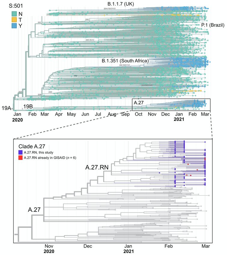
Fig. 3. Infectivity titers and RNA-infectivity ratios of different SARS-CoV-2 variants. Vero E6 cells
were infected with SARS-CoV-2 wild-type (BavPat) and the indicated variants. (A) Quantification of viral
RNA and infectivity. Vero E6 cells were infected with given isolates (MOI=0.05) and 48 h later culture
supernatants were harvested. Amounts of viral RNA contained in culture supernatants were assessed
by RT-qPCR and are given as RNA copies/ml (top panel). The titer of infectious virus was determined
by plaque assay and is given as plaque forming units per ml (middle panel). The ratio of RNA copies to
PFU (bottom panel) is an approximation for specific infectivity, which is reduced for all 3 variants relative
to the wild-type. The graphs show mean values and SD from two biological experiments, each
conducted in triplicates. (B) Representative images of wells of infected cells, stained with crystal violet
at 48 h post infection. Note the smaller plaques obtained with variant B.1.1.7.
12medRxiv preprint doi: https://doi.org/10.1101/2021.04.27.21254849; this version posted April 29, 2021. The copyright holder for this preprint
(which was not certified by peer review) is the author/funder, who has granted medRxiv a license to display the preprint in perpetuity.
It is made available under a CC-BY-NC-ND 4.0 International license .
This was reflected by a lower ratio of plaque-forming units to RNA copies for all three variants,
arguing for lower specific infectivity of the variant virus particles as compared to the wild-type
(Fig. 3A). Furthermore, we noted a consistently reduced plaque size in the case of the B.1.1.7
variant compared to the other strains arguing for less efficient spread of B.1.1.7 in this tissue
culture system (Fig. 3B).
Neutralization of variant A.27.RN is reduced compared to wild-type virus
To evaluate the effect of S gene mutations found in A.27.RN on neutralization capacity of
vaccine sera, we performed titration experiments with serum samples from BNT162b2 vaccine
recipients. Six samples collected 10-28 days after the second vaccine dose that displayed a
range of anti-S1 RBD IgG levels were chosen (Supplementary Table S3). Neutralization titers
were determined in infection experiments with the A.27.RN isolate and compared against the
wild-type control as well as B.1.1.7 and B.1.351 variants (Fig. 4).
Fig. 4. Antibody neutralization capacity of A.27.RN. Neutralizing titers were determined for the sera
from six donors vaccinated against SARS-CoV-2 in titration experiments using the indicated SARS-
CoV-2 variants on Vero E6 cells. Black: Wild-type (BavPat1/2020); red: A.27.RN; green: B.1.1.7; blue:
B.1.351. (A) Raw data from one experiment. Data represent mean values and SD from triplicate
measurements. The line is a non-linear regression fit to the data that yields the NT50 value, i.e. the
dilution at which virus infection is reduced to 50%. (B) NT50 dilution factors from three independent
experiments. Each data point refers to one independent measurement, lines display mean values.
(C) Differences of NT50 values of the three variants tested in comparison to wild-type BavPat1/2020.
13medRxiv preprint doi: https://doi.org/10.1101/2021.04.27.21254849; this version posted April 29, 2021. The copyright holder for this preprint
(which was not certified by peer review) is the author/funder, who has granted medRxiv a license to display the preprint in perpetuity.
It is made available under a CC-BY-NC-ND 4.0 International license .
Neutralizing titers of sera against the wild-type isolate varied from ~1:40 to 1:1,280, consistent
with the different levels of IgG reactivity determined by ELISA. Accordingly, neutralization
capacity against the A.27.RN varied somewhat between donors (Fig. 4A, B) but was notably
reduced compared to wild-type in all cases. Averaged values of the relative difference in NT50
values between variants against wild-type virus yielded a fold-difference and 95% confidence
interval (CI) of 2.6 (95% CI = 2.2 to 3.1) for A.27.RN, 0.9 (95% CI = 0.8 to 1.1) for B.1.1.7 and
4.3 (95% CI = 3.1 to 6.0) for B.1.351 (Fig. 4C). These values were mostly in line with a recent
study that also reports similar serum neutralization values for B.1.1.7 and wild-type but a
somewhat higher factor of 9-12 for B.1.351 (12, 49).
Conclusion
The mutational pattern of A.27.RN is different from other currently prevailing N501Y variants
as it combines N501Y and L452R and carries additional S protein and other mutations.
According to public databases, the L452R mutation has been acquired by several other
independent lineages across multiple countries and continents (5-7, 20, 46). Such repeatedly
emerging hot-spot mutations typically indicate strong and probably recent positive selection
(17). The spreading profile of A.27.RN on a population level suggests that its transmissibility
is higher than that of the SARS-CoV-2 wild-type strain but lower than that of the B.1.1.7 variant.
This was not reflected by the growth properties of the virus in tissue culture, where B.1.1.7
exhibited a smaller and A.27.RN a larger plaque phenotype compared to the wild-type
BavPat1/2020 reference. Notably, all three variants exhibited a lower specific infectivity,
indicating that the observed differences in tissue culture systems may not reflect or predict
their actual spread in the population where a variety of other factors come into play.
Our study revealed the regional and clustered appearance of a SARS-CoV-2 variant of
potential concern, which rapidly became dominant locally but subsequently was outcompeted
by another recently introduced variant on the population level. These results show that the
presence of certain signature mutations is not sufficient to predict the spread of different SARS-
CoV-2 variants. It can be speculated that the lack of the D614G mutation lowers the
transmissibility of A27.RN, since D614G is found in all of the other variants discussed here
(Table 1) and has been associated with an increased SARS-CoV-2 fitness (21, 22). Vaccine
sera were capable of neutralizing A.27.RN, albeit with reduced neutralization titers. This effect
was even more pronounced for B.1.351 in line with previous findings (12, 49). Accordingly, it
is expected that combinations of the phenotypically relevant L18F, K417N/T, L452R, E484R/Q,
N501Y and D614G mutations in the S protein discussed here, as well as newly emerging ones,
will become advantageous under immune selection. Continued and comprehensive
surveillance of emerging SARS-CoV-2 strains and mutations by unbiased whole genome
14medRxiv preprint doi: https://doi.org/10.1101/2021.04.27.21254849; this version posted April 29, 2021. The copyright holder for this preprint
(which was not certified by peer review) is the author/funder, who has granted medRxiv a license to display the preprint in perpetuity.
It is made available under a CC-BY-NC-ND 4.0 International license .
sequencing in combination with melting point PCR remains therefore of utmost importance for
controlling the current pandemic.
Acknowledgments
We thank Christian Drosten and Barbara Mühlemann from the German National Consultant
Laboratory for Coronaviruses for discussion on SARS-CoV-2 variant distribution. We gratefully
acknowledge the help of Maria Anders-Össwein with serological analyses and of Marie
Bartenschlager with the neutralization assays and thank all diagnostics employees and EMBL
GeneCore's NGS protocol-automation team for their support. This work was supported by the
program for surveillance and control of SARS-CoV-2 mutations of the state of Baden-
Württemberg, the German Federal Research Network Applied Surveillance and Testing (B-
FAST) within the Network University Medicine and the DKFZ@fightCOVID initiative.
Conflict of interest
None declared.
Author contributions
Conceptualization: HGK, KR, RB
Investigation: JPM, CB, HK, NW, KBö, KBa, KLJ, SP, BM, VB, NA, AT
Data Curation: CB, DH
Formal Analysis: SS, IL, TR, JPM, HK, KR
Writing – Original Draft Preparation: KR, JPM
Writing – Review & Editing: HGK, KR, JPM, RB, BM, NW, KB, CB, TR, VB, MB
Supervision: HGK, KR, RB, MB
15medRxiv preprint doi: https://doi.org/10.1101/2021.04.27.21254849; this version posted April 29, 2021. The copyright holder for this preprint
(which was not certified by peer review) is the author/funder, who has granted medRxiv a license to display the preprint in perpetuity.
It is made available under a CC-BY-NC-ND 4.0 International license .
References
1. Abdool Karim SS, de Oliveira T. New SARS-CoV-2 Variants - Clinical, Public Health, and
Vaccine Implications. N Engl J Med. 2021:published online 24 March 2021.
2. Rambaut A, Loman N, Pybus O, Barclay W, Barrett J, Carabelli A, et al. Preliminary genomic
characterisation of an emergent SARS-CoV-2 lineage in the UK defined by a novel set of spike
mutations. Virologicalorg. 2020:https://tinyurl.com/224c8pa9.
3. Tegally H, Wilkinson E, Giovanetti M, Iranzadeh A, Fonseca V, Giandhari J, et al.
Emergence and rapid spread of a new severe acute respiratory syndrome-related coronavirus
2 (SARS-CoV-2) lineage with multiple spike mutations in South Africa. medRxiv.
2020:2020.12.21.20248640.
4. Faria NR, Mellan TA, Whittaker C, Claro IM, Candido DDS, Mishra S, et al. Genomics and
epidemiology of a novel SARS-CoV-2 lineage in Manaus, Brazil. medRxiv.
2021:2021.02.26.21252554.
5. Zhang W, Davis BD, Chen SS, Sincuir Martinez JM, Plummer JT, Vail E. Emergence of a
Novel SARS-CoV-2 Variant in Southern California. JAMA. 2021;325(13):1324-6.
6. Deng X, Garcia-Knight MA, Khalid MM, Servellita V, Wang C, Morris MK, et al.
Transmission, infectivity, and antibody neutralization of an emerging SARS-CoV-2 variant in
California carrying a L452R spike protein mutation. medRxiv. 2021:2021.03.07.21252647.
7. Tchesnokova V, Kulakesara H, Larson L, Bowers V, Rechkina E, Kisiela D, et al. Acquisition
of the L452R mutation in the ACE2-binding interface of Spike protein triggers recent massive
expansion of SARS-Cov-2 variants. bioRxiv. 2021:2021.02.22.432189.
8. Public Health England. SARS-CoV-2 variants of concern and variants under investigation
in England. Technical briefing 9. 2021:tinyurl.com/3f2yavyb.
9. Robert-Koch-Institut. Bericht zu Virusvarianten von SARS-CoV-2 in Deutschland,
insbesondere zur Variant of Concern (VOC) B.1.1.7. Report from 21 April 2021.
2021:https://tinyurl.com/yfmaz5rv.
10. Plante JA, Mitchell BM, Plante KS, Debbink K, Weaver SC, Menachery VD. The variant
gambit: COVID-19's next move. Cell Host Microbe. 2021;29(4):508-15.
11. Lan J, Ge J, Yu J, Shan S, Zhou H, Fan S, et al. Structure of the SARS-CoV-2 spike
receptor-binding domain bound to the ACE2 receptor. Nature. 2020;581(7807):215-20.
12. Wang P, Nair MS, Liu L, Iketani S, Luo Y, Guo Y, et al. Antibody resistance of SARS-CoV-
2 variants B.1.351 and B.1.1.7. Nature. 2021:published online 8 March 2021.
13. Kustin T, Harel N, Finkel U, Perchik S, Harari S, Tahor M, et al. Evidence for increased
breakthrough rates of SARS-CoV-2 variants of concern in BNT162b2 mRNA vaccinated
individuals. medRxiv. 2021:2021.04.06.21254882.
14. Starr TN, Greaney AJ, Hilton SK, Ellis D, Crawford KHD, Dingens AS, et al. Deep
Mutational Scanning of SARS-CoV-2 Receptor Binding Domain Reveals Constraints on
Folding and ACE2 Binding. Cell. 2020;182(5):1295-310 e20.
15. Greaney AJ, Loes AN, Crawford KHD, Starr TN, Malone KD, Chu HY, et al. Comprehensive
mapping of mutations in the SARS-CoV-2 receptor-binding domain that affect recognition by
polyclonal human plasma antibodies. Cell Host Microbe. 2021;29(3):463-76 e6.
16. Grubaugh ND, Hodcroft EB, Fauver JR, Phelan AL, Cevik M. Public health actions to
control new SARS-CoV-2 variants. Cell. 2021;184(5):1127-32.
17. Martin DP, Weaver S, Tegally H, San EJ, Shank SD, Wilkinson E, et al. The emergence
and ongoing convergent evolution of the N501Y lineages coincides with a major global shift in
the SARS-CoV-2 selective landscape. medRxiv. 2021:2021.02.23.21252268.
16medRxiv preprint doi: https://doi.org/10.1101/2021.04.27.21254849; this version posted April 29, 2021. The copyright holder for this preprint
(which was not certified by peer review) is the author/funder, who has granted medRxiv a license to display the preprint in perpetuity.
It is made available under a CC-BY-NC-ND 4.0 International license .
18. Wise J. Covid-19: The E484K mutation and the risks it poses. BMJ. 2021;372:n359.
19. Li Q, Wu J, Nie J, Zhang L, Hao H, Liu S, et al. The Impact of Mutations in SARS-CoV-2
Spike on Viral Infectivity and Antigenicity. Cell. 2020;182(5):1284-94 e9.
20. Ranjan R, Sharma A, Verma MK. Characterization of the Second Wave of COVID-19 in
India. medRxiv. 2021:2021.04.17.21255665.
21. Hou YJ, Chiba S, Halfmann P, Ehre C, Kuroda M, Dinnon KH, 3rd, et al. SARS-CoV-2
D614G variant exhibits efficient replication ex vivo and transmission in vivo. Science.
2020;370(6523):1464-8.
22. Plante JA, Liu Y, Liu J, Xia H, Johnson BA, Lokugamage KG, et al. Spike mutation D614G
alters SARS-CoV-2 fitness. Nature. 2021;592(7852):116-21.
23. Grabowski F, Kochańczyk M, Lipniacki T. L18F substrain of SARS-CoV-2 VOC-202012/01
is rapidly spreading in England. medRxiv. 2021:2021.02.07.21251262.
24. Davies NG, Abbott S, Barnard RC, Jarvis CI, Kucharski AJ, Munday JD, et al. Estimated
transmissibility and impact of SARS-CoV-2 lineage B.1.1.7 in England. Science.
2021;372(6538):eabg3055.
25. Tyson JR, James P, Stoddart D, Sparks N, Wickenhagen A, Hall G, et al. Improvements
to the ARTIC multiplex PCR method for SARS-CoV-2 genome sequencing using nanopore.
bioRxiv. 2020:2020.09.04.283077.
26. Gohl DM, Garbe J, Grady P, Daniel J, Watson RHB, Auch B, et al. A rapid, cost-effective
tailed amplicon method for sequencing SARS-CoV-2. BMC Genomics. 2020;21(1):863.
27. Baker DJ, Aydin A, Le-Viet T, Kay GL, Rudder S, de Oliveira Martins L, et al. CoronaHiT:
high-throughput sequencing of SARS-CoV-2 genomes. Genome Med. 2021;13(1):21.
28. Banada P, Green R, Banik S, Chopoorian A, Streck D, Jones R, et al. A Simple RT-PCR
Melting temperature Assay to Rapidly Screen for Widely Circulating SARS-CoV-2 Variants.
medRxiv. 2021:2021.03.05.21252709.
29. Sascha T, Kathrin K, Markus V, Thorsten P, Stefan L, Christina LP, et al. Rapid base-
specific calling of SARS-CoV-2 variants of concern using combined RT-PCR melting curve
screening and SIRPH technology. medRxiv. 2021:2021.03.15.21253586.
30. Ewels PA, Peltzer A, Fillinger S, Patel H, Alneberg J, Wilm A, et al. The nf-core framework
for community-curated bioinformatics pipelines. Nat Biotechnol. 2020;38(3):276-8.
31. Patel H, Varona S, Monzón S, Espinosa-Carrasco J, Heuer ML, Gabernet G, et al. nf-
core/viralrecon. Zenodo. 2020:doi: 10.5281/zenodo.3901628.
32. Grubaugh ND, Gangavarapu K, Quick J, Matteson NL, De Jesus JG, Main BJ, et al. An
amplicon-based sequencing framework for accurately measuring intrahost virus diversity using
PrimalSeq and iVar. Genome Biol. 2019;20(1):8.
33. Wood DE, Lu J, Langmead B. Improved metagenomic analysis with Kraken 2. Genome
Biol. 2019;20(1):257.
34. Li H, Durbin R. Fast and accurate short read alignment with Burrows-Wheeler transform.
Bioinformatics. 2009;25(14):1754-60.
35. Li H, Handsaker B, Wysoker A, Fennell T, Ruan J, Homer N, et al. The Sequence
Alignment/Map format and SAMtools. Bioinformatics. 2009;25(16):2078-9.
36. Rausch T, Hsi-Yang Fritz M, Korbel JO, Benes V. Alfred: interactive multi-sample BAM
alignment statistics, feature counting and feature annotation for long- and short-read
sequencing. Bioinformatics. 2019;35(14):2489-91.
37. Garrison E, Marth G. Haplotype-based variant detection from short-read sequencing.
arXiv. 2012:1207.3907.
17medRxiv preprint doi: https://doi.org/10.1101/2021.04.27.21254849; this version posted April 29, 2021. The copyright holder for this preprint
(which was not certified by peer review) is the author/funder, who has granted medRxiv a license to display the preprint in perpetuity.
It is made available under a CC-BY-NC-ND 4.0 International license .
38. Li H. A statistical framework for SNP calling, mutation discovery, association mapping and
population genetical parameter estimation from sequencing data. Bioinformatics.
2011;27(21):2987-93.
39. Rambaut A, Holmes EC, O'Toole A, Hill V, McCrone JT, Ruis C, et al. A dynamic
nomenclature proposal for SARS-CoV-2 lineages to assist genomic epidemiology. Nat
Microbiol. 2020;5(11):1403-7.
40. Hadfield J, Megill C, Bell SM, Huddleston J, Potter B, Callender C, et al. Nextstrain: real-
time tracking of pathogen evolution. Bioinformatics. 2018;34(23):4121-3.
41. Cortese M, Lee JY, Cerikan B, Neufeldt CJ, Oorschot VMJ, Kohrer S, et al. Integrative
Imaging Reveals SARS-CoV-2-Induced Reshaping of Subcellular Morphologies. Cell Host
Microbe. 2020;28(6):853-66 e5.
42. Pape C, Remme R, Wolny A, Olberg S, Wolf S, Cerrone L, et al. Microscopy-based assay
for semi-quantitative detection of SARS-CoV-2 specific antibodies in human sera: A semi-
quantitative, high throughput, microscopy-based assay expands existing approaches to
measure SARS-CoV-2 specific antibody levels in human sera. Bioessays.
2021;43(3):e2000257.
43. Tonshoff B, Muller B, Elling R, Renk H, Meissner P, Hengel H, et al. Prevalence of SARS-
CoV-2 Infection in Children and Their Parents in Southwest Germany. JAMA Pediatr.
2021:published online 22 January 2021.
44. Shu Y, McCauley J. GISAID: Global initiative on sharing all influenza data - from vision to
reality. Euro Surveill. 2017;22(13).
45. Hodcroft EB, Zuber M, Nadeau S, Crawford KHD, Bloom JD, Veesler D, et al. Emergence
and spread of a SARS-CoV-2 variant through Europe in the summer of 2020. medRxiv.
2020:2020.10.25.20219063.
46. Simon-Loriere E. Potential new lineage causing a cluster in Mayotte. GitHub.
2021:https://github.com/cov-lineages/pango-designation/issues/11.
47. Robert-Koch-Institut. Aktualisierter Bericht zu Virusvarianten von SARS-CoV-2 in
Deutschland, insbesondere zur Variant of Concern (VOC) B.1.1.7. Report from 31 March 2021.
2021:https://tinyurl.com/evyjap8e.
48. Phillippine Genome Center. Detection and characterization of a new SARS-CoV-2 lineage
P.3, with spike protein mutations E484K, N501Y, P681H and LGV 141-143 deletion, from
samples sequenced through the intensified UP-PGC, UP-NIH and DOH biosurveillance
program. PGC SARS-CoV-2 Bulletin. 2021;7:https://pgc.up.edu.ph/sars-cov-2-bulletin-no-7.
49. Bates TA, Leier HC, Lyski ZL, McBride SK, Coulter FJ, Weinstein JB, et al. Neutralization
of SARS-CoV-2 variants by convalescent and vaccinated serum. medRxiv.
2021:2021.04.04.21254881.
50. Wrobel AG, Benton DJ, Xu P, Roustan C, Martin SR, Rosenthal PB, et al. SARS-CoV-2
and bat RaTG13 spike glycoprotein structures inform on virus evolution and furin-cleavage
effects. Nat Struct Mol Biol. 2020;27(8):763-7.
51. Humphrey W, Dalke A, Schulten K. VMD: visual molecular dynamics. J Mol Graph.
1996;14(1):33-8, 27-8.
18medRxiv preprint doi: https://doi.org/10.1101/2021.04.27.21254849; this version posted April 29, 2021. The copyright holder for this preprint
(which was not certified by peer review) is the author/funder, who has granted medRxiv a license to display the preprint in perpetuity.
It is made available under a CC-BY-NC-ND 4.0 International license .
Supplementary Information
Supplementary Tables
Table S1. A.27.RN mutations outside the S region
Region Mutation
N S202N
ORF1a P286L
ORF1a N3651S
ORF1b P1000L
ORF3a V50A
ORF3a D173G
ORF8 L84S
ORF3a del 258/259 (8 bp)
ORF8 del 119/120 (6 bp)
19medRxiv preprint doi: https://doi.org/10.1101/2021.04.27.21254849; this version posted April 29, 2021. The copyright holder for this preprint
(which was not certified by peer review) is the author/funder, who has granted medRxiv a license to display the preprint in perpetuity.
It is made available under a CC-BY-NC-ND 4.0 International license .
Table S2. Spreading of N501Y variants detected by sequencinga.
Calendar Total N501Y N501Y UK UK ZA ZA P.3 P.3 A.27.RN A.27.RN
week n n (%) n (%) n (%) n (%) n (%)
53, 2020 17 1 5.9 0 0 1 5.9 0 0 0 0
1, 2021 25 6 24 0 0 4 16 0 0 2 8
2, 2021 246 4 1.6 0 0 4 1.6 0 0 0 0
3, 2021 309 9 2.9 3 1.0 0 0 0 0 6 1.9
4, 2021 243 30 12.3 2 0.8 2 0.8 0 0 26 10.7
5, 2021 156 62 39.7 23 14.7 3 1.9 0 0 36 23.1
6,2021 129 50 38.8 20 15.5 1 0.8 0 0 29 22.5
7,2021 116 58 50 30 25.9 2 1.7 0 0 26 22.4
8,2021 88 65 73.8 42 47.7 4 4.5 1 1.1 18 20.4
9, 2021 64 53 82.8 43 67.2 0 0 2 3.1 8 12.5
10,2021 124 108 87.1 94 75.8 1 0.8 0 0 13 10.5
11,2021 26 24 92.3 21 80.8 1 3.8 0 0 2 7.7
total 1543 467 278 23 3 166
a
The calendar week when the sample was taken is given. UK, B.1.1.7; ZA, B.1.351; The number of a
samples n as well as the corresponding percentage fraction of the variants is listed.
20medRxiv preprint doi: https://doi.org/10.1101/2021.04.27.21254849; this version posted April 29, 2021. The copyright holder for this preprint
(which was not certified by peer review) is the author/funder, who has granted medRxiv a license to display the preprint in perpetuity.
It is made available under a CC-BY-NC-ND 4.0 International license .
Table S3: Reactivity of sera used for neutralization assays
ID # IgG antiS1RBD (Siemens sCOV CLIA) (ABU) Surrogate neutralization titer (IC50)
1 >3270 1:1150
2 >3270 1:740
3 >3270 1:650
4 3032,38 1:145
5 2905,94 1:90
6 948,3 1:88
Sera from healthy donors vaccinated with BNT162b2 were collected at day 10-28 after the
second vaccine dose. IgG reactivity was determined by CLIA (Siemens sCOV test kit) as
described in Materials and Methods. WHO antibody binding units (ABU) were calculated from
the index value obtained using the factor provided by the manufacturer. Interference with the
ACE2-RBD interaction was determined by a titration experiment using the GenScript SARS-
CoV-2 Surrogate Virus Neutralization Test Kit.
21medRxiv preprint doi: https://doi.org/10.1101/2021.04.27.21254849; this version posted April 29, 2021. The copyright holder for this preprint
(which was not certified by peer review) is the author/funder, who has granted medRxiv a license to display the preprint in perpetuity.
It is made available under a CC-BY-NC-ND 4.0 International license .
Supplementary Figures
Fig. S1. 3D structure of SARS-CoV-2 S protein with mutated residues mutated in
A.27.RN indicated. A.27.RN mutations were mapped either on the furin cleaved S protein
structure using pdb coordinates 6ZGG (50) and on the complex of the S protein RBD and the
ACE2 receptor (pdb coordinates 6MOJ) (11) and visualized with VMD viewer (51) (A)
Complete S protein after furin cleavage. (B) Complex of S protein RBD with ACE2 repressor
visualization of S protein structure with mutated residues. N501 directly interacts with N330 of
the receptor. In contrast, L452 is not in direct contact with ACE2 but creates as hydrophobic
patch together with F490 and L492 on the RBD surface.
22medRxiv preprint doi: https://doi.org/10.1101/2021.04.27.21254849; this version posted April 29, 2021. The copyright holder for this preprint
(which was not certified by peer review) is the author/funder, who has granted medRxiv a license to display the preprint in perpetuity.
It is made available under a CC-BY-NC-ND 4.0 International license .
Fig. S2. (A) Zoom into the A.27 clade as defined by the tool pangolin (39). A total of 162
sequences retrieved from GISAID are shown together with the A27.RN sequences with
coloring according to country. (B) The same phylogenetic tree as in the bottom panel of Fig. 2
but labeled according to the mutational status ORF3b:173. It can be seen that the A27.RN
specific D173G mutation in ORF3b (gold color) separates it from the D173 wild-type
background (cyan color) of the other A.27 sequences.
23medRxiv preprint doi: https://doi.org/10.1101/2021.04.27.21254849; this version posted April 29, 2021. The copyright holder for this preprint
(which was not certified by peer review) is the author/funder, who has granted medRxiv a license to display the preprint in perpetuity.
It is made available under a CC-BY-NC-ND 4.0 International license .
Fig. S3. Analysis of SARS-CoV-2 variants with N501Y by sequencing and melting curve
analysis. (A) Fraction of N501Y mutations present in the samples sequenced. It can be seen
that the variants with this mutation have become dominant and comprise 90% of all samples
in calendar week 11. (B) Samples were analyzed by Tm-PCR for N501Y, K417N and the
HV69/70 deletion in the S protein. Preliminary classification was done according to the
mutation pattern. K417T is supposed to be represented by a Tm-peak at a lower temperature
than 417K (wild-type) in the K417N-assay albeit at reduced specificity. Only samples with valid
results in all 3 assays were included. Samples with combinations not shown were classified as
“unspecified combination“. (C) Comparison of results obtained by sequencings with the Tm-
PCR analysis. From 101 matched samples assigned to the “N501Y only” category as a proxy
for A.27.RN, 98 samples were indeed this variant, whereas 3 samples were classified as P.3
based on the sequencing analysis.
24medRxiv preprint doi: https://doi.org/10.1101/2021.04.27.21254849; this version posted April 29, 2021. The copyright holder for this preprint
(which was not certified by peer review) is the author/funder, who has granted medRxiv a license to display the preprint in perpetuity.
It is made available under a CC-BY-NC-ND 4.0 International license .
Supplementary Data
Supplementary Data Set 1
The sequence of the A.27.RN variant as well as its A222V mutated from are provided in FASTA
format as a zip file.
25You can also read