Lymph Node Involvement in Advanced Gastric Cancer in the Era of Multimodal Treatment-Oncological and Surgical Perspective - MDPI
←
→
Page content transcription
If your browser does not render page correctly, please read the page content below
cancers
Review
Lymph Node Involvement in Advanced Gastric Cancer in the
Era of Multimodal Treatment—Oncological and
Surgical Perspective
Zuzanna Pelc, Magdalena Skórzewska, Karol Rawicz-Pruszyński *                              and Wojciech P. Polkowski

                                          Department of Surgical Oncology, Medical University of Lublin, Radziwiłłowska 13 St, 20-080 Lublin, Poland;
                                          zuzanna.torun@gmail.com (Z.P.); magdalenaskorzewska@umlub.pl (M.S.);
                                          wojciech.polkowski@umlub.pl (W.P.P.)
                                          * Correspondence: karolrawiczpruszynski@umlub.pl; Tel.: +48-81-531-81-26

                                          Simple Summary: Gastric cancer (GC) continues to be one of the major oncological challenges
                                          on a global scale. The role of neoadjuvant chemotherapy (NAC) in GC is to downstage primary
                                          tumour, eliminate potential micrometastases, and increase the chance for radical resection. Although
                                          systemic treatment prolongs the survival in advanced GC, persistent lymph node (LN) metastases
                                          indicate poor prognosis. Therefore, further identification of prognostic factors after NAC is urgent
                                          and could positively influence clinical outcomes. This article aimed to review the actual trends
                                          and future perspectives in multimodal therapy of advanced GC, with a particular interest in the
                                          post-neoadjuvant pathological nodal stage. Since downstaged and primarily node-negative patients
                                          show a similar prognosis, the main target for NAC in advanced GC should be nodal clearance.

                                   Adequate staging and personalised perioperative therapy seem to be of great importance in the
                                          multimodal treatment of GC.
Citation: Pelc, Z.; Skórzewska, M.;
Rawicz-Pruszyński, K.; Polkowski,
                                          Abstract: Gastric cancer (GC) continues to be one of the major oncological challenges on a global
W.P. Lymph Node Involvement in
                                          scale. The role of neoadjuvant chemotherapy (NAC) in GC is to downstage primary tumour, elim-
Advanced Gastric Cancer in the Era
of Multimodal Treatment—
                                          inate potential micrometastases, and increase the chance for radical resection. Although systemic
Oncological and Surgical Perspective.     treatment prolongs the survival in advanced GC, persistent lymph node (LN) metastases indicate
Cancers 2021, 13, 2509. https://          poor prognosis. Further identification of prognostic factors after NAC is urgent and could positively
doi.org/10.3390/cancers13102509           influence clinical outcomes. This article aimed to review the actual trends and future perspectives in
                                          multimodal therapy of advanced GC, with a particular interest in the post-neoadjuvant pathological
Academic Editor: Sachio Fushida           nodal stage. A favourable prognostic impact for ypN0 patients is observed, either due to truly
                                          negative LN before the start of therapy or because preoperative therapy achieved a pathologically
Received: 9 May 2021                      complete nodal response. Ongoing trials investigating the extent of lymphadenectomy after neoad-
Accepted: 19 May 2021
                                          juvant therapy will standardise the LN dissection from the multimodal therapy perspective. Since
Published: 20 May 2021
                                          downstaged and primarily node-negative patients show a similar prognosis, the main target for
                                          NAC in advanced GC should be nodal clearance. Adequate staging and personalised perioperative
Publisher’s Note: MDPI stays neutral
                                          therapy seem to be of great importance in the multimodal treatment of GC.
with regard to jurisdictional claims in
published maps and institutional affil-
                                          Keywords: advanced gastric cancer; neoadjuvant chemotherapy; lymph node metastases
iations.

                                          1. Introduction
Copyright: © 2021 by the authors.
Licensee MDPI, Basel, Switzerland.
                                               Gastric cancer (GC) continues to be one of the major oncological challenges on a global
This article is an open access article
                                          scale. According to GLOBOCAN 2020 data, GC remains the fifth most common cancer
distributed under the terms and           and the third most deadly neoplasm causing nearly 769,000 deaths in 2020 [1]. Curative
conditions of the Creative Commons        management in early GC patients reaches nearly 90%. Unfortunately, lymph node (LN)
Attribution (CC BY) license (https://     metastases significantly decrease the 5-year overall survival (OS) to 70–80% in stage N1/N2,
creativecommons.org/licenses/by/          and to 30% in stage N3 [2,3]. A 5-years OS in advanced GC patients treated with optimal
4.0/).                                    multimodal therapy, based on systemic chemotherapy and surgery does not exceed 38%,

Cancers 2021, 13, 2509. https://doi.org/10.3390/cancers13102509                                              https://www.mdpi.com/journal/cancers
Cancers 2021, 13, 2509 2 of 14
as shown in FNCLCC and FFCD Multicenter Phase III Trial [4]. However, the role of
neoadjuvant chemotherapy (NAC) in nodal metastasis remains unknown and constituted
the aim of JCOG trials [5,6]
Currently, perioperative and adjuvant chemotherapy (CTH) is indicated in advanced
GC (stage IB-III) [7]. The role of neoadjuvant chemotherapy (NAC) is to downstage pri-
mary tumour, eliminate potential micrometastases, and increase the chance for radical
resection [1,8,9]. Pathologic response to NAC is treated as an independent predictor of
OS and 3-year disease-specific survival [10–12]. Although systemic treatment prolongs
the survival in advanced GC, persistent LN metastases indicates poor prognosis. There-
fore, further identification of prognostic factors after NAC is urgent and could positively
influence clinical outcomes [2]. The American Joint Committee on Cancer (AJCC) in its 8th
edition created a separate staging system for GC patients who underwent preoperative
therapy [13]. Post-neoadjuvant pathological stage (yp) is considered as important survival
predictor [14]. It allowed distinguishing several groups of patients by nodal involve-
ment: cN0/ypN0 (node-negative), cN+/ypN0 (downstaged N0) and ypN+ (node-positive).
Indeed, one of the most valid prognostic factors in advanced GC is lymph node (LN)
involvement [1,15,16]. Despite multimodal treatment, the prognosis for ypN+ patients
is poor, suggesting further insight into the treatment strategy. Recognition of occult LN
metastases in a preoperative setting remains a challenge, since preoperative independent
predictive factors produce area under the receiver operating characteristics curve of only
0.660 [17]. Thus surgeons need to be aware of limitations in preoperative prediction of the
LN metastasis. Efficient verification of LN status is essential considering the clinical issues
of undertreatment and overtreatment.
This article aimed to review the actual trends and future perspectives in multimodal
therapy of advanced GC, with particular interest on post-neoadjuvant pathological nodal
stage.
2. Preoperative Verification of LN Status
Verification of LN is essential while determining the treatment pathway, followed
by extent of lymphadenectomy during gastrectomy. The role of appropriate preoperative
assessment is crucial in the era of multidisciplinary and multimodal treatment. In order
to establish a new golden standard for nodal staging, several ongoing trials aim to as-
sess imaging characteristics for nodal staging [18] (NCT04028375), compare radiological
techniques with histopathological verification [19] (NCT04440605), and determine the
sensitivity of sentinel lymph node sampling [20] (NCT03049345).
2.1. Computed Tomography
Computed tomography (CT) is widely recognized as the primary method for staging
and detecting distant metastases, determining surgical treatment [21]. Accuracy of pre-
operative cN staging is strongly dependent on cT stage and histological type of GC [22].
In early GC, LN metastases are detected with low sensitivity of only 34% [23]. The prin-
cipal criterion of LN invasion in CT is nodal diameter, whereas other include: circular
shape, heterogeneous enhancement, necrosis or decline of fatty LN hilum [24]. However,
undefined cutoff values represent one of the primary limitations. Many radiologists per-
forming multidetector CT arbitrarily accept a threshold of 6 mm for the celiac axis LN’s
diameter and 8 mm for perigastric LN [25]. A German study showed that even 55% of
cancerous-infiltrated LN remain smaller than 5 mm [26], which implies high possibility of
false-negative and false-positive results. Limitations are related to poor visualisation of
nodal engagement [21,27], nodal location dependency and sensitivity rate of 44.4% [22].
Moreover, in up to 30% of negative CT scans, endoscopic ultrasound (EUS) reveals nodal
involvement [27,28]. Another restriction is associated with ambiguous restaging before
potential gastrectomy. According to Gertsen et al., restaging CT prevents only 1% of unnec-
essary laparotomies, although preoperative assessment was performed before completing
NAC [29]. High volume centres present contrasting numbers with negative prognosticCancers 2021, 13, 2509 3 of 14
value (NPV) of LN involvement at a level of 90.1% among patients with early GC [21,30].
However, in advanced GC Kagedan et al. estimated NPV of LN metastases at 43.3%,
emphasizing the issue of cN understaging [31]. On the other hand, Yamamoto et al. in-
vestigated a correlation between the histological type of GC and accuracy of preoperative
LN assessment: well-differentiated GC patients had more frequent overdiagnosis of LN
involvement than undifferentiated GC (50% vs. 13.3%) [22].
2.2. Endoscopic Ultrasonography (EUS)
The effectiveness of nodal staging in EUS varies from 30% to 90% [10]. Perigastric
LN suspected of metastasis occur as enlarged, round, hypoechoic and homogeneous
structures. Unfortunately, lymphadenitis provides similar clinical image. An effective
manner to distinguish those two processes is fine-needle aspiration (FNA) for cytological
assessment, which should be performed unless the primary tumour and large vessels
are in close proximity [10]. Another limitation results from suboptimal visualisation of
regional LNs and poor quality of restaging, particularly after chemoradiotherapy [32,33].
Moreover, Ikoma et al. suggested that EUS could be eliminated from routinely clinical
staging due to the low predictive value of cN assessment for patients undergoing NAC [17].
Extensive differences in sensitivity (16.7–96.8%) and specificity (48.4–100.0%) classify EUS
as unreliable in detecting and excluding LN metastases [25]. However, according to recent
data, the role of EUS in clinical staging of advanced GC should be maintained, underlying
its correlation with further patient’s prognosis [33].
2.3. Magnetic Resonance Imaging
In terms of N staging, Magnetic Resonance Imaging (MRI) remains inferior to CT
or EUS [34]. Restraints arise from costs and time of examination [18]. Sensitivity and
specificity rates reach 85% and 67%, respectively and remain steady over time [35,36].
Additionally, there is no standardised protocol for defining LN metastases [37]. How-
ever, the role of MRI in nodal verification may change—diffusion-weighted (DW) MRI
establishes LN involvement not by diameter but based on the integrity of cell membranes
and tissue consistency [18]. Nevertheless, MRI is currently not recommended for nodal
assessment [10].
2.4. Fluorodeoxyglucose Positron Emission Tomography
Fluorodeoxyglucose positron emission tomography (FDG PET) is not a universal
method in GC imaging since up to 30% of non-FDG avid primary tumour (particularly
mucinous type) [38]. Its sensitivity and specificity in the detection of LN metastases
have been reported to be 41–51% and 86–100%, respectively [39]. FDG PET sensitivity
rates, in particular, are lower compared to CT and EUS (56% vs. 78% and 50% vs. 73%,
respectively) [40]. However, PET compiled with CT (PET-CT) could be valuable in detecting
occult metastases helping to avoid redundant gastrectomy [41]. Fluorodeoxythymidine
(F-FLT) PET is limited in its ability for detecting LN metastasis due to insufficient spatial
resolution of PET component [42]. The primary use of PET-CT in clinical trials refers to
an assessment of metabolic response after NAC identifying patients who do not benefit
from a preoperative setting (e.g., cN+ units) and should be referred to surgery or undergo
treatment modification [32,43]. Currently, the clinical usefulness of FDG PET after NAC
in locally advanced GC is limited [44]. MUNICON trial was the first one proving the
role of PET in verifying initial response to NAC and underlying prognostic rather than
predictive role [45]. Since no firm conclusions can be made on solely performing FDG-
PET/CT [46], the results of the PLASTIC study should be awaited [47], which hypothesized
that performing PET and staging laparoscopy(SL) for locally advanced GC results in a
change of treatment strategy in 27% of patients.Cancers 2021, 13, 2509 4 of 14
2.5. Staging Laparoscopy
SL with lavage cytology is a valuable staging procedure to tailor the treatment strategy
and avoid unnecessary laparotomy in advanced GC [48]. Despite great value of SL in
peritoneal metastases assessment [49,50], it has a potential disadvantage for N staging, since
it cannot provide complete exploration of the regional lymph nodes [51]. On the other hand,
direct visualization and laparoscopic ultrasound (LUS) of loco-regional LNs may allow
removal of an entire LN for an extensive pathological examination [52].Presence of bulky
regional lymph nodes in diagnostic imaging suggest a high potential for peritoneal spread,
which should be considered as an additional indication for SL [53,54]. In case of suspected
extra-regional LN metastases (stage IV) additional biopsies can be performed [54].
The comparison of selected imaging techniques for nodal staging in advanced GC is
shown in Table 1.
Table 1. Comparison of selected imaging techniques for nodal staging in advanced GC.
Imaging
Sensitivity Specificity PPV NPV Accuracy
Technique
CT 44.4 [22]–92% [21] 78.8 [35]–93.4% [22] 62.8–83.1% [21] 80.00% [22] 43.3 [31]–90.1% [21]
MRI 86% [36] 67% [36] 78% [55] 64% [56] 77.8% [56]
PET 40.3–61% [21] 97.7% [35] 69.9 [57]–85.8% [58] 95.2% [57] 46.1% [57]
EUS 63–83% [21] 80–95% [21] 75% [21] 98% [59] 94.1% [59]
SL 53% [60] 91% [60] 88% [60] 53% [60] 90% [60]
CT—computed tomography, MRI—magnetic resonance imaging, PET—positron emission tomography, EUS—endoscopic ultrasonography,
SL—staging laparoscopy, PPV—positive predictive value; NPV—negative predictive value.
3. Nodal Status from an Oncological Perspective
Neoadjuvant Chemotherapy
NAC is used in patients with localised and resectable GC and aims to allow radical
resection and reduce possible microscopic spread. Its efficacy was proved in two flagship
trials: MAGIC and FNCLCC. Based on the ECF scheme (epirubicin, cisplatin, and 5-
fluorouracil), patients with stage II-III GC recorded an increase in 5-year SR and less
advanced nodal disease when compared to surgery alone [61]. FNCLCC and FFCD
Multicenter Phase III Trial verified the concept of perioperative 5-fluorouracil and cisplatin
doublet revealing improvement of 5-y OS (24% vs. 38%) and irrelevant decrease in LN
invasion [4].
MAGIC and FNCLCC studies set the stage for the multimodal treatment of advanced
GC in the West. However, the golden standard, superior to ECF/ECX regimen, remains
FLOT scheme (5-fluorouracil, folinic acid, oxaliplatin, and docetaxel) after publishing
the clinically meaningful results of the AIO-FLOT trial [62]. Importantly, Al-Batran et al.
achieved higher rate of ypN0 patients in the FLOT group (49% vs. 41%, p = 0.025), although
despite this encouraging progress, cure rates oscillating at 40% are still insufficient.
Therefore new studies are being conducted, such as GASTRODOC Regimen (doc-
etaxel, oxaliplatin and capecitabine), where Monti et al. veryfied whether four cycles of
NAC were superior to perioperative CTH for locally advanced GC [63], providing higher
progression-free survival (PFS) and nodal response rates (from 22% of cN0 to 34% of ypN0)
in experimental group.
Management of signet ring GC remains a great challange due to its poor response to
NAC. The unsatisfying outcomes result from higher prevalence of nodal disease at initial
stage of treatment and lack of improvement in LN downstaging after NAC [64]. However,
the French PRODIGE 19 trial comparing adjuvant CTH vs. perioperative CTH for resectable
signet ring GC revealed higher OS and DFS in the perioperative CTH group [65]. Similar
conclusions were introduced in the FLOT4-AIO study confirming greater effectiveness
of the FLOT-4 regimen vs. ECF/ECX in histopathological regression of GC [66,67]. TheCancers 2021, 13, 2509 5 of 14
outcomes from II-III ADCI 002 trial are awaited and may provide new insight for NAC
efficacy and its influence for cN+ status in signet ring GC [65].
4. Immunotherapy
Targeted management is required for human epidermal receptor 2 (HER-2) positive
GC patients. The addition of trastuzumab to NAC induces better OS and improves ypN
stage comparing to CTH alone [68,69]. German phase II PETRARCA trial analysed standard
FLOT regimen vs. FLOT with the addition of trastuzumab and pertuzumab in perioperative
management [70]. The study additionally revealed higher nodal response and complete
pathological response (pCR) in patients treated with CTH with immunotherapy. Whether
the anti-HER-2 dual blockade is superior to trastuzumab alone will be revealed after the
publication of the results of the INNOVATION trial [69]. Limitations of anti-HER-2 therapy
include frequent dose modification due to leucopenia or diarrhea [70] and the necessity to
reevaluate HER2 status after completion of neoadjuvant treatment.
Phase III KEYNOTE-585 trial assessed value of adding pembrolizumab to periopera-
tive CTH in GC patients [71]. The study included patients with localised GC or esophageal-
gastric junction (EGJ) adenocarcinoma and additionally evaluated pathological nodal
response. The appliance of immunochemotherapy and achieving pathologic complete
response is also analysed in the ICONIC trial (avelumab with FLOT regimen for operable
GC) [72].
Patients with microsatellite instability (MSI) may benefit from nivolumab therapy due
to more common PD-L1 expression [73], frequently present in patients with LN metastases.
Typically, MSI+ GC is characterised by different biology, unfavourable outcome and is
classified as prognostic biomarker in GC [74], indicating higher 5-years OS and DFS.
MSI+ GC shows no clinical benefit from both NAC and adjuvant CTH. However, there
is a possible gain in usage of immune checkpoint inhibitors (ICI), such as nivolumab or
pembrolizumab. Partial regression of LN metastases was observed in a Japanese case
report after 23 courses of nivolumab [75]. Currently, MSI testing is advocated for newly
diagnosed GC patients regardless of the clinical stage [10,32]. The results of clinical trials
show benefits from adding biological agents to the currently recommended perioperative
treatment [67]. However, the results of EORTC-1707 VESTIGE trial119 are awaited. The
research investigates the role of adjuvant immunotherapy with nivolumab and ipilimumab
in patients with higher risk of relapse(e.g., ypN+) [76].
5. Status of Radio- and Radiochemo-Therapy in Perioperative Setting
Radiochemo-therapy (RCTH) in a preoperative setting clinically impacts tumour re-
gression grade, and increases the chance for radical resection. Nevertheless, LN status
remains the most valid prognostic factor of survival after neoadjuvant therapy and gastrec-
tomy. The addition of RT by its Abscopal effect [34,77] results in less adequate detection of
LN in histopathological examination [34].
The results of CROSS trial had a significant impact on OS in patients with esophageal
and EGJ cancer who underwent neoadjuvant RCTH followed by radical surgical treat-
ment [78]. Australian TOPGEAR trial investigates preoperative RCTH (MAGIC regimen)
for patients with locally advanced GC [79,80]. Sada et al. in a retrospective cohort study
aimed to verify the role of preoperative RT in GC treatment in patients with clinically
positive LN [81]. More than one-third of cN+ GC showed pathologic nodal response with
preoperative treatment. It was concluded that RT is associated with a higher response than
CTH. However, patients with ypN+ disease had worse survival, regardless of whether they
received adjuvant therapy. Analysis of data from the National Cancer Database (NCDB)
outlined similar conclusions: nodal regression after NAC was 30% compared to 47% after
neoadjuvant RCTH [82].
In the context of ypN+ patients, the role of adjuvant RCTH is limited. ARTIST trial
verified the effect of RCTH versus CTH alone among GC patients after D2 gastrectomy [83].
Early results proved statistically insignificant improvement in 3-years DFS. Noteworthy,Cancers 2021, 13, 2509 6 of 14
RCTH resulted in improved DFS when compared to CTH alone in subgroup of ypN+
patients (77.5% vs. 72.3%). 7 years later ARTIST-2 trial systematised these results, showing
no advantage in RCTH, apart from ypN+ patients maintaining DFS advantage [84].
Successively, the Dutch study CRITICS-II compared perioperative CTH with preop-
erative CTH and postoperative RCTH in patients with resectable GC [85]. Postoperative
RCTH did not improve OS compared to postoperative CTH. Additionally, poor postop-
erative patients’ compliance in both treatment groups was found, implicating a need for
optimising preoperative treatment strategies.
6. Outcome of Neoadjuvant Therapy on LN Status
6.1. cN0/ypN0—Natural N0
Despite historically known inaccuracy of preoperative evaluation of cN status [86],
The proportion of cN0 patients increases due to more sensitive diagnostic procedures and
earlier identification of GC [87]. Natural N0 patients are reported with a similar prognosis
as those with cN+/ypN0 [86], indicating ypN0 status is an important hallmark representing
a successful preoperative treatment of GC regardless of pre-treatment cN status. However,
primarily N0 groups are correlated with lower cT category than downstaged patients
(60.1% cT3–4 vs. 76.8% cT3–4) [82]. On the other hand, ypN0 patients had markedly better
overall survival than did ypN+ patients regardless of ypT status [86].Unfortunately, even
26% of primarily cN0 patients are pN+, further known as occult metastases [22]. Moreover,
41% of cases diagnosed as ypN0 reveal micrometastases, resulting in higher risk of GC
recurrence [87]. LN micrometastases may remain unidentified during typical H+E staining.
However, immunohistochemical technique based on cytokeratin evaluation is a successful
method of LN micrometastases detection [88]. In the 7th edition of TNM classification LN
micrometastases are classified as N+, although isolated tumour cells present in LN are
categorized as N0 [89].
6.2. cN+/ypN0—Downstaged N0
In the MAGIC trial, patients with good response to NAC (ypN0) who underwent
R0 resection had most favourable outcome in the entire cohort. The median OS in that
subgroup of patients was not reached, since it was longer than the predicted censoring
period. Brazilian research group proved that cN0 and cN+/ypN0 patients have similar
prognosis [81]. Surprisingly, LN regression impacted DFS and decreased the risk of death
more significantly than downstaging the primary tumour. Similar results were obtained
by the Japanese research group showing no difference in 5-year OS between cN0 and
cN+/ypN0 (72% vs. 69%) [86]. Moreover, the addition of anti-HER-2 agents to periop-
erative treatment resulted in a higher rate of ypN0 status when compared to CTH alone
(68% vs. 39%) [70]. According to Stark et al., neoadjuvant RCTH caused more frequent
nodal downstaging simultaneously with inferior survival benefit when compared to NAC
alone [82]. Therefore, negative LN status is the most promising independent favourable
prognostic factor for patients with GC.
6.3. ypN+—Node-Positive
There are discrepancies between cN+ GC patients treatment in the East and the West.
In Asia, cN+ status implies the necessity of surgical assessment followed by adjuvant
chemotherapy [90]. In the West, perioperative approach is recommended. Interestingly,
the concept of multimodal treatment is gaining recognition also in the United States, where
the Asian guidelines have been respected so far [91].
In ypN+ patients high risk of recurrence is observed, regardless of ypT status. This
issue was addressed in the MAGIC trial. Patients who did not achieve pathological re-
sponse after the perioperative treatment had poor prognosis. Furthermore, the adjuvant
CTH compliance rate was 22.9%. Thus it was unclear whether the favourable outcome of
perioperative treatment was achieved through adjuvant CTH or NAC alone [32] The impact
of postoperative CTH among ypN+ patients shows no survival benefit [40]. Sada et al. sug-Cancers 2021, 13, 2509 7 of 14
gested that cN+/ypN+ patients should be treated with second-line regimen or additional
radiotherapy [81].
7. Nodal Status from the Surgical Perspective
Since LN involvement is one of the most important prognostic factors in advanced
GC, technique and extent of lymphadenectomy is of crucial importance from surgical
prespective [92]. The extent of LN dissection depends on primary tumour location and
size, together with substantial risk factors for LN metastases: lymphovascular invasion,
submucosal infiltration, poor differentiation, large tumour size and ulceration [7,10,93]. D2
lymphadenectomy is generally accepted as standard procedure during curative gastrec-
tomy. While in the East it has been performed with satisfying outcomes for decades [93],
in the West a consensus was reached relatively lately, indicating superiority of the D2
procedure [7,10]. It requires LN removal from the perigastric area (N1) along with the
lymph nodes at the coeliac axis and its branches (N2). The benefit of an extended (D2) LN
dissection is based upon four pillars: reliable staging with increased number of resected
nodes; removal of potential LN metastases; risk reduction of loco-regional recurrence, and
improvement of overall survival, albeit not shown in any of the previous randomised
trials [92,94,95]. However, D2 lymphadenectomy may improve disease-specific survival in
advanced GC with LN metastases, as recently shown by Italian trial [96].
If the number of harvested LNs is not adequate (less than 15 LNs pathologically
examined), the proportion of positive LNs to the total LN harvest, called the LN ratio
(LNR), is valuable prognostic factor [97]. A meta-analysis of 27 studies confirmed LNR
as an independent prognostic factor in GC patients undergoing upfront surgery, where
higher LNR was significantly related to poor long-term outcomes [98]. In patients treated
with multimodal therapy, tumour diameter, Laurén intestinal subtype, no pathological
tumour response, serosal infiltration and distant metastases were significantly associated
with higher ypLNR [99]. More extensive (>D2) lymphadenectomy is suggested only in the
experimental setting [100] or during gastrectomy with extra-regional LN metastases [93].
Ongoing trials investigating the extent of lymphadenectomy after neoadjuvant therapy
will allow to standardize the LN dissection from the multimodal therapy perspective
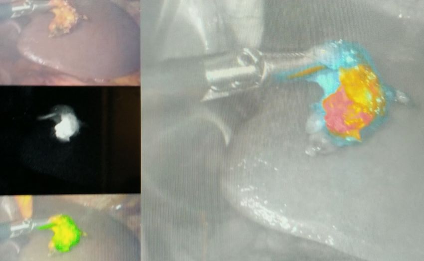
(NCT02139605, NCT03961373). Fluorescence-guided lymphadenectomy is a new, feasible
technique which allows to detect LN metastases within fluorescent LN stations [101,102]
(Figure 1). Indocyanine green can improve the LN harvest and reduce LN noncompliance
Cancers 2021, 13, 2509 8 of 14
without increased complications in patients undergoing D2 lymphadenectomy [103].
Figure 1. Fluorescence-guided LN dissection in GC patients (Department of Surgical Oncology, Medical University of Lublin).
Figure 1. Fluorescence-guided LN dissection in GC patients (Department of Surgical Oncology,
Medical University of vs.
8. East Lublin).
West Perspective
The differences in incidence and survival between Eastern and Western GC patients
result from the disease’s biological characteristics and different screening and treatment
strategy [104]. With an extended lymphadenectomy, more commonly performed in the
East, more LNs are retrieved with a higher chance of detecting a positive node, resulting
in a stage migration phenomenon [90]. However, the differences in surgical practice forCancers 2021, 13, 2509 8 of 14
8. East vs. West Perspective
The differences in incidence and survival between Eastern and Western GC patients
result from the disease’s biological characteristics and different screening and treatment
strategy [104]. With an extended lymphadenectomy, more commonly performed in the
East, more LNs are retrieved with a higher chance of detecting a positive node, resulting
in a stage migration phenomenon [90]. However, the differences in surgical practice for
GC between the East and the West have lessened and become standardized [105]. A recent
study on the impact of the introduction of formal D2 lymphadenectomy in a Western setting
resulted in improved LN sampling, decreased postoperative complications and improved
survival of patients undergoing surgery for GC [106]. However, a wide variation remains
in the multimodal treatment concept. In contrast to the perioperative approach in the
West, adjuvant chemotherapy with S1 or XELOX regimen are used [104]. The collaboration
between the East and the West will allow for a better understanding of the specific subtypes
of GC and will facilitate future studies to improve treatment strategies [91].
9. Nodal Regression Grade
Histologic downstage of cN+ status results in prolonged survival [107,108]. Moreover,
regression of LN status is more significant than regression of the primary tumour in
predicting GC recurrence [107]. Additionally, LN regression, regardless of the percent of
tumour cells in the primary site, independently influences the survival in patients who
underwent NAC [109]. Factors increasing chances for higher nodal regression grade are
lack of venous, lymphatic and perineural invasion, lower primary tumour depth and
diameter, regardless of its location. Nodal regression grade might be applied as one of the
endpoints in clinical trials verifying the efficacy of neoadjuvant treatment. Unnecessary
reporting of nodal regression grade due to the higher prognostic value of LN’s residual
tumour assessment in predicting the prognosis after NAC was highlighted in a study from
the East [108]. Despite usage of similar classification of regressive changes, this result stays
in contradiction with European findings [110]. Both, nodal response and the quality of
that response correlates with long-term survival. A favourable prognostic impact for ypN0
patients is observed, either due to truly negative LN before the start of therapy or because
preoperative therapy achieved a pathologically complete nodal response [111].
10. Conclusions
Since downstaged and primarily node-negative patients show similar prognosis, the
main target for NAC in advanced GC should be the nodal clearance. More adequate staging
and personalized perioperative therapy seem to be of great importance in the multimodal
treatment of GC.
Author Contributions: Conceptualization, Z.P. and K.R.-P.; methodology, Z.P., K.R.-P., M.S. and
W.P.P.; validation, K.R.-P. and W.P.P.; writing—original draft preparation, Z.P. and K.R.-P.; writing—
review and editing, K.R.-P., M.S. and W.P.P.; supervision, M.S. and W.P.P. All authors have read and
agreed to the published version of the manuscript.
Funding: This research received no external funding.
Institutional Review Board Statement: Not applicable.
Informed Consent Statement: Not applicable.
Data Availability Statement: Not applicable.
Conflicts of Interest: The authors declare no conflict of interest.
References
1. Smyth, E.C.; Nilsson, M.; Grabsch, H.I.; van Grieken, N.C.; Lordick, F. Gastric cancer. Lancet 2020, 396, 635–648. [CrossRef]
2. Wagner, A.D.; Lordick, F.; Grabsch, H.I.; Terashima, M.; Terada, M.; Yoshikawa, T.; Boku, N.; Kataoka, K.; Smyth, E.C.; Mauer, M.;
et al. Multidisciplinary management of stage II–III gastric and gastro-oesophageal junction cancer. Eur. J. Cancer 2020, 124, 67–76.
[CrossRef] [PubMed]Cancers 2021, 13, 2509 9 of 14
3. Degiuli, M.; De Manzoni, G.; Di Leo, A.; D’Ugo, D.; Galasso, E.; Marrelli, D.; Petrioli, R.; Polom, K.; Roviello, F.; Santullo, F.; et al.
Gastric cancer: Current status of lymph node dissection. World J. Gastroenterol. 2016, 22, 2875–2893. [CrossRef]
4. Ychou, M.; Boige, V.; Pignon, J.P.; Conroy, T.; Bouche, O.; Lebreton, G.; Ducourtieux, M.; Bedenne, L.; Fabre, J.M.; Saint-Aubert, B.;
et al. Perioperative chemotherapy compared with surgery alone for resectable gastroesophageal adenocarcinoma: An FNCLCC
and FFCD multicenter phase III trial. J. Clin. Oncol. 2011, 29, 1715–1721. [CrossRef]
5. Katayama, H.; Tsuburaya, A.; Mizusawa, J.; Nakamura, K.; Katai, H.; Imamura, H.; Nashimoto, A.; Fukushima, N.; Sano, T.;
Sasako, M. An integrated analysis of two phase II trials (JCOG0001 and JCOG0405) of preoperative chemotherapy followed by
D3 gastrectomy for gastric cancer with extensive lymph node metastasis. Gastric Cancer 2019, 22, 1301–1307. [CrossRef] [PubMed]
6. Ito, S.; Ito, Y.; Misawa, K.; Shimizu, Y.; Kinoshita, T. Neoadjuvant chemotherapy followed by surgery in gastric cancer patients
with extensive lymph node metastasis. World J. Clin. Oncol. 2015, 6, 291–294. [CrossRef] [PubMed]
7. Smyth, E.C.; Verheij, M.; Allum, W.; Cunningham, D.; Cervantes, A.; Arnold, D.; Committee, E.G. Gastric cancer: ESMO Clinical
Practice Guidelines for diagnosis, treatment and follow-up. Ann. Oncol. 2016, 27, v38–v49. [CrossRef]
8. Hashemzadeh, S.; Pourzand, A.; Somi, M.H.; Zarrintan, S.; Javad-Rashid, R.; Esfahani, A. The effects of neoadjuvant chemotherapy
on resectability of locally-advanced gastric adenocarcinoma: A clinical trial. Int. J. Surg. 2014, 12, 1061–1069. [CrossRef]
9. Eto, K.; Hiki, N.; Kumagai, K.; Shoji, Y.; Tsuda, Y.; Kano, Y.; Yasufuku, I.; Okumura, Y.; Tsujiura, M.; Ida, S.; et al. Prophylactic
effect of neoadjuvant chemotherapy in gastric cancer patients with postoperative complications. Gastric Cancer 2018, 21, 703–709.
[CrossRef]
10. NCCN Gastric Cancer Guidelines, Version 2.2021; National Comprehensive Cancer Network: Plymouth, PA, USA, 2021.
11. Lowy, A.M.; Mansfield, P.F.; Leach, S.D.; Pazdur, R.; Dumas, P.; Ajani, J.A. Response to neoadjuvant chemotherapy best predicts
survival after curative resection of gastric cancer. Ann. Surg. 1999, 229, 303–308. [CrossRef]
12. Mansour, J.C.; Tang, L.; Shah, M.; Bentrem, D.; Klimstra, D.S.; Gonen, M.; Kelsen, D.P.; Brennan, M.F.; Coit, D.G. Does graded
histologic response after neoadjuvant chemotherapy predict survival for completely resected gastric cancer? Ann. Surg. Oncol.
2007, 14, 3412–3418. [CrossRef]
13. Amin, M.B.; Greene, F.L.; Edge, S.B.; Compton, C.C.; Gershenwald, J.E.; Brookland, R.K.; Meyer, L.; Gress, D.M.; Byrd, D.R.;
Winchester, D.P. The Eighth Edition AJCC Cancer Staging Manual: Continuing to build a bridge from a population-based to a
more “personalized” approach to cancer staging. CA Cancer J. Clin. 2017, 67, 93–99. [CrossRef] [PubMed]
14. Ikoma, N.; Blum, M.; Estrella, J.S.; Das, P.; Hofstetter, W.L.; Fournier, K.F.; Mansfield, P.; Ajani, J.A.; Badgwell, B.D. Evaluation of
the American Joint Committee on Cancer 8th edition staging system for gastric cancer patients after preoperative therapy. Gastric
Cancer 2018, 21, 74–83. [CrossRef]
15. Park, J.M.; Ryu, W.S.; Kim, J.H.; Park, S.S.; Kim, S.J.; Kim, C.S.; Mok, Y.J. Prognostic factors for advanced gastric cancer:
Stage-stratified analysis of patients who underwent curative resection. Cancer Res. Treat. 2006, 38, 13–18. [CrossRef] [PubMed]
16. Hu, K.; Wang, S.; Wang, Z.; Li, L.; Huang, Z.; Yu, W.; Chen, Z.; Wu, Q.F. Clinicopathological risk factors for gastric cancer: A
retrospective cohort study in China. BMJ Open 2019, 9, e030639. [CrossRef] [PubMed]
17. Nakagawa, M.; Choi, Y.Y.; An, J.Y.; Chung, H.; Seo, S.H.; Shin, H.B.; Bang, H.J.; Li, S.; Kim, H.I.; Cheong, J.H.; et al. Difficulty of
predicting the presence of lymph node metastases in patients with clinical early stage gastric cancer: A case control study. BMC
Cancer 2015, 15, 943. [CrossRef] [PubMed]
18. Borggreve, A.S.; Goense, L.; Brenkman, H.J.F.; Mook, S.; Meijer, G.J.; Wessels, F.J.; Verheij, M.; Jansen, E.P.M.; van Hillegersberg,
R.; van Rossum, P.S.N.; et al. Imaging strategies in the management of gastric cancer: Current role and future potential of MRI. Br.
J. Radiol. 2019, 92, 20181044. [CrossRef] [PubMed]
19. Fukagawa, T.; Katai, H.; Mizusawa, J.; Nakamura, K.; Sano, T.; Terashima, M.; Ito, S.; Yoshikawa, T.; Fukushima, N.; Kawachi, Y.;
et al. A prospective multi-institutional validity study to evaluate the accuracy of clinical diagnosis of pathological stage III gastric
cancer (JCOG1302A). Gastric Cancer 2018, 21, 68–73. [CrossRef]
20. Mueller, C.L.; Lisbona, R.; Sorial, R.; Siblini, A.; Ferri, L.E. Sentinel Lymph Node Sampling for Early Gastric Cancer-Preliminary
Results of A North American Prospective Study. J. Gastrointest. Surg. 2019, 23, 1113–1121. [CrossRef]
21. Berlth, F.; Chon, S.H.; Chevallay, M.; Jung, M.K.; Monig, S.P. Preoperative staging of nodal status in gastric cancer. Transl.
Gastroenterol. Hepatol. 2017, 2, 8. [CrossRef]
22. Yamamoto, A.; Kawaguchi, Y.; Shiraishi, K.; Akaike, H.; Shimizu, H.; Furuya, S.; Hosomura, N.; Amemiya, H.; Kawaida, H.;
Sudo, M.; et al. The impact of histological type on the accuracy of preoperative N staging in patients with gastric cancer. World J.
Surg. Oncol. 2019, 17, 130. [CrossRef] [PubMed]
23. Luo, M.; Lv, Y.; Guo, X.; Song, H.; Su, G.; Chen, B. Value and impact factors of multidetector computed tomography in diagnosis
of preoperative lymph node metastasis in gastric cancer: A PRISMA-compliant systematic review and meta-analysis. Medicine
2017, 96, e7769. [CrossRef] [PubMed]
24. Choi, J.I.; Joo, I.; Lee, J.M. State-of-the-art preoperative staging of gastric cancer by MDCT and magnetic resonance imaging.
World J. Gastroenterol. 2014, 20, 4546–4557. [CrossRef] [PubMed]
25. Kwee, R.M.; Kwee, T.C. Imaging in assessing lymph node status in gastric cancer. Gastric Cancer 2009, 12, 6–22. [CrossRef]
26. Monig, S.P.; Zirbes, T.K.; Schroder, W.; Baldus, S.E.; Lindemann, D.G.; Dienes, H.P.; Holscher, A.H. Staging of gastric cancer:
Correlation of lymph node size and metastatic infiltration. AJR Am. J. Roentgenol. 1999, 173, 365–367. [CrossRef] [PubMed]
27. Sanjeevaiah, A.; Park, H.; Fangman, B.; Porembka, M. Gastric Cancer with Radiographically Occult Metastatic Disease: Biology,
Challenges, and Diagnostic Approaches. Cancers 2020, 12, 592. [CrossRef]Cancers 2021, 13, 2509 10 of 14
28. Power, D.G.; Schattner, M.A.; Gerdes, H.; Brenner, B.; Markowitz, A.J.; Capanu, M.; Coit, D.G.; Brennan, M.; Kelsen, D.P.; Shah,
M.A. Endoscopic ultrasound can improve the selection for laparoscopy in patients with localized gastric cancer. J. Am. Coll. Surg.
2009, 208, 173–178. [CrossRef]
29. Gertsen, E.C.; de Jongh, C.; Brenkman, H.J.F.; Mertens, A.C.; Broeders, I.; Los, M.; Boerma, D.; Ten Bokkel Huinink, D.; van
Leeuwen, L.; Wessels, F.J.; et al. The additive value of restaging-CT during neoadjuvant chemotherapy for gastric cancer. Eur. J.
Surg. Oncol. 2020, 46, 1247–1253. [CrossRef]
30. Ahn, H.S.; Lee, H.J.; Yoo, M.W.; Kim, S.G.; Im, J.P.; Kim, S.H.; Kim, W.H.; Lee, K.U.; Yang, H.K. Diagnostic accuracy of T and N
stages with endoscopy, stomach protocol CT, and endoscopic ultrasonography in early gastric cancer. J. Surg. Oncol. 2009, 99,
20–27. [CrossRef]
31. Kagedan, D.J.; Frankul, F.; El-Sedfy, A.; McGregor, C.; Elmi, M.; Zagorski, B.; Dixon, M.E.; Mahar, A.L.; Vasilevska-Ristovska,
J.; Helyer, L.; et al. Negative predictive value of preoperative computed tomography in determining pathologic local invasion,
nodal disease, and abdominal metastases in gastric cancer. Curr. Oncol. 2016, 23, 273–279. [CrossRef]
32. Fornaro, L.; Spallanzani, A.; de Vita, F.; D’Ugo, D.; Falcone, A.; Lorenzon, L.; Tirino, G.; Cascinu, S.; on behalf of GAIN (GAstric
Cancer Italian Network). Beyond the Guidelines: The Grey Zones of the Management of Gastric Cancer. Consensus Statements
from the Gastric Cancer Italian Network (GAIN). Cancers 2021, 13, 1304. [CrossRef] [PubMed]
33. Hoibian, S.; Giovannini, M.; Autret, A.; Pesenti, C.; Bories, E.; Ratone, J.P.; Dahel, Y.; Dermeche, S.; Meillat, H.; Guiramand, J.;
et al. Preoperative EUS evaluation of the response to neoadjuvant therapy for gastric and esophagogastric junction cancer is
correlated with survival: A single retrospective study of 97 patients. Endosc. Ultrasound 2021. [CrossRef] [PubMed]
34. MingHua, Z.; KeCheng, Z.; ZhenYu, C.; Lin, C.; ChunXi, W.; ZeLong, Y. Impact of Lymph Nodes Examined on Survival in ypN0
Gastric Cancer Patients: A Population-Based Study. J. Gastrointest. Surg. 2021, 25, 919–925. [CrossRef] [PubMed]
35. Seevaratnam, R.; Cardoso, R.; McGregor, C.; Lourenco, L.; Mahar, A.; Sutradhar, R.; Law, C.; Paszat, L.; Coburn, N. How useful
is preoperative imaging for tumor, node, metastasis (TNM) staging of gastric cancer? A meta-analysis. Gastric Cancer 2012, 15
(Suppl. 1), 3–18. [CrossRef] [PubMed]
36. Huang, Z.; Xie, D.H.; Guo, L.; Hu, C.H.; Fang, X.; Meng, Q.; Ping, X.X.; Lu, Z.W. The utility of MRI for pre-operative T and N
staging of gastric carcinoma: A systematic review and meta-analysis. Br. J. Radiol. 2015, 88, 20140552. [CrossRef] [PubMed]
37. Zhang, Y.; Yu, J. The role of MRI in the diagnosis and treatment of gastric cancer. Diagn. Interv. Radiol. 2020, 26, 176–182.
[CrossRef]
38. Chen, J.; Cheong, J.H.; Yun, M.J.; Kim, J.; Lim, J.S.; Hyung, W.J.; Noh, S.H. Improvement in preoperative staging of gastric
adenocarcinoma with positron emission tomography. Cancer 2005, 103, 2383–2390. [CrossRef] [PubMed]
39. Kim, E.Y.; Lee, W.J.; Choi, D.; Lee, S.J.; Choi, J.Y.; Kim, B.T.; Kim, H.S. The value of PET/CT for preoperative staging of advanced
gastric cancer: Comparison with contrast-enhanced CT. Eur. J. Radiol. 2011, 79, 183–188. [CrossRef]
40. Wieder, H.A.; Krause, B.J.; Herrmann, K. PET and PET-CT in esophageal and gastric cancer. Methods Mol. Biol. 2011, 727, 59–76.
[CrossRef]
41. Smyth, E.; Schoder, H.; Strong, V.E.; Capanu, M.; Kelsen, D.P.; Coit, D.G.; Shah, M.A. A prospective evaluation of the utility of
2-deoxy-2-[(18) F]fluoro-D-glucose positron emission tomography and computed tomography in staging locally advanced gastric
cancer. Cancer 2012, 118, 5481–5488. [CrossRef]
42. Nakajo, M.; Kajiya, Y.; Jinguji, M.; Nakabeppu, Y.; Nakajo, M.; Nihara, T.; Yoshiura, T. Current clinical status of (18)F-FLT PET or
PET/CT in digestive and abdominal organ oncology. Abdom. Radiol. 2017, 42, 951–961. [CrossRef] [PubMed]
43. Schneider, P.M.; Eshmuminov, D.; Rordorf, T.; Vetter, D.; Veit-Haibach, P.; Weber, A.; Bauerfeind, P.; Samaras, P.; Lehmann, K.
(18)FDG-PET-CT identifies histopathological non-responders after neoadjuvant chemotherapy in locally advanced gastric and
cardia cancer: Cohort study. BMC Cancer 2018, 18, 548. [CrossRef] [PubMed]
44. Morgagni, P.; Bencivenga, M.; Colciago, E.; Tringali, D.; Giacopuzzi, S.; Framarini, M.; Saragoni, L.; Mura, G.; Graziosi, L.;
Marino, E.; et al. Limited Usefulness of 18F-FDG PET/CT in Predicting Tumor Regression After Preoperative Chemotherapy for
Noncardia Gastric Cancer: The Italian Research Group for Gastric Cancer (GIRCG) Experience. Clin. Nucl. Med. 2020, 45, 177–181.
[CrossRef] [PubMed]
45. Lordick, F.; Ott, K.; Krause, B.J.; Weber, W.A.; Becker, K.; Stein, H.J.; Lorenzen, S.; Schuster, T.; Wieder, H.; Herrmann, K.; et al. PET
to assess early metabolic response and to guide treatment of adenocarcinoma of the oesophagogastric junction: The MUNICON
phase II trial. Lancet Oncol. 2007, 8, 797–805. [CrossRef]
46. Gertsen, E.C.; Borggreve, A.S.; Brenkman, H.J.F.; Verhoeven, R.H.A.; Vegt, E.; van Hillegersberg, R.; Siersema, P.D.; Ruurda, J.P.;
Dutch Upper Gastrointestinal Cancer Audit, G. Evaluation of the Implementation of FDG-PET/CT and Staging Laparoscopy for
Gastric Cancer in The Netherlands. Ann. Surg. Oncol. 2021, 28, 2384–2393. [CrossRef]
47. Brenkman, H.J.F.; Gertsen, E.C.; Vegt, E.; van Hillegersberg, R.; van Berge Henegouwen, M.I.; Gisbertz, S.S.; Luyer, M.D.P.;
Nieuwenhuijzen, G.A.P.; van Lanschot, J.J.B.; Lagarde, S.M.; et al. Evaluation of PET and laparoscopy in STagIng advanced
gastric cancer: A multicenter prospective study (PLASTIC-study). BMC Cancer 2018, 18, 450. [CrossRef]
48. Leake, P.A.; Cardoso, R.; Seevaratnam, R.; Lourenco, L.; Helyer, L.; Mahar, A.; Law, C.; Coburn, N.G. A systematic review of the
accuracy and indications for diagnostic laparoscopy prior to curative-intent resection of gastric cancer. Gastric Cancer 2012, 15
(Suppl. 1), S38–S47. [CrossRef]Cancers 2021, 13, 2509 11 of 14
49. Mizrak Kaya, D.; Nogueras-Gonzalez, G.M.; Harada, K.; Amlashi, F.G.; Roy-Chowdhuri, S.; Estrella, J.S.; Das, P.; Lee, J.H.; Weston,
B.; Bhutani, M.S.; et al. Risk of peritoneal metastases in patients who had negative peritoneal staging and received therapy for
localized gastric adenocarcinoma. J. Surg. Oncol. 2018, 117, 678–684. [CrossRef]
50. Ramos, R.F.; Scalon, F.M.; Scalon, M.M.; Dias, D.I. Staging laparoscopy in gastric cancer to detect peritoneal metastases: A
systematic review and meta-analysis. Eur. J. Surg. Oncol. 2016, 42, 1315–1321. [CrossRef]
51. Fukagawa, T. Role of staging laparoscopy for gastric cancer patients. Ann. Gastroenterol. Surg. 2019, 3, 496–505. [CrossRef]
[PubMed]
52. Bintintan, V.V.; Cordos, A.; Chira, R.; Cocu, S.; Rus, P.; Bintintan, A.; Nagy, G.; Ciule, L.; Cata, E.; Pop, A.; et al. The Value of
Staging Laparoscopy for Optimal Multidisciplinary Treatment in Patients with Gastric Cancer. Chirurgia 2018, 113, 789–798.
[CrossRef] [PubMed]
53. Hosogi, H.; Shinohara, H.; Tsunoda, S.; Hisamori, S.; Sumida, H.; Hida, K.; Obama, K.; Okabe, H.; Sakai, Y. Staging laparoscopy
for advanced gastric cancer: Significance of preoperative clinicopathological factors. Langenbecks Arch. Surg. 2017, 402, 33–39.
[CrossRef]
54. Machairas, N.; Charalampoudis, P.; Molmenti, E.P.; Kykalos, S.; Tsaparas, P.; Stamopoulos, P.; Sotiropoulos, G.C. The value of
staging laparoscopy in gastric cancer. Ann. Gastroenterol. 2017, 30, 287–294. [CrossRef] [PubMed]
55. Vergadis, C.; Schizas, D. Is Accurate N—Staging for Gastric Cancer Possible? Front. Surg. 2018, 5, 41. [CrossRef] [PubMed]
56. Zhong, J.; Zhao, W.; Ren, F.; Qi, S.; Wang, X.; Lv, T.; Su, Z.; Yin, H.; Ren, J.; Huan, Y. Lymph node metastasis in patients with
gastric cancer: A multi-modality, morphologic and functional imaging study. Am. J. Transl. Res. 2016, 8, 5601–5609.
57. Wang, X.; Wei, Y.; Xue, Y.; Lu, P.; Yu, L.; Shen, B. Predictive Role of the Number of 18F-FDG-Positive Lymph Nodes Detected by
PET/CT for Pre-Treatment Evaluation of Locally Advanced Gastric Cancer. PLoS ONE 2016, 11, e0166836. [CrossRef] [PubMed]
58. Kawanaka, Y.; Kitajima, K.; Fukushima, K.; Mouri, M.; Doi, H.; Oshima, T.; Niwa, H.; Kaibe, N.; Sasako, M.; Tomita, T.; et al.
Added value of pretreatment (18)F-FDG PET/CT for staging of advanced gastric cancer: Comparison with contrast-enhanced
MDCT. Eur. J. Radiol. 2016, 85, 989–995. [CrossRef] [PubMed]
59. Hallinan, J.T.; Venkatesh, S.K. Gastric carcinoma: Imaging diagnosis, staging and assessment of treatment response. Cancer
Imaging 2013, 13, 212–227. [CrossRef] [PubMed]
60. Kakroo, S.M.; Rashid, A.; Wani, A.A.; Akhtar, Z.; Chalkoo, M.A.; Laharwal, A.R. Staging Laparoscopy in Carcinoma of Stomach:
A Comparison with CECT Staging. Int. J. Surg. Oncol. 2013, 2013, 674965. [CrossRef]
61. Cunningham, D.; Allum, W.H.; Stenning, S.P.; Thompson, J.N.; Van de Velde, C.J.; Nicolson, M.; Scarffe, J.H.; Lofts, F.J.; Falk, S.J.;
Iveson, T.J.; et al. Perioperative chemotherapy versus surgery alone for resectable gastroesophageal cancer. N. Engl. J. Med. 2006,
355, 11–20. [CrossRef]
62. Al-Batran, S.E.; Homann, N.; Pauligk, C.; Goetze, T.O.; Meiler, J.; Kasper, S.; Kopp, H.G.; Mayer, F.; Haag, G.M.; Luley, K.; et al.
Perioperative chemotherapy with fluorouracil plus leucovorin, oxaliplatin, and docetaxel versus fluorouracil or capecitabine
plus cisplatin and epirubicin for locally advanced, resectable gastric or gastro-oesophageal junction adenocarcinoma (FLOT4): A
randomised, phase 2/3 trial. Lancet 2019, 393, 1948–1957. [CrossRef] [PubMed]
63. Monti, M.; Morgagni, P.; Nanni, O.; Framarini, M.; Saragoni, L.; Marrelli, D.; Roviello, F.; Petrioli, R.; Fumagalli Romario, U.;
Rimassa, L.; et al. Preoperative or Perioperative Docetaxel, Oxaliplatin, and Capecitabine (GASTRODOC Regimen) in Patients
with Locally-Advanced Resectable Gastric Cancer: A Randomized Phase-II Trial. Cancers 2020, 12, 2790. [CrossRef] [PubMed]
64. Messager, M.; Lefevre, J.H.; Pichot-Delahaye, V.; Souadka, A.; Piessen, G.; Mariette, C.; FREGAT Working Group. The impact of
perioperative chemotherapy on survival in patients with gastric signet ring cell adenocarcinoma: A multicenter comparative
study. Ann. Surg. 2011, 254, 684–693; discussion 693. [CrossRef] [PubMed]
65. Piessen, G.; Messager, M.; Le Malicot, K.; Robb, W.B.; Di Fiore, F.; Guilbert, M.; Moreau, M.; Christophe, V.; Adenis, A.; Mariette, C.
Phase II/III multicentre randomised controlled trial evaluating a strategy of primary surgery and adjuvant chemotherapy versus
peri-operative chemotherapy for resectable gastric signet ring cell adenocarcinomas—PRODIGE 19—FFCD1103—ADCI002. BMC
Cancer 2013, 13, 281. [CrossRef] [PubMed]
66. Al-Batran, S.E.; Hofheinz, R.D.; Pauligk, C.; Kopp, H.G.; Haag, G.M.; Luley, K.B.; Meiler, J.; Homann, N.; Lorenzen, S.;
Schmalenberg, H.; et al. Histopathological regression after neoadjuvant docetaxel, oxaliplatin, fluorouracil, and leucovorin
versus epirubicin, cisplatin, and fluorouracil or capecitabine in patients with resectable gastric or gastro-oesophageal junction
adenocarcinoma (FLOT4-AIO): Results from the phase 2 part of a multicentre, open-label, randomised phase 2/3 trial. Lancet
Oncol. 2016, 17, 1697–1708. [CrossRef] [PubMed]
67. Petrillo, A.; Pompella, L.; Tirino, G.; Pappalardo, A.; Laterza, M.M.; Caterino, M.; Orditura, M.; Ciardiello, F.; Lieto, E.; Galizia, G.;
et al. Perioperative Treatment in Resectable Gastric Cancer: Current Perspectives and Future Directions. Cancers 2019, 11, 399.
[CrossRef] [PubMed]
68. He, Q.; Chen, J.; Zhou, K.; Jin, C.; Wang, A.; Ji, K.; Ji, X.; Zhang, J.; Wu, X.; Li, X.; et al. Effect of Additional Trastuzumab
in Neoadjuvant and Adjuvant Treatment for Patients with Resectable HER2-Positive Gastric Cancer. Ann. Surg. Oncol. 2021.
[CrossRef]Cancers 2021, 13, 2509 12 of 14
69. Wagner, A.D.; Grabsch, H.I.; Mauer, M.; Marreaud, S.; Caballero, C.; Thuss-Patience, P.; Mueller, L.; Elme, A.; Moehler, M.H.;
Martens, U.; et al. EORTC-1203-GITCG—The “INNOVATION”-trial: Effect of chemotherapy alone versus chemotherapy plus
trastuzumab, versus chemotherapy plus trastuzumab plus pertuzumab, in the perioperative treatment of HER2 positive, gastric
and gastroesophageal junction adenocarcinoma on pathologic response rate: A randomized phase II-intergroup trial of the
EORTC-Gastrointestinal Tract Cancer Group, Korean Cancer Study Group and Dutch Upper GI-Cancer group. BMC Cancer 2019,
19, 494. [CrossRef]
70. Hofheinz, R.D.; Haag, G.M.; Ettrich, T.J.; Borchert, K.; Kretzschmar, A.; Teschendorf, C.; Siegler, G.M.; Ebert, M.P.; Goekkurt, E.;
Welslau, M.; et al. Perioperative trastuzumab and pertuzumab in combination with FLOT versus FLOT alone for HER2-positive
resectable esophagogastric adenocarcinoma: Final results of the PETRARCA multicenter randomized phase II trial of the AIO. J.
Clin. Oncol. 2020, 38, 4502. [CrossRef]
71. Bang, Y.J.; Van Cutsem, E.; Fuchs, C.S.; Ohtsu, A.; Tabernero, J.; Ilson, D.H.; Hyung, W.J.; Strong, V.E.; Goetze, T.O.; Yoshikawa, T.;
et al. KEYNOTE-585: Phase III study of perioperative chemotherapy with or without pembrolizumab for gastric cancer. Future
Oncol. 2019, 15, 943–952. [CrossRef]
72. Mansukhani, S.; Davidson, M.; Gillbanks, A.; Peckitt, C.; Musallam, A.; Begum, R.; Morganstein, D.; Wotherspoon, A.; Riddell,
A.M.; Kinross, J.M.; et al. Iconic: Peri-operative immuno-chemotherapy in operable oesophageal and gastric cancer. J. Clin. Oncol.
2018, 36, TPS4139. [CrossRef]
73. Gu, L.; Chen, M.; Guo, D.; Zhu, H.; Zhang, W.; Pan, J.; Zhong, X.; Li, X.; Qian, H.; Wang, X. PD-L1 and gastric cancer prognosis: A
systematic review and meta-analysis. PLoS ONE 2017, 12, e0182692. [CrossRef]
74. Pietrantonio, F.; Miceli, R.; Raimondi, A.; Kim, Y.W.; Kang, W.K.; Langley, R.E.; Choi, Y.Y.; Kim, K.M.; Nankivell, M.G.; Morano, F.;
et al. Individual Patient Data Meta-Analysis of the Value of Microsatellite Instability As a Biomarker in Gastric Cancer. J. Clin.
Oncol. 2019, 37, 3392–3400. [CrossRef]
75. Yasuta, S.; Yamauchi, J.; Miyazaki, K.; Sato, M.; Ikeda, T.; Fujita, S.; Shirasaki, K.; Kobayashi, S.; Ajiki, T.; Tsuchihara, K.; et al.
A Case of Advanced Gastric Cancer with Extensive Lymph Node Metastases Treated by Capecitabine plus Cisplatin plus
Trastuzumab Chemotherapy, Followed by Conversion Surgery. Gan Kagaku Ryoho 2016, 43, 1923–1925.
76. Smyth, E.; Knodler, M.; Giraut, A.; Mauer, M.; Nilsson, M.; Van Grieken, N.; Wagner, A.D.; Moehler, M.; Lordick, F. VESTIGE:
Adjuvant Immunotherapy in Patients with Resected Esophageal, Gastroesophageal Junction and Gastric Cancer Following
Preoperative Chemotherapy with High Risk for Recurrence (N+ and/or R1): An Open Label Randomized Controlled Phase-2-
Study. Front. Oncol. 2019, 9, 1320. [CrossRef] [PubMed]
77. Kuhara, Y.; Ninomiya, M.; Hirahara, S.; Doi, H.; Kenji, S.; Toyota, K.; Yano, R.; Kobayashi, H.; Hashimoto, Y.; Yokoyama, Y.; et al.
A long-term survival case of unresectable gastric cancer with multidisciplinary therapy including immunotherapy and abscopal
effect. Int. Cancer Conf. J. 2020, 9, 193–198. [CrossRef] [PubMed]
78. Shapiro, J.; van Lanschot, J.J.B.; Hulshof, M.; van Hagen, P.; van Berge Henegouwen, M.I.; Wijnhoven, B.P.L.; van Laarhoven,
H.W.M.; Nieuwenhuijzen, G.A.P.; Hospers, G.A.P.; Bonenkamp, J.J.; et al. Neoadjuvant chemoradiotherapy plus surgery versus
surgery alone for oesophageal or junctional cancer (CROSS): Long-term results of a randomised controlled trial. Lancet Oncol.
2015, 16, 1090–1098. [CrossRef]
79. Leong, T.; Smithers, B.M.; Michael, M.; Gebski, V.; Boussioutas, A.; Miller, D.; Simes, J.; Zalcberg, J.; Haustermans, K.; Lordick, F.;
et al. TOPGEAR: A randomised phase III trial of perioperative ECF chemotherapy versus preoperative chemoradiation plus peri-
operative ECF chemotherapy for resectable gastric cancer (an international, intergroup trial of the AGITG/TROG/EORTC/NCIC
CTG). BMC Cancer 2015, 15, 532. [CrossRef] [PubMed]
80. Leong, T.; Smithers, B.M.; Haustermans, K.; Michael, M.; Gebski, V.; Miller, D.; Zalcberg, J.; Boussioutas, A.; Findlay, M.; O’Connell,
R.L.; et al. TOPGEAR: A Randomized, Phase III Trial of Perioperative ECF Chemotherapy with or Without Preoperative
Chemoradiation for Resectable Gastric Cancer: Interim Results from an International, Intergroup Trial of the AGITG, TROG,
EORTC and CCTG. Ann. Surg. Oncol. 2017, 24, 2252–2258. [CrossRef] [PubMed]
81. Sada, Y.H.; Smaglo, B.G.; Tan, J.C.; Tran Cao, H.S.; Musher, B.L.; Massarweh, N.N. Prognostic Value of Nodal Response After
Preoperative Treatment of Gastric Adenocarcinoma. J. Natl. Compr. Cancer Netw. 2019, 17, 161–168. [CrossRef] [PubMed]
82. Stark, A.P.; Blum, M.M.; Chiang, Y.J.; Das, P.; Minsky, B.D.; Estrella, J.S.; Ajani, J.A.; Badgwell, B.D.; Mansfield, P.; Ikoma, N.
Preoperative Therapy Regimen Influences the Incidence and Implication of Nodal Downstaging in Patients with Gastric Cancer.
J. Gastric Cancer 2020, 20, 313–327. [CrossRef]
83. Lee, J.; Lim, D.H.; Kim, S.; Park, S.H.; Park, J.O.; Park, Y.S.; Lim, H.Y.; Choi, M.G.; Sohn, T.S.; Noh, J.H.; et al. Phase III trial
comparing capecitabine plus cisplatin versus capecitabine plus cisplatin with concurrent capecitabine radiotherapy in completely
resected gastric cancer with D2 lymph node dissection: The ARTIST trial. J. Clin. Oncol. 2012, 30, 268–273. [CrossRef]
84. Park, S.H.; Zang, D.Y.; Han, B.; Ji, J.H.; Kim, T.G.; Oh, S.Y.; Hwang, I.G.; Kim, J.H.; Shin, D.; Lim, D.H.; et al. ARTIST 2: Interim
results of a phase III trial involving adjuvant chemotherapy and/or chemoradiotherapy after D2-gastrectomy in stage II/III
gastric cancer (GC). J. Clin. Oncol. 2019, 37, 4001. [CrossRef]
85. Cats, A.; Jansen, E.P.M.; van Grieken, N.C.T.; Sikorska, K.; Lind, P.; Nordsmark, M.; Meershoek-Klein Kranenbarg, E.; Boot, H.;
Trip, A.K.; Swellengrebel, H.A.M.; et al. Chemotherapy versus chemoradiotherapy after surgery and preoperative chemotherapy
for resectable gastric cancer (CRITICS): An international, open-label, randomised phase 3 trial. Lancet Oncol. 2018, 19, 616–628.
[CrossRef]You can also read