Mechanical adaptation of brachiopod shells via hydration-induced structural changes
←
→
Page content transcription
If your browser does not render page correctly, please read the page content below
ARTICLE
                  https://doi.org/10.1038/s41467-021-25613-4                 OPEN

                  Mechanical adaptation of brachiopod shells via
                  hydration-induced structural changes
                  Johannes Ihli1 ✉, Anna S. Schenk2, Sabine Rosenfeldt2, Klaus Wakonig 1,3, Mirko Holler                                         1,   Giuseppe Falini 4,
                  Luca Pasquini5, Eugénia Delacou 6, Jim Buckman7, Thomas S. Glen8, Thomas Kress                                                9,    Esther H. R. Tsai10,
                  David G. Reid9, Melinda J. Duer9, Maggie Cusack11 & Fabio Nudelman 6 ✉
1234567890():,;

                  The function-optimized properties of biominerals arise from the hierarchical organization of
                  primary building blocks. Alteration of properties in response to environmental stresses
                  generally involves time-intensive processes of resorption and reprecipitation of mineral in the
                  underlying organic scaffold. Here, we report that the load-bearing shells of the brachiopod
                  Discinisca tenuis are an exception to this process. These shells can dynamically modulate their
                  mechanical properties in response to a change in environment, switching from hard and stiff
                  when dry to malleable when hydrated within minutes. Using ptychographic X-ray tomo-
                  graphy, electron microscopy and spectroscopy, we describe their hierarchical structure and
                  composition as a function of hydration to understand the structural motifs that generate this
                  adaptability. Key is a complementary set of structural modifications, starting with the swelling
                  of an organic matrix on the micron level via nanocrystal reorganization and ending in an
                  intercalation process on the molecular level in response to hydration.

                  1 Photon Science Division, Paul Scherrer Institut, Villigen PSI, Switzerland. 2 Department of Chemistry, Faculty of Biology, Chemistry & Earth Sciences,

                  University of Bayreuth, and Bavarian Polymer Institute, Universitaetsstrasse 30, Bayreuth, Germany. 3 ETH and University of Zürich, Institute for Biomedical
                  Engineering, 8093, Zürich, Switzerland. 4 Dipartimento di Chimica “Giacomo Ciamician”, Alma Mater Studiorum Università di Bologna, via F. Selmi 2,
                  Bologna, Italy. 5 Department of Physics and Astronomy, University of Bologna, viale Berti-Pichat 6/2, Bologna, Italy. 6 School of Chemistry, the University of
                  Edinburgh, Joseph Black Building, Edinburgh, UK. 7 Institute of GeoEnergy Engineering, School of Energy, Geoscience, Infrastructure and Society, Heriot-Watt
                  University, Riccarton, Edinburgh, UK. 8 School of Physics and Astronomy, University of Edinburgh, Edinburgh, UK. 9 Department of Chemistry, University of
                  Cambridge, Cambridge, UK. 10 Center for Functional Nanomaterials, Brookhaven National Laboratory, Upton, NY, USA. 11 Munster Technological University,
                  Bishopstown, Cork, T12 P928 & Tralee, Kerry, Cork, Ireland. ✉email: Johannes.ihli@psi.ch; Fabio.nudelman@ed.ac.uk

                  NATURE COMMUNICATIONS | (2021)12:5383 | https://doi.org/10.1038/s41467-021-25613-4 | www.nature.com/naturecommunications                                    1
ARTICLE                                                                 NATURE COMMUNICATIONS | https://doi.org/10.1038/s41467-021-25613-4

F
      or hundreds of millions of years, nature has evolved a large              Using a combination of ptychographic X-ray tomography,
      assortment of organic–inorganic hybrid materials such as               electron microscopy, small- and wide-angle X-ray scattering,
      bone, teeth, and shells. Each of these biominerals exhibits            solid-state nuclear magnetic resonance spectroscopy, and
material properties that have been optimized to aid a particular             mechanical testing, we characterized the shell’s hierarchical
function, such as navigation, protection, or mechanical                      structure and composition as a function of hydration covering the
support1,2. These properties arise from a three-dimensional                  micro- and nanoscales and provide an insight into molecular
multi-scale organization of the biomineral’s primary building                changes. We demonstrate that water absorption by the shell
blocks, e.g., inorganic nanocrystals, specialized proteins, and              induces a complementary set of structural modifications, starting
polysaccharides, from the molecular to the millimeter scale3–5.              with the swelling of an organic matrix on the micron level, via
Biominerals with load-bearing functions are optimized, in parti-             nanocrystal reorganization and restructuring, and ending in the
cular, with respect to their mechanical properties, so as to provide         intercalation of water between the organic framework and the
sufficient stiffness to support the typical mechanical loads in the           mineral on the molecular level. In combination, we propose that
biomineral’s environment and enough toughness to resist crack                these changes endow the shell with its mechanical adaptability.
propagation3. This optimization is achieved, first, by incorpor-              We envisage that these observations will aid/ inspire the design of
ating organic biopolymers within the inorganic phase, which                  novel synthetic materials with properties that can be modulated
increases the toughness of the inherently brittle mineral6, and              in real-time.
second by organizing the basic building blocks of the tissue into
higher-order structures7. This hierarchical organization creates a
                                                                             Results
large number of internal interfaces that help to avoid crack
                                                                             Global compositional analysis. The shells of D. tenuis (Fig. 1a)
propagation and significantly increases fracture toughness. A
                                                                             are an organic-inorganic composite material, where the mineral
further advantage of a hierarchical structure is that it endows the
                                                                             phase constitutes about 68 wt% of the dry shell11. The mineral
organism with an additional level of constructional control, where
                                                                             phase is composed predominantly of carbonate-substituted
the basic building blocks can be assembled into different struc-
                                                                             fluorapatite crystals in the form of francolite18 (Supplementary
tural motifs of different mechanical properties8. Altering the
                                                                             Fig. 1) with minor contributions of amorphous calcium phos-
material properties of the biomineral in response to environ-
                                                                             phate, octacalcium phosphate, and tricalcium phosphate19. The
mental stresses, generally requires active restructuring by remo-
                                                                             remaining ~32 wt% of the shell consist of various organic frac-
deling by the organism. A time- and energy-consuming process
                                                                             tions, of which chitin, glycosaminoglycans, and proteins make up
that involves the resorption of the existing biomineral, followed
                                                                             the dominant portion11,19–21. While these shells do not exhibit
by the precipitation of new tissue with a different structure and
                                                                             discernible structural motifs at the micron scale (Fig. 1b), high-
composition9,10.
                                                                             angle annular dark-field scanning transmission electron micro-
   In this paper, we report that the load-bearing shells of the
                                                                             scopy (HAADF-STEM) has shown that they are hierarchically
brachiopod Discinisca tenuis11 are able to dynamically modulate
                                                                             structured consisting of a laminated brick-work-like structure.
their mechanical properties in response to a change in the
                                                                             This structure is arranged normal to the shell height, where
environment without the need for remodeling via resorption and
                                                                             francolite crystals (Fig. 1c, bright objects) are enwrapped by a
regeneration of the tissue, i.e., they switch from hard and stiff
                                                                             network of chitin and proteins (Fig. 1c, dark regions).
when dry to malleable when hydrated within minutes. Impor-
                                                                                Importantly, these shells which are hard and brittle when dry,
tantly, when hydrated the shell can freely bend to the point that it
                                                                             significantly increase in flexibility upon hydration to the point
can be folded in two without fracturing.
                                                                             where they can be folded in half without fracturing as seen in
   The effects that water and organic matrix hydration degree
                                                                             Fig. 1d and supplementary Movie 1. This process is reversible so
have on the mechanical properties of biominerals are well
                                                                             that shells can be cycled multiple times through hard/brittle and
recognized12. Water, as a component of most biominerals, is
                                                                             soft/flexible by dehydrating/rehydrating.
known to increase the flexibility of materials such as bone, teeth,
                                                                                Thermogravimetric analysis (TGA) was used to determine the
and shells. Modulation of hardness and elastic modulus/
                                                                             water content of an atmospherically dry shell stored in the air as
flexibility13 by passive control of the water content of the tissue
                                                                             compared to a fully hydrated shell after immersion in H2O for
has been suggested to occur in non-mineralized insect cuticle14
                                                                             24 h (Supplementary Fig. 2). The dry shell displayed a gradual
and in mineralized crustacean cuticles, both of which contain
                                                                             water weight loss of 5% from ambient temperature to 200 °C. The
organic matrices composed of chitin and proteins15,16. In these
                                                                             hydrated shell exhibits a water weight loss of 24% across the same
cases, the changes in mechanical properties are due to the plas-
                                                                             temperature range. Notably, in the hydrated shell, these losses
ticizing role of water and do not involve major changes in the
                                                                             occur in two distinct steps, the first at 60 °C, corresponding to the
structure of the tissue12,16. However, none of these aforemen-
                                                                             loss of physisorbed water accounting for ~18 wt% H2O and a
tioned mineralized tissues exhibit flexibility that is comparable to
                                                                             more gradual secondary loss between ~100 and 200 °C (~6 wt%
that of the mineralized D. tenuis shell when in their natural,
                                                                             H2O). Further weight loss steps, observed for both the dry and the
hydrated state.
                                                                             hydrated shell, occurring above 287 and 400 °C, are related to the
   We hypothesized that the extreme flexibility of the hydrated D.
                                                                             pyrolysis of the organic components22,23, while decarbonation
tenuis shell cannot be accounted for solely by the plasticizing
                                                                             occurs above 700 °C and results in the formation of fluorapatite
effect of water as in these other examples. Rather, such reversi-
                                                                             (Supplementary Fig. 3).
bility between stiff and flexible as a function of hydration must
have its origins in the structure of the D. tenuis shell with water
promoting structural changes at different hierarchical levels. The           Mechanical behavior characterization. Depth-sensing nanoin-
mechanisms that underpin these changes in mechanical proper-                 dentation was used to determine the mechanical properties of the
ties as a function of hydration are unknown. Chemically con-                 shell as a function of the degree of hydration. Due to practical and
trollable material properties and the causal structural motives are          geometrical constraints, i.e., the brachiopod’s shell is curved, has
of significant interest in the design of stimuli-responsive synthetic         an irregular surface and a thickness of 50–500 µm that is both
materials17. As such, there is imperative to determine how                   shell- and location-dependent, depth-dependent dynamic
hydration alters the structure of the D. tenuis shell, and how these         nanoindentation measurements were performed on shell cross-
changes facilitate the modulation in mechanical properties.                  sections. These cross-sections expose the laminated structure

2                NATURE COMMUNICATIONS | (2021)12:5383 | https://doi.org/10.1038/s41467-021-25613-4 | www.nature.com/naturecommunications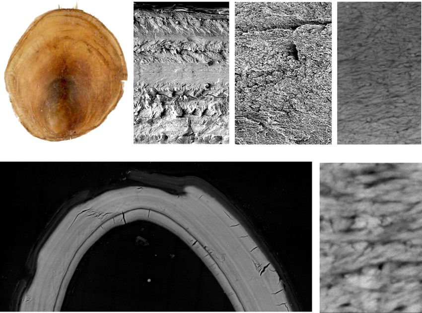
NATURE COMMUNICATIONS | https://doi.org/10.1038/s41467-021-25613-4                                                                           ARTICLE

                     a)                            b)                                                                   c)

                      y                              z

                     z       x
                                                            y

                     d)

                         z
                             y

Fig. 1 Hierarchical structure of a brachiopod Discinisca tenuis shell. a Top-down optical micrograph of a dry brachiopod Discinisca tenuis shell. Scale bar is
2 mm. b Cross-sectional scanning electron microscopy images of the dry shell at increasing magnification. Left: low magnification image showing the cross-
section across the z-axis. Right: high magnification of the area marked by the dotted square. Scale bars are 20 µm (left), 2 µm (right). c High-angle annular
dark-field scanning-transmission electron microscopy (HAADF-STEM) images of a thin section from a dry shell. Scale bars are 200 and 20 nm. White
arrows point to the organic matrix component (dark areas) surrounding the mineral (bright areas). d Cross-sectional electron micrograph acquired with
backscattered electrons (BSE) of a fully hydrated shell thin section folded in two. Scale bar 50 µm. Source data for this figure are available at the University
of Edinburgh DataShare, data identifier https://doi.org/10.7488/ds/305667.

                                                                                    (Fig. 1b), i.e., the indentation direction is in the plane of the
            12                                                                      laminae. Measurements were performed on atmospherically dry
                                                                  600               and fully hydrated shells on the same sample, at the center of the
                                                    Dry shell                       cross-section or shell diameter.
            10                                                                         Depth-sensing nanoindentation measurements show that both
                                                                  500               Young’s modulus (EIT) and hardness (HIT) drop drastically when
                                                                                    the shell becomes hydrated (Fig. 2). At the maximum tested load
                                                                        HIT (MPa)
EIT (GPa)

             8                                                                      of 30 mN, EIT is about 26% of the dry value and HIT shows a
                                                                  400
                                                                                    similar reduction down to 22% (Supplementary Table 1). The
                                                                                    greater deformability of the hydrated sections under the same
             6
                                                                  300               applied load is demonstrated also by the increase of the
                                                                                    maximum indentation depth from ~1.5 to ~3.5 µm at 30 mN
                                              Hydrated shell                        (Supplementary Fig. 4), as well as by the larger residual
             4                                                    200               indentation imprint (Supplementary Fig. 5).

             2                                                    100               Micron-scale characterization. Ptychographic X-ray computed
                 0   500 1000 1500 2000 2500 3000 3500                              tomography (PXCT) was used to characterize the shell structure
                                 Depth (nm)                                         at increasing levels of hydration on the micron and sub-micron
                                                                                    levels. PXCT provided quantitative electron density tomograms
Fig. 2 Depth-dependent dynamic nanoindentation measurements of D.                   (neÅ−3)24–26, with a half-period spatial resolution of roughly
tenuis brachiopod shell samples at different hydration levels. Plotted is           85 nm (Supplementary Fig. 6). These tomograms allow a struc-
the dependence of shell hardness HIT (red, right) and Young’s modulus EIT           tural evaluation of the shell and provide information such as local
(blue, left) as a function of indentation depth for an atmospherically dry          variations in swelling behavior and the hydration degree of spe-
shell sample (top, filled symbols) and a fully hydrated shell sample (bottom,        cific components in the sample, e.g., organic- and mineral-rich
open symbols). Measurements were conducted in continuous stiffness                  regions.
mode up to a maximum force of 30 mN. Shown is the average over eight                   Sample cylinders, greater than 15 µm in diameter, were
indentation measurements.                                                           extracted along the shell width (z-axis in Fig. 1b) and prepared

NATURE COMMUNICATIONS | (2021)12:5383 | https://doi.org/10.1038/s41467-021-25613-4 | www.nature.com/naturecommunications                                     3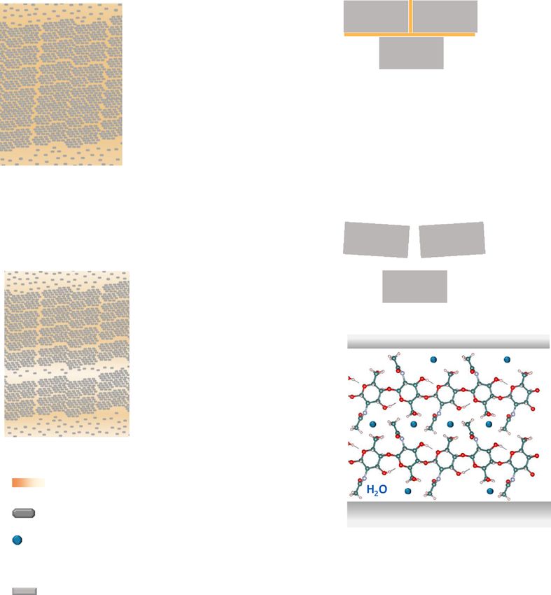
ARTICLE NATURE COMMUNICATIONS | https://doi.org/10.1038/s41467-021-25613-4
a) b) Vacuum Dried c) Hydrated 70% RH d) Hydrated 100% RH
0.4 0.63
Electron Density / n eÅ-3
z z z
y y y
e)
f)
Hydrated 100% RH
Ice
Ice
Ice
Chitin
Chitin
Chitin
Vacuum Dried
Francolite
Francolite
Francolite
Hydrated 70% RH
FrequencyN
FrequencyN
FrequencyN
0.3 0.4 0.5 0.6 0.7 0.8 0.3 0.4 0.5 0.6 0.7 0.8 0.3 0.4 0.5 0.6 0.7 0.8
Electron density / neÅ-3 Electron density / neÅ-3 Electron density / neÅ-3
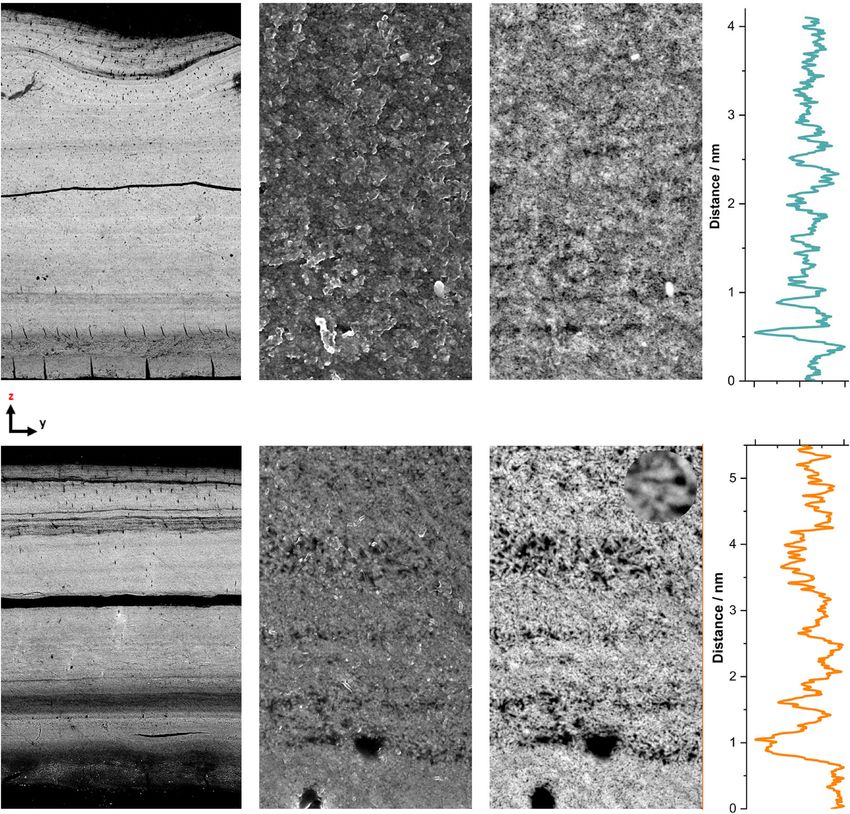
Fig. 3 Electron density tomograms of D. tenuis brachiopod shell samples at increasing hydration level. a Example volume rendering of the imaged,
cylindrical, sample pillars. Sagittal cut slices through the center of b a vacuum dried sample, c a sample incubated at 70% RH, and d a sample incubated at
100% RH. The cutting plane is represented by the yellow line shown in (a). Scale bars are 2 µm. Common to all cuts is a single color scale ranging from
white to yellow representative of electron density values. Shown in e are sample plane averaged electron density line profiles normal to the laminae
structure (pink arrow) alongside secondary derivatives highlighting major fluctuations in electron density. Sample corresponding frequency normalized (N)
electron density histograms are shown in (f). Further provided in f are the theoretical electron density values of the shell main components: francolite
0.78 neÅ−3, high molecular weight polysaccharides approximated using chitin ~0.46 neÅ−3, and low-density amorphous ice, 0.31 neÅ−3. PCXT
measurements were conducted under cryogenic conditions. The voxel size of all tomograms is (38.8 nm)3. Source data for this figure are available at the
University of Edinburgh DataShare, data identifier https://doi.org/10.7488/ds/305667.
using a nitrogen-cooled micro-lath (Supplementary Movie 2)27. and 0.46 neÅ−3 (chitin), the measured electron densities can be
These cylinders were then either vacuum-dried or incubated at 70 used to determine the shell’s composition globally and locally in
% or 100 % relative humidity (RH) for 36 h28,29, resulting in consideration of partial volume effects24,30. Partial volume effects
samples of increasing hydration level. Lastly, samples were flash- refer to the occupation of a volume element, e.g., a voxel, by
frozen in liquid nitrogen and analyzed using cryo-PXCT at multiple components, leading to a fractional occupancy-related
−180 °C. electron density. For example, the average electron density of the
PXCT-derived electron density tomograms are shown in Fig. 3. vacuum-dried sample of 0.59 neÅ−3, suggests that the vacuum-
As the vacuum-dried sample can be described as a two- dried shell consists of ~58 vol% organics or roughly 33 wt%, as
component system of mineral and organic fractions, each with previously reported11,31. Moreover, using the vacuum-dried shell
approximately known electron densities of 0.78 neÅ−3 (francolite) as a compositional reference point, the average water content in
4 NATURE COMMUNICATIONS | (2021)12:5383 | https://doi.org/10.1038/s41467-021-25613-4 | www.nature.com/naturecommunicationsNATURE COMMUNICATIONS | https://doi.org/10.1038/s41467-021-25613-4 ARTICLE the hydrated samples can be estimated, i.e., observable changes in Nanometer-scale characterization. As PXCT measurements are electron density are attributed to the incorporation of water into limited in spatial-resolution, e.g., a single voxel is occupied by the shell structure. In detail, whereas the shell sample stored at multiple organic matrix-coated francolite crystals (Fig. 1c), we 70% RH contains roughly 17 vol% water or ~4 wt%, the sample used backscattered electron-scanning electron microscopy (BSE- incubated at 100% RH possesses up to 50 vol% water or ~12 wt%. SEM), offering a higher resolving power, to confirm and expand Although the hydration level of the sample stored at 70% RH is on these observations. Further BSE-SEM allowed us to resolve the comparable with that of the atmospherically dry sample shown in shell’s fine structure and probe the hydration behavior on the Fig. 1c, a lower hydration level was measured for the 100% RH nanoscale. Cross-sections of a fully hydrated (fixed in 4% for- sample by PCXT when compared to the shell sample fully maldehyde) and a dry shell stored in the air were prepared immersed in water shown in Supplementary Fig. 2. This through a series of dehydration, critical-point drying, resin discrepancy is a result of the sample cylinder hydration process, embedding, and mechanical polishing (details in Methods). which was used to avoid structural alteration of the shell during Overview micrographs confirm both the presence of organic- the freezing process. rich layers of varying thickness and their volume expansion upon Figure 3a provides an example volume rendering of one of the hydration (Fig. 4a, b). The volume expansion is suggested to imaged sample cylinders. Sagittal slices through the acquired occur through the uptake of physisorbed water. In addition, given electron density tomograms are presented in Fig. 3b–d and the available cross-sectional view of the entire shell height in these Supplementary Movies 3–5. These slices reveal a progressively micrographs, it is evident that major organic-rich layers are more defined laminar structure of alternating high electron predominantly located toward both the outer and inner surfaces. density, mineral-rich layers and low electron density, organic-rich Equally visible in these micrographs is a sparse network of layers normal to the shell height from the dry to the fully transport channels,
ARTICLE                                                                     NATURE COMMUNICATIONS | https://doi.org/10.1038/s41467-021-25613-4

      a)
                                                                                                                                         Dry

                                                   SE                                      BSE                                         Gray LevelN

       b)

                                                                                                                                         Hydrated

Fig. 4 Scanning electron microscopy of polished shell samples. Shown in the left panels are SEM-BSE cross-section micrographs of a a dry shell stored in
air and b a fully hydrated shell fixed with 4% formaldehyde. Blue arrows highlight fracture lines incurred during sample preparation. Red arrows are used to
point out example areas of high organic content. Blue circles are used to indicate pores in the shell. Scale bars are 20 µm. Provided in the central panels are
images at a higher magnification acquired either with secondary electrons (SE) to stress variations in morphology and surface topography or acquired with
backscattered electrons (BSE) used to highlight compositional/elemental contrast. Scale bars are 200 nm. The inset highlights the francolite bundle
dimension. The scale bar is 50 nm. In the far right panels are average line profiles of the grayscale within the colored boxes in (a) and (b), normal to the
laminae direction. Source data for this figure are available at the University of Edinburgh DataShare, data identifier https://doi.org/10.7488/ds/305667.

ppm and the N-acetyl C=O 13C around 175 ppm. We assign the                       hydrated shell sample, due to short T2 relaxation times indicate
signals at 22.8 and 174.4 ppm which sharpen with increasing                      that hydration results in a significant increase in molecular
hydration to chitin (or other glycans) N-acetyl groups that                      mobility of all organic components of the shell material. Further,
become more mobile with hydration of the shell.                                  signals from methyl groups at 16.9 and 22.8 ppm show a
   To examine the interface between the organic matrix and                       reduction in intensity in the dry REDOR spectrum (orange
mineral, 13C{31P} rotational-echo double resonance (REDOR)                       spectrum in Fig. 5b), along with both amide carbonyl signals, a
NMR spectra were collected to determine which organic                            broad signal at ~185 ppm from carboxylate groups, and a set of
functional groups are in closest contact with the phosphate                      signals corresponding in the chemical shift to primary or
anions of the francolite crystals (Fig. 5b). Signals that have a                 secondary amine 13Cs (50–60 ppm, as labeled in Fig. 5b).
reduced intensity in the REDOR spectrum compared to the                          Interestingly, there is no reduction in intensity in the REDOR
reference spectrum of the dry shell (orange spectrum in Fig. 5b)                 spectrum of signals from chitin/glycan ring carbons, suggesting
are indicative of 13C sites that are within 0.8–1 nm of 31P. An                  that these carbons are more than 0.8–1 nm from phosphate. The
insufficient signal intensity even in the reference spectrum of the               amide carbonyl signals are from glycan N-acetyl and/or

6                   NATURE COMMUNICATIONS | (2021)12:5383 | https://doi.org/10.1038/s41467-021-25613-4 | www.nature.com/naturecommunications
NATURE COMMUNICATIONS | https://doi.org/10.1038/s41467-021-25613-4 ARTICLE
with the suggested hydration process. Not only does the DSC
signal of an atmospherically dry shell (5 wt% H2O) display two
endothermic peaks, one at 156 °C and a second at 200 °C, it also
matches the expected stepwise transition of β-chitin dihydrate to
its anhydrous form32. Importantly, these transitions are recorded
at significantly reduced temperatures, 137 and 175 °C (Fig. 5c) in
the case of a fully hydrated shell (24 wt% H2O). These
observations imply that water molecules intercalate between the
polysaccharide chains of dihydrate chitin and decrease the inter-
chain interactions, making the molecules more mobile. Chemi-
cally this can be explained by the preference of the hydroxyl
groups in the pyranose ring to make hydrogen bonds with the
more mobile solvent molecules rather than with a neighboring
sugar residue33.
Lastly, to characterize the effect that hydration has on the
mineral, 2D 1H–31P heteronuclear correlation solid-state NMR
spectra were collected on shell fragments (Fig. 6). Spectra from
atmospherically dry shells show multiple distinct 1H environ-
ments correlated with phosphate 31P signals, evident of a well-
structured mineral on the molecular length scale. The 1H
environment near 4.7 ppm is similar to that previously observed
for water in the amorphous hydrated surface layer of nanocrystal-
line hydroxyapatite34. The 1H signals between ~10 and 15 ppm
are from mineral hydrogen phosphate groups with 1H chemical
shifts that are similar to those found in the amorphous hydrated
surface layer of synthetic nanocrystalline hydroxyapatite35 and in
the hydrated layers of octacalcium phosphate, and in the hydrated
calcium hydrogen phosphate phases, monocalcium monohydrate
and brushite36. There is an additional intriguing 1H signal around
7.5 ppm; a similar 1H chemical shift was observed in
hydroxyapatite samples and has previously been tentatively
assigned as hydroxyapatite-associated hydrogen phosphate36,
and 2D 1H–31P correlation spectra of synthetic hydroxyapatites
contain intensity in this spectral region as well34 (Fig. 6a). Upon
hydration, the 1H spectrum is dominated by a single water signal
which has shifted to being centered at 5.9 ppm (Fig. 6b),
indicating a significant change in the mineral water environment;
the shift to a higher frequency for the water 1H resonance
suggests the water is in a more strongly hydrogen-bonded
environment, such as that in the hydrated layers of OCP or the
crystalline water in brushite36. These observations suggest that in
the hydrated shell, water associated with the mineral is in smaller
width channels than in the dry shell. These spectral changes are
Fig. 5 Solid-state nuclear magnetic resonance spectroscopy and
consistent with a relaxation of strain in the mineral structure
differential scanning calorimetry of D. tenuis brachiopod shell samples. a
upon hydration, as suggested by wide-angle-X-ray-scattering
13C Cross-polarization magic angle spinning (CP MAS) NMR spectra of an measurements of a shell in its dry and hydrated form
atmospherically dry and hydrated shell. b Rotational-echo double
(Supplementary Fig. 10) and possibly the result of cracking or
resonance (REDOR) NMR on an atmospherically dry shell, cyan: reference
partial hydrolysis of mineral crystals and admission of water into
spectrum; orange: REDOR dephasing spectrum. c Differential scanning
the resulting cracks/ hydrolyzed regions.
calorimetry measurements of an atmospherically dry and hydrated
specimen and of β-chitin extracted from a brachiopod shell.
Discussion
protein–peptide bond carbonyls and the 22.8 ppm signal is from We show that water absorption causes structural changes in the
the N-acetyl methyl 13C in chitin or other glycans, suggesting that shell at three levels: (1) At the microscopic level, where organic-
the N-acetyl moieties of chitin/glycan molecules are associated rich laminae swell due to the uptake of physisorbed water; (2) at
with the mineral. the nanoscopic level, where the organic matrix, surrounding
In summary, the ssNMR data indicate that chitin is organized mineral bundles, swells, and (3) at the molecular level, where the
in layers with its N-acetyl groups facing the mineral where chitin network that surrounds the mineral crystals within each
possible, creating inter-layer channels that allow the intercalation bundle become hydrated. This results in a mobility increase of the
of water molecules during hydration. This picture is consistent macromolecular chains of the polysaccharide and in the inter-
with the chitin network surrounding the crystals absorbing water. calation of water molecules between chitin and the mineral. These
The result is increased mobility of the macromolecular chains and insights allowed us to develop a hypothesis as to how such
thus flexibility of this particular part of the shell when hydrated. structural changes translate into the observed mechanical
DSC measurements of entire shells and chitin extracted from adaptability. Our proposed model and the wider significance of
D. tenuis shells, presented in Fig. 5c, are in general agreement the material properties of the shell are discussed below.
NATURE COMMUNICATIONS | (2021)12:5383 | https://doi.org/10.1038/s41467-021-25613-4 | www.nature.com/naturecommunications 7ARTICLE NATURE COMMUNICATIONS | https://doi.org/10.1038/s41467-021-25613-4
Dry Hydrated
-5 -5 1
a) b)
0 0 0.8
(1H) / ppm
(1H) / ppm
5 5 0.6
10 10
0.4
15 15
0.2
20 20
0
10 8 6 4 2 0 -2 -4 10 8 6 4 2 0 -2 -4
(31P) / ppm (31P) / ppm
Fig. 6 1H–31P heteronuclear correlation solid-state NMR spectra of D. tenuis brachiopod shell samples. Shown are changes in the chemical structure of
the atmospherically dry shell (a) upon hydration (b). The correlation degree follows a normalized color map ranging from red to white (0–1).
responsible for the inherent high toughness of the shell38. We
Dry propose that the swelling of the organic components at this level,
i ii iii increasing the disorder in the arrangement of these bundles,
facilitates the movement of these structural building blocks when
a load is applied (Fig. 7ii). At the molecular level, hydration
reduces the stiffness of chitin39 by breaking stabilizing hydrogen
8 nm
bonds between the sugar residues33, perhaps analogous to how
water breaks the inter-peptide bonds in collagen in dentine,
decreasing the hardness and stiffness of the latter. It is therefore
200 nm conceivable that this reduction in the stiffness of chitin, together
50 μm with the increase in mobility of the polysaccharide side chains,
iii helps to dissipate mechanical stress40. In addition, we propose
Fully Hydrated that the intercalation of water molecules between the poly-
i ii saccharide and the mineral decreases the interaction between
these two components so that they can slide/move with respect to
each other when under a load (Fig. 7iii). In combination, these
structural changes could explain the mechanical adaptability of
the shell as a function of the surrounding environment.
iv H2O
In view of the passive, rapid, and repeated adaptability of the
shell, a key factor facilitating the hydration process is the efficient
transport of water into and out of the shell. As the mechanical
O properties of the shell change within minutes after immersion in
N
C or removal from water, diffusion through the mineral layers is
Hydraon Degree H
unlikely to be the dominant transport mechanism. As shown in
Mineral Bundle Fluorapate 5Å Fig. 4a, b and Supplementary Fig. 9, the shell is permeated by
H2O pores that run predominantly normal to the laminae structure. As
Chin these pores become filled with water in the hydrated sample
Franolite Crystal (Supplementary Fig. 9) and are interconnected with the protein
and chitin networks, as discussed by Williams et al.11, it is con-
Fig. 7 Hydration scheme of a D. tenuis brachiopod shell. Schematic of the ceivable that they serve as hydration channels in the mineralized
proposed hydration mechanism across length scales from the micron (i), tissue.
sub-micron (ii), nano (iii), and molecular level (iv)33, 68, 69. In (iii) and (iv), In terms of how hydration affects the mechanical properties of
we propose that the intercalation of water between the mineral and chitin the shell, absorption of water causes a reduction in elastic mod-
enables the mineral units to move more freely when a load is applied. ulus (E) and hardness by factors of around four. Considering that
hardness is generally proportional to yield stress (σy), and the
shell diameter or thickness (h) is nearly unchanged following
To explain the structure–property relationship of the shells of hydration, this suggests that both the dry and hydrated shell
D. tenuis we propose the following. At the microscopic level, the possess roughly the same flexibility (f) as defined by Peng and
shell has organic-rich regions which swell when hydrated Snyder (2019), ƒ = (2/h)(σy/E)13. Therefore, the increased flex-
(Fig. 7i). This results in thicker laminae of low stiffness inter- ibility observed upon hydration does not originate from a larger
calated with high-stiffness mineral-rich regions, which provides decrease in E compared to σy.
higher flexibility and fracture toughness37. At the nano-scale, the A decrease in elastic modulus by a factor of four implies that a
mineral-rich regions are composed of francolite crystals assem- four times higher elastic strain can be imposed on the hydrated
bled into rod-like bundles ca. 25 nm in diameter and 100 nm in shell compared to the dry one under the same load, possibly
length surrounded by a network of chitin. This intercalation of explaining why it becomes so easy to bend. Yet, the observed
two different materials with different elastic moduli—low-stiff- change in hardness, positively correlated with a yield stress,
ness chitin surrounding high-stiffness mineral crystals—is suggests that plastic deformation develops under a four times
8 NATURE COMMUNICATIONS | (2021)12:5383 | https://doi.org/10.1038/s41467-021-25613-4 | www.nature.com/naturecommunicationsNATURE COMMUNICATIONS | https://doi.org/10.1038/s41467-021-25613-4 ARTICLE smaller stress. In other words, when the bending force is arranged into higher-order structures—such as unidirectional increased, the hydrated shell material enters the plastic defor- ordered fibrils or plywood structures, further arranged into super- mation regime at about four times smaller stress compared to the structures3,44. This hierarchical organization results in a material dry one. with high stiffness that resists deformation when under a load. The fact that much larger deformations can be achieved in the Similarly, other biominerals such as the nacreous layer in mol- hydrated state compared to the dry state can only be explained by luscs have their mineral building blocks arranged such that they considering the presence of some plastic deformation together cannot move much with respect to each other when under a with a much higher ductility upon hydration (In general, plasti- bending load. The nacre of bivalves, for example, is made of city at smaller stress brings about an enhanced ductility, i.e., the aragonite tablets that are significantly larger than the crystalline ability to withstand larger plastic deformation without fracture). units of the D. tenuis shells—500 nm in thickness and 5–15 μm in If the situation were otherwise, it would be possible to achieve the diameter46—and are staggered with respect to each other which same deformation in the dry shell simply by applying a four times readily prevents any deformation in the same scale as reported higher force. This is not the case as the dry shell fractures at small here for the brachiopod shell. In addition, while these tissues are strains. naturally hydrated, they have not been reported to further uptake In summary, we suggest: (1) The large deformations in the significant amounts of water as the D. tenuis shell does. As hydrated shell are never purely elastic but include a certain degree of demonstrated, an increase in water content is a prerequisite to plasticity; (2) the hydrated shell has a much higher ductility and increase their flexibility. As for the non-mineralized insect cuticle does not fracture immediately when entering the plastic deformation and the mineralized crustacean cuticles, these tissues are com- regime, in contrast to the brittle dry shell; (3) the combined changes posed of chitin-protein fibers aligned in parallel arrays forming of elastic modulus, hardness and ductility with hydration/dehydra- horizontal planes that are stacked vertically with the gradual tion determines the macroscopic mechanical behavior of the shell. rotation of the long axis of the fibers around the normal axis of It is interesting to compare this behavior with that of other the cuticle, leading to a twisted plywood structure16,39. Their biominerals. Dentine, for example, possesses a similar ratio of hardness and stiffness depend on the stacking density of the organic to mineral content, yet displays a decrease in elastic chitin–protein layers and on the degree of mineralization47. These modulus by a factor of ~1.5, and hardness by a factor of ~3 upon two structural factors do not change upon hydration and hydration41. The crustacean endocuticle, which has a thickness dehydration. that is more comparable to that of the D. tenuis shell (ca. In summary, we conclude that the responsiveness of the 200–300 µm16, whereas the shell is 50–500 µm thick), and a more mechanical properties of the D. tenuis shell to hydration, when comparable passive change in flexibility with changes in hydra- compared to other biominerals, is a combination of several factors: tion, displays a decrease in stiffness by a factor of ~1.4 upon (1) the high amount of organic content; (2) the plasticizing role of hydration, while the yield stress changes by a factor of ~416. In water on the organic matrix and mineral; (3) the weakening of the the latter, these changes are driven mainly by the interaction of interaction between the mineral and chitin upon hydration, water with the protein that is associated with the chitin fibers39, allowing the former to move more freely under a load; and (4) the and ingress of water breaking hydrogen bonds between macro- unique hierarchical structure of the shell, with crystals surrounded molecular chains33 or inter-peptide bonds41 is a common by a chitin matrix at the nanoscale, and organic-rich layers at the mechanism by which water increases the flexibility of bio- micron scale. While factors (1) and (2) are common among other minerals. In these cases, water chiefly acts as a plasticizer, biominerals, factors (3) and (4) are unique to the D. tenuis shell. increasing the viscoelasticity and plasticity of the organic matrix To address the lingering question as to why D. tenuis bra- components12 through changes in intermolecular hydrogen chiopods evolved and currently possess shells of such mechanical bonding in the tissue as discussed above. What sets the D. tenuis adaptability further investigations are needed. Nonetheless, one shells apart from other biominerals is the extent of the flexibility can draw a parallel between the brachiopod Lingula anatina, caused by hydration, which is not seen in other mineralized tis- which also has a phosphatic shell. A certain degree of mechanical sues, and the speed of the structural reorganization underlying adaptability and flexibility in the shells of L. anatina was pro- the change in flexibility. To put it simply, mollucs shells, bone and posed to be needed for the burrowing of the animal into dentine cannot bend in half without breaking, as the hydrated D. sediment37, for its infaunal habitat. It is likely that the mechanical tenuis shell can, no matter what their water content. properties of D. tenuis shells are similarly suitable to their Considering that bone and dentine have similar amounts of environment. Large clusters of D. tenuis specimens, attached only organic and inorganic contents as the D. tenuis shell (60–70% to each other, inhabit the inter-tidal zone. In such a high-energy inorganic and 30–40% organic), it is clear that the ability of the environment, with extreme ranges of hydration throughout shell to freely bend is due not only to its high organic content and diurnal tidal cycles, as mimicked in the presented experiments, the plasticizing effect of water. Their differences in mechanical environment-adapting flexibility could be advantageous to pre- behavior must also be due to how the building blocks of each vent shell damage and therefore could be key to the survival of the material are organized. As described above, the arrangement of animals. Thus, differences between the ecological niches between the francolite nanocrystals into discrete bundles enwrapped by a the two species of phosphatic brachiopods, D. tenuis and L. layer of the organic matrix provides the shell with separate blocks anatina, or even when compared to calcitic-shelled species, could that can move with respect to each other once a load is applied, mean different mechanical requirements for their shells and and the stiffness of chitin and the chitin–mineral interactions are hence explain the reason D. tenuis shells have higher flexibility weakened by hydration. Moreover, the mineral itself appears when hydrated than other brachiopods. from solid-state NMR measurements, to restructure reversibly In conclusion, we report on the mechanical behavior of the upon hydration/dehydration. We speculate that the activation shells of D. tenuis. The former displays passive adaptability energy for such restructuring comes from the relaxation of crystal among binominerals in mechanical properties as a function of strain upon water ingress and the resulting formation of hydrated hydration/the environment it finds itself in. Mechanical testing channels or layers with dimensions of the scale of the water and characterization of the structure of the shell as a function of channels and layers in OCP or brushite, for instance. Bone, on the hydration level, at several length scales, from the micro- to the other hand, is made of collagen fibrils with intra- and extra- molecular level, revealed that these shells conform to a hier- fibrillar mineral42–45. The mineralized collagen fibrils are further archical, non-uniform construction, wherein water absorption NATURE COMMUNICATIONS | (2021)12:5383 | https://doi.org/10.1038/s41467-021-25613-4 | www.nature.com/naturecommunications 9
ARTICLE                                                                            NATURE COMMUNICATIONS | https://doi.org/10.1038/s41467-021-25613-4

within distinct environments facilitates structural adaptation to a                     zirconia rotors in a Bruker double-resonance MAS probe on a Bruker AVANCE II
changing environment. The discovered design motifs and mod-                             400 MHz wide-bore spectrometer. CP-MAS: MAS frequency 10 kHz, 1H 90° pulse
                                                                                        2.5 μs, contact time of 2.5 ms with a ramped pulse on 1H and square pulse on 13C
ifications thereof upon water absorption, underpinning the                               70 kHz spin lock field strength, 100 kHz field strength SPINAL64 decoupling
properties of this natural composite material, will help material                       during acquisition with 4.4 μs pulses, recycle delay 2 s. Heteronuclear correlation
scientists to design and synthesize novel stimuli-response mate-                        (Hetcor) spectra were recorded at 400 MHz, 10 kHz MAS and 290 K. NMR
rials that are as tough and adaptable as these brachiopod shells.                       parameters were: 1H 90° pulse length and decoupling 86 kHz, 1H contact pulse
                                                                                        54 kHz, 31 P contact pulse 44 kHz, 200 µs contact time. Lee–Goldburg (LG) RF
                                                                                        field was set to 50 kHz, with an offset for proton evolution under LG of −2000 Hz.
Methods
Materials. Brachiopod D. tenuis shells were collected in Swakopmind, Namibia by
Sir Alwyn Williams. The soft tissue was removed, and shells were stored in the air.     Depth-dependent dynamic nanoindentation. To prepare the physical cross-
                                                                                        sections, the two large shell surfaces were first covered with a thin layer of plasticine
                                                                                        (about 1 mm in thickness) before the sample was embedded in epoxy resin and cut
Electron microscopy (EM). SEM was performed either on a Quanta 650 FEG SEM              to size using a rotating diamond blade. The exposed cross-sections were polished
or on a Zeiss Crossbeam 550 cryoFIB-SEM. HAADF TEM was performed on a                   with silicon carbide sandpaper of two decreasing grit sizes (P600 and P1220) and
Thermofisher Scientific Scios Dual Beam FIB-SEM.                                          finally using alumina colloidal suspensions with grain sizes of 3-1 and 0.05
                                                                                        microns. Finally, the plasticine was removed with the help of a thin curved dis-
Sample preparation for electron microscopy. Shell fragments were incubated in           secting needle generating two cavities (Supplementary Fig. 11). These cavities were
MilliQ water overnight at room temperature, followed by fixation in 4% for-              filled with double-distilled water for the measurements in hydrated conditions. The
maldehyde for 4 h. The specimens were then gradually dehydrated in ethanol              same sample was then used for the measurements in dry conditions upon water
following a dilution series (50%, 70%, 96%, and 100% ethanol), followed by critical     removal and overnight air drying.
point drying using a Polaron critical point dryer. Subsequently, the shells were            Depth-dependent mechanical properties of dry and fully hydrated shell sections
embedded in resin, polished, and coated with carbon. To check that the sample           were measured with a nanoindentation tester (model NHT-TTX by CSM
preparation procedure does not introduce artifacts such as local sample shrinkage,      Instruments) equipped with a Berkovich diamond tip. The instrument was
control experiments on non-treated samples were performed. A comparison                 operated in continuous stiffness mode up to a maximum applied load of 30 mN.
between dry shells in their native state and after sample preparation showed no         During the 60 s loading phase, the oscillatory force modulation had an amplitude
discernible differences. Further, electron microscopy observations, where possible,     equal to 10% of the current force value and a frequency of 5 Hz, while the
are cross-validated and confirmed by cryo-PXCT. Moreover, both dry and                   unloading phase was carried out linearly in 15 s. The instrumented values of the
hydrated samples underwent the same sample preparation, i.e., samples would be          elastic Young’s modulus EIT and hardness HIT were determined as a function of the
largely equally affected by potential preparation artifacts.                            indentation depth by Oliver–Pharr dynamic analysis50 of the loading phase. The
                                                                                        mechanical properties of the tested shell samples were completely reversible in
                                                                                        response to the applied hydration-drying cycle.
Thermogravimetric analysis (TGA). TGA was performed using a Thermal
Analysis SDT Q600 instrument. A 5–10 mg of ground shell, were subjected to a
heating rate of 10 °C/min under nitrogen.                                               Ptychographic X-ray computed tomography (PXCT). Sample preparation for
                                                                                        PXCT. Brachiopod shells were first mechanically fractured and cut into mm-sized
                                                                                        pieces. Pieces from adjacent areas taken from the environment facing side along the
Fourier transform infrared spectroscopy (FT-IR). FT-IR spectroscopy mea-
                                                                                        shell width were then glued onto individual custom-build tomography pins51. The
surements were performed using an FTIR Nicolet iS10. Approximately, 2 wt% of            epoxy was pre-cured and only applied to the top of the tomography pin to avoid
ground shell fragments were mixed with KBr and pressed into a transparent disk48.
                                                                                        sample contamination. The sample-loaded pins were mounted on a custom-built
Data were acquired between 4000–400 cm−1 with a spectral resolution of 4 cm−1.
                                                                                        micro-lath and milled under cryogenic conditions27. The resulting cylindrical
                                                                                        pillars had a diameter of ~20–40 microns and a sample height of ~50–80 microns.
Differential scanning calorimetry. DSC measurements were performed using a              The prepared pillars were then either vacuum dried or incubated in desiccators
Thermal Analysis SDT Q600 instrument. A 2–5 mg of ground shell was analyzed.            containing salt solutions to create an atmosphere of 70% relative humidity or 100%
Samples were heated up to 240 °C with a rate of 10 °C/min. β-chitin extracted from      relative humidity for 36 h28,29. The pillars were following frozen using liquid
D. tenuis shells was used as a standard. Chitin extraction was done by decalcifying     nitrogen to lock the set hydration level in place for the duration of the PXCT
10–15 mg of shells in 0.55 M HCl twice for 30 min, then once for 1 h at room            measurement. No signs of crystalline ice on the surface of prepared pillars were
temperature. The remaining organic material was then incubated in 0.3 M NaOH            recorded.
at 80 °C under reflux for 1 h. The extracted chitin was dried at 80 °C for 1 h49.            PXCT setup and data acquisition. PXCT experiments were carried out at the
                                                                                        cSAXS beamline of the SLS. The photon energy was 6.2 keV. The horizontal
                                                                                        aperture of slits located 22 m upstream of the sample was set to 20 μm in width to
Powder X-ray diffraction (PXRD). Diffraction patterns were collected using an X′
                                                                                        coherently illuminate a Fresnel zone plate, the latter being 220 μm in diameter with
Celerator detector fitted on a PANalytical X′Pert Pro diffractometer, using Cu-Kα
                                                                                        an outermost zone width of 60 nm52. Coherent diffraction patterns were acquired
radiation generated at 40 kV and 40 mA. Data were collected within the 2θ range
                                                                                        using a 500k Eiger detector53 with a 75 μm pixel size, 7.284 m downstream of the
from 5° to 70° with a step size of 0.02° and a counting time of 1200 s. Fixed anti-
                                                                                        sample. A flight tube was positioned between sample and detector to reduce air
scatter and divergence slits of 1/16° were used with a 10 mm beam mask.
                                                                                        scattering and absorption. Measurements were carried out using the positioning
                                                                                        instrumentation described in Holler et al.54,55. The samples were imaged in an in-
Small-angle X-ray scattering (SAXS). Monochromatic radiation with a wave-               vacuum version of this setup at a temperature of −180 °C in a vacuum. Sampling
length of λ = 1.54 Å was produced by a rotating Cu anode (MicroMax 007HF).              positions were set using a Fermat spiral scanning grid56 with an average step size of
Scattering patterns were acquired using a Dectris PILATUS 300 K, with a pixel size      2 μm. Tomography projections were acquired using a binary acquisition strategy as
of 172 μm, placed at a different sample to detector distances between 0.5 and 1.6 m     described by Kaestner et al.57 with two nests of projections. Around 600–1200
for each dataset. The obtained 2D SAXS patterns were azimuthally integrated,            projections were acquired depending on the sample diameter. Each projection was
normalized with respect to the incident beam intensity and acquisition time, and        obtained by a ptychographic scan of ~400–800 to diffraction patterns, each with an
then merged to construct a single 1D intensity profile I(q) vs. q covering an            exposure time of 0.1 s.
effective scattering vector range of 0.0035 to 1 Å−1. For each sample, at least three       Ptychographic image and tomogram reconstruction. From each diffraction
SAXS datasets were collected across the shell width. Two representative intensity       pattern, a region of 512 × 512 pixels was used in the ptychographic reconstruction
profiles, i.e., one per sample, are shown in Supplementary Fig. 8.                       of acquired projections. The resulting pixel size is (38.8 nm)2. Reconstructions were
                                                                                        obtained with 300 iterations of the difference map algorithm58 followed by 300
                                                                                        iterations of maximum likelihood refinement using 2 probes modes59,60.
Wide-angle X-ray scattering (WAXS). WAXS data were collected at the 11-BM
                                                                                        Reconstructions were performed using the PtychoShelves package61. Prior to
Complex Materials Scattering (CMS) beamline at National Synchrotron Light
                                                                                        tomography reconstructions, the complex-valued projections were aligned and
Source II (NSLS-II), Brookhaven National Laboratory. Data were collected at
                                                                                        processed as described in Guizar-Sicairos et al.62. Horizontal alignment was
13.5 keV with a beam footprint on the sample of 0.2 × 0.2 mm. The point acqui-
                                                                                        ensured based on tomographic consistency63. Tomographic reconstruction of
sition time was 10 s. WAXS patterns were recorded with a Pilatus800k placed
                                                                                        phase projections was performed using a modified filtered back-projection
0.26 m downstream of the sample. The obtained 2D WAXS patterns were azi-
                                                                                        algorithm (FBP)62. To mitigate noise in the reconstruction, a Hanning filter was
muthally integrated, normalized with respect to the incident beam intensity and
                                                                                        used. The tomograms provide the 3D distribution of the refractive index
acquisition time. The resulting 1D intensity profiles are shown in Supplementary
                                                                                        decrement, δ(r), and electron density away from sample relevant absorption edges
Fig. 10.
                                                                                        as in the present case24,25.
                                                                                            PXCT dose estimation. The X-ray dose imparted to a shell sample during
Solid-state nuclear magnetic resonance spectroscopy. All experiments were               tomogram acquisition was estimated to be on the order of 106 to 107 Gy. The
carried out on shells that had been stored at −80 °C, packed into inserts for 4 mm      estimated dose is based on the average area flux density of each scan and the

10                    NATURE COMMUNICATIONS | (2021)12:5383 | https://doi.org/10.1038/s41467-021-25613-4 | www.nature.com/naturecommunications
NATURE COMMUNICATIONS | https://doi.org/10.1038/s41467-021-25613-4 ARTICLE
assumed mass density of the specimen64. Here, the specimen was assumed to 15. Paris, O., Hartman, M. A. & Fritz-Popovsi, G. in Materials Design Inspired by
consist of hydroxyapatite and chitin. Nature: Function Through Inner Architecture (eds Peter Fratzl, John W. C.
Estimation of spatial resolution. The half-period spatial resolution of Dunlop, & Richard Weinkamer) (Royal Society of Chemistry, 2013).
ptychographic tomograms was estimated by Fourier shell correlation (FSC)65. The 16. Fabritius, H.-O., Sachs, C., Triguero, P. R. & Raabe, D. Influence of structural
full dataset of angular projections used for the tomographic reconstructions was principles on the mechanics of a biological fiber-based composite material
divided in half, and two independent tomograms with double angular spacing were with hierarchical organization: the exoskeleton of the lobster Homarus
reconstructed independently. Then, the correlation between these two tomograms americanus. Adv. Mater. 21, 391–400 (2009).
in the Fourier domain was calculated and the resolution was estimated based on the 17. Ye, X. L., Liu, L. Z. & Jin, H. J. Responsive nanoporous metals: recoverable
intersection with a set threshold. The threshold criteria for the FSC was the ½ bit modulations on strength and shape by watering. Nanotechnology 27, 325501
criteria65. FSC line plots of are shown in, Supplementary Fig. 6. (2016).
Tomogram analysis. Owed to the superior spatial-resolution, the analysis 18. Legeros, R. Z., Pan, C. M., Suga, S. & Watabe, N. Crystallo-chemical properties
focused on the retrieved phase respectively electron density tomograms30. To of apatite in atremate brachiopod shells. Calcif. Tissue Int. 37, 98–100 (1985).
exclude any potential sample preparation artifacts we extracted sub-volumes 19. Agbaje, O. B. A., George, S. C., Zhang, Z., Brock, G. A. & Holmer, L. E.
(Fig. 3a) from the center of the imaged volume. Figure 3e, electron density line Characterization of organophosphatic brachiopod shells: spectroscopic
profiles normal to the laminae structure were obtained by calculating the radially assessment of collagen matrix and biomineral components. RSC Adv. 10,
averaged electron density of the identifiable layers. The average layer thickness was
38456–38467 (2020).
calculated using a parallel plate model. Overall sample composition and hydration
20. Williams, A., Cusack, M., Buckman, J. O. & Stachel, T. Siliceous tablets in the
were based on linear combination fitting using the theoretical electron density
larval shells of apatitic discinid brachiopods. Science 279, 2094–2096 (1998).
values of known shell components as well as the measured electron densities of the
21. Williams, A., Luter, C. & Cusack, M. The nature of siliceous mosaics forming
fully dry shell as reference points. Equally, component matching was achieved by
comparing calculated electron densities of known shell components, i.e., francolite, the first shell of the brachiopod discinisca. J. Struct. Biol. 134, 25–34 (2001).
organic matrix (approximated using molecular weight and density of chitin), and 22. Kaya, M. et al. On chemistry of γ-chitin. Carbohydr. Polym. 176, 177–186
water/ice, with the measured electron densities of manually isolated, i.e., visual (2017).
pure, components in the tomogram where possible. Supplementary Fig. 7 shows 23. Tõnsuaadu, K., Gross, K. A., Plūduma, L. & Veiderma, M. A review on the
local variations in hydration level for the fully hydrated sample calculated from the thermal stability of calcium apatites. J. Therm. Anal. Calorim. 110, 647–659
respective electron density tomogram using the average electron density of the dry (2012).
sample and the electron density of amorphous ice as reference values. The volume 24. Diaz, A. et al. Quantitative x-ray phase nanotomography. Phys. Rev. B 85,
percentages obtained were converted to water weight percent using tabulated 020104 (2012).
density values of shell components. The resulting hydration tomogram was 25. Dierolf, M. et al. Ptychographic X-ray computed tomography at the nanoscale.
threshold segmented for visualization purposes and to determine the swelling Nature http://www.nature.com/nature/journal/v467/n7314/abs/
degree of structurally coherent layers as a function of hydration level following nature09419.html#supplementary-information (2010).
using thickness analysis26,66. 26. Ihli, J. et al. A three-dimensional view of structural changes caused by
deactivation of fluid catalytic cracking catalysts. Nat. Commun. 8, 809 (2017).
27. Holler, M. et al. A lathe system for micrometre-sized cylindrical sample
Data availability preparation at room and cryogenic temperatures. J. Synchrotron Radiat.
The electron microscopy and X-ray computed ptychography generated in this study can https://doi.org/10.1107/S1600577519017028 (2020).
be retrieved from the University of Edinburgh DataShare, https://doi.org/10.7488/ds/ 28. Rockland, L. B. Saturated salt solutions for static control of relative humidity
3056. The remaining data that support the findings reported in this study are available between 5° and 40 °C. Anal. Chem. 32, 1375–1376 (1960).
within the paper and its supplementary information files. 29. Stokes, R. H. & Robinson, R. A. Standard solutions for humidity control at
25 °C. Ind. Eng. Chem. 41, 2013–2013 (1949).
Received: 12 March 2021; Accepted: 16 August 2021; 30. Ihli, J. et al. Resonant ptychographic tomography facilitates three-dimensional
quantitative colocalization of catalyst components and chemical elements. J.
Phys. Chem. C 122, 22920–22929 (2018).
31. Ihli, J. et al. Ptychographic X-ray tomography reveals additive zoning in
nanocomposite single crystals. Chem. Sci. 11, 355–363 (2020).
32. Saito, Y., Kumagai, H., Wada, M. & Kuga, S. Thermally reversible hydration of
beta-chitin. Biomacromolecules 3, 407–410 (2002).
References 33. Sawada, D. et al. Water in crystalline fibers of dihydrate beta-chitin results in
1. Lowenstam, H. A. & Weiner, S. On Biomineralization. (Oxford University unexpected absence of intramolecular hydrogen bonding. PLoS ONE 7,
Press, 1989). e39376 (2012).
2. Meldrum, F. C. & Colfen, H. Controlling mineral morphologies and structures 34. Wang, Y. et al. The predominant role of collagen in the nucleation, growth,
in biological and synthetic systems. Chem. Rev. 108, 4332–4432 (2008). structure and orientation of bone apatite. Nat. Mater. 11, 724–733 (2012).
3. Dunlop, J. W. C. & Fratzl, P. Biological composites. Annu. Rev. Mater. Res. 40, 35. Jager, C., Welzel, T., Meyer-Zaika, W. & Epple, M. A solid-state NMR
1–24 (2010). investigation of the structure of nanocrystalline hydroxyapatite. Magn. Reson.
4. Wegst, U. G. K., Bai, H., Saiz, E., Tomsia, A. P. & Ritchie, R. O. Bioinspired Chem. 44, 573–580 (2006).
structural materials. Nat. Mater. 14, 23–36 (2015). 36. Yesinowski, J. P. & Eckert, H. Hydrogen environments in calcium phosphates:
5. Fratzl, P., Kolednik, O., Fischer, F. D. & Dean, M. N. The mechanics of proton MAS NMR at high spinning speeds. J. Am. Chem. Soc. 109, 6274–6282
tessellations—bioinspired strategies for fracture resistance. Chem. Soc. Rev. 45, (1987).
252–267 (2016). 37. Merkel, C. et al. Mechanical properties of modern calcite- (Mergerlia truncata)
6. Smith, B. L. et al. Molecular mechanistic origin of the toughness of natural and phosphate-shelled brachiopods (Discradisca stella and Lingula anatina)
adhesives, fibres and composites. Nature 399, 761–763 (1999). determined by nanoindentation. J. Struct. Biol. 168, 396–408 (2009).
7. Aizenberg, J. et al. Skeleton of Euplectella sp.: structural hierarchy from the 38. Fratzl, P., Gupta, H. S., Fischer, F. D. & Kolednik, O. Hindered crack
nanoscale to the macroscale. Science 309, 275–278 (2005). propagation in materials with periodically varying Young’s modulus - Lessons
8. Weiner, S. & Wagner, H. D. The material bone: structure mechanical function from biological materials. Adv. Mater. 19, 2657–2661 (2007).
relations. Annu. Rev. Mater. Res. 28, 271–298 (1998). 39. Vincent, J. F. & Wegst, U. G. Design and mechanical properties of insect
9. Dunlop, J. W. C., Hartmann, M. A., Brechet, Y. J., Fratzl, P. & Weinkamer, R. cuticle. Arthropod. Struct. Dev. 33, 187–199 (2004).
New suggestions for the mechanical control of bone remodeling. Calcif. Tissue 40. Duer, M. J., McDougal, N. & Murray, R. C. A solid-state NMR study of the
Int. 85, 45–54 (2009). structure and molecular mobility of alpha-keratin. Phys. Chem. Chem. Phys. 5,
10. Frost, H. M. Bone “mass” and the “mechanostat”: a proposal. Anat. Rec. 219, 2894–2899 (2003).
1–9 (1987). 41. Guidoni, G., Denkmayr, J., Schoberl, T. & Jager, I. Nanoindentation in teeth:
11. Williams, A., Cusack, M. & Buckman, J. O. Chemico-structural phylogeny of influence of experimental conditions on local mechanical properties. Philos.
the discinoid brachiopod shell. Philos. Trans. R. Soc. B 353, 2005–2038 (1998). Mag. 86, 5705–5714 (2006).
12. Bayerlein, B. et al. Inherent role of water in damage tolerance of the prismatic 42. Grandfield, K., Vuong, V. & Schwarcz, H. P. Ultrastructure of bone:
mineral–organic biocomposite in the shell of Pinna nobilis. Adv. Funct. Mater. hierarchical features from nanometer to micrometer scale revealed in focused
26, 3663–3669 (2016). ion beam sections in the TEM. Calcif. Tissue Int. 103, 606–616 (2018).
13. Peng, J. & Snyder, G. J. A figure of merit for flexibility. Science 366, 690 (2019). 43. McNally, E. A., Schwarcz, H. P., Botton, G. A. & Arsenault, A. L. A model for
14. Klocke, D. & Schmitz, H. Water as a major modulator of the mechanical the ultrastructure of bone based on electron microscopy of ion-milled
properties of insect cuticle. Acta Biomater. 7, 2935–2942 (2011). sections. PLoS ONE 7, e29258 (2012).
NATURE COMMUNICATIONS | (2021)12:5383 | https://doi.org/10.1038/s41467-021-25613-4 | www.nature.com/naturecommunications 11You can also read