Metabolomic profiling identifies a novel mechanism for heat stroke related acute kidney injury
←
→
Page content transcription
If your browser does not render page correctly, please read the page content below
MOLECULAR MEDICINE REPORTS 23: 241, 2021

                  Metabolomic profiling identifies a novel mechanism
                     for heat stroke‑related acute kidney injury
                            LING XUE1*, WENLI GUO2*, LI LI2, SANTAO OU2, TINGTING ZHU2,
                                    LIANG CAI3, WENFEI DING2 and WEIHUA WU2

                   Departments of 1Urology and 2Nephrology, Sichuan Clinical Research Center for Nephropathy,
                    The Affiliated Hospital of Southwest Medical University; 3Department of Nuclear Medicine,
                   The Affiliated Hospital of Southwest Medical University, Luzhou, Sichuan 646000, P.R. China

                                          Received April 13, 2020; Accepted August 20, 2020

                                                      DOI: 10.3892/mmr.2021.11880

Abstract. Heat stroke can induce a systemic inflammatory                 factor mitochondria‑associated 2 (Aifm2), high‑mobility group
response, which may lead to multi‑organ dysfunction including            box 1 (HMGB1) and receptor for advanced glycosylation end
acute kidney injury (AKI) and electrolyte disturbances. To               products (RAGE). Liquid chromatography‑mass spectrometry
investigate the pathogenesis of heat stroke (HS)‑related AKI,            analysis was conducted to find differential metabolites and
a mouse model of HS was induced by increasing the animal's               signaling pathways. The HS mouse model was built success‑
core temperature to 41˚C. Blood samples obtained from the                fully, with significantly increased creatinine levels detected
tail vein were used to measure plasma glucose and creati‑                in the serum of HS mice compared with controls, whereas
nine levels. Micro‑positron emission tomography‑computed                 micro‑PET/CT revealed active metabolism in the whole
tomography (micro‑PET/CT), H&E staining and trans‑                       body of HS mice. H&E and TUNEL staining revealed that
mission electron microscopy were conducted to examine                    the kidneys of HS mice exhibited signs of hemorrhage and
metabolic and morphological changes in the mouse kidneys.                apoptosis. IHC and western blotting demonstrated significant
Immunohistochemistry (IHC) and western blot analyses were                upregulation of Aifm2, HMGB1 and RAGE in response to
performed to investigate the expression of apoptosis‑inducing            HS. Finally, 136 differential metabolites were screened out,
                                                                         and enrichment of the ‘biosynthesis of unsaturated fatty acids’
                                                                         pathway was detected. HS‑associated AKI is the renal manifes‑
                                                                         tation of systemic inflammatory response syndrome, and may
Correspondence to: Dr Weihua Wu, Department of Nephrology,               be triggered by the HMGB1/RAGE pathway. Metabolomics
Sichuan Clinical Research Center for Nephropathy, The Affiliated         indicated increased adrenic acid, docosahexaenoic acid and
Hospital of Southwest Medical University, 25 Taiping Street,             eicosapentaenoic acid may serve as metabolic biomarkers for
Luzhou, Sichuan 646000, P.R. China                                       AKI in HS. The findings suggested that a correlation between
E‑mail: 12390369@qq.com                                                  the HMGB1/RAGE pathway and biosynthesis of unsaturated
                                                                         fatty acids may contribute to the progression of HS‑related
*
    Contributed equally
                                                                         AKI.
Abbreviations: 18FDG, 18F‑deoxyglucose; ALS, amyotrophic lateral
sclerosis; AKI, acute kidney injury; ARF, acute renal failure; DIC,      Introduction
disseminated intravascular coagulation; DHA, docosahexaenoic
acid; EHS, exertional heat stroke; EPA, eicosapentaenoic acid;           Heat stroke (HS) is a potentially fatal medical condition, and
HMGB1, high‑mobility group box 1; HS, heat stroke; IHC,                  its incidence and mortality rates are predicted to increase
immunohistochemistry; LC‑MS, liquid chromatography‑mass                  due to global warming (1). Between 2006 and 2010, ~3,000
spectrometry; LPS, lipopolysaccharides; micro‑PET/CT, micro‑positron     HS‑related deaths were reported in the USA according to an
emission tomography‑computed tomography; MODS, multiple                  epidemiological analysis (2). In 2003, ~30,000 HS‑related
organ dysfunction syndrome; NEHS, non‑exertional HS; OPLS‑DA,            deaths were caused by a European heat wave event (3).
orthogonal partial least squares discriminant analysis; PCA, principal
                                                                         Additionally, ~1,600 HS‑related deaths are reported in India
component analysis; RAGE, receptor for advanced glycosylation end
                                                                         annually (4). A study estimated that HS‑related deaths could
products; SIRS, systemic inflammatory response syndrome; TEM,
transmission electron microscopy                                         increase to ~2.5 times the current rate (5).
                                                                             HS is characterized by a core body temperature in excess
                                                                         of 40˚C, skin that feels hot and dry to the touch, dysfunction
Key words: heat stroke, metabolomics, acute kidney injury,               of the central nervous system and occasionally multiple organ
unsaturated fatty acids, HMGB1, RAGE                                     dysfunction syndrome (MODS) (6‑8). HS is classified into
                                                                         exertional HS (EHS) and non‑exertional HS (NEHS). EHS
                                                                         primarily affects young individuals who perform physical
                                                                         activities in high temperature and humid environments, such
2                                     XUE et al: METABOLOMIC PROFILING OF HEAT STROKE

as soldiers and athletes, whereas NEHS primarily affects            Metabolomics analysis was performed to investigate the
elderly individuals, or those with preexisting conditions such      underlying mechanisms of HS. A total of 136 differential
as diabetes and heart disease (9). Regardless of the type, HS       metabolites were screened out and analyzed, and several
affects the temperature regulation system of the body, trig‑        important signaling pathways that may play important roles in
gering damage to important body organs, such as the lungs,          HS development, including ‘biosynthesis of unsaturated fatty
liver, brain and kidneys (10‑12); this series of events may         acids’, were determined to be enriched.
progress to MODS and induce death. The most common
complication of HS is acute kidney injury (AKI), which can          Materials and methods
rapidly develop into acute renal failure (ARF), with a high
mortality (13,14).                                                  Animals. The present study was conducted on 20 8‑week‑old
    Although advances in medical science have resulted in the       male C57BL/6J mice (weight, 21‑23 g; Beijing HFK Bioscience
development of effective treatment/management strategies, such      Co., Ltd.). The mice were housed in a specific pathogen‑free
as rapid cooling, blood purification to stabilize organ functions   and 40‑50% humidity environment at the animal laboratory of
and supportive therapies, the incidence and mortality rates of      the Affiliated Hospital of Southwest Medical University (free
severe HS have not decreased in previous decades (15‑17). A         access to food and water; 12‑h light/12‑h dark cycle; 24˚C).
number of studies have identified certain biochemical markers       All animal experiments complied with the guidelines of the
as prognostic factors for HS. For example, Ye et al (18) found      Animal Care and Use Committee of the Southwest Medical
that increased creatine kinase and decreased blood platelet         University; the study protocol was approved by the same
count are useful in assessing the progression of HS, thereby        committee.
enabling timely prediction of complications. Zhao et al (19)
reported that disseminated intravascular coagulation (DIC)          Experimental design. The mice were randomly divided into
and AKI are the only two significant prognostic factors for         control (no heat exposure) and HS groups (heat exposure).
HS among all its complications, including MODS, that predict        The experimental design was based on a previously described
increased HS‑related mortality. Another study demonstrated          method (31‑33). After being subjected to 6 h of fasting, the HS
that AKI occurred during the early stages of HS and lasted for      group was housed in a temperature‑controlled environment
a sustained period of time, before progressing to ARF if left       at 41.0˚C (relative humidity, 60%). An infrared thermom‑
untreated (20). Moreover, a study also found that occurrence        eter (MC818A; Shenzhen MileSeey Technology Co., Ltd.)
of AKI has been associated with increased myeloperoxi‑              was used to detect the shell temperature of the mice every
dase, tumor necrosis factor‑α and interleukin‑6 levels in the       15 min until the temperature reached 41˚C. Electrodes were
kidneys (21). A study on CASQ1‑knockout mice indicated that         then inserted into the rectum of each mouse to re‑detect the
calsequestrin‑1 could be a novel candidate gene for malignant       temperature. The onset of HS was defined as when the core
hyperthermia and EHS (22). Unfortunately, the identification        temperature exceeded 41˚C (34). In the experimental group, all
of all of these parameters associated with HS has resulted in       mice were immediately withdrawn from the heat environment
only limited improvements in the treatment of HS. Therefore,        and showered with cold water to rapidly decrease their core
understanding the potential mechanisms underlying HS is             temperature to 37.0˚C. This temperature was maintained by
important for improving the treatment of HS.                        placing the mice in ambient conditions (26˚C). In the control
    Several mechanisms involved in HS are currently                 group, the core temperature of the mice was maintained at
accepted. Systemic inflammation and MODS are reported to            36˚C using a folded heating pad, if needed. Control animals
play key roles in the pathophysiology of HS (23,24). HS causes      were housed in the same temperature‑controlled environment
dysfunction of the intestinal barrier (25), leading to intercel‑    at the HS group during the protocol. After model construction,
lular penetration of harmful substances such as bacteria and        two mice from each group were selected for a micro‑positron
endotoxins within the gut lumen. The bacteria and endotoxins        emission tomography‑computed tomography (micro‑PET/CT)
then seep into the circulation, and cause inflammation and           scan. All mice were then anesthetized with 3% pentobarbital
cytokine secretion, which will eventually result in systemic        sodium (30 mg/kg) by intraperitoneal injection and sacrificed
inflammatory response syndrome (SIRS) and MODS (26,27).             via cervical dislocation. Death was ascertained based on pupil
Metabolomics profiling, an emerging research methodology            dilation and an inability to palpate the carotid pulse. Blood
of systems biology, enables the comprehensive and quantita‑         samples were then collected, and the kidneys were dissected
tive analysis of all metabolites in a biological sample and         and fixed in 4% paraformaldehyde for 30 min at room temper‑
the identification of their direct associations with biological     ature for follow‑up examinations.
phenotypes, such as responses to a disease or a drug treatment
or intervention (28,29). In contrast to genomics and proteomics,    Plasma glucose and creatinine levels. Before sacrificing the
which focus on intermediate media, metabolomics measures            mice, peripheral plasma glucose levels were measured with a
end products, which helps in accurate identification of disease     blood glucose meter (Roche Applied Science). The tails were
pathogenesis. HS increases glucose utilization, which reduces       removed to ensure that 18F‑deoxyglucose (18FDG), which was
the availability of the metabolic substrates required for proper    injected into the tail vein for micro‑PET/CT, did not influence
function of the brain, heart and muscles (30).                      the peripheral plasma glucose level measurements. Cardiac
    In the present study, a mouse model of HS was                   blood samples were collected immediately after cervical
constructed to examine whether the high‑mobility group              dislocation. Subsequently, peripheral and cardiac blood was
box 1 (HMGB1)/receptor for advanced glycosylation end               centrifuged at 2,000 x g for 30 min at room temperature to
products (RAGE) pathway is activated in mouse kidneys.              isolate the plasma, which was stored at ‑80˚C. The obtained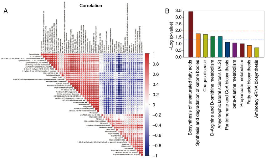
MOLECULAR MEDICINE REPORTS 23: 241, 2021                                             3

plasma was then transported to a diagnostic laboratory for the     activity was visualized using 3,3'‑diaminobenzidine at room
measurement of creatinine levels using an Indiko™ system           temperature for 5 min. The relative expression density and
(Thermo Fisher Scientific, Inc.).                                  location of Aifm2 and HMGB1 in the mouse kidney tissues
                                                                   were quantified at x400 magnification per 30 fields, according
Micro‑PET/CT scanning. 18FDG is a commonly used radio‑             to the Envision method (36), by two pathologists blinded to
nuclide imaging agent that is actively absorbed by cells as an     the experiment. The relative expression of proteins located
energy substrate and indirectly reflects the energy metabolism     mainly in the cytoplasm was calculated using the relative
of the body. 18FDG (3.7 MBq/kg), obtained from an Eclipse          mean absorbance according to the Envision method. The rela‑
HP/RD accelerator (Siemens AG), was injected into the              tive expression of proteins located mainly in the nucleus was
tail vein 20 min before the scan. The radioisotope uptake          determined as the number of positive cells per 200 cells in a
of the tissues, which reflects the status of the body's energy     high‑power field (magnification, x400). All IHC sections were
metabolism, was detected using an Inveon™ micro‑PET/CT             examined with a phase‑contrast microscope. Image‑Pro Plus 6
system (Siemens AG). The test parameters were set as follows:      software (Media Cybernetics, Inc.) was used for analysis.
Resolution of transverse, longitudinal and axial directions of
the PET module, 1.5 mm; collection timing window, 3.4 nsec;        Transmission electron microscopy (TEM). Dissected mouse
energy window, 350‑650 keV; and collection time, 10 min. The       kidneys were fixed in 2.5% glutaraldehyde at 4˚C overnight,
filtered back projection algorithm was used for reconstruction.    washed three times with 0.1 M PBS and stained with 1%
The images were analyzed using Inveon image acquisition            osmium acid at 4˚C for 3 h. The sections were then washed
(IAW 1.5; Siemens AG). The PET value was set at 1.8‑9.9, and       3 times with PBS, dehydrated in ethanol and subsequently
the CT window width at 500‑1000 HU.                                propylene oxide, embedded in Spurr resin at room tempera‑
                                                                   ture overnight and finally polymerized at 70˚C overnight. The
H&E staining. Paraformaldehyde‑fixed kidney tissues were           embedded sections were cut into 70‑nm slices. Slides of these
paraffin‑embedded, cut into 4‑µm sections, deparaffinized,         slices were prepared and stained with lead citrate and uranium
and serially rehydrated using xylene, an ethanol gradient          dioxide acetate at 80˚C for 15 min, and photographed under a
series (100, 96, 80 and 70% ethanol) and finally H2O. Next, the    transmission electron microscope.
sections were stained with 10% hematoxylin for 5 min at room
temperature (Sigma‑Aldrich; Merck KGaA), washed, stained           TUNEL assay. Sections (2‑3 µm) of mouse kidney tissues
with 1% eosin (Sigma‑Aldrich; Merck KGaA) supplemented             were incubated for 2 min with 0.1% Triton X‑100 and washed
with 0.2% glacial acetic acid for 5 mins at room tempera‑          using ice‑cold PBS. Cell apoptosis was assayed using TUNEL
ture, immediately washed again, and dehydrated using an            reagent for 60 min at 37˚C (Roche Applied Science), according
ethanol gradient (70, 80, 96 and 100%) and xylene. Finally,        to the manufacturer's protocols. Cells with yellow‑stained
the sections were examined under an inverted phase‑contrast        nuclei were defined as positive cells at 25˚C for 10 min
microscope (DMi1; Leica Microsystems GmbH). The renal              (Concentrated DAB reagent kit; OriGene Technologies, Inc.).
tubule injury score based on the Parller system was calculated     The positive cells in 200 randomly selected cells were counted
to analyze renal injury (35). A total of 10 renal tubules within   using a phase‑contrast microscope (magnification, x100; 10
10 non‑overlapping high magnification fields were randomly         fields of view).
selected for a total of 100 renal tubules, which were then
evaluated to determine the renal tubule injury score as follows:   Western blot analysis. The dissected kidney tissues were
Normal structure of the epithelial cells, 0 points; obvious        lysed and sonicated (20 sec per 5 min) for 30 min on ice in
renal tubule expansion, loss of brush border, epithelial cell      radioimmunoprecipitation assay buffer (Beyotime Institute
edema, vacuolar degeneration, granular degeneration and/or         of Biotechnology) supplemented with a protease inhibitor
nuclear deflation, 1 point; and foaming of the cell membrane,      and a phosphatase inhibitor (Sigma‑Aldrich; Merck KGaA)
cell necrosis and/or tubular appearance in the tubule lumen,       on an ice bath for 30 min. The tissues were then centrifuged
2 points. The sum of the scores for all 100 tubules was used as    for 15 min at 13,000 x g and 4˚C. The supernatant thus
the Parller score for each animal.                                 obtained was subjected to the bicinchoninic acid assay for
                                                                   protein quantitation, and bovine serum albumin (Beyotime
Immunohistochemistry (IHC) analysis. In brief, 4‑µm                Institute of Biotechnology,) was used as the standard. In
paraffin‑embedded sections were deparaffinized, subjected          brief, 40 µg protein extracts were denatured, separated via
to antigen retrieval using citrate buffer (pH 6.0) at 95˚C for     10% SDS‑PAGE, and transferred to Hybond® polyvinyli‑
10 min and treated with 0.3% H 2O2 to block their endog‑           dene difluoride membranes. The membranes were washed
enous peroxidase activity at room temperature for 10 min.          and blocked using 0.1% TBS‑Tween buffer and 5% non‑fat
Next, the sections were continuously treated with blocking         milk (BD Biosciences) at room temperature for 2 h and
reagent QuickBlock™ (cat. no. P0260; Beyotime Institute            subsequently probed with RAGE (1:2,000; cat. no. ab3611;
of Biotechnology), then incubated with the appropriate             Abcam), HMGB1 (1:2,000; cat. no. ab227168; Abcam) and
primary antibodies, such as apoptosis‑inducing factor mito‑        β‑actin (1:5,000; cat. no. ab8226; Abcam) primary antibodies
chondria‑associated 2 (Aifm2; 1:1,000; cat. no. GXP20453;          at 4˚C overnight, and secondary antibodies for 1 h at room
GenXspan) and HMGB1 (1:100; cat. no. ab227168; Abcam) at           temperature [1:5,000; Biotinylated Goat anti rabbit IgG
4˚C overnight, and then with the secondary antibody (1:5,000;      (H + L); cat. no. ab6721; Abcam] Protein bands were evalu‑
cat. no. TA130017 Biotinylated Goat anti rabbit; OriGene           ated using enhanced chemiluminescence reagent (Bio‑Rad
Technologies, Inc.) for 1 h at room temperature. Peroxidase        Laboratories, Inc.) and Biomax MR films (Kodak). Relative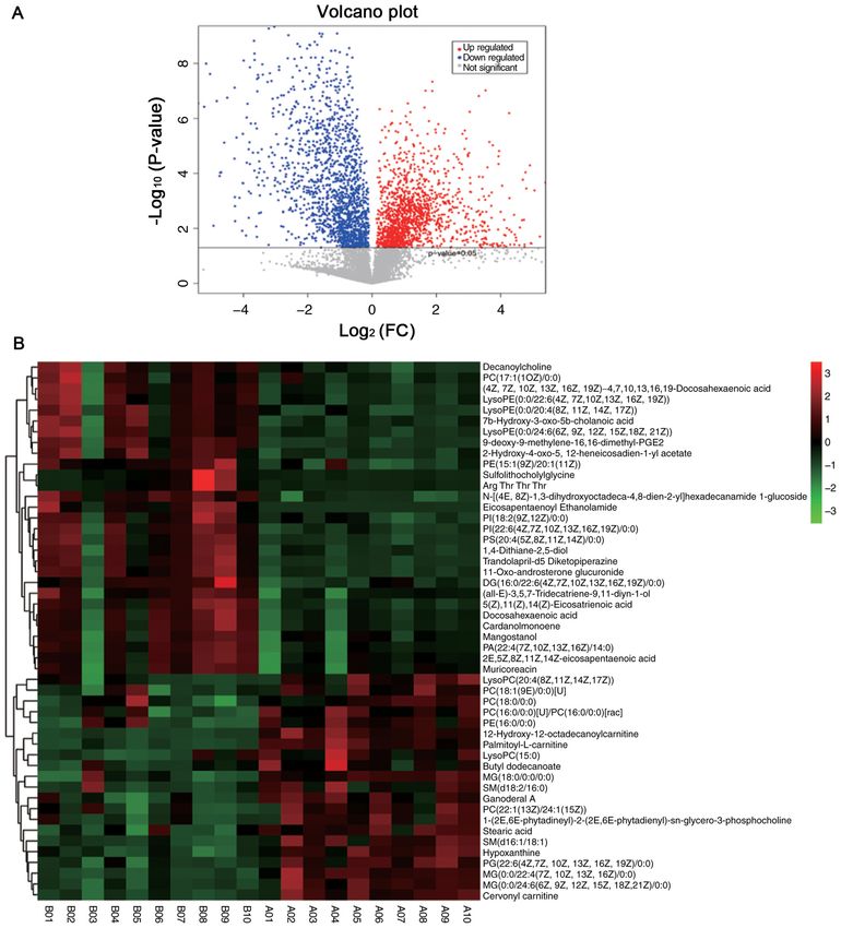
4                                     XUE et al: METABOLOMIC PROFILING OF HEAT STROKE

protein abundance of the indicated proteins was determined         Table I. Change in blood glucose and creatinine levels in the
using ChemiDoc XRS+ System/Image Lab Software                      study groups.
version 2.0 (Bio‑Rad Laboratories, Inc.).
                                                                   Group            Glucose, mmol/l        Serum creatinine, umol/l
Liquid chromatography‑mass spectrometry (LC‑MS)
analysis. All chemicals and solvents used were of analytical or    Control             5.26±1.63                  11.47±3.91
high‑performance liquid chromatography grade. Acetonitrile,        Heat stroke         5.34±1.92                  20.53±4.75a
formic acid, methanol and water were acquired from CNW
                                                                   P1.0 (obtained using the OPLS‑DA model) and
added. The samples were frozen to ‑80˚C for 2 min, ground for      P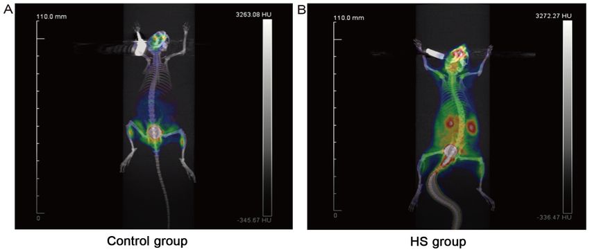
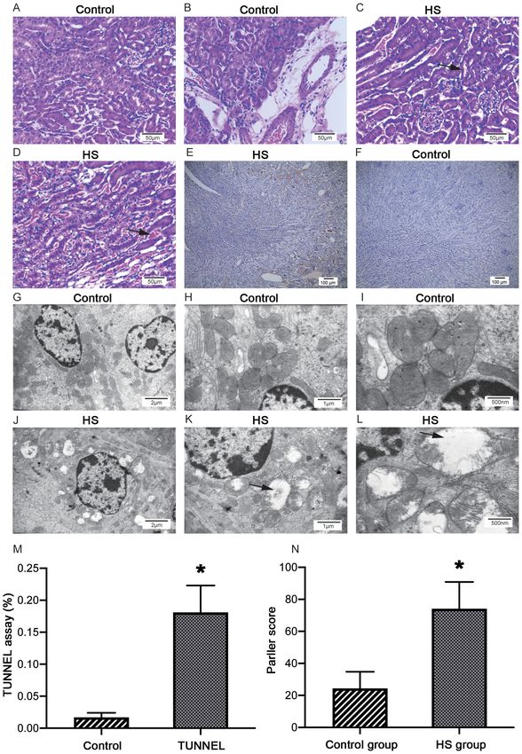
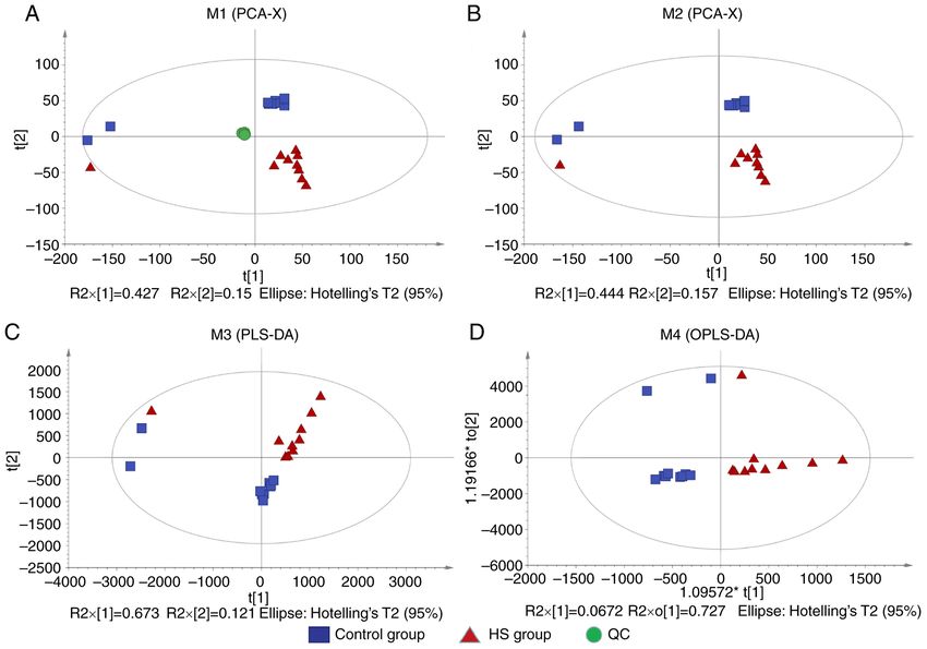
MOLECULAR MEDICINE REPORTS 23: 241, 2021                                                                5

Figure 1. 18FDG‑labeled micro‑PET/CT scanning. Mice were injected with 18FDG and subjected to micro‑PET/CT to visualize energy metabolism. (A) Control
group (n=2). (B) HS group (n=2). Red indicates high uptake and dispersion of the radionuclide imaging agent, whereas green indicates low uptake and disper‑
sion of the radionuclide imaging agent. HS, heat stroke; micro‑PET/CT, micro‑positron emission tomography‑computed tomography; 18FDG, 18F‑deoxyglucose.

indicates a high risk of hemorrhage (Fig. 2C and D). All these                 Identification of differential metabolites and signaling
changes were not present in the control group (Fig. 2A and B).                 pathways. OPLS‑DA models were used to identify differ‑
The TUNEL assay revealed yellow‑stained nuclei of apoptotic                    ences in metabolites between the control and HS groups. The
cells primarily located in the renal tubule area in the HS group.              HS‑group cluster was distant from the control‑group cluster
The number of apoptotic cells was significantly increased in                   (Fig. 4). The results revealed that of the 136 metabolites that
the HS group compared with in the control group (P
6                                             XUE et al: METABOLOMIC PROFILING OF HEAT STROKE

Figure 2. HS triggers acute kidney injury in mice. (A‑D) Representative H&E‑stained images of the kidneys in both groups. HS group (n=8) displays glomerular
swelling, inflammatory cell infiltration, vacuolar degeneration of endothelial cells and red blood cells in the renal tubules (C and D). These changes were not
found in the control group (n=8) (A and B). Scale bar, 50 µm. Representative TUNEL assay images of the kidneys in the (E) HS (n=8) and (F) control groups
(n=8). Yellow nuclear staining indicates apoptotic cells in the kidney tissues. scale bar, 100 µm. Representative transmission electron microscope images of
the kidneys in the (G‑I) control (n=8) and (J‑L) HS group (n=8). Scale bar, 2 µm in G and J, 1 µm in H and K, and 500 nm in I and L. (M) Statistical analysis
of the TUNEL assay. *P
MOLECULAR MEDICINE REPORTS 23: 241, 2021                                                                  7

Figure 3. HS activates the HMGB1/RAGE pathway. (A) Representative IHC staining of Aifm2 protein expression in the kidneys of HS (n=5) and control
group (n=5) mice. Aifm2 is mainly located in the cytoplasm of kidney tissues. Scale bar, 40 µm. (B) Statistical analysis of the IHC staining of Aifm2. *P
8                                              XUE et al: METABOLOMIC PROFILING OF HEAT STROKE

Figure 4. Scatter plots of scores established by PCA. (A) Multivariate statistical comparison of PCA between the two groups with quality control. (B) Multivariate
statistical comparison of PCA between the two groups without quality control. (C) PLS‑DA. (D) OPLS‑DA. PCA, principal component analysis; OPLS‑DA,
orthogonal partial least squares discriminant analysis; PLS‑DA, partial least squares discriminant analysis; HS, heat stroke; QC, quality control.

The present study aimed to acquire a deeper understanding of                       HMGB1/RAGE pathway may be involved in this process (62),
the mechanisms underlying HS‑induced AKI, which has been                           but the specific mechanism needs to be elucidated.
rarely studied and could provide novel insight.                                        In present study, metabolomics analysis of the animal
    HMGB1, also known as alarmin, is a gene transcription                          model was conducted to further explore the pathogenesis of
cofactor and a damage‑associated molecular pattern molecule                        HS. Of the 136 differential metabolites, 80 metabolites were
that mediates proinflammatory effects by activating multiple cell                  upregulated and 56 were downregulated. The top 15 differ‑
surface receptors, such as RAGE and Toll‑like receptors (56).                      ential metabolites were associated with fatty acid metabolism
This proinflammatory role is relevant to the pathogenesis of                       and other associated metabolic pathways, which may induce
several kidney diseases, including AKI (57). HMGB1 is also                         early changes in the kidneys during HS. Additionally, the
considered to be involved in the pathogenesis of HS, as increased                  correlation among a number of the top 50 metabolites was
serum HMGB1 levels indicate poor prognosis in HS (48).                             strong, indicating their probable interplay in inducing AKI
Inhibiting the HMGB1/RAGE pathway may aid in the treatment                         in response to HS. Enrichment analysis of all the differential
of AKI, although its role in HS‑related AKI is unclear (58). It was                metabolites was performed, and ‘biosynthesis of unsaturated
observed that both HMGB1 and RAGE were significantly upreg‑                        fatty acids̓, ‘synthesis and degradation of ketone bodies̓,
ulated in the kidneys of HS mice. Furthermore, both the TUNEL                      ‘Chagas disease’, ‘D‑arginine and D‑ornithine metabolism’
assay and TEM scanning suggested that mitochondrial‑depen‑                         and ‘ALS’ were the only five significantly enriched signaling
dent apoptosis had occurred in the kidneys of HS mice, which                       pathways between the two groups. Adrenic acid, DHA and
was supported by the upregulation of Aifm2, a gene involved in                     EPA, which are involved in the ‘biosynthesis of unsaturated
mitochondrial‑dependent apoptosis (59). As the HMGB1/RAGE                          fatty acids̓ pathway, were upregulated in the HS group. This
pathway may trigger mitochondria‑related apoptosis in inflam‑                      may appear to be in contrast to a previous study, which reported
matory states (60), it is hypothesized that the apparent increase                  the synergistic functioning of DHA and EPA to protect intes‑
in renal tubular epithelial cell apoptosis observed in the present                 tinal barrier integrity in HS (63). However, these are, in fact,
study was caused, at least partly, by the HMGB1/RAGE pathway.                      two phases of a process. HS can damage the intestine barrier
However, pathological examination indicated renal tubular                          to allow the seepage of unsaturated fatty acids, which are
hyperemia and edema, instead of apoptosis. A previous study                        important components of cell membranes, into the urinary
has reported the occurrence of severe pulmonary hemorrhage in                      system, leading to an increase in their levels in the urinary
patients who died of HS; however, kidney injury due to HS was                      system. As DHA and EPA treatment prevents the disruption
not mentioned (61). Hence, it is proposed that bleeding, rather                    of the intestinal barrier, fewer metabolites, such as unsaturated
than apoptosis, may be a characteristic feature of HS‑associated                   fatty acids, flow into the circulation to initiate inflamma‑
AKI. Moreover, HS‑induced SIRS and secondary hyperfibri‑                           tory processes, and the damage caused by HS is limited. As
nolysis may be other reasons for hyperemia and edema in the                        reported previously, the HMGB1/RAGE pathway may be
kidneys, which were different from the reported manifestations                     a potential target for unsaturated fatty acids or regulate the
of common acute tubular necrosis (23). Dysregulation of the                        concentration of unsaturated fatty acids in other diseases via
MOLECULAR MEDICINE REPORTS 23: 241, 2021 9 Figure 5. Identification of differential metabolites. (A) Volcano plot of the total metabolites. Red indicates upregulated metabolites, and blue indicates down‑ regulated metabolites. (B) Heat map of the top 50 differential metabolites in the heat stroke group (A1‑10) compared with the control group (B1‑10). Red indicates upregulated metabolites, and green indicates downregulated metabolites. FC, fold change. Figure 6. Identification of enriched signaling pathways. (A) Correlation analysis of top 50 differential metabolites. Red indicates positive correlation, and blue indicates negative correlation. (B) Histogram of top 10 significantly enriched signaling pathways between the two groups.
10                                      XUE et al: METABOLOMIC PROFILING OF HEAT STROKE

various means (64,65). At present, the relationship between the       Competing interests
HMGB1/RAGE pathway and unsaturated fatty acids remains
unclear, and further studies are required.                            The authors declare that they have no competing interests.
    The present study has certain limitations. First, for a
more in‑depth investigation of the mechanisms underlying              References
HS‑related AKI, multiple observation time points should                1. Laitano O, Murray KO and Leon LR: Overlapping mechanisms
be used, such as 15 and 30 min, and 1 and 2 h; this will be               of exertional heat stroke and malignant hyperthermia: Evidence
explored in subsequent studies. Second, the correlations                  vs.conjecture. Sports Med 50: 1581‑1592, 2020.
between metabolic changes and alterations in signaling path‑           2. Gaudio FG and Grissom CK: Cooling methods in heat stroke.
                                                                          J Emerg Med 50: 607‑616, 2016.
ways should be verified using gain‑or‑loss experiments. Third,         3. Barriopedro D, Fischer EM, Luterbacher J, Trigo RM and
as HS is a complex disease that involves alterations in diverse           Garcia‑Herrera R: The hot summer of 2010: Redrawing the
signaling pathways, therapeutic interventions targeting the               temperature record map of Europe. Science 332: 220‑224, 2011.
                                                                       4. Kovats RS and Kristie LE: Heatwaves and public health in
HMGB1/RAGE pathway or biosynthesis of unsaturated fatty                   Europe. Eur J Public Health 16: 592‑599, 2006.
acids could be added in future studies into HS.                        5. Argaud L, Ferry T, Le QH, Marfisi A, Ciorba D, Achache P,
    In summary, the present study suggested that HS‑associated            Ducluzeau R and Robert D: Short‑ and long‑term outcomes of
                                                                          heatstroke following the 2003 heat wave in Lyon, France. Arch
AKI is the most common result of HS‑induced SIRS, and may                 Intern Med 167: 2177‑2183, 2007.
be associated with the HMGB1/RAGE pathway. Furthermore,                6. Roux‑Buisson N, Monnier N, Sagui E, Abriat A, Brosset C,
a feedback loop between the HMGB1/RAGE pathway and the                    Bendahan D, Kozak‑Ribbens G, Gazzola S, Quesada JL,
                                                                          Foutrier‑Morello C, et al: Identification of variants of the ryano‑
biosynthesis of unsaturated fatty acids may contribute to the             dine receptor type 1 in patients with exertional heat stroke and
progression of HS‑related AKI. Therefore, it is of great impor‑           positive response to the malignant hyperthermia in vitro contrac‑
tance to elucidate the relationship between the HMGB1/RAGE                ture test. Br J Anaesth 116: 566‑568, 2016.
                                                                       7. Yang M, Zhang Y, Zhao Y and Kang H: Research progress in
pathway and disordered unsaturated fatty acid metabolism in               the multiple organ dysfunction syndrome caused by heat stroke.
the pathogenesis of HS‑associated AKI.                                    Zhonghua Wei Zhong Bing Ji Jiu Yi Xue 29: 188‑192, 2017 (In
                                                                          Chinese).
                                                                       8. Chen CM, Hou CC, Cheng KC, Tian RL, Chang CP and Lin MT:
Acknowledgements                                                          Activated protein C therapy in a rat heat stroke model. Crit Care
                                                                          Med 34: 1960‑1966, 2006.
Not applicable.                                                        9. Ren MQ, Kazman JB, Abraham PA, Atias‑Varon D, Heled Y
                                                                          and Deuster PA: Gene expression profiling of humans under
                                                                          exertional heat stress: Comparisons between persons with and
Funding                                                                   without exertional heat stroke. J Therm 85: 102423, 2019.
                                                                      10. Lumlertgul D, Chuaychoo B, Thitiarchakul S, Srimahachota S,
                                                                          Sangchun K and Keoplung M: Heat stroke‑induced multiple
The study was supported by National Natural Science Foundation            organ failure. Ren Fail 14: 77‑80, 1992.
of China (grant no. 81200533), Sichuan Clinical Research              11. Deutsch M, Koskinas J, Emmanuel T, Kountouras D and
Center for Nephropathy (grant no. 2019YFS0537‑4) and                      Hadziyannis S: Heat stroke and multi‑organ failure with liver involve‑
                                                                          ment in an asylum‑seeking refugee. J Emerg Med 31: 255‑257, 2006.
Luzhou Municipal Government‑Southwest Medical University              12. McGeehin MA and Mirabelli M: The potential impacts of
Strategic Cooperation Fund (grant no. 2016LZXNYD‑J20).                    climate variability and change on temperature‑related morbidity
                                                                          and mortality in the United States. Environ Health Perspect 109
                                                                          (Suppl 2): S185‑S189, 2001.
Availability of data and materials                                    13. Bouchama A and Knochel JP: Heat stroke. N Engl J Med 346:
                                                                          1978‑1988, 2002.
The datasets used and/or analyzed during the current study are        14. Woodrow G, Brownjohn AM and Turney JH: The clinical and
available from the corresponding author on reasonable request.            biochemical features of acute renal failure due to rhabdomy‑
                                                                          olysis. Ren Fail 17: 467‑474, 1995.
                                                                      15. Chen GM, Chen YH, Zhang W, Yu Y, Chen JH and Chen J:
Authors' contributions                                                    Therapy of severe heatshock in combination with multiple
                                                                          organ dysfunction with continuous renal replacement therapy: A
                                                                          clinical study. Medicine (Baltimore) 94: e1212, 2015.
LX and WW designed the study and participated in animal               16. Inoue N, Sato A, Ikawa Y, Shimizu M, Okajima M, Taniguchi T
experiments. LX and WG participated in the study design and               and Yachie A: Successful treatment of exertional heat stroke using
drafting the manuscript. LL and SO were responsible for statistical       continuous plasma diafiltration. J Clin Apher 31: 490‑492, 2016.
                                                                      17. Hamaya H, Hifumi T, Kawakita K, Okazaki T, Kiridume K,
analysis of the metabolomic data. TZ performed in vivo animal             Shinohara N, Abe Y, Takano K, Hagiike M and Kuroda Y:
experiments. LC and WD performed PET/CT scanning and data                 Successful management of heat stroke associated with
analysis. All authors read and approved the final manuscript.             multiple‑organ dysfunction by active intravascular cooling. Am J
                                                                          Emerg Med 33: 124.e5‑e7, 2015.
                                                                      18. Ye J, Mo W, Chen Y and Yang A: An analysis of laboratory results of
Ethics approval and consent to participate                                parameters of organ function in patients with heat stroke. Zhonghua
                                                                          Wei Zhong Bing Ji Jiu Yi Xue 27: 658‑661, 2015 (In Chinese).
                                                                      19. Zhao JJ, Zhou JJ, Hu J, Zhou FH, Kang HJ, Liu H, Pan L and Song Q:
All animal experiments complied with the guidelines of the                Analysis of risk factors affecting prognosis of exertional heat stroke.
Animal Care and Use Committee of the Southwest Medical                    Zhonghua Wei Zhong Bing Ji Jiu Yi Xue 25: 515‑518, 2013.
University. The study protocol was approved by the same               20. Segev G, Bruchim Y, Berl N, Cohen A and Aroch I: Effects of
                                                                          fenoldopam on kidney function parameters and its therapeutic
committee.                                                                efficacy in the management of acute kidney injury in dogs with
                                                                          heatstroke. J Vet Intern Med 32: 1109‑1115, 2018.
Patient consent for publication                                       21. Peng N, Geng Y, Zhang S, Tang Y, Wen Q, Tong H, Liu Y,
                                                                          Liu Z and Su L: Correlation of kidney injury and inflammatory
                                                                          response in rats with classic severe heatstroke. Zhonghua Wei
Not applicable.                                                           Zhong Bing Ji Jiu Yi Xue 27: 327‑331, 2015 (In Chinese).
MOLECULAR MEDICINE REPORTS 23: 241, 2021 11
22. Protasi F, Paolini C and Dainese M: Calsequestrin‑1: A new 45. Gathiram P, Wells MT, Raidoo D, Brock‑Utne JG and Gaffin SL:
candidate gene for malignant hyperthermia and exertional/environ‑ Portal and systemic plasma lipopolysaccharide concentrations in
mental heat stroke. J Physiol 587: 3095‑3100, 2009. heat‑stressed primates. Circ Shock 25: 223‑230, 1988.
23. Leon LR and Helwig BG: Heat stroke: Role of the systemic inflam‑ 46. Gathiram P, Wells MT, Brock‑Utne JG and Gaffin SL:
matory response. J Appl Physiol (1985) 109: 1980‑1988, 2010. Antilipopolysaccharide improves survival in primates subjected to
24. Roberts GT, Ghebeh H, Chishti MA, Al‑Mohanna F, El‑Sayed R, heat stroke. Circ Shock 23: 157‑164, 1987.
Al‑Mohanna F and Bouchama A: Microvascular injury, thrombosis, 47. Huisse MG, Pease S, Hurtado‑Nedelec M, Arnaud B, Malaquin C,
inflammation, and apoptosis in the pathogenesis of heatstroke: Wolff M, Gougerot‑Pocidalo MA, Kermarrec N, Bezeaud A,
A study in baboon model. Arterioscler Thromb Vasc Biol 28: Guillin MC, et al: Leukocyte activation: The link between inflam‑
1130‑1136, 2008. mation and coagulation during heatstroke. A study of patients
25. Rosendal L, Langberg H, Skov‑Jensen A and Kjaer M: Incidence during the 2003 heat wave in Paris. Crit Care Med 36: 2288‑2295,
of injury and physical performance adaptations during military 2008.
training. Clin J Sport Med 13: 157‑163, 2003. 48. Tong HS, Tang YQ, Chen Y, Qiu JM, Wen Q and Su L: Early
26. Yang PC, He SH and Zheng PY: Investigation into the signal trans‑ elevated HMGB1 level predicting the outcome in exertional heat‑
duction pathway via which heat stress impairs intestinal epithelial stroke. J Trauma 71: 808‑814, 2011.
barrier function. J Gastroenterol Hepatol 22: 1823‑1831, 2007. 49. Pozner RG, Ure AE, Jaquenod de Giusti C, D'Atri LP, Italiano JE,
27. Mittal R and Coopersmith CM: Redefining the gut as the motor of Torres O, Romanowski V, Schattner M and Gómez RM: Junín virus
critical illness. Trends Mol Med 20: 214‑223, 2014. infection of human hematopoietic progenitors impairs in vitro
28. Clayton TA, Lindon JC, Cloarec O, Antti H, Charuel C, proplatelet formation and platelet release via a bystander effect
Hanton G, Provost JP, Le Net JL, Baker D, Walley RJ, et al: involving type I IFN signaling. PLoS Pathog 6: e1000847, 2010.
Pharmaco‑metabonomic phenotyping and personalized drug treat‑ 50. Chen HS, Tong HS, Zhao Y, Hong CY, Bin JP and Su L: Differential
ment. Nature 440: 1073‑1077, 2006. expression pattern of exosome long non‑coding RNAs (lncRNAs)
29. Kork F, Holthues J, Hellweg R, Jankowski V, Tepel M, Ohring R, and MicroRNAs (miRNAs) in vascular endothelial cells under heat
Heuser I, Bierbrauer J, Peters O, Schlattmann P, et al: A possible stroke. Med Sci Monit 24: 7965‑7974, 2018.
new diagnostic biomarker in early diagnosis of Alzheimer's disease. 51. Thulesius O: Thermal reactions of blood vessels in vascular stroke
Curr Alzheimer Res 6: 519‑524, 2009. and heatstroke. Med Princ Pract 15: 316‑321, 2006.
30. Laitano O, Garcia CK, Mattingly AJ, Robinson GP, Murray KO, 52. Fan H, Zhao Y, Zhu JH, Song FC, Ye JH, Wang ZY and Le JW:
King MA, Ingram B, Ramamoorthy S, Leon LR and Clanton TL: Thrombocytopenia as a predictor of severe acute kidney injury in
Delayed metabolic dysfunction in myocardium following exertional patients with heat stroke. Ren Fail 37: 877‑881, 2015.
heat stroke in mice. J Physiol 598: 967‑985, 2020. 53. Michels S, Buchholz HG, Rosar F, Heinrich I, Hoffmann MA,
31. Li Y, Zhu X, Zhang M, Tong H and Su L: Heatstroke‑induced hepa‑ Schweiger S, Tüscher O and Schreckenberger M: 18F‑FDG
tocyte exosomes promote liver injury by activating the NOD‑like PET/CT: An unexpected case of Huntington's disease. BMC
receptor signaling pathway in mice. PeerJ 7: e8216, 2019. Neurol 19: 78, 2019.
32. Tai PA, Chang CK, Niu KC, Lin MT, Chiu WT and Lin JW: 54. Wan Y, Sun SS, Fu HY, Xu YK, Liu Q, Yin JT and Wan B: Adjuvant
Reduction of ischemic and oxidative damage to the hypothalamus by rhubarb alleviates organs dysfunction and inhibits inflammation in
hyperbaric oxygen in heatstroke mice. J Biomed Biotechnol 2010: heat stroke. Exp Ther Med 16: 1493‑1498, 2018.
609526, 2010. 55. Yang TH, Shih MF, Wen YS, Ho WY, Leu KL, Wang MY and
33. Umemura Y, Ogura H, Matsuura H, Ebihara T, Shimizu K and Liu CC: Attenuation of circulatory shock and cerebral ischemia
Shimazu T: Bone marrow‑derived mononuclear cell therapy can injury in heat stroke by combination treatment with dexamethasone
attenuate systemic inflammation in rat heatstroke. Scand J Trauma and hydroxyethyl starch. Exp Transl Stroke Med 2: 19, 2010.
Resusc Emerg Med 26: 97, 2018. 56. Kurts C, Panzer U, Anders HJ and Rees AJ: The immune system
34. Yeh CH, Chen ZC, Hsu CC, Lin MT and Chen CC: Protection and kidney disease: Basic concepts and clinical implications. Nat
in rats with heatstroke: Hyperbaric oxygen vs activated protein C Rev Immunol 13: 738‑753, 2013.
therapy. Eur J Pharmacol 635: 103‑108, 2010. 57. Chen Q, Guan X, Zuo X, Wang J and Yin W: The role of high
35. Yayi H, Yeda X, Huaxin W, Yang W, Qian S and Zhongyuan X: mobility group box 1 (HMGB1) in the pathogenesis of kidney
Toll‑like receptor 7 involves the injury in acute kidney isch‑
emia/reperfusion of STZ‑induced diabetic rats. Acta Cir Bras 31: diseases. Acta Pharm Sin B 6: 183‑188, 2016.
448‑455, 2016. 58. Liu GQ, Zuo XH, Jiang LN, Zhang YP, Zhang LM, Zhao ZG and
36. Satirapoj B, Kongthaworn S, Choovichian P and Supasyndh O: Niu CY: Inhibitory effect of post‑hemorrhagic shock mesenteric
Electrolyte disturbances and risk factors of acute kidney injury lymph drainage on the HMGB1 and RAGE in mouse kidney. Ren
patients receiving dialysis in exertional heat stroke. BMC Fail 38: 131‑136, 2016.
Nephrol 17: 55, 2016. 59. Miriyala S, Thippakorn C, Chaiswing L, Xu Y, Noel T, Tovmasyan A,
37. López‑Ibáñez J, Pazos F and Chagoyen M: MBROLE 2.0‑func‑ Batinic‑Haberle I, Vander Kooi CW, Chi W, Latif AA, et al: Novel
tional enrichment of chemical compounds. Nucleic Acids Res 44 role of 4‑hydroxy‑2‑nonenal in AIFm2‑mediated mitochondrial
(W1): W201‑W204, 2016. stress signaling. Free Radic Biol Med 91: 68‑80, 2016.
38. Kanehisa M, Sato Y, Furumichi M, Morishima K and Tanabe M: 60. Qin S, Wang H, Yuan R, Li H, Ochani M, Ochani K,
New approach for understanding genome variations in KEGG. Rosas‑Ballina M, Czura CJ, Huston JM, Miller E, et al: Role of
Nucleic Acids Res 47 (D1): D590‑D595, 2019. HMGB1 in apoptosis‑mediated sepsis lethality. J Exp Med 203:
39. Alele F, Malau‑Aduli B, Malau‑Aduli A and Crowe M: Systematic 1637‑1642, 2006.
review of gender differences in the epidemiology and risk factors of 61. Adato B, Dubnov‑Raz G, Gips H, Heled Y and Epstein Y: Fatal
exertional heat illness and heat tolerance in the armed forces. BMJ heat stroke in children found in parked cars: Autopsy findings. Eur
Open 10: e031825, 2020. J Pediatr 175: 1249‑1252, 2016.
40. Li L, Tan H, Zou Z, Gong J, Zhou J, Peng N, Su L, Maegele M, 62. Xu L, Zhao K, Shen X, Fan XX, Ding K, Liu RM and Wang F:
Cai D and Gu Z: Preventing necroptosis by scavenging ROS Blockade of extracellular high‑mobility group box 1 attenuates
production alleviates heat stress‑induced intestinal injury. Int J systemic inflammation and coagulation abnormalities in rats with
Hyperthermia 37: 517‑530, 2020. acute traumatic coagulopathy. Med Sci Monit 22: 2561‑2570, 2016.
41. Thongprayoon C, Qureshi F, Petnak T, Cheungpasitporn W, 63. Xiao G, Yuan F, Geng Y, Qiu X, Liu Z, Lu J, Tang L, Zhang Y and
Chewcharat A, Cato LD, Boonpheng B, Bathini T, Hansrivijit P, Su L: Eicosapentaenoic acid enhances heatstroke‑impaired intes‑
Vallabhajosyula S and Kaewput W: Impact of acute kidney injury tinal epithelial barrier function in rats. Shock 44: 348‑356, 2015.
on outcomes of hospitalizations for heat stroke in the United States. 64. Wei W, Chen M, Zhu Y, Wang J, Zhu P, Li Y and Li J:
Diseases 8: 28, 2020. Down‑regulation of vascular HMGB1 and RAGE expression by
42. Weigand K, Riediger C, Stremmel W, Flechtenmacher C and n‑3 polyunsaturated fatty acids is accompanied by amelioration of
Encke J: Are heat stroke and physical exhaustion underestimated chronic vasculopathy of small bowel allografts. J Nutr Biochem 23:
causes of acute hepatic failure? World J Gastroenterol 13: 306‑309, 1333‑1340, 2012.
2007. 65. Ying S, Xiao X, Chen T and Lou J: PPAR ligands function as
43. Hu J, Kang HJ, Liu C, Hu P, Yang MM and Zhou FH: Response of suppressors that target biological actions of HMGB1. PPAR
regulatory T cells to classic heat stroke in mice. Exp Ther Med 16: Res 2016: 2612743, 2016.
4609‑4615, 2018. This work is licensed under a Creative Commons
44. Shapiro Y, Alkan M, Epstein Y, Newman F and Magazanik A:
Attribution-NonCommercial-NoDerivatives 4.0
Increase in rat intestinal permeability to endotoxin during hyper‑
thermia. Eur J Appl Physiol Occup Physiol 55: 410‑412, 1986. International (CC BY-NC-ND 4.0) License.You can also read