Molecular characterization of the recurrent unbalanced translocation der(1;7)(q10;p10)
←
→
Page content transcription
If your browser does not render page correctly, please read the page content below
NEOPLASIA

Molecular characterization of the recurrent unbalanced translocation
der(1;7)(q10;p10)
Lili Wang, Seishi Ogawa, Akira Hangaishi, Ying Qiao, Noriko Hosoya, Yasuhito Nanya, Kazuma Ohyashiki,
Hideaki Mizoguchi, and Hisamaru Hirai

An unbalanced translocation der(1;7)(q10;          on chromosome 7, were almost invariably            these patients were randomly distributed
p10) is a nonrandom chromosomal aber-              reduced on the derivative chromosome               over several megabase pairs within each
ration commonly observed in myelodys-              compared with those on their normal                alphoid cluster except for its extreme end
plastic syndrome and acute myeloid                 counterparts. These results suggest that           to the short arm. Our results provide a
leukemia. We molecularly analyzed the              this translocation results from the recom-         novel insight into the structural basis for
breakpoints of der(1;7)(q10;p10) by quan-          bination between the 2 alphoids, which             generation of this translocation as well as
titative fluorescent in situ hybridization         was further confirmed by fiber FISH ex-            its leukemogenic roles. (Blood. 2003;102:
(FISH) analyses using centromeric satel-           periments. Because the relative reduc-             2597-2604)
lite DNAs mapped to chromosomes 1 and              tion in the intensities of D1Z7 and D7Z1
7 as probes. We found that the signal              signals on the derivative chromosomes
intensities of 2 centromere alphoid                was highly variable among patients, it
probes, D1Z7 on chromosome 1 and D7Z1              was estimated that the breakpoints in              © 2003 by The American Society of Hematology

Introduction
The unbalanced whole-arm translocation of chromosome 1 (chr1)               chromosome in this translocation. However, no further molecular
and chromosome 7 (chr7), der(1;7)(q10;p10), is a nonrandom                  analysis, especially concerning exact locations of the breakpoints
chromosomal abnormality first described by Geraedts et al.1 It is           and their distribution, has been conducted to date.
commonly found in myelodysplastic syndrome (MDS) and acute                      In this paper, in order to disclose the genomic mechanism
myeloid leukemia (AML) and less frequently in myeloproliferative            generating der(1;7)(q10;p10), we focused on the centromere al-
disorders, involving all subgroups of MDS and AML. Since more               phoid sequences on chr1 and chr7 and dissected the structural
than half of the patients had a history of previous antitumor               alterations associated with this translocation by the 2-color FISH
chemotherapy (especially containing alkylating agents) and/or               method with simultaneous measurements of FISH signals.
radiotherapy or occupational exposure to toxic agents, it has been
causally related to secondary or therapy-related MDS or AML.2-9 In
MDS, as many as 6% of the patients are reported to have this                Patients, materials, and methods
karyotypic abnormality. They typically present trilineage morpho-
                                                                            Clinical samples
logic myelodysplasia in the bone marrow and pancytopenia in the
peripheral blood and have a high rate of progression to AML with            A total of 27 bone marrow samples were collected from patients carrying
generally poor prognosis.10,11                                              der(1;7)(q10;p10) and subjected to FISH analysis. The patients’ profiles are
    This unbalanced translocation, typically described as 46, XY (or        summarized in Table 1. All patients gave their informed consent to the
XX), ⫹1, der(1;7)(q10;p10), has 2 prominent cytogenetic charac-             sample collection and to the biologic analyses included in the present study
                                                                            according to the Declaration of Helsinki. Giemsa trypsin G-banding
teristics: (1) it retains only 1 of 2 possible derivative chromosomes,
                                                                            (GTG-banding) analysis was performed according to the standard protocol.15
containing 1q and 7p; and (2) there are 2 copies of apparently
normal chr1 and only 1 copy of normal chr7, resulting in trisomy of         DNA probes
1q and monosomy of 7q (Figure 1). In the early fluorescent in situ
                                                                            The screening of bacterial artificial chromosome (BAC) or P1-based
hybridization (FISH) studies, several authors pointed out the
                                                                            artificial chromosome (PAC) clones was previously described.16 DNA from
dicentric nature of this translocation, in a sense that the signals of
                                                                            BACs or PACs was extracted using the Large-Construct Kit (QIAGEN,
the 2 alphoids from chr1 and chr7 are colocalized on the derivative         Tokyo, Japan). A D1Z7 clone, pE25.a, was purchased from American Type
chromosome. Alitalo et al12 reported that FISH signals of D1Z5 and          Culture Collection (ATCC; Manassas, VA). A clone of D7Z1, called
D7Z2 coexisted on the der(1;7)(q10;p10), and others13,14 also               D7Z16mer, was obtained from polymerase chain reaction (PCR) amplifica-
described that D1Z5 and D7Z1 cohybridized to the derivative                 tion with primers spanning the dimer and the tetramer of the published

From the Department of Hematology and Oncology, Graduate School of          Reprints: Hisamaru Hirai, Department of Hematology & Oncology, Graduate
Medicine, University of Tokyo, Japan; the First Department of Internal      School of Medicine, University of Tokyo 7-3-1 Hongo, Bunkyo-ku, Tokyo 113-
Medicine, Tokyo Medical University, Japan; and the Department of Hema-      8655, Japan; e-mail: hhirai-tky@umin.ac.jp.
tology, Tokyo Women’s Medical University, Japan.
                                                                            The publication costs of this article were defrayed in part by page charge
Submitted January 6, 2003; accepted June 5, 2003. Prepublished online as
                                                                            payment. Therefore, and solely to indicate this fact, this article is hereby
Blood First Edition Paper, June 19, 2003; DOI 10.1182/blood-2003-01-0031.
                                                                            marked ‘‘advertisement’’ in accordance with 18 U.S.C. section 1734.
Supported in part by Grant-in-Aid for Scientific Research (KAKENHI
13307029).                                                                  © 2003 by The American Society of Hematology

BLOOD, 1 OCTOBER 2003 䡠 VOLUME 102, NUMBER 7                                                                                                       2597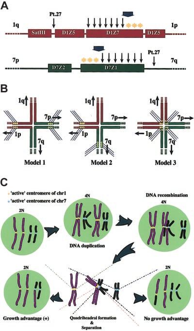
2598      WANG et al                                                                                      BLOOD, 1 OCTOBER 2003 䡠 VOLUME 102, NUMBER 7

                                                                                      heim, Germany) and anti–digoxigenin-rhodamine Fab fragments (Roche),
                                                                                      respectively. For fiber FISH experiments, the released chromatin fibers
                                                                                      were prepared as described18 and hybridized with 2 directly labeled probes,
                                                                                      CEP 1/5 and CEP 7, following the procedures recommended by the
                                                                                      manufacturer. Samples were examined with a Nikon E800 epi-fluorescence
                                                                                      microscope (Tokyo, Japan) at ⫻ 1000 magnification, and the FISH images
                                                                                      were captured to 8-bit–depth image files using a Kodak KAF1400
Figure 1. Partial karyotype of der (1;7)(q10;p10). GTG-banding analysis from Pt 1     thermoelectronically cooled charge-coupled device (CCD) camera (Roper
shows that the whole long arm of chromosome 1 (chr1) and the whole short arm of
                                                                                      Scientific, Tucson, AZ) and the SimpleVision software (Digital Scientific,
chromosome 7 (chr7) fuse to constitute der(1;7)(q10;p10) (der).
                                                                                      Cambridge, United Kingdom) through a triple–band-pass filter, which
                                                                                      allowed rhodamine, FITC, and DAPI (4,6 diamidino-2-phenylindole)
D7Z1 sequences (National Center for Biotechnology Information [NCBI]                  signals to be captured without any image displacement. For each experi-
accession: M16087, M16101). The sequences of the primers were 5⬘-                     ment, more than 30 metaphase images taken under exact focus conditions
TCCACTTGCAAATTCCACAA-3⬘ and 5⬘-TGGATATATGGACCG-                                       were stored. The whole intensity of each signal was automatically
CATTG-3⬘. The probes of satellite III (SatIII) and D7Z2 were also                     computed as the sum of pixel intensities under the indicated area by using
generated by PCR. Primers for SatIII (GenBank database X60726) were                   the IP lab/PathVysion Extras software (Scanalytics, Fairfax, VA), and
5⬘-TCCATTCCAGTCCATTCGAT-3⬘ and 5⬘-AATCATCATCCAACG-                                    finally the average was taken from the 30 measurements together with
GAAGC-3⬘ and those for D7Z2 (NCBI accession: G31642) were 5⬘-                         calculation of the standard deviation. The allelic origin of the 7p and 1q
CTGGAGGCGGATATTAGGGT-3⬘ and 5⬘-CTGGGAATACTTCTGTC-                                     components of the der(1;7)(q10;p10) was determined by comparing the
TAT-3⬘. All the plasmid DNAs were extracted using QIAGEN Plasmid                      signal intensity of polymorphic satellite elements on chr1 (SatIII) and on
Maxi Kit (QIAGEN). Three directly labeled probes, CEP 1/5 (Spectrum                   chr7 (D7Z2), which were safely apart from the breakpoints and their
Orange), CEP 1 (Spectrum Orange), and CEP 7 (Spectrum Green), which                   sequences were expected to be completely reserved on the der(1;7)(q10;
detect the sequences of D1Z7, D1Z5, and D7Z1, respectively, were                      p10) in tumor cells.
purchased from VYSIS (Downers Grove, IL).
                                                                                      Southern blot analysis
FISH analysis
                                                                                      Neo1 and An4, Neo7 and NT18, Neo2, Neo16, and Neo19, were monochro-
FISH experiments on metaphase chromosomes and interphase nuclei were                  mosomal human-mouse hybrid cell lines containing human chromosome 1,
performed as described17 with some modifications in posthybridization                 7, 2, 16, and 19, respectively, and were purchased from the JCRB Cell Bank
washes, which consisted of 2 ⫻ standard saline citrate (SSC)/50% form-                (Japanese Collection of Research Bioresources, Osaka, Japan). Genomic
amide at 37°C for 15 minutes followed by 2 ⫻ SSC and 1 ⫻ SSC at room                  DNAs were extracted from the cell lines19 and subjected to Southern blot
temperature for 15 minutes each. Biotin- and digoxigenin-labeled probes               analysis essentially as described.20 High-stringency hybridization was done
were detected with avidin–fluorescein isothiocyanate (FITC; Roche, Mann-              at 53°C for 16 to 18 hours in hybridization solution containing 50%

Table 1. Cytogenetic profile of patients
Patient    Age, y/sex       Diagnosis                                                               Karyotype

   1          45/M        MDS (RA)          46,XY,⫹1,der(1;7)(q10;p10),add(10)(q?)[20]
   2          74/M        MDS (RA)          46,XY,⫹1,der(1;7)(q10;p10)[18]
   3          67/M        MDS (RA)          46,XY,⫹1,der(1;7)(q10;p10)[9]/46,XY[6]
   4          74/M        MDS (RA)          46,XY,⫹1,der(1;7)(q10;p10),del(20)(q11)[12]/45,idem,⫺20[4]
   5          66/M        MDS, MF           46,XY,⫹1,der(1;7)(q10;p10)[5]/47,idem,⫹8[13]
   6*         58/M        MDS (RA)          46,XY,⫹1,der(1;7)(q10;p10)[3]/46,XY[18]
   7*         53/F        MDS               46,XX,⫹1,der(1;7)(q10;p10)[12]/46,X,⫺X,⫹1,der(1;7)(q10;p10),⫹der(1;7)(q10;p10), ⫺2,⫹5,⫹6,⫺8,⫺12,⫹14,⫺16,⫺17,⫹18,
                                              ⫹21[1]/46,XX[7]
   8*         70/M        MDS (RA)          46,XY,⫹1,der(1;7)(q10;p10)[18]/46,XY[2]
   9*         67/M        MDS (RA)          46,XY,⫹1,der(1;7)(q10;p10)[1]/47,idem,⫹82⁄20/46,idem,del(20)(q11)[2]/47,idem,⫹8,del(20)(q11)[15]
  10          51/M        MDS (RAEB)        46,XY,⫹1,der(1;7)(q10;p10)[19]/45,X,⫺Y,idem[1]
  11          53/M        MDS               46,XY,⫹1,der(1;7)(q10;p10)[20]
  12          68/M        MDS               46,XY,⫹1,der(1;7)(q10;p10)[10]/46,XY[10]
  13          49/M        MDS (RA)          46,XY,⫹1,der(1;7)(q10;p10)[12]/46,XY[8]
  14          49/M        AML (M0)          46,XY,⫹1,der(1;7)(q10;p10)[20]
  15*         68/M        AML (M2)          46,XY,⫹1,der(1;7)(q10;p10)[5]/47,idem,⫹8[16]
  16          88/M        MDS (RA)          47,XY,⫹1,der(1;7)(q10;p10),del(20)(q?),del(20)[9]/46,XY[10]
  17          56/M        MDS (RA)          46,XY,⫹1,der(1;7)(q10;p10)[18]/46,XY[4]
  18*         47/M        MF                46,XY,⫹1,der(1;7)(q10;p10)[16]/46,XY[2]
  19          58/M        MDS (RA)          46,XY,⫹1,der(1;7)(q10;p10)[3]/47,idem,⫹8[16]/46,XY[2]
  20*         74/M        MDS (RA)          46,XY,⫹1,der(1;7)(q10;p10)[13]/46,XY[1]
  21*         68/M        MDS (RAEB-t)      46,XY,⫹1,der(1;7)(q10;p10)[14]/46,XY[6]
  22          54/M        MDS (RA)          46,XY,⫹1,der(1;7)(q10;p10)[2]/46,XY[18]
  23          72/M        MF                46,XY,⫹1,der(1;7)(q10;p10)[1]/46,XY,del(13)(q10q21)[4]/46,XY[14]
  24*         57/M        MM                46,XY,⫹1,der(1;7)(q10;p10)[5]/46,XY[16]
  25*         70/M        MDS (RA)          46,XY,⫹1,der(1;7)(q10;p10)[9]
  26          72/M        AML (M0)          46,XY,⫹1,der(1;7)(q10;p10)[17]/45,idem,⫺14[1]/45,idem,⫺21[1]/46,XY[1]
  27          58/M        MDS/AML           47,XY,⫹1,der(1;7)(q10;p10),⫹8[4]/47,idem,del(20)(q11)[1]/46,XY[1]

   RA indicates refractory anemia; MF, myelofibrosis; RAEB, refractory anemia with excess blasts; M0, minimally differentiated acute myeloblastic leukemia; M2, acute
myeloblastic leukemia with maturation; RAEB-t, refractory anemia with excess blasts in transformation; and MM, multiple myeloma.
   *Therapy-related cases.
BLOOD, 1 OCTOBER 2003 䡠 VOLUME 102, NUMBER 7                                                       BREAKPOINT ANALYSIS OF der(1;7)(q10;p10)                      2599

formamide, 5 ⫻ SSC, and 1 ⫻ Denhaldt solution. The final wash was
performed at 65°C in 0.1 ⫻ SSC/0.1% sodium dodecyl sulfate (SDS) for
20 minutes.

Results
Breakpoint mapping of der(1;7)(q10;p10) by FISH

Initially, in order to map the breakpoint of der(1;7)(q10;p10), we
repeatedly performed FISH experiments using a series of probes
already mapped to, or newly isolated from, the pericentric regions
of chr1 and chr7. Finally, the breakpoints were mapped between
D1Z1 and D1S3445 on chr1 and between D7Z2 and sWSS295 on
chr7, when no more BAC probes could be obtained to extend the
contigs because of a heavy load of repetitive sequences around
these regions (data not shown). It was noted, however, that the
FISH signals of D1Z7 (probe pE25.a) on the derivative chromo-
some always looked weaker compared with those on normal chr1s
in most cases, raising a possibility that the breakpoints on chr1
might be within the D1Z7 alphoid region. On the other hand,
several BAC clones mapped to the pericentromeric region of chr7
seemed to produce a reduced FISH signal on the derivative
chromosome when compared with the signal on the normal chr7 in
tumor cells. Subsequent database analyses and Southern blot
experiments revealed that these BAC clones characteristically had
varying contents of D7Z1 alphoid sequences (data not shown),
suggesting that the breakpoint on chr7 might be associated with the
D7Z1 alphoid cluster.
Identification of the allelic chr1 and chr7 counterparts
of the derivative chromosome

Both D1Z7 and D7Z1 belong to the centromere alpha satellite
DNAs (or alphoid DNA), which are characterized by a large block
of tandem repeats (a subset) spanning from 250 kilobase (kb) to
5000 kb, depending on chromosomes.19,21-24 Each alphoid subset is
composed of highly ordered repeats of multimers, which consist of         Figure 2. Representative results of 2-color FISH experiments and comparison
                                                                          of signal intensities in centromere alphoids. Two-color FISH was performed with
homologous 171–base pair (bp) monomer units. According to their           SatIII (green) and D1Z7 (red) probes for normal (A-C) and abnormal (D-F) meta-
sequence homology and array structure, alphoid subsets can be             phases in Pt 6 and with D7Z2 (red), 97G24 (red), and D7Z1 (green) probes for normal
divided into 5 different suprachromosomal families.21 Because             (G-I) and abnormal (J-L) metaphases in Pt 16. 97G24 is a PAC probe located on 1q13
                                                                          that helps detect trisomy of 1q. Images for FITC (B,E,H,K), Rhodamin (C,F,I,L), and
alphoid DNAs typically show extensive polymorphism regarding
                                                                          both (A,D,G,J) were separately captured through a single triple–band-pass filter with
the number of their tandem repeats,25-27 we had to determine the          an appropriate first-pass filter using a synchronized wheel filter device. 1 and 1⬘, 7
exact allelic origin of the derivative chromosome before properly         and 7⬘, and d indicate different alleles of chr1, chr7, and derivative chromosome,
evaluating the reduction of the D1Z7 and D7Z1 signals on this             respectively. Captured images were subjected to measurement of signal intensities.
                                                                          Original magnification, ⫻ 1000. Intensity of the D1Z7 signal on derivative chromosome (*)
chromosome, as the intensity of these FISH signals directly               is grossly reduced from its original intensity (**), as determined using SatIII intensity as a
depends on their cluster length.                                          reference in Pt 6 (M) and Pt 8 (N). Likewise, D7Z1 signal on derivative chromosome (*) is
    To this end, we used the other 2 repetitive sequences as the          also shown to decrease when compared with its original signal (**), as determined using
                                                                          D7Z2 intensity in Pt 16 (O). Arrows in panels A-L indicate the centromeres of chromosome
allelic references, SatIII on 1q11 to q1221,28,29 and D7Z2 on the         1, chromosome 7, or the derivative chromosome. Arrowheads in panel L point to the D7Z2
short-arm side of D7Z1 on chr7,30,31 which also exhibit a prominent       signals in order to distinguish these signals from the red signals of 97G24. Error bars
allelic polymorphism. When FISH was performed using D1Z7 and              represent the SD from 30 measurements in 1 patient.
SatIII probes, as shown in Figure 2A-F and 2M, the origins of the 2
homologous chr1s could be distinguished by the relative intensity         these measurements, we could determine the origins of chr1 and
of their SatIII signals, which were uniform among all the meta-           chr7 portions of the derivative chromosome using SatIII and D7Z2
phases from normal as well as abnormal (tumor) cells (Table 2),           as allelic markers, respectively, and thus quantitatively evaluate the
indicating that the 2 apparently normal chr1s in tumor cells have         alteration of the signal intensities on the derivative chromosome on
different allelic origins and did not result from duplication of either   an allelic basis.
of the 2. Of note is that the SatIII signal on the derivative             Reduction of the D1Z7 and D7Z1 signal intensities
chromosome always had the same intensity as either of the 2 SatIII        on the derivative chromosome
signals on the normal chr1s. Similarly, using D7Z2 probes, relative
intensities of the 2 D7Z2 signals in abnormal and, if existed,            Comparison of the D1Z7 signals based on the SatIII marker
normal metaphases were also uniform (Figure 2G-L,O; Table 3),             showed that the signal intensity of D1Z7 on the derivative
showing that the short-arm portion of the derivative chromosome           chromosome was clearly reduced from that of the normal counter-
and the remaining normal chr7 had different allelic origins. Given        part to the varying extent in all but one (patient [Pt] 27) of the 27
2600 WANG et al BLOOD, 1 OCTOBER 2003 䡠 VOLUME 102, NUMBER 7
Table 2. Relative intensities of D1Z7 and SatIII signals on the derivative chromosome and its allelic equivalent
D1Z7 SatIII
Normal cells Tumor cells Normal cells Tumor cells
Patient chr1* chr1* der chr1* chr1* der
1 ND 124.9 (18.5) 3.8 (2.6) ND 166.6 (64.2) 168.2 (59.5)
2 ND 80.8 (9.5) 17.5 (5.5) ND 50.2 (17.4) 48.9 (11.5)
3 190.0 (27.3) 188.3 (24.5) 37.9 (10.7) 57.9 (8.8) 57.2 (10.8) 56.0 (10.3)
4 ND 127.7 (17.6) 15.9 (7.9) ND 41.3 (8.7) 41.8 (10.2)
5 ND 128.0 (19.1) 7.0 (4.7) ND 68.7 (13.3) 68.3 (12.6)
6 68.5 (9.2) 64.3 (7.8) 5.7 (3.3) 56.3 (9.0) 51.5 (8.4) 53.7 (11.4)
7 163.4 (24.5) 158.2 (39.0) 28.8 (14.7) 46.7 (5.3) 45.9 (9.2) 46.1 (7.6)
8 225.5 (46.9) 233.8 (57.2) 104.9 (37.0) 129.9 (23.4) 131.9 (25.4) 130.1 (22.0)
9 ND 75.6 (8.3) 43.5 (7.0) ND 65.0 (4.8) 69.0 (6.3)
10 ND 191.6 (41.2) 31.3 (12.9) ND 48.1 (6.6) 48.9 (7.7)
11 ND 64.3 (9.6) 37.4 (8.9) ND 266.5 (59) 265.6 (52.2)
12 124.7 (14.8) 122.2 (14.5) 15.6 (4.2) 52.3 (6.5) 49.5 (6.5) 49.0 (8.7)
13 152.6 (15.8) 149.2 (12.4) 51.7 (9.3) 171.5 (24.4) 174.1 (30.6) 176.1 (30.5)
14 ND 140.2 (13.1) 50.7 (7.7) ND 21.2 (4.6) 19.3 (7.1)
15 ND 149.5 (13.3) 56.6 (9.7) ND 269.3 (43.3) 270.1 (38.0)
16 78.2 (6.1) 78.1 (8.4) 33.3 (8.3) 167.5 (11.6) 169.3 (18.5) 170.4 (18.1)
17 130.8 (9.5) 129.2 (9.8) 8.4 (2.7) 75.2 (6.5) 72.1 (7.1) 72.1 (6.8)
18 ND 70.7 (7.1) 10.2 (7.0) ND 63.2 (8.2) 63.2 (8.0)
19 ND 149.7 (14.9) 24.8 (6.6) ND 60.9 (10.3) 62.1 (9.8)
20 64.7 (9.1) 65.3 (6.5) 33.4 (3.7) 45.3 (7.0) 43.4 (6.5) 43.1 (8.4)
21 153.6 (8.7) 158.5 (18.1) 69.1 (10.0) 51.7 (4.2) 50.8 (10.0) 50.8 (9.3)
22 149.4 (10.8) 143.4 (19.0) 31.3 (12.6) 20.8 (2.8) 21.7 (3.6) 20.6 (4.7)
23 185.0 (10.7) 179.0 (17.5) 45.3 (13.9) 138.9 (6.5) 137.1 (13.1) 136.7 (17.1)
24 137.4 (6.1) 142.8 (13.0) 58.5 (14.2) 34.9 (2.8) 35.2 (4.4) 35.8 (6.3)
25 ND 160.5 (16.0) 51.6 (7.9) ND 52.9 (7.5) 52.6 (6.6)
26 ND 192.9 (8.7) 22.7 (6.0) ND 22.0 (1.8) 22.7 (2.7)
27 52.9 (4.9) 53.8 (5.5) 0 45.7 (3.1) 45.1 (6.1) 47.4 (6.2)
The allelic equivalent of the derivative chromosome for chr1 was determined by comparing the relative intensities of SatIII signals within normal and abnormal metaphases.
Then the intensities of D1Z7 and SatIII on the derivative chromosome and its equivalent normal chr1 were expressed as relative values to those on their homologous chr1,
where the latter were set to 100. The mean values calculated from 30 measurements for normal and abnormal metaphases are presented, and the standard deviations are
given in parentheses.
der indicates the derivative chromosome; and chr1*, the normal chr1 from which the derivative chromosome was generated. ND indicates that there is either no detectable
normal metaphase in the samples or that the normal metaphases are too little to get a reliable statistical estimation.
samples examined (the ratios of D1Z7 and SatIII signals are one of the 2 doublets (Figure 3B). This indicated that the smaller
summarized in Table 2). Even when the D1Z7 signal on one of the D1Z5 cluster was lost on the derivative chromosome and that the
normal chr1s seemed weaker than that on the derivative chromo- breakpoint of der(1;7)(q10.p10) exists between the 2 D1Z5 clus-
some, the signal reduction in the latter could also be detected ters, most likely within the D1Z7 cluster.
when compared properly with its authentic normal counterpart
(Figure 2N). In the same manner the reduction of the D7Z1 Breakpoint in Pt 27
signal on the derivative chromosome was also demonstrated, Pt 27 was considered an exceptional case because no visible D1Z7
except for Pt 27, using D7Z2 as an allelic reference, though the
or D1Z5 signal was detected on the derivative chromosome,
comparison was more complicated and, in principle, only
whereas the D7Z1 and SatIII signals seemed completely preserved
possible for those samples that contain normal metaphases
(Tables 2-3; data not shown). This suggested that, in this particular
(Table 3). These observations strongly suggest that the break-
case, the breakpoint should be mapped between the larger cluster of
points of der(1;7)(q10:p10) are located within the D1Z7 alphoid
D1Z5 and SatIII on chr1 and between D7Z1 and the 7q tail on chr7
cluster on chr1 and D7Z1 on chr7.
(Figure 4A).
FISH analysis of D1Z5 alphoid clusters
Interphase and fiber FISH analysis
D1Z5 is another kind of alphoid on the centromere of chr1 and is
clustered on both sides of the D1Z7 alphoid.17,32 Results from As expected from these results, pE25.a (D1Z7) and D7Z16mer
interphase FISH and fiber FISH experiments suggest that there (D7Z1) signals were observed largely overlapped on metaphase
seems to be no intervening sequences between these alphoids.17 chromosomes as well as in interphase nuclei of der(1;7)(q10;p10)
When the interphase nuclei from normal samples were hybridized samples in dual-color FISH experiments (Figure 3C-D). To im-
with a D1Z5 probe (CEP1), the 2 separate clusters of D1Z5 could prove the FISH resolution and to further confirm the direct
be clearly recognized as a pair of doublet signals with different recombination between D1Z7 and D7Z1 in der(1;7)(q10;p10), we
signal intensities (Figure 3A). In contrast, in all other tumor performed fiber FISH analysis. As shown in Figure 3E, the D1Z7
samples except for Pt 27, in addition to the 2 pairs of the doublet (the red beads) and the D7Z1 (the green beads) signals were found
signals corresponding to the 2 normal chr1s, there was another directly connected on the same DNA fiber in 10 tumor samples
signal having the intensity comparable to that of the larger signal in examined (Pt 2-Pt 9, Pt 12, and Pt 20).BLOOD, 1 OCTOBER 2003 䡠 VOLUME 102, NUMBER 7 BREAKPOINT ANALYSIS OF der(1;7)(q10;p10) 2601
Table 3. Relative intensities of D7Z1 and D7Z2 signals on the
derivative chromosome and its allelic equivalent
D7Z1 D7Z2
Patient norm der norm der
3 130.2 (11.3) 72.2 (9.1) 283.0 (40.4) 277.5 (38.5)
6 303.7 (9.2) 182.4 (19.3) 69.7 (8.3) 72.1 (8.2)
7 133.0 (14.7) 46.4 (8.4) 49.5 (6.9) 51.8 (7.5)
8 115.0 (11.1) 88.6 (5.9) 48.0 (15.1) 48.6 (9.3)
12 80.2 (9.0) 55.4 (8.7) 364.5 (74.8) 364.2 (70.4)
13 222.6 (16.6) 168.8 (16.5) 245.1 (23.0) 254.3 (29.2)
16 293.3 (25.1) 144.6 (8.3) 293.0 (30.3) 290.3 (25.0)
17 231.4 (22.0) 210.2 (8.6) 233.0 (19.4) 246.1 (16.0)
20 220.7 (15.3) 133.3 (4.0) 64.4 (4.0) 62.8 (2.0)
21 225.5 (18.9) 103.1 (12.3) 177.0 (8.7) 173.1 (14.8)
22 76.3 (4.0) 61.8 (1.6) 181.9 (16.9) 182.3 (9.7)
23 246.8 (22.1) 118.7 (6.7) 55.2 (4.3) 54.8 (5.5)
24 144.2 (5.1) 84.1 (4.0) 74.3 (3.8) 75.5 (2.0)
27 50.1 (4.6) 49.3 (4.7) 172.3 (19.8) 179.6 (27.8)
The allelic equivalent of the derivative chromosome for chr7 was determined by
comparing the relative intensities of D7Z2 signals within normal and abnormal
metaphases. Then the intensities of D7Z1 and D7Z2 on the derivative chromosome
and its equivalent normal chr7 were expressed as relative values to those on their
homologous chr7, where the latter were set to 100. The mean values calculated from
30 measurements for normal and abnormal metaphases are presented, and the
standard deviations are given in parentheses. We were able to obtain the information
regarding the signal reduction of D7Z1 from only 14 of the 27 patients because in the
remaining cases, there were either no normal metaphases in their samples or
the numbers of normal metaphase were too small to make a reliable
statistical assessment.
der indicates the derivative chromosome; and norm, the normal chr7 from which
the derivative chromosome was generated.
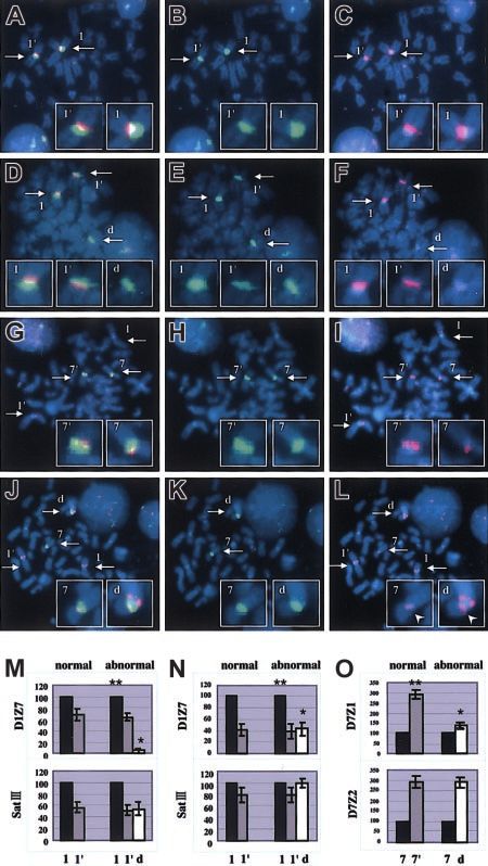
Distribution of the breakpoints within the alphoid regions
Figure 4. Breakpoint mapping in der(1;7)(q10;p10) and proposed mechanism
To investigate the characteristics of this translocation more in generating this translocation. (A) Breakpoints are widely distributed within D1Z7
detail, we focused on the residual proportions of D1Z7 and D7Z1 and D7Z1. The ends to the short arm within both alphoids (*) are free from
recombinations. A broad arrow indicates the hypothetical critical point on each
signals on the derivative chromosome, which could provide a gross alphoid. A recombination that occurs beyond this point might compromise segrega-
tion of recombined chromosomes and eventually result in their loss. Thin arrows
represent the locations of the breakpoints; yellow stars indicate the hypothetic active
centromere. (B) Three possible models for the quadriheadral formation with relation
to relative location of active centromeres to the breakpoint. Active centromeres may
be either contralaterally (Model 1), ipsilaterally (Model 2), or centrally (Model 3)
positioned. For the sake of stable maintenance of sister chromatids, the contralateral
model might be favored, and only this seems to be compatible with the real
distribution of the breakpoints and with allelic distribution in this unbalanced
translocation. (C) Proposed mechanism of generation of 46, XY (or XX), ⫹1,
der(1;7)(q10;p10). Active centromere sequences on chr1 and chr7 are indicated as
yellow and blue circles, respectively. For simplicity, some features of chromosomes
are not always as they really are. For example, sister chromatids are depicted
separately from before recombination, which should be tightly paired and stuck to
each other through cohesion molecules.
estimation for the relative locations of the breakpoints within each
cluster. As summarized in Figure 5, the relative reduction in D1Z7
and D7Z1 sequences on the derivative chromosome was different
from patient to patient. Considering each alphoid cluster is
estimated to be several megabase pairs in length, our results
suggest that the breakpoints on chr1 and chr7 are not clustered but
widely distributed within the D1Z7 and D7Z1 sequences among
these patients.
Similarity of the 2 involved alphoid subsets
Figure 3. FISH analysis with the patients using various centromeric alphoid
When hybridized to the human genomic DNA at high stringency,
probes. Interphase FISH with a D1Z5 probe, CEP1, in normal (A) and abnormal (B) D1Z7 (pE25.a) and D7Z1 (D7Z16mer) produced almost an identical
cells. Two-color FISH with pE25.a (green) and D7Z16mer (red) with tumor cells, hybridization pattern (Figure 6A). To separately evaluate the
showing almost completely overlapped signals (arrows) on metaphase (C) and
specific hybridization of each probe to different homologous
interphase (D) nuclei. Original magnification, ⫻ 1000. (E) Fiber FISH analysis of
der(1;7)(q10;p10) using a D1Z7 probe, CEP1/5 (red), and a D7Z1 probe, CEP7 alphoids on different human chromosomes, the DNAs from a series
(green), visualizing direct connection of both alphoids on the same DNA fiber. of monochromosomal human-mouse hybrid cells were examined2602 WANG et al BLOOD, 1 OCTOBER 2003 䡠 VOLUME 102, NUMBER 7
Figure 5. Relative reduction of D1Z7 and D7Z1 con-
tents on the derivative chromosome in different
samples. The remaining proportions of D1Z7 (A) and
D7Z1 (B) alphoids are depicted based on the measure-
ments of FISH signals. They show great variations from
patient to patient, indicating the wide distribution of the
breakpoints within each alphoid in der(1;7)(q10;p10).
Note that the extreme ends to the short arm within both
alphoids are retained, and therefore are devoid of break-
points. As indicated by the arrows, the 1p portion of D1Z7
and the 7q portion of D7Z1 are lost on the derivative
chromosome.
by Southern blot analysis using both probes. Under the same and the alphoid length might not be linear, it was still expected to
stringent condition, D1Z7 probe strongly hybridized to chr1 but be monotonic. Given this monotonic relationship, we could grossly
also, to a lesser extent, to chr7, creating the similar hybridization estimate the relative reduction in the length of both involved
pattern with 3 major bands of 340-bp dimer, 680-bp tetramer, and alphoid clusters. According to this estimation, the proportion of
1020-bp hexamer (Figure 6B). D7Z1 also created the similar shortening of each alphoid sequence is highly variable, indicating
hybridization pattern with stronger hybridization to chr7 than to that the breakpoints of der(1;7)(q10;p10) translocation were widely
chr1. While chr16 and chr19 contain alphoids of the same distributed within D1Z7 on chr1 and D7Z1 on chr7, although they
suprachromosomal family 1 as D1Z7 and D7Z1,20 the hybridiza- seemed to spare the extreme end to the short arm within both
tion patterns in chr16 and chr19 were substantially different. No alphoids (Figure 5). Therefore, it seems unlikely that there exists a
hybridization was detected in chr2, which has no suprachromo- unique gene target at or near those breakpoints, and the leukemo-
somal family 1 alphoid. These results indicated that D1Z7 and genic potential of this translocation may well be ascribed to altered
D7Z1 were more similar than other alphoids of suprachromosomal gene dosages resulting from trisomy 1q and/or monosomy 7q,
family 1, not only in their sequence contents but also in their well-known chromosomal abnormalities found in MDS/AML
higher-order array structures. (7q⫺) as well as many solid cancers (⫹1q), even though the critical
gene targets for these deletion and duplication remain to be unveiled.
FISH analysis in this study also provided additional information
Discussion as to the structure of the alphoids on chr1, their order being
1p-D1Z5 (smaller cluster)-D1Z7-D1Z5 (larger cluster)-1q. Of
On exploring the centromeric fusion in this unbalanced transloca- interest is that this translocation generates a derivative chromo-
tion, the conventional methods for identification of chromosomal some that contains 4 alphoid subsets at its centromeric region, 2
breakpoints, such as Southern blot analysis, were not available due from chr1 and the other 2 from chr7 (mapped as 7p-D7Z2-partial
to the highly repetitive and polymorphic features of the pericentric D7Z1-partial D1Z7-the larger cluster of D1Z5-1q). This is the first
sequences. Nevertheless, it was these features that allowed us the report in the literature that 4 alphoid subsets coexist on a single
other approach to molecularly delineate the structure of der(1;7)(q10; centromere. Further investigations will be required to understand
p10); variations of the large cluster lengths of the involved alphoid how this multi-alphoid centromere can function and be maintained
sequences can be easily detected and compared on an allelic basis in eukaryotic cells.
by measuring the intensity of polymorphic satellite signals. The With regard to the exact sequences that participate in this
reproducibility of our signal measurements was satisfactory enough centromeric fusion, our fiber FISH results strongly indicated that
to get a reliable estimation on the alterations of the signal intensity.
D1Z7 and D7Z1 were directly involved in the DNA recombination.
Although the relationship between the intensity of FISH signals
This is also supported by the fact that the centromere alphoid
clusters are highly ordered tandem arrays without interruptions by
other elements19,32,33 except at their marginal regions of the
cluster.34,35 In addition, both alphoids have extremely high struc-
tural homology, which is estimated to be about 90% at a unit
component level. Both are composed of dimer and tetramer repeat
units, each of which is defined by EcoRI sites. This extreme
similarity in their higher-order array structures was also confirmed
by our Southern blot experiment (Figure 6). These particular
similarities in the unit component as well as higher-order structures
seem to make both alphoids especially prone to be recombined to
each other.
Although the exact mechanism through which this alphoid
recombination takes place is still unclear, we may postulate that it
Figure 6. Similarity of the 2 involved alphoid subsets. Southern blot analysis of
is mediated by an error that occurred during DNA repair processes
D1Z7 (pE25.a) and D7Z1 (D7Z16mer) alphoids in total human genome (A) digested
with BamHI (b), EcoRI (e), and HindIII (h), as well as in DNAs from human because, clinically, der(1;7)(q10;p10) has been closely associated
monochromosomal mouse hybrid cells containing chromosome 1 (Neo1 and An4), 7 with secondary MDS/AML that arises after heavy doses of
(Neo7 and NTI8), 2 (Neo2), 16 (Neo16), or 19 (Neo19) digested with EcoRI (B). chemotherapy, especially of alkylating agents, the well-known
Locations of dimmer (340 bp), tetramer (680 bp), and hexamer (1020 bp) are
indicated to the left. D1Z7 and D7Z1 created a similar hybridization pattern with some antitumor drugs that cause DNA double-strand breaks (DSBs).36,37
cross hybridization with each other. As the excessive accumulation of DSBs imposed on either D1Z7 orBLOOD, 1 OCTOBER 2003 䡠 VOLUME 102, NUMBER 7 BREAKPOINT ANALYSIS OF der(1;7)(q10;p10) 2603
D7Z1, cells would recruit the repair processes, which are thought to sequence ensures the complete centromere functions including
erroneously mediate recombination between these highly homolo- kinetocore attachment, spindle formation, and successful chroma-
gous alphoid sequences. Given that the initiating DSBs occur tid separation, little has been known about the essential part of
randomly, it might be expected that the chromosome having a mammalian centromeres. In order to explain the unique karyotypic
larger centromere alphoid content would be involved more fre- feature of der(1;7)(q10;p10), we hypothesize existence of the
quently. In fact, in our case series, the der(1;7) tended to be sequences related to this active centromere function and kinetocore
originated from the homologous chromosome with a larger alphoid formation to the centromere region polarized to the short arm of
content. Participation of the chromosome allele with a larger each chromosome. There is no definite evidence for this hypothesis
amount of D1Z7 and D7Z1 was observed in 19 of 26 and 11 of 13 but there are some rationales: first of all, this gives us the simplest
samples, respectively. explanation. Once the quadriradial structure is formed, the cytoge-
From a cytogenetic point of view, der(1;7)(q10;p10) has a netic configuration of der(1;7)(q10;p10) under interest will be
consistent feature in that it contains 2 apparently normal chr1s and directly generated without involvement of any other abnormalities
only one copy of normal chr7, and our analysis has clearly in mitotic machinery. Second, our model also holds true for Pt 27,
demonstrated that the 2 apparently normal chr1s as well as the an exceptional case in terms of the location of the breakpoints,
remaining chr7 and the 7p of the derivative chromosome in the because the quadriradial chromatids (Figure 4A-B, Model 1) in Pt
tumor cells always have different allelic origins. To explain this 27 are expected to retain the whole of the D7Z1 and D7Z2 clusters
prominent cytogenetic feature we could assume that the recombina- on its 7p arm and the whole of the D1Z7 and D1Z5 clusters on its
tion occurs during or after DNA synthesis (Figure 4). In this model, 1p arm. In this case we could more safely conclude that the active
a double-stranded DNA breakage taking place on one of the centromeres are localized to the 7p and 1p arms. Finally, as shown
just-duplicated daughter chromatids of chr1 or chr7 evokes a in Figure 5, the breakpoints strangely spare the short-arm ends of
recombination repair process, during which an error occurs to both D1Z7 and D7Z1, where we presumed the hypothetical active
misconnect the D1Z7 and D7Z1 sequences and leads to the centromere elements are localized.
quadriradial formation involving the 2 pairs of daughter chromatids In conclusion, we disclosed the molecular characteristics of
(Figure 4B-C). At this stage, there may be several possibilities as to der(1;7)(q10:p10), one of the most common forms of chromosomal
the way of kinetocore formation and separation of duplicated abnormalities found in secondary MDS/AML. It directly involves
chromatids, as shown in Figure 4B. This process (mitosis) is 2 centromere alphoids of its participating chromosomes, and the
thought to involve a number of well-coordinated interactions of underlying mechanism that gives rise to this translocation seems to
DNA and proteins, and, for the time being, we could not know its be closely related to the structural similarities of both alphoids and
exact mechanism. However, the cytogenetic profile of the der(1; their association with centromere functions. It requires further
7)(q10;p10) seems to be most simply explained by Model 1 shown investigation of human centromere structure and its functions to
in Figure 4B, in which “active” or “critical” centers of kinetocore fully understand the entire pathogenesis, which may serve as
formation on the replicated chr1 and chr7 are both polarized to the prevention of this translocation with poor prognosis.
short arm of each chromosome. According to this model, the
quadriradial daughter chromatids, also previously proposed by
Morrison-DeLap et al,5 will be separated following the usual
mechanism of mitosis. Of the 2 possible ways of chromatid Acknowledgment
distributions only one will result in the unbalanced translocation
with uneven distribution of chromosomal materials, which might We are grateful to Dr Huntington F. Willard for providing us the
confer a growth advantage to the cells that inherit der(1;7)(q10; chromosome 7 alpha satellite probes that were used to confirm the
p10) (Figure 4C). Although in the budding yeast, only a 125-bp results in our paper.
References
1. Geraedts JP, den Ottolander GJ, Ploem JE, 1;7 in four cases of myeloid disorders. Cancer myelodysplastic syndromes is strongly modified
Muntinghe OG. An identical translocation be- Genet Cytogenet. 1989;38:49-52. by sex. Br J Haematol. 2001;113:347-356.
tween chromosome 1 and 7 in three patients with 7. Yokoo H, Okada Y, Tominaga K, et al. der(1;7) 12. Alitalo T, Willard HF, de la Chapelle A. Determina-
myelofibrosis and myeloid metaplasia. Br J (q10;p10) in three patients with malignant hema- tion of the breakpoints of 1;7 translocations in my-
Haematol. 1980;44:569-575. tologic disorders. Rinsho Ketsueki. 1992;33: elodysplastic syndrome by in situ hybridization
2. Pedersen-Bjergaard J, Philip P, Mortensen BT, et 1829-1833. using chromosome-specific alpha satellite DNA
al. Acute nonlymphocytic leukemia, preleukemia, 8. Pedersen-Bjergaard J, Timshel S, Andersen MK, from human chromosomes 1 and 7. Cytogenet
and acute myeloproliferative syndrome second- Andersen AS, Philip P. Cytogenetically unrelated Cell Genet. 1989;50:49-53.
ary to treatment of other malignant diseases: clones in therapy-related myelodysplasia and
clinical and cytogenetic characteristics and re- acute myeloid leukemia: experience from the 13. Kibbelaar RE, Mulder JW, van Kamp H, et al.
sults of in vitro culture of bone marrow and HLA Copenhagen series updated to 180 consecutive Nonradioactive in situ hybridisation of the translo-
typing. Blood. 1981;57:712-723. cases. Genes Chromosomes Cancer. 1998;23: cation t(1;7) in myeloid malignancies. Genes
3. Mecucci C, Ghione F, Tricot G, Van den Berghe 337-349. Chromosomes Cancer. 1992;4:128-134.
H. Combined trisomy 1q and monosomy 7q due 9. Andersen MK, Pedersen-Bjergaard J. Increased 14. Fonseca R, Rajkumar SV, Ahmann GJ, et al.
to translocation 1;7 in myelodysplastic syn- frequency of dicentric chromosomes in therapy- FISH demonstrates treatment-related chromo-
dromes. Cancer Genet Cytogenet. 1985;18:193- related MDS and AML compared to de novo dis- some damage in myeloid but not plasma cells in
197. ease is significantly related to previous treatment primary systemic amyloidosis. Leuk Lymphoma.
4. Scheres JM, Hustinx TW, Geraedts JP, Leeksma with alkylating agents and suggests a specific 2000;39:391-395.
CH, Meltzer PS. Translocation 1;7 in hematologic susceptibility to chromosome breakage at the
15. Seabright M. A rapid banding technique for hu-
disorders: a brief review of 22 cases. Cancer centromere. Leukemia. 2000;14:105-111.
man chromosomes. Lancet. 1971;2:971-972.
Genet Cytogenet. 1985;18:207-213. 10. Greenberg P, Cox C, LeBeau MM, et al. Interna-
5. Morrison-DeLap SJ, Kuffel DG, Dewald GW, Le- tional scoring system for evaluating prognosis in 16. Yang Y, Kiss H, Kost-Alimova M, et al. A 1-Mb
tendre L. Unbalanced 1;7 translocation and myelodysplastic syndromes. Blood. 1997;89: PAC contig spanning the common eliminated re-
therapy-induced hematologic disorders: a pos- 2079-2088. gion 1 (CER1) in microcell hybrid-derived SCID
sible relationship. Am J Hematol. 1986;21:39-47. tumors. Genomics. 1999;62:147-155.
11. Mauritzson N, Johansson B, Rylander L, et al.
6. Sheppard DM, Richkind KE, Bull M. Translocation The prognostic impact of karyotypic subgroups in 17. Finelli P, Antonacci R, Marzella R, Lonoce A,2604 WANG et al BLOOD, 1 OCTOBER 2003 䡠 VOLUME 102, NUMBER 7
Archidiacono N, Rocchi M. Structural organiza- Powers VE, England SB. Detection of restriction some 7, using double-labeling hybridizations in
tion of multiple alphoid subsets coexisting on hu- fragment length polymorphisms at the centro- fluorescence and electron microscopy on a mela-
man chromosomes 1, 4, 5, 7, 9, 15, 18, and 19. meres of human chromosomes by using chromo- noma cell line. Cancer Genet Cytogenet. 1997;
Genomics. 1996;38:325-330. some-specific alpha satellite DNA probes: impli- 96:17-22.
18. Tapia-Paez I, Kost-Alimova M, Hu P, et al. The cations for development of centromere-based 32. Waye JS, Durfy SJ, Pinkel D, et al. Chromosome-
position of t(11;22)(q23;q11) constitutional trans- genetic linkage maps. Proc Natl Acad Sci U S A. specific alpha satellite DNA from human chromo-
location breakpoint is conserved among its car- 1986;83:5611-5615. some 1: hierarchical structure and genomic orga-
riers. Hum Genet. 2001;109:167-177. 26. Wevrick R, Willard HF. Long-range organization nization of a polymorphic domain spanning
19. Waye JS, England SB, Willard HF. Genomic or- of tandem arrays of alpha satellite DNA at the several hundred kilobase pairs of centromeric
ganization of alpha satellite DNA on human chro- centromeres of human chromosomes: high-fre- DNA. Genomics. 1987;1:43-51.
mosome 7: evidence for two distinct alphoid do- quency array-length polymorphism and meiotic 33. Schueler MG, Higgins AW, Rudd MK, Gustashaw
mains on a single chromosome. Mol Cell Biol. stability. Proc Natl Acad Sci U S A. 1989;86:9394- K, Willard HF. Genomic and genetic definition of a
1987;7:349-356. 9398. functional human centromere. Science. 2001;
27. Alexandrov I, Kazakov A, Tumeneva I, Shepelev 294:109-115.
20. Ogawa S, Toyoshima H, Kozutsumi H, et al. The
V, Yurov Y. Alpha-satellite DNA of primates: old 34. Auriche C, Donini P, Ascenzioni F. Molecular and
C-terminal SH3 domain of the mouse c-Crk pro-
and new families. Chromosoma. 2001;110:253- cytological analysis of a 5.5 Mb minichromosome.
tein negatively regulates tyrosine-phosphoryla-
266. EMBO Rep. 2001;2:102-107.
tion of Crk associated p130 in rat 3Y1 cells. On-
cogene. 1994;9:1669-1678. 28. Cooke HJ, Hindley J. Cloning of human satellite 35. Wevrick R, Willard VP, Willard HF. Structure of
III DNA: different components are on different DNA near long tandem arrays of alpha satellite
21. Lee C, Wevrick R, Fisher RB, Ferguson-Smith
chromosomes. Nucleic Acids Res. 1979;6:3177- DNA at the centromere of human chromosome 7.
MA, Lin CC. Human centromeric DNAs. Hum
3197. Genomics. 1992;14:912-923.
Genet. 1997;100:291-304.
29. Tagarro I, Fernandez-Peralta AM, Gonzalez- 36. Pedersen-Bjergaard J, Philip P, Larsen SO, et al.
22. Willard HF. Centromeres: the missing link in the
Aguilera JJ. Chromosomal localization of human Therapy-related myelodysplasia and acute my-
development of human artificial chromosomes.
satellites 2 and 3 by a FISH method using oligo- eloid leukemia: cytogenetic characteristics of 115
Curr Opin Genet Dev. 1998;8:219-225.
nucleotides as probes. Hum Genet. 1994;93:383- consecutive cases and risk in seven cohorts of
23. Jorgensen AL. Alphoid repetitive DNA in human 388. patients treated intensively for malignant dis-
chromosomes. Dan Med Bull. 1997;44:522-534. 30. Wevrick R, Willard HF. Physical map of the cen- eases in the Copenhagen series. Leukemia.
24. Carine K, Jacquemin-Sablon A, Waltzer E, Mas- tromeric region of human chromosome 7: rela- 1993;7:1975-1986.
carello J, Scheffler IE. Molecular characterization tionship between two distinct alpha satellite ar- 37. Pedersen-Bjergaard J, Pedersen M, Roulston D,
of human minichromosomes with centromere rays. Nucleic Acids Res. 1991;19:2295-2301. Philip P. Different genetic pathways in leukemo-
from chromosome 1 in human-hamster hybrid 31. Fetni R, Richer CL, Malfoy B, Dutrillaux B, Le- genesis for patients presenting with therapy-re-
cells. Somat Cell Mol Genet. 1989;15:445-460. mieux N. Cytologic characterization of two distinct lated myelodysplasia and therapy-related acute
25. Willard HF, Waye JS, Skolnick MH, Schwartz CE, alpha satellite DNA domains on human chromo- myeloid leukemia. Blood. 1995;86:3542-3552.You can also read