Multimodal magnetic resonance imaging analysis in the characteristics of Wilson's disease: A case report and literature review - De Gruyter
←
→
Page content transcription
If your browser does not render page correctly, please read the page content below
Open Life Sciences 2021; 16: 793–799
Case Report
Yun Wang#, Zejin Jia#, Yuelei Lyu#, Qian Dong, Shujuan Li, Wenli Hu*
Multimodal magnetic resonance imaging
analysis in the characteristics of Wilson’s
disease: A case report and literature review
https://doi.org/10.1515/biol-2021-0071 Keywords: multimodal MRI, Wilson’s disease, copper,
received July 17, 2020; accepted June 11, 2021 susceptibility-weighted imaging, arterial spin labeling
Abstract: Wilson’s disease (WD) is an inherited disorder
of copper metabolism. Multimodal magnetic resonance
imaging (MRI) has been reported to provide evidence of
the extent and severity of brain lesions. However, there 1 Introduction
are few studies related to the diagnosis of WD with multi-
modal MRI. Here, we reported a WD patient who was Wilson’s disease (WD) is an autosomal recessive inherited
subjected to Sanger sequencing, conventional MRI, and disorder of copper metabolism, with a lifetime prevalence of
multimodal MRI examinations, including susceptibility- 20–100% [1]. Adolescents and adults with WD may develop
weighted imaging (SWI) and arterial spin labeling (ASL). neurological and psychiatric diseases, including movement
Sanger sequencing demonstrated two pathogenic muta- disorders (Parkinson, ataxia, and dystonia), cognitive impair-
tions in exon 8 of the ATP7B gene. Slit-lamp examination ment, depression, psychosis, and schizophrenia, which
revealed the presence of Kayser–Fleischer rings in both may be due to the different locations and concentrations
eyes, as well as low serum ceruloplasmin and high 24-h of copper ions in various organs, resulting in excess copper
urinary copper excretion on admission. Although the accumulation in the brain, liver, kidneys, and cornea [2,3].
substantia nigra, red nucleus, and lenticular nucleus Grover et al. have found that a young WD patient developed
on T1-weighted imaging and T2-weighted imaging were psychotic symptoms characterized by irritability, delusion
normal, SWI and ASL showed hypointensities in these of persecution, and decreased sleep [1]. WD is potentially
regions. Besides, decreased cerebral blood flow was found curable, suggesting immediate diagnostic evaluation and
in the lenticular nucleus and the head of caudate nucleus. early treatment initiation of the disease [4].
The patient recovered well after 1 year and 9 months of Brain magnetic resonance imaging (MRI) is a crucial
follow-up, with only a Unified Wilson Disease Rating Scale tool that can provide evidence of the morphological char-
score of 1 for neurological symptom. Brain multimodal MRI acteristics and functional changes of brain lesions, and
provided a thorough insight into the WD, which might make abnormalities in brain MRI are present in more than 90%
up for the deficiency of conventional MRI. of neurological WD patients [5]. Multimodal MRI techni-
ques, such as susceptibility-weighted imaging (SWI),
arterial spin labeling (ASL), magnetic resonance spectro-
scopy (MRS), resting-state functional MRI, and diffusion
# These authors contributed equally to the work.
tensor imaging (DTI), have been widely used for the cli-
nical diagnosis of cancers, cerebral infarction, and neural
degenerative diseases [6–8]. The MRI features of untreated
* Corresponding author: Wenli Hu, Department of Neurology,
Beijing Chao-Yang Hospital, Capital Medical University, No. 8 WD cases are central pontine myelinolysis-like abnorm-
Gongtinan Road, Chaoyang District, Beijing 100020, China, ality, tectal plate hyperintensity, giant panda face, and
e-mail: doctorbjcy@hotmail.com synchronous signal changes in thalamus, brain stem,
Yun Wang, Zejin Jia, Qian Dong, Shujuan Li: Department of and basal ganglia [9]. Previous research has pointed out
Neurology, Beijing Chao-Yang Hospital, Capital Medical University,
that the damage to thalamus in WD patients can be
No. 8 Gongtinan Road, Chaoyang District, Beijing 100020, China
Yuelei Lyu: Department of Imaging, Beijing Chao-Yang Hospital,
detected using DTI prior to the abnormal signals on con-
Capital Medical University, No. 8 Gongtinan Road, Chaoyang ventional MRI [10,11]. However, the applications of multi-
District, Beijing 100020, China modal MRI in WD are rarely studied.
Open Access. © 2021 Yun Wang et al., published by De Gruyter. This work is licensed under the Creative Commons Attribution 4.0
International License.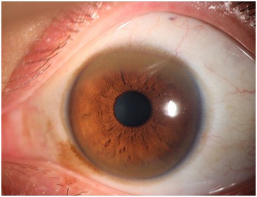
794 Yun Wang et al.
In this report, we described the case of a 26-year-old exon 8 of ATP7B gene, namely c.2333G>T (Arg778Leu) and
WD man who underwent brain multimodal MRI and c.2294A>G (Asp765Gly), of which the former mutation was
reviewed the relevant literature with respect to multi- inherited from his mother and the latter was from his
modal MRI in WD patients. father. Overall, the patient was diagnosed with WD based
on a Leipzig score of 12, with 2 scores for K–F rings, 2
scores for ceruloplasmin, 2 scores for severe neuropsychia-
tric symptoms, and 4 scores for disease-causing mutation
2 Case presentation (two chromosomes). Moreover, he was examined using the
Unified Wilson Disease Rating Scale (UWDRS), and the
A 26-year-old man, presented to the hospital with a mood results showed that his total score was 110 on admission,
of gloom and wretchedness, has been diagnosed with including neurological score of 91, psychiatric score of 19,
depression and treated with antidepressants. It should and liver score of 0.
be noted that this patient had no history of liver disease MRI observations of the WD case were recorded via
or mental illness. After 1 month, the patient developed MRI semiquantitative scale. Brain MRI showed abnormal
slow movement, slurred speech, and hand tremors, and findings and was characterized by evidence of atrophy
even occasionally felt irritable. After 4 months, he was re- and signal intensity changes, with a high MRI score of
admitted to the hospital due to stiffness of extremities 11 (Table 1). The assessment of T2-weighted imaging-fluid
that caused difficulty in ambulation and tremors, with attenuated inversion recovery (T2WI-FLAIR)/SWI signal
worsening slurred speech. On admission, the neuro- intensity changes was performed subjectively, while the
logical examination revealed that the patient had diffi- degree of atrophy was visually assessed based on the
culty in speaking, slight weakness (left lower limb), sulcal and ventricular enlargement. Conventional brain
increased muscle tone (trunk and extremities), tremors MRI (General Electric Company, USA) results showed
(head and extremities), limb ataxia (left), hypoalgesia symmetrical hyperintensity in the midbrain and cerebral
(left lower limb), and positive Babinski sign (left). Slit- peduncle and hypointensity in the substantia nigra and
lamp examination showed the presence of Kayser–Fleischer red nucleus on T2WI-FLAIR (Figure 2a–c), hyperintense
(K–F) rings in both eyes (zigzag score: 2) (Figure 1). Besides, in the pons on T2WI-FLAIR (Figure 2d), and hypointen-
his serum ceruloplasmin was 4.6 mg/dL and the 24-h sity in the midbrain on T1-weighted imaging (T1WI)
urinary copper excretion was 204 μg/24 h. The blood cell (Figure 2e). SWI showed marked hypointensity in the
count, serum aspartate aminotransferase level, alanine substantia nigra, red nucleus, and lenticular nucleus
aminotransferase level, and creatinine of the patient were (Figure 3a and b). ASL-MRI showed a slight decrease
all normal. in cerebral blood flow (CBF) to the lenticular nucleus, with
Subsequently, the patient was subjected to genetic test. the left and right putamen being 40.4 mL/100 g/min and
Sanger sequencing revealed two heterozygous mutations in 38.5 mL/100 g/min, respectively (Figure 3c and d). The
lenticular nucleus had no obvious abnormality on T1WI
and T2WI (Figure 3e and f) (Table 1). The MRS changes of
the patient in the lenticular nucleus and midbrain were
unremarkable. A contrast-enhanced MRI of the abdomen
revealed splenomegaly.
The patient was administered intravenously with
dimercaptopropane sulfonate in the hospital. After dis-
charge, the UWDRS score of the patient was 53 consisting
of neurological score of 47, psychiatric score of 6, and
liver score of 0. He continued oral treatment with a com-
bination of D-penicillamine and zinc sulfate for long-term
therapy and was restricted to a copper diet. After 3 months of
therapy, the symptoms of speech difficulty, tremors, and
stiffening of extremities remarkably improved, but the MRI
remained unchanged. After 1 year and 9 months of follow-
up, except for slow speech speed and slightly unclear speech,
the patient recovered well and had returned to work, with
Figure 1: K–F rings in the patient (left eye). only a UWDRS score of 1 for neurological symptom.The diagnosis of neurologic Wilson’s disease 795
Table 1: Brain magnetic resonance imaging severity scale for Wilson disease
Parameters Grade
Caudate nucleus Right 1
Putamen SWI hypointensity 1
Internal capsule Normal 0
Thalamus Normal 0
Midbrain Atrophy + T2 hyperintensity + SWI hypointensity (substantia nigra and red nucleus) and giant 3
panda face
Pons Atrophy + central pontine myelinosis-like changes 3
Medulla obligation Atrophy 1
Cerebellum Atrophy 1
White matter Normal 0
Cortex Atrophy 1
Total MRI score (0–30) — 11
SWI: susceptibility-weighted imaging. The anatomic distribution of abnormalities was noted and severity was graded based on the changes
in signal intensity of focal lesions and associated atrophy: 0 = no abnormality, 1 = change in signal intensity with no atrophy or atrophy
without signal change, 2 = change in signal intensity with mild or moderate atrophy, and 3 = change in signal intensity with severe atrophy.
The grading system provided a score of 0–30, with 0 being the normal scan and 30 indicating the most severe changes.
Informed consent: Informed consent has been obtained Ethical approval: The research related to human use has
from all individuals included in this study. been complied with all the relevant national regulations,
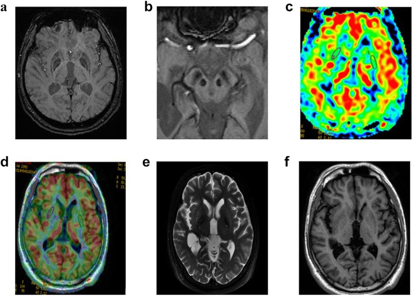
Figure 2: Conventional brain MRI of WD. Lesions were hyperintense in the midbrain and cerebral peduncle and hypointense in the
substantial nigra and red nucleus on (a) axial and (b and c) coronal T2WI-FLAIR, (d) hyperintense in the pons on T2WI-FLAIR, and (e)
hypointensity in the midbrain on axial T1WI.796 Yun Wang et al.
Figure 3: SWI and ASL-MRI of WD. (a and b) SWI showed decreased signal intensities in putamen, globus pallidus, substantia nigra, and red
nucleus. (c and d) ASL-MRI demonstrated the reduction of cerebral blood flow in the bilateral putamen. (e) T1WI, and (f) T2WI showed
substantially normal in the putamen, globus pallidus, substantial nigra, and red nucleus.
institutional policies, and in accordance with the tenets study, accompanied with low serum ceruloplasmin and
of the Helsinki Declaration, and has been approved by high urine copper level. Moreover, two heterozygous muta-
the Ethics Committee of the Beijing Chao-Yang Hospital, tions in exon 8 of ATP7B gene were also found in the
Capital Medical University. patient, namely c.2333G>T (Arg778Leu) and c.2294A>G
(Asp765Gly). Genetic testing is also a typical diagnostic
technique, whereas its clinical application is limited by
high costs and a large number of mutations [14].
3 Discussion Studies have reported that MRI is of great significance
for the clinical observation and prognosis evaluation
WD is an autosomal recessive disorder of copper metabo- of WD patients presenting with neurological symptoms
lism caused by ATP7B gene mutation [12]. According to [10]. Nevertheless, the conventional MRI had a certain
the criterion of the European Commission on Public omission diagnostic rate [15]. Multimodal MRI refers to
Health and Institute of Human Genetics, WD is considered the combination of conventional MR scanning sequences
a rare disease with a prevalence of 0.0018–0.003%, sug- and multiple functional MRI techniques, thereby achiev-
gesting the importance of intervention for WD patients ing the complementary functions of multiple scanning
[13]. The traditional WD testing depends on the assessment sequences, and provides more detailed information for the
of urine and liver copper levels, K–F ring, ceruloplasmin, diagnosis of diseases [16]. Although multimodal MRI has
and liver-related histological changes [9]. Likewise, K–F not been used as a diagnostic criterion for the WD due to
rings were noticed in both eyes of the patient in this its late emergence, evidence indicates that it exerts higherThe diagnosis of neurologic Wilson’s disease 797
sensitivity and specificity in the detection of WD when com- aspartate/creatine in MRS between WD patients and con-
pared with conventional MRI [6,8]. In this study, multi- trol groups, while no abnormalities were noticed in brain
modal MRI techniques, including ASL and SWI, were used MRI, indicating that MRS can assist MRI in the assess-
to describe the characteristics of neurologic WD. ment of WD [6]. In the current research, no significant
SWI takes advantage of differences in magnetic sus- change was observed in the lenticular nucleus and mid-
ceptibility between tissues to show deposition of para- brain of the WD patient.
magnetic material and is particularly sensitive to iron At present, the main available drugs for WD treatment
[17]. An increasing number of studies have pointed out include zinc salts and copper chelators (D-penicillamine,
a complex relationship between copper and iron metabo- trientine, dimercaptopropane sulfonate, and dimercapto-
lisms in WD [18,19]. The deletion of the coding cerulo- succinic acid) [28]. As the first orally administered che-
plasmin gene can cause large amounts of iron deposition lating agent, D-penicillamine is effective for WD. Both
in the liver and brain [20]. Several researchers have found D-penicillamine and zinc sulfate are the first choice for
that the liver biopsies of WD patients after long-term the diagnosis and treatment of WD in China [29]. However,
decoppering therapy showed a significant reduction in up to 20% of WD patients have reported paradoxical wor-
copper and an increase in iron, suggesting that iron over- sening of neurological symptoms in the early stage of
load might be associated with aceruloplasminemia [21,22]. therapy, which might be associated with D-penicillamine
Yang et al. have claimed that paramagnetic mineralization administration [30]. Thus, the international WD diagnostic
deposition exists in the brain gray nuclei of WD patients, guidelines recommend trientine for patients intolerant to
and SWI is an effective approach to assess these structures, D-penicillamine [31], but trientine has not been used clini-
suggesting that SWI could be used as a potential bio- cally in China. In addition, dimercaptopropane sulfonate
marker for WD diagnosis [23]. The SWI results of the combined with zinc has been proved to be an optimal
case showed marked dark-signal intensities in the sub- therapeutic approach for neurological WD [32]. In this
stantia nigra, red nucleus, and lenticular nucleus despite research, the patient was given dimercaptopropane sulfo-
normal T1 and T2 signals, indicating abnormal paramag- nate intravenously during hospitalization and received
netic substance deposition in his brain. long-term therapy of D-penicillamine and zinc sulfate after
ASL perfusion MRI sequences can be adopted for discharge. In China, additional dimercaptosuccinic acid is
MRI-based CBF quantification without the requirement needed for WD patients with D-penicillamine allergy or
for contrast administration [24]. Ishida et al. found dif- intolerance. Our patient had no intolerance or allergy after
fuse cerebral perfusion reduction including basal ganglia taking D-penicillamine orally, so dimercaptosuccinic acid
in WD patients via single-photon emission computed was not added.
tomography [25]. In addition, the association between
CBF and functional connectivity strength in WD patients
was significantly reduced in the basal ganglia and cerebellum
and slightly increased in the prefrontal cortex and thalamus 4 Conclusion
compared with healthy controls. These findings suggested
that aberrant coupling between resting-state CBF and func- In this study, substantia nigra, red nucleus, and lenti-
tional connectivity may be a potential neural mechanism cular nucleus are the most involved areas, and the sig-
underlying the pathophysiology of WD [26]. Furthermore, nals of SWI and ASL decreased in these lesions despite
the decrease in CBF in basal ganglia may be the result of T1 and T2 signals are normal, suggesting that SWI and
neuronal loss due to copper deposition in WD. The ASL result ASL may be the most sensitive sequence for neurologic
of the patient showed slightly decreased CBF in the lenticular WD. This is the first article to retrospect multimodal MRI
nucleus and the head of the caudate nucleus. features in the diagnosis of WD, illustrating the imaging
In contrast to MRI, MRS can be used to assess the characteristics of WD and enriching our knowledge with
concentration of different metabolites in tissues and to the brain multimodal MRI results. In summary, brain
monitor the neurochemistry of the brain. It has been sug- multimodal MRI may make up for the shortcomings of
gested that copper-induced cell injury results in reduced conventional MRI, and that it is useful for the diagnosis
N-alanine aspartate/creatine ratio in WD patients, which of WD. More cases with brain multimodal MRI are required
may be partially reversed after chelation treatment [27]. to replicate these findings.
A study conducted by Alkhalik Basha et al. found
that there were significant differences in the mean values Funding information: This work was funded by the National
of N-alanine aspartate, choline, creatine, and N-alanine Natural Science Foundation of China (No. 81271309).798 Yun Wang et al.
Author contributions: Study concept and initial design: Wilson’s disease with diffusion tensor imaging. Neural Plast.
Y.W. and Z.J.J. Study design and statistical analysis: Y.W., 2017;2017:7323121.
Y.L.L., and Q.D. Acquisition of data and data analysis and [11] Li G, Zhou X, Xu P, Pan X, Chen Y. Microstructure assessment
of the thalamus in Wilson’s disease using diffusion tensor
interpretation: S.J.L. and W.L.H. All authors read and
imaging. Clin Radiol. 2014;69:294–8.
approved the final manuscript. The authors applied the [12] Treepongkaruna S, Pienvichit P, Phuapradit P, Kodcharin P,
SDC approach for the sequence of authors. Wattanasirichaigoon D. Mutations of ATP7B gene in two Thai
siblings with Wilson disease. Asian Biomed. 2018;4:163–9.
Conflict of interest: The authors state no conflict of [13] Lalioti V, Tsubota A, Sandoval IV. Disorders in hepatic copper
secretion: Wilson’s disease and pleomorphic syndromes.
interest.
Sem Liver Dis. 2017;37:175–88.
[14] Weitzman E, Pappo O, Weiss P, Frydman M, Haviv-Yadid Y, Ben
Data availability statement: The datasets generated during Ari Z. Late onset fulminant Wilson’s disease: a case report and
and/or analyzed during the current study are available review of the literature. World J Gastroenterol.
from the corresponding author upon reasonable request. 2014;20:17656–60.
[15] Hegde S, Sinha S, Rao SL, Taly AB, Vasudev MK. Cognitive
profile and structural findings in Wilson’s disease: a neuro-
psychological and MRI-based study. Neurol India.
2010;58:708–13.
[16] Soltaninejad M, Yang G, Lambrou T, Allinson N, Ye X.
References Supervised learning based multimodal MRI brain tumour
segmentation using texture features from supervoxels.
[1] Grover S, Sarkar S, Jhanda S, Chawla Y. Psychosis in an ado- Comput Methods Prog Biomed. 2018;157:69–84.
lescent with Wilson’s disease: a case report and review of the [17] Lopatina A, Ropele S, Sibgatulin R, Reichenbach JR, Güllmar D.
literature. Indian J Psychiatry. 2014;56:395. Investigation of deep-learning-driven identification of multiple
[2] Lu CX, Qing L, Huang WQ, Tzeng CM. New mutations and sclerosis patients based on susceptibility-weighted images
polymorphisms of the ATP7B gene in sporadic Wilson disease. using relevance analysis. Front Neurosci. 2020;14:609468.
Eur J Med Genet. 2014;57:498–502. [18] Gromadzka G, Wierzbicka D, Litwin T, Przybyłkowski A. Iron
[3] Antczak-Kowalska M, Członkowska A, Litwin T, Nehring P, metabolism is disturbed and anti-copper treatment improves
Niewada M, Przybyłkowski A. Gastropathy in patients with but does not normalize iron metabolism in Wilson’s disease.
Wilson disease. Scand J Gastroenterol. 2020;55:14–7. Biometals. 2021;34:407–14.
[4] Porlas RV, de Castillo LL, Dioquino CP. Neurologic Wilson [19] Tatsumi Y, Kato A, Kato K, Hayashi H. The interactions between
disease: case series on a diagnostic and therapeutic emer- iron and copper in genetic iron overload syndromes and pri-
gency. Dialogues Clin Neurosci. 2018;20:341–5. mary copper toxicoses in Japan. Hepatol Res. 2018;48:679–91.
[5] Dezortova M, Lescinskij A, Dusek P, Herynek V, Acosta- [20] Ayton S, Lei P, Adlard PA, Volitakis I, Cherny RA, Bush AI, et al.
Cabronero J, Bruha R, et al. Multiparametric quantitative brain Iron accumulation confers neurotoxicity to a vulnerable
MRI in neurological and hepatic forms of Wilson’s disease. population of nigral neurons: implications for Parkinson’s
J Magn Reson Imaging. 2020;51:1829–35. disease. Mol Neurodegen. 2014;9:27.
[6] Alkhalik Basha MA, Refaat R, Ahmed AF, Yousef HY, [21] Wang B, Wang XP. Does ceruloplasmin defend against neuro-
Alsowey AM, Metwally MI, et al. Brain magnetic resonance degenerative diseases? Curr Neuropharmacol.
spectroscopy (MRS) as a diagnostic tool for detecting early 2019;17:539–49.
neurological changes in children with Wilson’s disease. [22] Dubbioso R, Ruggiero L, Esposito M, Tarantino P, De Angelis M,
Eur J Radiol. 2019;111:41–6. Aruta F, et al. Different cortical excitability profiles in heredi-
[7] Puig J, Ellis MJ, Kornelsen J, Figley TD, Figley CR, Daunis IEP, tary brain iron and copper accumulation. Neurol Sci.
et al. Magnetic resonance imaging biomarkers of brain 2020;41:679–85.
connectivity in predicting outcome after mild traumatic [23] Yang J, Li X, Yang R, Yu X, Yu C, Qian Y, et al. Susceptibility-
brain injury: a systematic review. J Neurotrauma. weighted imaging manifestations in the brain of Wilson’s
2020;37:1761–76. disease patients. PLoS One. 2015;10:e0125100.
[8] Zhong W, Huang Z, Tang X. A study of brain MRI characteristics [24] Telischak NA, Detre JA, Zaharchuk G. Arterial spin labeling MRI:
and clinical features in 76 cases of Wilson’s disease. J Clin clinical applications in the brain. J Magn Reson Imaging.
Neurosci. 2019;59:167–74. 2015;41:1165–80.
[9] Nagral A, Sarma MS, Matthai J, Kukkle PL, Devarbhavi H, [25] Ishida S, Doi Y, Yamane K, Sugino M, Kimura F, Hanafusa T,
Sinha S, et al. Wilson’s disease: clinical practice guidelines of et al. Resolution of cranial MRI and SPECT abnormalities in a
the Indian national association for study of the liver, the Indian patient with Wilson’s disease following oral zinc monotherapy.
society of pediatric gastroenterology, hepatology and nutri- Intern Med (Tokyo, Jpn). 2012;51:1759–63.
tion, and the movement disorders society of India. J Clin Exp [26] Hu S, Wu H, Xu C, Wang A, Wang Y, Shen T, et al. Aberrant
Hepatol. 2019;9:74–98. coupling between resting-state cerebral blood flow and func-
[10] Wang A, Wu H, Xu C, Tang L, Lee J, Wang M, et al. Study on tional connectivity in Wilson’s disease. Front Neural Circuits.
lesion assessment of cerebello-thalamo-cortical network in 2019;13:25.The diagnosis of neurologic Wilson’s disease 799
[27] Pulai S, Biswas A, Roy A, Guin DS, Pandit A, Gangopadhyay G, [30] Mohr I, Weiss KH. Current anti-copper therapies in manage-
et al. Clinical features, MRI brain, and MRS abnormalities of ment of Wilson disease. Ann Transl Med. 2019;7:S69.
drug-naïve neurologic Wilson’s disease. Neurol India. [31] Zhou X, Xiao X, Li XH, Qin HL, Pu XY, Chen DB, et al. A study of
2014;62:153–8. susceptibility-weighted imaging in patients with Wilson dis-
[28] Członkowska A, Litwin T. Wilson disease – currently used ease during the treatment of metal chelator. J Neurol.
anticopper therapy. Handb Clin Neurol. 2017;142:181–91. 2020;267:1643–50.
[29] Neurogenetics Group of Neurology Society of Chinese Medical [32] Zhang J, Xiao L, Yang W. Combined sodium
Association. Chinese guidelines for the diagnosis and treat- Dimercaptopropanesulfonate and zinc versus d-penicillamine
ment of hepatolenticular degeneration 2021. Chin J Neurol. as first-line therapy for neurological Wilson’s disease.
2021;54:310–9. BMC Neurol. 2020;20:255.You can also read


