Nanoporous gold nanoleaf as tunable metamaterial - Nature
←
→
Page content transcription
If your browser does not render page correctly, please read the page content below
www.nature.com/scientificreports
OPEN Nanoporous gold nanoleaf
as tunable metamaterial
Sangeeta Rout1, Zhen Qi2, Monika M. Biener2, Devon Courtwright1, Jakeem C. Adrien1,
Ezekiel Mills3, Mohammad Shahabuddin1, Natalia Noginova1 & Mikhail A. Noginov1*
We have studied optical properties of single-layer and multi-fold nanoporous gold leaf (NPGL)
metamaterials and observed highly unusual transmission spectra composed of two well-resolved
peaks. We explain this phenomenon in terms of a surface plasmon absorption band positioned on the
top of a broader transmission band, the latter being characteristic of both homogeneous “solid” and
inhomogeneous “diluted” Au films. The transmission spectra of NPGL metamaterials were shown to
be controlled by external dielectric environments, e.g. water and applied voltage in an electrochemical
cell. This paves the road to numerous functionalities of the studied tunable and active metamaterials,
including control of spontaneous emission, energy transfer and many others.
Complex metal-dielectric environments, including metamaterials1–3, plasmonic structures4–6, waveguides7–9 and
cavities10–12, have been demonstrated to control scores of physical phenomena including s pontaneous13–22 and
stimulated23–27 emission, Förster energy transfer11,28,29, van der Waals interactions30,31, and chemical reactions32–37.
Of particular interest are tunable and active metamaterials allowing one to manipulate light and other physical
processes in real t ime38–40.
Transmission is one of the most important characteristics of optical materials. An extraordinary large
transmission41–43, facilitated by plasmons, has been observed in arrays of subwavelength holes in metallic films.
Other nanostructured composite metal/dielectric materials with remarkable properties include, among others,
arrays of metallic nanowires grown in channels of porous alumina membranes44,45, nanoporous Au foams46,47,
gyroidal structures48, etc.
At this time, we report studies and control of optical properties of nanoporous gold leafs (NPGLs)—
unique inch-size single-layer and multi-fold (meta)materials, whose thickness can be varied between ~ 0.1
and ~ 1 µm. Composite materials with nominally similar composition and morphology have been studied in
the literature46,49–53. In particular, it has been shown that their optical responses can be tuned by external stimuli
and environments49,50, enabling multiple applications of active and tunable metamaterials. At the same time, the
optical properties of NPGLs reported by different research g roups49,50,52,54 were quite different from each other
and the intriguing spectra of these porous materials, featuring e.g. the double-headed transmission band, were
never clearly understood. This motivated our studies of NPGLs reported below.
Experimental samples
The NPGL samples have been prepared from one layer of 12 K “white gold” leaf (50 wt.% Au and 50 wt.% Ag
alloy, from Gold Leaf Company) through dealloying in 10 ml of concentrated H NO3 (68%, ACS, VWR) solution
at room temperature for one hour. After complete removal of Ag, the volume filling factor of Au was 35 vol.%.
The area of the nanoporous Au leafs was of the order of several centimeters squared.
The scanning electron microscope (SEM) image of the single-layer Au nanoleaf sample is depicted in Fig. 1.
The dealloying parameters for all samples in this work were nominally the same, resulting in ligaments’ diameters
in different samples studied (determined using the Image J software) to range from 20 ± 3 nm to 27 ± 3 nm. The
NPG leafs were rinsed thoroughly in deionized (DI) water for at least three times after the dealloying process
before being transferred onto microscope glass slides. Different thicknesses of the fabricated samples, ranging
from 84 to 943 nm, were achieved by using a single NPGL layer or folding the gold leaf once (two layers), twice
(four layers), and three times (eight layers). The film’s thickness was measured using the stylus profilometer
(DekTak XT form Bruker). The fabricated samples were dried in air before taking any measurements.
1
Center for Materials Research, Norfolk State University, Norfolk, VA 23504, USA. 2Lawrence Livermore
National Laboratory, Livermore, CA 94550, USA. 3Virginia State University, Petersburg, VA 23806, USA. *email:
mnoginov@nsu.edu
Scientific Reports | (2021) 11:1795 | https://doi.org/10.1038/s41598-021-81128-4 1
Vol.:(0123456789)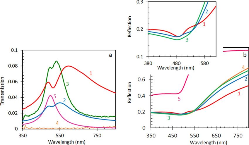
www.nature.com/scientificreports/
Figure 1. Scanning electron microscope (SEM) image of the single layer nanoporous gold leaf sample.
Figure 2. Transmission (a) and reflection (b) spectra of a single-layer (trace 1), two-layers (trace 2), four-layers
(multiplied by 100, trace 3) and eight-layers (multiplied by 100, trace 4) NPGLs. Control sample: 90 nm Au film
(trace 5). Inset: zoomed part of (b).
Results and discussion
Transmission and reflection. The results of the transmission and reflection measurements of the single-
layer and multi-fold Au nanoleafs studied (taken using the Lambda 900 spectrophotometer from PerkinElmer)
are summarized in Fig. 2a, b. The reference sample, 90 nm thick homogeneous gold film deposited on glass
(using the thermal vapor deposition apparatus, Nano 36 from Kurt J Lesker), has the transmission maximum
at 505 nm, where the real part of the dielectric permittivity of Au, ε′, approaches the epsilon-near-zero (ENZ)
range55,56. At the same time, Au nanoleaf samples have two transmission peaks. The first one has its maximum
almost at the same wavelength as the transmission peak in a thermally deposited smooth and homogeneous Au
film, while the second one is shifted to longer wavelengths (up to 605 nm), Fig. 2a. The characteristic “wiggles”
could be seen in the reflection spectra of the nanoleaf samples at the wavelengths corresponding to both trans-
mission maxima, Fig. 2b.
Scientific Reports | (2021) 11:1795 | https://doi.org/10.1038/s41598-021-81128-4 2
Vol:.(1234567890)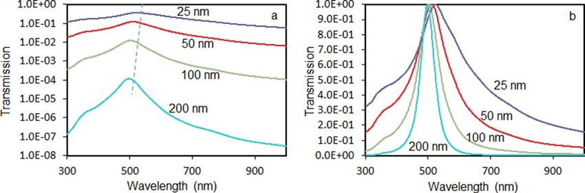
www.nature.com/scientificreports/
Figure 3. (a) Wavelength positions of the maxima of the transmission peaks; red squares—calculation done
(using the online solver57) for homogeneous Au films; black circles—experimental measurements in porous Au
nanoleafs. (b) Red circles: Transmission as the function of the thickness of the NPGL sample, experimentally
measured in the maximum at λ ~ 590 nm. Open blue squares: Transmission as the function of the thickness
of the homogeneous Au film (characterized by the dielectric permittivities)55,56, calculated57 at the spectral
position of the long-wavelength transmission maximum. Solid blue diamonds: Transmission as the function
of the thickness of the homogeneous Au film (characterized by the dielectric permittivities)55,56, calculated57 at
λ = 498 nm. Green triangle: transmission measured in the maximum (at λ = 505 nm) in the homogeneous 90 nm
Au film.
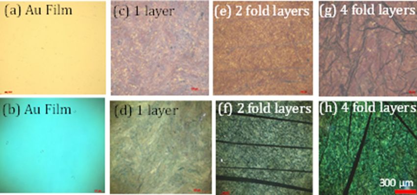
Figure 4. Microscopic images of the smooth 90 nm Au film (a,b), single-layer Au nanoleaf (c,d), two-fold Au
nanoleaf (e,f), and four-fold Au nanoleaf (g,h) taken in the transmission (b,d,f,h) and reflection (a,c,e,g) modes
of the optical microscope.
Note that a qualitatively similar double-peak transmission spectra were obtained when Au films were depos-
ited onto nanoporous anodic alumina membranes (pore diameter ~ 30 nm, Au film thickness 54 nm, membrane
thickness ~ 50 µm, commercially acquired from Redox Inc.), Supplementary Information Fig. SI1. However, this
phenomenon was not universal, and the transmission spectra of Au films deposited onto polymeric (PMMA)
films with high concentration of microscopic air bubbles did not have two maxima in their transmission spectra
(to be published elsewhere).
With increase of the number of layers and the overall thickness of the porous Au nanoleaf samples, the width
of the long-wavelength transmission peak got reduced and its spectral position shifted to shorter wavelengths,
Fig. 3a. The spectral shifts of the short-wavelength transmission peak and the dip between the two peaks were less
significant. The transmission (measured in the maximum of the long-wavelength peak at λ ~ 590 nm) decreased
with the increase of the sample’s thickness d almost exponentially, with the characteristic penetration depth of
l = 53 nm, Fig. 3b.
The transmission and reflection spectra depicted in Fig. 2a, b are in a good agreement with the color images
of the same NPGL samples taken using the AxioVision 4 optical microscope (from Carl Zeiss) operating in the
transmission and reflection regimes, Fig. 4. In fact, the light transmitted by the homogeneous thermally depos-
ited Au film and four-fold Au nanoleaf (featuring short-wavelength transmission peaks) has a greenish-bluish
Scientific Reports | (2021) 11:1795 | https://doi.org/10.1038/s41598-021-81128-4 3
Vol.:(0123456789)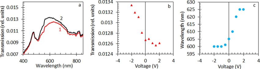
www.nature.com/scientificreports/
Figure 5. Transmission spectra of the Au nanoleaf in air (trace 1) and in water (trace 2).
Figure 6. (a) Transmission spectra of Au nanoleaf in a two-electrode electrochemical cell at positive voltage
(+ 1.5 V, trace 1) and negative voltage (− 2 V, trace 2) applied to the NPGL working electrode. (b) Transmission
peak intensity as a function of the applied voltage. (c) The wavelength of the transmission peak as a function of
the applied voltage.
color, while the light transmitted by the single-layer Au nanoleaf (characterized by a relatively long-wavelength
transmission) is more yellowish. Dark lines, which are particularly pronounced in Fig. 4f–h are wrinkles in
multi-fold samples.
Effect of the dielectric environment. Our next particular experiment was aimed at control of the optical
properties of NPGLs with external dielectric environments. When Au nanoleaf was placed in water, its long-
wavelength absorption peak moved slightly (by ~ 10 nm) to shorter wavelengths, while the short-wavelength
peak and the dip between the two peaks practically did not change their spectral positions, Fig. 5. This result
is in a qualitative agreement with the literature50. However, the spectral shift in our studies is smaller than that
reported by Lang et. al.50 On the other hand, the two transmission peaks in Ref.50 could be observed only when
the NPGL samples were immersed in a liquid, and only one transmission peak was seen in a dry sample, without
any liquid. The effect of water on the transmission of NPGLs can be explained by change in the effective medi-
um’s dielectric permittivity and/or spectral shift of surface plasmon resonances (supported by Au ligaments) in
the presence of water.
Control of transmission with applied voltage. To tune the optical properties of NPGLs in real time,
we placed the sample in the two-electrode electrochemical cell. The NPGL sample, with attached copper tape
conduit, served as a working electrode, while a platinum wire played the role of a counter electrode. Following
Ref.49, the electrolyte was 0.7 M solution of NaF in water. The applied voltage varied between − 2 V and + 2 V
(applied in a sequence 0 V, + 0.5 V, − 0.5 V, + 1 V, − 1 V, …), caused changes in both the strength (~ 10%) and the
wavelength position (Δλ ~ 20 nm) of the long-wavelength transmission peak, Fig. 6a–c. No permanent damage
occurred to the sample until + 2.5 V, after which (at − 2.5 V) the Au nanoleaf got damaged and detached from the
glass substrate. The voltage-induced changes in the optical spectra were, presumably, due to change in the car-
Scientific Reports | (2021) 11:1795 | https://doi.org/10.1038/s41598-021-81128-4 4
Vol:.(1234567890)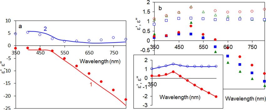
www.nature.com/scientificreports/
Figure 7. (a) Spectra of the real (red trace 1 and solid circles) and imaginary (blue trace 2 and open circles)
parts of the dielectric permittivity of Au. Solid line—the data from Refs.55,56, characters—the data derived from
the experiment. (b) Spectra of real (closed characters) and imaginary (open characters) parts of dielectric
permittivity of a single-layer Au nanoleaf (164 nm, red circles), two-fold Au nanoleaf (230 nm, blue squares) and
four-fold Au nanoleaf (404 nm, green triangles). Inset: average of the three spectra of dielectric permittivities
depicted in the main frame; red closed circles—real parts of dielectric permittivity ε′, blue open circles—
imaginary parts of dielectric permittivity ε″.
Figure 8. Normal-incidence transmission spectra of Au films of different thickness, computed using the online
solver57, for the dielectric permittivities of gold reported in Refs.55,56, (a) semi-logarithmic vertical scale, dashed
line indicates the shift of the transmission maxima; (b)—linear vertical scale, emission maxima are normalized
to unity.
riers’ density49, which, in turn, affected the effective medium’s dielectric permittivity and plasmonic resonances
of Au ligaments. Note that the largest spectral changes in our experiment were observed when negative (rather
than positive)49 voltage was applied to the gold leaf. This is not the same as (although not in direct contradiction
with) the result of Ref.49, where the strongest changes occurred in the transmission spectra, when the applied
potential (measured relative to the Ag/AgCl pseudo-reference electrode) was positive. This intriguing phenom-
enon requires more studies, which results will be published elsewhere.
Familiar and unfamiliar gold. Before trying to understand the rather unusual double-headed transmis-
sion spectra of NPGLs, let us review a seemingly familiar transmission spectrum of a smooth and homogeneous
Au film deposited on glass. We assumed gold to have the dielectric permittivities similar to those of Refs.55,56
(Fig. 7a) and calculated, using the online solver57, the series of normal-incidence transmission spectra for Au
films, whose thicknesses ranged between 25 and 200 nm, Fig. 8a,b. As one can see, the transmission spectra
of homogeneous gold films are characterized by a single peak, whose magnitude (Figs. 3b and 8a) and width
(Fig. 8b) are getting smaller and the spectral position shifts to shorter wavelengths (Figs. 3a and 8a) with increase
of the film’s thickness. Therefore, the only important difference between the transmission spectra of homogene-
ous thermally deposited Au films and porous Au nanoleafs is the number of peaks. Otherwise, the behavior of
the λ ~ 590 nm transmission peak observed in NPGL is qualitatively similar to that of the transmission peak in
a homogeneous Au film.
In the homogeneous gold film, the thickness dependence of the maximal transmission intensity (and nearly
similar to it transmission measured at λ = 498 nm) is nearly exponential, with the penetration depth equal to
22 nm, Fig. 3b. (The green triangle in Fig. 3b is the experimental point.) Besides the number of the transmission
Scientific Reports | (2021) 11:1795 | https://doi.org/10.1038/s41598-021-81128-4 5
Vol.:(0123456789)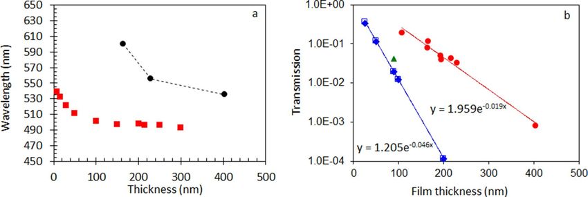
www.nature.com/scientificreports/
Figure 9. Spectra of dielectric permittivities used in the toy model; 1a and 1b—Drude terms (Eq. 1); 2a and
2b Drude and single Lorenzian; 3a and 3b—Drude and two Lorentzians. 1a, 2a, and 3a—real parts of dielectric
permittivities; 1b, 2b, and 3b—imaginary parts of dielectric permittivities.
peaks (one or two), this behavior is in a qualitative agreement with that of NPGLs, Fig. 3b. The fact that the
sample’s extinction is fairly described by an exponential Beer’s law and, at the same time, the extinction spectral
maximum changes with an increase of the film’s thickness, is counterintuitive. It can be explained by the fact that
the extinction, which is measured in the experiment, is determined not only by the sample’s absorption, but also
by the strong reflection, which does not follow the Beer’s law.
Dielectric permittivities of porous Au nanoleafs. The reflection R (λ) and transmission T(λ) spectra
of three-layered sandwich-like structures can be calculated using the analytical formulas derived in Ref.5 and
knowing the complex dielectric permittivities (ε′ + i ε″) of all three constituent media: ε′, ε″ → R, T. Solving
(numerically) the inverse problem, one can derive the spectra of real and imaginary parts of dielectric permit-
tivities, ε′ (λ) and ε″ (λ), if R(λ) and T(λ) are known: R, T → ε′, ε″. Note that this point-by-point calculation does
not depend on any knowledge or assumptions regarding the relationships between the dielectric permittivities at
different wavelengths and/or Drude versus Lorentz nature of the spectra. When we applied this technique to the
90 nm homogeneous Au film deposited on glass, we obtained a good agreement between the derived dielectric
permittivities and those of Refs.55,56, compare characters and solid lines in Fig. 7a. The minor mismatch between
the latter two datasets can have many possible reasons; one of them is that our Au films were not exactly the same
as the gold surfaces studied in reference55.
We further applied the same extraction method to a single-layer (164 nm), two-fold (230 nm), and four-fold
(404 nm) porous Au nanoleafs and obtained the spectra of dielectric permittivities, which could be described as
the spectra of “diluted” gold, compare Fig. 7a,b. Expectedly, the spectra of ε′ and ε″ practically did not depend on
the number of layers or the overall thickness of the NPGL samples. The spectra of the real and imaginary parts
of dielectric permittivity, averaged over three samples studied, are depicted in the inset of Fig. 7b. Note that the
derived spectra of ε′ and ε″ are in a good agreement with those of uncoated and not annealed thick (~ 200 µm)
Au nanofoam samples, fabricated using a similar de-alloying technique47.
The toy model of NPGL. As we have discussed above, many optical properties of NPGLs are qualitatively
similar to those of homogeneous Au films. Thus, with increase of the film’s thickness, the transmission peak nar-
rows and shifts to shorter wavelengths, its intensity decreases nearly exponentially, and the spectra of dielectric
permittivities of NPGLs resemble those of “diluted” gold. The only vast difference between the transmission
spectra of homogeneous and porous Au films is that the former has one peak while the latter has two. Although
surface plasmons have been named in the literature as a plausible reason for the double-peaked transmission
spectra of NPGLs49,50,52, the underlining physics of this intriguing phenomenon has never been clearly explained.
Below, we introduce a simple toy model qualitatively explaining the double-headed transmission spectra of
porous Au nanoleafs. We start with the Drude model for Au (first two terms on the right-hand side of Eq. 1),
2
ωDp L1 L2
ε = εb + + + 2 (1)
−ω2 − iωγD ωL21 0 2
− ω − iωγL1 ωL2 0 − ω2 − iωγL2
where εb = 9 is the bulk dielectric permittivity of Au, ω is the angular frequency, ωDp = 1.37 × 10¹⁶ rad/s is the
Drude plasma frequency (determined by the concentration of free electrons), and γD = 1 × 10¹⁴ rad/s is the free
electrons’ dissipation rate58. The corresponding spectra of real and imaginary parts of dielectric permittivity are
shown in Fig. 9 traces 1a and 1b). The real part of the dielectric permittivity ε′ crosses zero at λ = 415 nm, caus-
ing a very large reflection and, correspondingly, very small transmission at longer wavelength, Fig. 10 (trace
1). At the same time, both absorption and reflection are small at shorter wavelengths, resulting in a very broad
and strong transmission band, which does not resemble the transmission spectra of neither homogeneous nor
nanoporous Au films.
Scientific Reports | (2021) 11:1795 | https://doi.org/10.1038/s41598-021-81128-4 6
Vol:.(1234567890)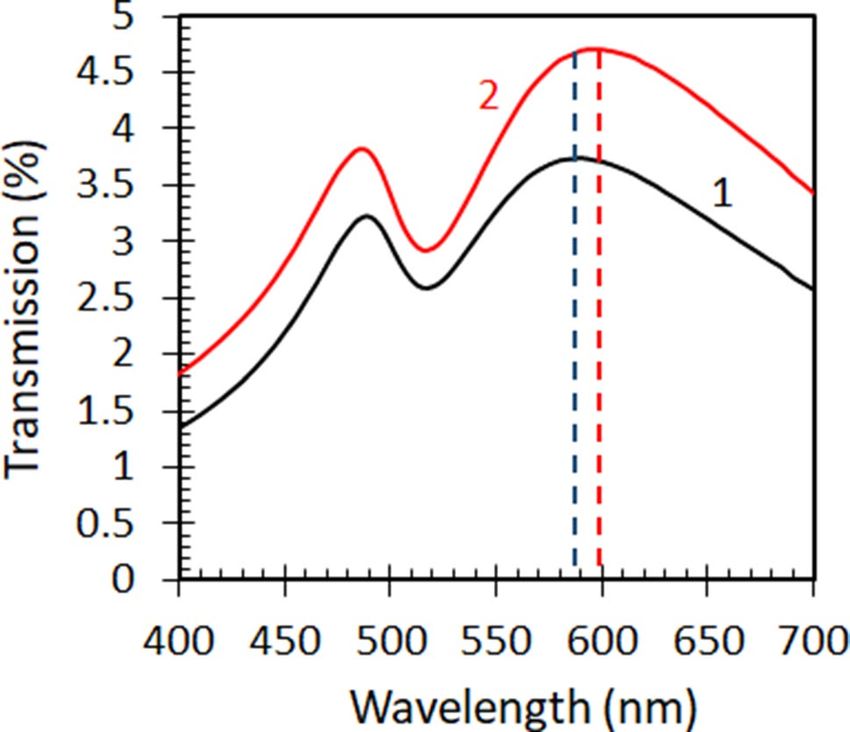
www.nature.com/scientificreports/
Figure 10. Transmission spectra calculated using the dielectric permittivities determined by the Drude model
(trace 1), Drude + single Lorenzian model (trace 2), and Drude + two Lorenzians model (trace 3).
The latter shortcoming of the Drude model (caused by neglecting bound state electron transitions, which are
particularly strong in the blue-to-ultraviolet range of the spectrum) is well known in the literature. In order to
overcome it, we added to the model the Lorenzian band (third term in the right-hand side of Eq. 1) simulating
the bound state transitions, with the maximum at λ = 377 nm, Fig. 9 (traces 2a and 2b). (Here ωL21 0 is the natural
frequency of oscillation, γL1 is the dissipation rate, and L1 is the amplitude factor determined by the concentration
of the oscillating electrons and the oscillator strengths.) Now the strong Lorentzian absorption band restricts the
transmission at short wavelengths, while the strong reflection at λ ≥ 415 nm still limits it at long wavelengths.
The narrow spectral window determined by these two boundaries constitutes the (single-headed) transmission
peak observed in homogeneous Au films, Fig. 10 (trace 2).
In order to explain the origin of the two maxima in the transmission spectra of porous Au nanoleafs, let us
recall that gold nanospheres and nanorods support localized surface plasmon resonances in vicinity of 500 nm49,
and gold ligaments composing NPGLs should not be an exception. Therefore, in accord with Ref.49, a surface plas-
mon absorption band is expected in the spectral range of the gold’s transmission window. If the latter absorption
band is strong enough and narrow enough, it can create a dip in the middle of a broader transmission spectrum,
causing the double-headed transmission band observed it in the spectra of Au nanoleafs.
In order to illustrate this, we added to Eq. (1) the second Lorenzian band with the maximum at 480 nm, the
fourth term in the right-hand side (the notations are the same as above). This allowed us to model the spectrum
of dielectric permittivities Fig. 9 (traces 3a and 3b), which resulted in the dip in the middle of the broader trans-
mission band, Fig. 10 (trace 3). This shape, a broad band with a dip in the middle, can be alternatively described
as a double-headed transmission peak. (Note that the width of the second Lorenzian band in the model was
commeasurable with that determined experimentally.52) Although the Lorenzian shape of the latter surface
plasmon absorption band is not well justified, the maximum at λ ~ 500 nm can be seen in the experimental
spectra of dielectric permittivities of NPGLs, see inset of Fig. 7b. (We infer that almost any bell-shape absorption
band can be used in the calculations for illustration purposes.) Although the toy model above is not rigorous
or comprehensive, it qualitatively interprets the highly unusual double-headed transmission band of NPGLs as
one broad transmission peak with the dip in the middle.
Summary
To summarize, we have studied optical properties of single and multi-fold nanoporous gold leafs (NPGLs) of
different thickness and found that with increase of the leaf ’s thickness, the transmission spectrum narrows and
shifts to shorter wavelengths and its intensity decreases nearly exponentially—the behavior, which is quali-
tatively similar to that of homogeneous gold films. Furthermore, the dispersion of dielectric permittivities of
NPGLs resembles that of “diluted” gold. The vast difference between the transmission spectra of homogeneous
and porous Au films is that the former has one maximum while the latter has two. We explain this intriguing
phenomenon in terms of a relatively narrow surface plasmon absorption band positioned on top of the broader
transmission band. We further demonstrated that the optical properties of NPGLs can be controlled by applied
voltage in an electrochemical cell (presumably due to change in the electron d ensity49) and by change of the
ater50. Although we were able to qualitatively explain
dielectric environment, e.g., by immersing Au nanoleafs in w
the major experimental results, the underlying fundamental physics and applications of the transmission, reflec-
tion, dispersion, and emission (not discussed here) of NPGLs require further studies to be published elsewhere.
Received: 12 October 2020; Accepted: 4 January 2021
References
1. Engheta, N. & Ziolkowski, R. W. Metamaterials: Physics and Engineering Explorations (Wiley, New York, 2006).
Scientific Reports | (2021) 11:1795 | https://doi.org/10.1038/s41598-021-81128-4 7
Vol.:(0123456789)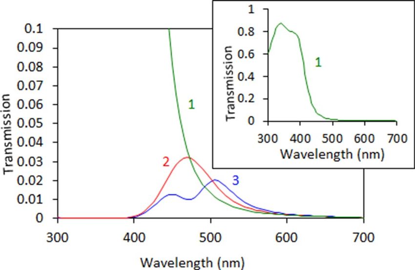
www.nature.com/scientificreports/

                                             2.   Noginov, M. A. & Podolskiy, V. A. Tutorials in Metamaterials (CRC Press, Boca Raton, 2011).
                                             3.   Cai, W. & Shalaev, V. M. Optical Metamaterials Vol. 10 (Springer, New York, 2010).
                                             4.   Maier, S. A. Plasmonics: Fundamentals & Applications (Springer, New York, 2007).
                                             5.   Raether, H. Springer Tracts in Modern Physics Vol. 111, 1–133 (Springer, New York, 1988).
                                             6.   Atwater, H. A. The promise of plasmonics. Sci. Am. 296, 56–63 (2007).
                                             7.   Bozhevolnyi, S. I., Volkov, V. S., Devaux, E., Laluet, J. Y. & Ebbesen, T. W. Channel plasmon subwavelength waveguide components
                                                  including interferometers and ring resonators. Nature 440, 508–511 (2006).
                                             8.   Jun, Y. C., Kekatpure, R. D., White, J. S. & Brongersma, M. L. Nonresonant enhancement of spontaneous emission in metal-
                                                  dielectric-metal plasmon waveguide structures. Phys. Rev. B. 78, 153111 (2008).
                                             9.   Wen, J., Romanov, S. & Peschel, U. Excitation of plasmonic gap waveguides by nanoantennas. Opt. Express. 17, 5925–5932 (2009).
                                            10.   Santhosh, K., Bitton, O., Chuntonov, L. & Haran, G. Vacuum Rabi splitting in a plasmonic cavity at the single quantum emitter
                                                  limit. Nat. Commun. 7, 1–5 (2016).
                                            11.   Andrew, P. & Barnes, W. L. Förster energy transfer in an optical microcavity. Science 290, 785–788 (2000).
                                            12.   Ebbesen, T. W. Hybrid light–matter states in a molecular and material science perspective. Acc. Chem. Res. 49, 2403–2412 (2016).
                                            13.   Krishnamoorthy, H. N., Jacob, Z., Narimanov, E., Kretzschmar, I., Menon, V. M Metamaterial based broadband engineering of
                                                  quantum dot spontaneous emission. arXiv preprint arXiv. 0912, 2454 (2009).
                                            14.   Noginov, M. A. et al. Controlling spontaneous emission with metamaterials. Opt. Lett. 35, 1863–1865 (2010).
                                            15.   Jacob, Z. et al. Engineering photonic density of states using metamaterials. Appl. Phys. B. 100, 215–218 (2010).
                                            16.   Tumkur, T. et al. Control of spontaneous emission in a volume of functionalized hyperbolic metamaterial. Appl. Phys. Lett. 99,
                                                  151115 (2011).
                                            17.   Jacob, Z., Smolyaninov, I. I. & Narimanov, E. E. Broadband Purcell effect: Radiative decay engineering with metamaterials. Appl.
                                                  Phys. Lett. 100, 181105 (2012).
                                            18.   Krishnamoorthy, H. N., Jacob, Z., Narimanov, E., Kretzschmar, I. & Menon, V. M. Topological transitions in metamaterials. Science
                                                  336, 205–209 (2012).
                                            19.   Poddubny, A. N., Belov, P. A. & Kivshar, Y. S. Spontaneous radiation of a finite-size dipole emitter in hyperbolic media. Phys. Rev.
                                                  A. 84, 023807 (2011).
                                            20.   Galfsky, T. et al. Active hyperbolic metamaterials: enhanced spontaneous emission and light extraction. Optica 2, 62–65 (2015).
                                            21.   Lu, D., Kan, J. J., Fullerton, E. E. & Liu, Z. Enhancing spontaneous emission rates of molecules using nanopatterned multilayer
                                                  hyperbolic metamaterials. Nat. Nanotechnol. 9, 48 (2014).
                                            22.   Gu, L., Tumkur, T. U., Zhu, G. & Noginov, M. A. Blue shift of spontaneous emission in hyperbolic metamaterial. Sci. Rep. 4, 4969
                                                  (2014).
                                            23.   Sudarkin, A. N. & Demkovich, P. A. Excitation of surface electromagnetic waves on the boundary of a metal with an amplifying
                                                  medium. Žurnal Tehn. Fiziki. 59, 86–90 (1989).
                                            24.   Seidel, J., Grafström, S. & Eng, L. Stimulated emission of surface plasmons at the interface between a silver film and an optically
                                                  pumped dye solution. Phys. Rev. Lett. 94, 177401 (2005).
                                            25.   Noginov, M. A. et al. Stimulated emission of surface plasmon polaritons. Phys. Rev. Lett. 101, 226806 (2008).
                                            26.   Kitur, J. K., Podolskiy, V. A. & Noginov, M. A. Stimulated emission of surface plasmon polaritons in a microcylinder cavity. Phys.
                                                  Rev. Lett. 106, 183903 (2011).
                                            27.   Kitur, J. K., Gu, L., Tumkur, T., Bonner, C. & Noginov, M. A. Stimulated emission of surface plasmons on top of metamaterials
                                                  with hyperbolic dispersion. ACS Photonics. 2, 1019–1024 (2015).
                                            28.   Blum, C. et al. Nanophotonic control of the Förster resonance energy transfer efficiency. Phys. Rev. Lett. 109, 203601 (2012).
                                            29.   Tumkur, T. U. et al. Control of Förster energy transfer in the vicinity of metallic surfaces and hyperbolic metamaterials. Faraday
                                                  Discuss. 178, 395–412 (2015).
                                            30.   Rodriguez, A. W., Capasso, F. & Johnson, S. G. The Casimir effect in microstructured geometries. Nat. Photonics 5, 211 (2011).
                                            31.   Noginov, M. A. et al. Long-range wetting transparency on top of layered metal-dielectric substrates. Sci. Rep. 6, 27834 (2016).
                                            32.   Schwartz, T., Hutchison, J. A., Genet, C. & Ebbesen, T. W. Reversible switching of ultra-strong light-molecule coupling. Phys. Rev.
                                                  Lett. 106, 196405 (2011).
                                            33.   Hutchison, J. A., Schwartz, T., Genet, C., Devaux, E. & Ebbesen, T. W. Modifying chemical landscapes by coupling to vacuum
                                                  fields. Angew. Chem. Int. Ed. 51, 1592–1596 (2012).
                                            34.   Peters, V. N., Tumkur, T. U., Zhu, G. & Noginov, M. A. Control of a chemical reaction (photodegradation of the p3ht polymer)
                                                  with nonlocal dielectric environments. Sci. Rep. 5, 14620 (2015).
                                            35.   Peters, V. N. et al. Effect of strong coupling on photodegradation of the semiconducting polymer P3HT. Optica 6, 318–325 (2019).
                                            36.   Lee, K. J. et al. Charge-transfer dynamics and nonlocal dielectric permittivity tuned with metamaterial structures as solvent ana-
                                                  logues. Nat. Mater. 16, 722–729 (2017).
                                            37.   Peters, V. N., Yang, C., Prayakarao, S. & Noginov, M. A. Effect of metal–dielectric substrates on chemiluminescence kinetics. J.
                                                  Opt. Soc. Am. B. 36, E132–E138 (2019).
                                            38.   Boardman, A. D. et al. Active and tunable metamaterials. Laser Photonics Rev. 5, 287–307 (2011).
                                            39.   Dicken, M. J. et al. Frequency tunable near-infrared metamaterials based on VO2 phase transition. Opt. Express 17, 18330–18339
                                                  (2009).
                                            40.   Driscoll, T. et al. Memory metamaterials. Science 325, 1518–1521 (2009).
                                            41.   Ebbesen, T. W., Lezec, H. J., Ghaemi, H. F., Thio, T. & Wolff, P. A. Extraordinary optical transmission through sub-wavelength hole
                                                  arrays. Nature 391, 667–669 (1998).
                                            42.   Bonod, N., Enoch, S., Li, L., Popov, E. & Nevière, M. Resonant optical transmission through thin metallic films with and without
                                                  holes. Opt. Express. 11, 482–490 (2003).
                                            43.   Liu, H. & Lalanne, P. Microscopic theory of the extraordinary optical transmission. Nature 452, 728–731 (2008).
                                            44.   Kabashin, A. V. et al. Resonances in complementary metamaterials and nanoapertures. Nat. Mater. 8, 867–871 (2009).
                                            45.   Lee, S. J. & Moskovits, M. Remote sensing by plasmonic transport. J. Am. Chem. Soc. 134, 11384–11387 (2012).
                                            46.   Biener, J. et al. Nanoporous plasmonic metamaterials. Adv. Mater. 20, 1211–1217 (2008).
                                            47.   Asane, J. K., Qi, Z., Biener, M. M., Liberman, V. & Noginov, M. A. Optical properties of nanoporous gold foams. AIP Adv. 8, 095302
                                                  (2018).
                                            48.   Hur, K. et al. Three-dimensionally isotropic negative refractive index materials from block copolymer self-assembled chiral gyroid
                                                  networks. Angew. Chem. Int. Ed. 50, 11985–11989 (2011).
                                            49.   Jalas, D. et al. Electrochemical tuning of the optical properties of nanoporous gold. Sci. Rep. 7(1), 1–8 (2017).
                                            50.   Lang, X., Qian, L., Guan, P., Zi, J. & Chen, M. Localized surface plasmon resonance of nanoporous gold. Appl. Phys. Lett. 98, 093701
                                                  (2011).
                                            51.   Dixon, M. C. et al. Preparation, structure, and optical properties of nanoporous gold thin films. Langmuir 23, 2414–2422 (2007).
                                            52.   Yu, F., Ahl, S., Caminade, A. M., Majoral, J. P. & Knoll, W. Erlebacher, Simultaneous excitation of propagating and localized surface
                                                  plasmon resonance in nanoporous gold membranes. J. Anal. Chem. 78, 7346–7350 (2006).
                                            53.   Qian, L. H. & Chen, M. W. Ultrafine nanoporous gold by low-temperature dealloying and kinetics of nanopore formation. Appl.
                                                  Phys. Lett. 91, 083105 (2007).

          Scientific Reports |   (2021) 11:1795 |                     https://doi.org/10.1038/s41598-021-81128-4                                                                     8

Vol:.(1234567890)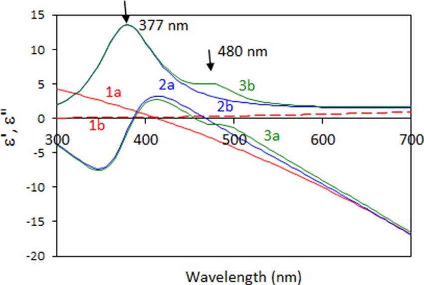
www.nature.com/scientificreports/
54. Chen, H. A., Long, J. L., Lin, Y. H., Weng, C. J. & Lin, H. N. Plasmonic properties of a nanoporous gold film investigated by far-field
and near-field optical techniques. J. Appl. Phys. 110, 054302 (2011).
55. Johnson, P. B. & Christy, R. W. Optical constants of the noble metals. Phys. Rev. B. 6, 4370 (1972).
56. Ni, X., Liu, Z., Kildishev A. V. Photonics DB: Optical Constants (2018). https://nanohub.org/resources/photonicsdb. https://doi.
org/10.4231/D3D795D1M.
57. Ishii, S., Chettiar, U. K., Ni, X., Kildishev A. V. PhotonicsRT: Wave Propagation in Multilayer Structures (2014). https://nanoh
ub.org/resources/photonicsrt. https://doi.org/10.4231/D3MK6588C.
58. Shalaev, V. M. et al. Negative index of refraction in optical metamaterials. Opt. Lett. 30, 3356–3358 (2005).
Acknowledgements
This work was supported by the NSF EiR Grants 1830886 and 1856515, DoD Grant W911NF1810472, and
AFOSR Grant FA9550-18-1-0417. Work at LLNL was performed under the auspices of the U.S. Department of
Energy by LLNL under Contract DE-AC52-07NA27344.
Author contributions
S.R. performed the experiment, analyzed the data, and participated in writing the manuscript. Z.Q. and M.B.
fabricated the gold nanoleaf samples and contributed to the scientific discussions. D.C., J.A. and E.M. contributed
to data analysis. M.S. and N.N. did experiment and data acquisition. M.N. designed the experiment, participated
in the experimental studies, and took part in writing the manuscript.
Competing interests
The authors declare no competing interests.
Additional information
Supplementary Information The online version contains supplementary material available at https://doi.
org/10.1038/s41598-021-81128-4.
Correspondence and requests for materials should be addressed to M.A.N.
Reprints and permissions information is available at www.nature.com/reprints.
Publisher’s note Springer Nature remains neutral with regard to jurisdictional claims in published maps and
institutional affiliations.
Open Access This article is licensed under a Creative Commons Attribution 4.0 International
License, which permits use, sharing, adaptation, distribution and reproduction in any medium or
format, as long as you give appropriate credit to the original author(s) and the source, provide a link to the
Creative Commons licence, and indicate if changes were made. The images or other third party material in this
article are included in the article’s Creative Commons licence, unless indicated otherwise in a credit line to the
material. If material is not included in the article’s Creative Commons licence and your intended use is not
permitted by statutory regulation or exceeds the permitted use, you will need to obtain permission directly from
the copyright holder. To view a copy of this licence, visit http://creativecommons.org/licenses/by/4.0/.
© The Author(s) 2021
Scientific Reports | (2021) 11:1795 | https://doi.org/10.1038/s41598-021-81128-4 9
Vol.:(0123456789)You can also read