Neuropathology of the Common Forms of Dementia - BINASSS
←
→
Page content transcription
If your browser does not render page correctly, please read the page content below
N e u ro p a t h o l o g y o f t h e
C o mmon F o r ms o f D eme nti a
                          a,b,                                     a,b,c
Rupal I. Mehta,      MD          *, Julie A. Schneider,      MD, MS

 KEYWORDS
  Cardiovascular disease  Cerebrovascular disease  Dementia  Mixed dementia
  Neurodegenerative disease  Resilience  Resistance  Proteinopathy

 KEY POINTS
  Dementia is a nonspecific term that corresponds to a group of heterogeneous, mostly
   age-related neurologic disorders that cause impaired cognition and deterioration of brain
   functions.
  This neurologic condition is a manifestation of various processes that primarily or second-
   arily affect the brain organ.
  Underlying neuropathologic substrates encompass various protein inclusions, vascular
   brain injuries, and cerebrovascular, traumatic, and metabolic brain changes.
  Protein inclusions and metabolic abnormalities accumulate in distinct stereotyped brain
   regions, whereas trauma and vascular-associated substrates may occur in patchy and/
   or irregular distribution(s).
  The burden and location(s) of brain lesions influence the type and degree of clinical symp-
   toms, which establish the dementia syndrome.

INTRODUCTION

Dementia is an umbrella clinical term that refers to a range of debilitating neurologic
conditions, and its incidence is increasing. Alzheimer disease (AD) dementia is the
most common form of dementia.1 However, a variety of neuropathological lesions
are found in persons who succumb with dementia, including AD dementia.2 Due to
the chronic nature of dementing diseases and the range of potential causes and brain
lesions, accurate diagnosis of dementia subtypes requires targeted postmortem brain
dissection with comprehensive histologic evaluation and incorporation of ancillary
testing. However, knowledge on complex causes for dementias also remains

 a
   Rush Alzheimer’s Disease Center, Rush University Medical Center, Chicago, IL 60612, USA;
 b
   Department of Pathology, Rush University Medical Center, 1750 West Harrison Street, Chi-
 cago, IL 60612, USA; c Department of Neurological Sciences, Rush University Medical Center,
 Chicago, IL 60612, USA
 * Corresponding author. Department of Pathology, Rush University, Alzheimer’s Disease Center,
 1750 West Harrison Street, Chicago, IL 60612.
 E-mail address: rupal_mehta@rush.edu

 Clin Geriatr Med 39 (2023) 91–107
 https://doi.org/10.1016/j.cger.2022.07.005                                            geriatric.theclinics.com
 0749-0690/23/ª 2022 Elsevier Inc. All rights reserved.

           Descargado para Irene Ramírez (iramirez@binasss.sa.cr) en National Library of Health and Social
          Security de ClinicalKey.es por Elsevier en enero 26, 2023. Para uso personal exclusivamente. No se
          permiten otros usos sin autorización. Copyright ©2023. Elsevier Inc. Todos los derechos reservados.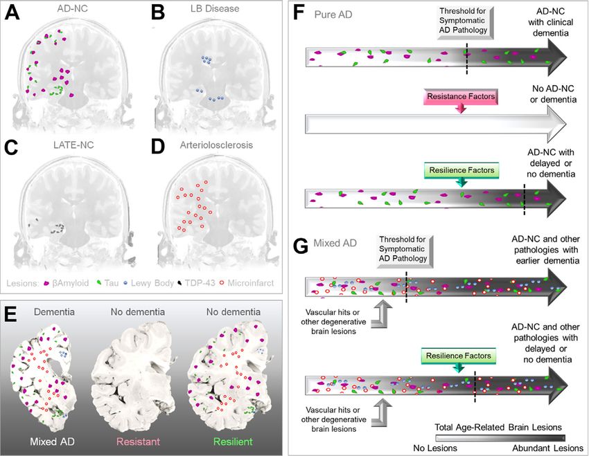
92   Mehta & Schneider

     incomplete and, over the years, neurobiological and epidemiologic evidence have
     revised criteria used for neuropathologic diagnosis, scoring, and staging schemes.
     Similarly, neuropathological studies continue to inform on the significance of epidemi-
     ologic data and new forms of disease. Therefore, classification of this group of dis-
     eases is evolving.
        A century of research has shown that various misfolded protein species accumulate
     in different brain regions and account for distinct proteinopathies that involve different
     susceptible cell types1 Here, we explore the range of lesions that are documented
     within brains of persons who have died with a clinical diagnosis of dementia and sum-
     marize the causes, classification, topographies, and staging of these lesions. Although
     AD pathologic condition is the most common pathologic condition encountered over-
     all, persons with AD type dementia (eg, amnestic dementia) may harbor multiple brain
     changes including various vascular pathologies and other non-AD neuropathologies,
     in addition to typical AD lesions. Other common forms of neurodegenerative diseases,
     such as those characterized by Lewy bodies and transactive response DNA-binding
     protein 43 kD (TDP-43) pathologic condition, as well as less common processes
     such as Creutzfeldt-Jacob disease and chronic traumatic encephalopathy (CTE) are
     described briefly. We also summarize nonproteinopathic diseases and mixed lesions
     that are linked to dementia syndromes. These other brain “hits” are recognized to
     lower the threshold for dementia in proteinopathic diseases.3 Although summarizing
     current knowledge on dementia pathologies, this review also highlights knowledge
     gaps and emerging concepts pertaining to cognitive reserve.

     PROTEINOPATHIC DISEASES

     Proteinopathic diseases, also known as proteopathies or proteinopathies, are a het-
     erogeneous group of protein misfolding disorders that manifest grossly as brain atro-
     phy and histologically as accumulation of aberrant deposits within cortical or
     subcortical brain regions. These deposits selectively damage vulnerable neurons
     and/or glial cell populations in distinct brain regions, disturbing neural pathways and
     thereby altering brain structure and functions. Multiple mechanisms are involved,
     including neuronal and glial cell death pathways, organelle damage, and secondary
     physiologic disturbances including reactive astrogliosis and microgliosis.4 Neuropath-
     ologic lesions accumulate chronically over time, thereby precipitating dementia syn-
     dromes. Primary diseases are listed in Table 1 (upper panel) and are summarized
     below and in Fig. 1A, B, D.

     Alzheimer Disease
     Microscopically, AD neuropathologic change (AD-NC) is defined by the intraparenchy-
     mal accrual of 2 abnormal proteins, amyloid-beta (bA) in the form of extracellular pla-
     ques and irregularly phosphorylated forms of the microtubule-associated protein tau
     (MAPT), in the form of intraneuronal neurofibrillary tangles (NFTs).5,6 On routine stains,
     these protein aggregates may seem as atypical eosinophilic inclusions within brain
     gray matter (for bA), or as irregular or flame-shaped intraneuronal inclusions (for
     NFTs; see Fig. 1A). These neuropathologic markers may be imperceptible on routine
     brain examination but are easily visualized on silver stains (eg, Bielschowsky, Gallyas,
     and Bodian) and/or via immunohistochemistry panels that incorporate primary anti-
     bodies targeted toward the aberrant bA or tau protein (eg, thioflavin S, AT8, Tau;
     see Fig. 1A). The abundance of these aggregates (especially tau protein) generally
     corresponds with the severity of cognitive impairment because they are related to var-
     iable degree of cortical brain atrophy, particularly in vulnerable medial-temporal lobe

           Descargado para Irene Ramírez (iramirez@binasss.sa.cr) en National Library of Health and Social
          Security de ClinicalKey.es por Elsevier en enero 26, 2023. Para uso personal exclusivamente. No se
          permiten otros usos sin autorización. Copyright ©2023. Elsevier Inc. Todos los derechos reservados.
Neuropathology of Dementia 93
Table 1
Summary of common diseases that lead to neurodegenerative lesions in persons with
dementia
Etiologic
Disease Inclusion(s) Primary Brain Regions(s) Involved
Proteinopathic Diseases Associated with Chronic Cell Loss
AD-NC b-Amyloid Cerebral cortex
Tau(3R and 4R) Cerebral cortex
LB Disease a-Synuclein Brainstem and limbic region,
including substantia nigra
LATE-NC TDP-43 Hippocampus and temporal lobe
FTLD-Tau Tau Frontal and temporal lobes
FTLD-TDP TDP-43 Frontal and temporal lobes
FTLD-Atypical Ubiquitin, FUSb Frontal and temporal lobes
PSP Tau(4R) Subcortical nuclei and brainstem,
including substantia nigra
CBD Tau(4R) Cerebral cortex and subcortical
nuclei, including substantia nigra
CJD PrP Hemispheric gray matter
HD mHTT Basal ganglia
PART Tau Hippocampus
CTE Tau Cerebral cortex
ARTAG Tau Cerebral cortex
CAAa b-Amyloid Cerebral cortex
Hippocampal sclerosis TDP-43c Hippocampus
Other Diseases Associated with Acute/Subacute or Chronic Cell Loss
Cardiac and extracranial N/A Global/multilobar/lobar, variable
CVD
Intracranial CVD (large vessel) N/A Global/multilobar/lobar, variable
Intracranial CVD (small vessel) N/A Subcortical gray or white matter, variable
Hippocampal sclerosis N/Ac Hippocampus and white matter
Wernicke Korsakoff syndrome N/Ad Mammillary body and white matter
Carbon monoxide poisoning N/Ae Globus pallidus and white matter
a
CAA may occur with or without AD-NC.
b
FTLD-Atypical is characterized by FUS, ubiquitin, and neuronal intermediate filament inclusions,
or basophilic inclusions.
c
Hippocampal sclerosis may or may not be associated with TDP-43 and LATE-NC.
d
The cause of Wernicke-Korsakoff syndrome is vitamin B1 (thiamine) deficiency.
e
The cause of carbon monoxide poisoning is oxygen deficiency.
structures. Cerebral cortical and subcortical tissue loss leads to variable degree of
symmetric sulcal widening, hippocampal atrophy, and lateral ventriculomegaly. AD-
NC may also be characterized by the loss of neuromelanin-containing neurons in
the locus ceruleus.
Extracellular plaques are formed by the accumulation and aggregation of bA with 40
(1-40bA) or 42 (1-42bA) amino acids. Ab is a normal peptide product derived from cellular
metabolism of the amyloid precursor protein (APP), which may be a neuroprotective
molecule although its functions are yet unknown. APP undergoes sequential b-secre-
tase and g-secretase mediated enzymatic cleavage to produce amyloidogenic iso-
forms. Based on its accumulation pattern, bA-containing plaques are classified into
Descargado para Irene Ramírez (iramirez@binasss.sa.cr) en National Library of Health and Social
Security de ClinicalKey.es por Elsevier en enero 26, 2023. Para uso personal exclusivamente. No se
permiten otros usos sin autorización. Copyright ©2023. Elsevier Inc. Todos los derechos reservados.94 Mehta & Schneider
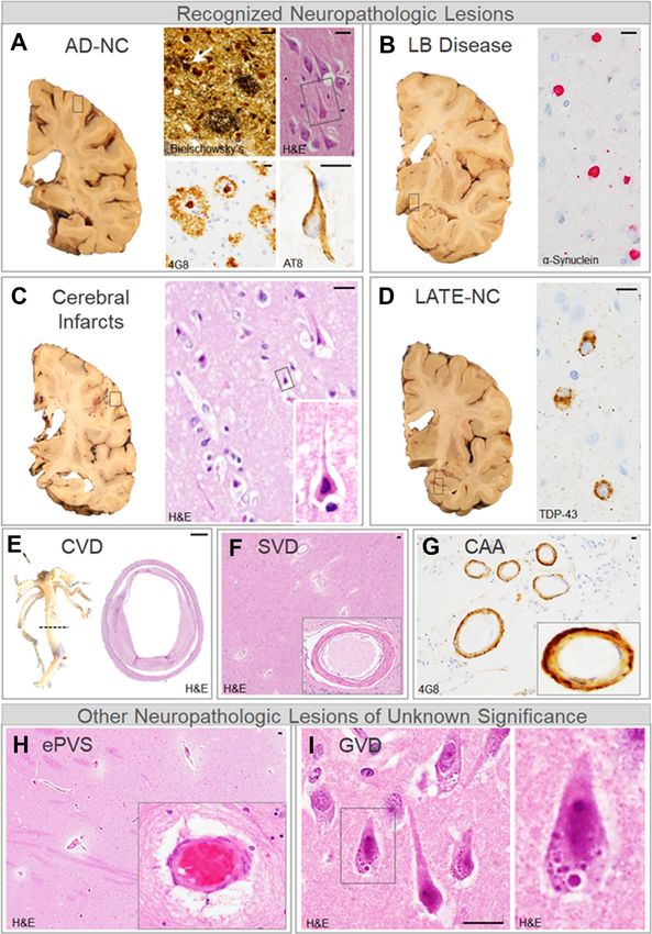
Fig. 1. Histologic features of common neurodegenerative disease lesions. (A) AD-NC is clas-
sically associated with frontoparietal atrophy (left). Etiologic lesions, shown at right (clock-
wise from upper left), include diffuse and neuritic plaques with neurofibrillary tangles
(Bielschowsky’s silver stain), neurofibrillary tangles (H&E stain), neurofibrillary tangle (phos-
pho-Tau AT8 stain), and diffuse and neuritic plaques (4G8 amyloid stain). (B) LB disease is
characterized by mild cortical atrophy with evidence of a-synuclein-positive LBs. (C) Macro-
scopic cerebral infarcts are characterized by cerebral edema with reperfusion-related
Descargado para Irene Ramírez (iramirez@binasss.sa.cr) en National Library of Health and Social
Security de ClinicalKey.es por Elsevier en enero 26, 2023. Para uso personal exclusivamente. No se
permiten otros usos sin autorización. Copyright ©2023. Elsevier Inc. Todos los derechos reservados.Neuropathology of Dementia 95
either diffuse plaques (DPs) or neuritic plaque (NP) varieties. NPs are composed of
aggregated bA protein that may accumulate as dense cores and/or dystrophic neu-
rites, which correspond to degenerated neuronal cell processes.1–40 bA is enriched
in NPs, whereas1–42 bA is enriched in DPs. In addition to1–40 bA, NPs also harbor var-
iable numbers of astrocytic and microglial processes. bA NPs usually deposit in layers
II–V of the neocortex but in advanced cases can also be noted in neocortical layers I
and VI and in subcortical white matter.7
Tau protein, which is produced from MAPT gene, supports neuronal microtubule
stability and is the primary constituent of NFTs. Alternate splicing of exon 10 generates
tau species with either 3 or 4 conserved w32 amino acid repeats (R), leading to either
3R or 4R tau on biochemical and immunohistochemical characterization. In AD, tau
undergoes abnormal phosphorylation that promotes its aggregation into paired helical
filaments.8 These tau inclusions accumulate within neuronal cell soma as large
rounded bodies and in neuronal dendrites and axons as neuropil threads.8 The
morphology of NFTs is variable because they acquire the shape of involved neurons.
In cortical neurons, NFTs may be flame or triangular shaped (see Fig. 1A) but in
subcortical or brainstem neurons, they are generally rounded and/or globose. Pre-
NFTs exhibit diffuse, low-level tau label within the cytoplasm of intact neurons,
whereas mature NFTs exhibit cytoplasmic filamentous tau aggregates that displace
neuronal nuclei and extend into dendrites. Late-stage “ghost” NFTs seem entombed
within nonviable, nucleus-devoid neurons.8
The successive accumulation of NFTs within the aging brain follows a stereotyped
pattern that has been depicted in 6 stages.7 Initially, NFTs are found only in the trans-
entorhinal cortex (stage I and II). With disease progression, they gradually involve the
entorhinal cortex and hippocampus (stage III and IV) and neocortical brain regions
(stage V and VI; Fig. 2A). Similar to NFTs, bA accumulation also follows a stereotyped
pattern of brain accumulation that is distinct from tau accumulation and has been
described along 5 stages.9 Initially, bA deposits within neocortex (stage I). With disease
progression, they spread into allocortical brain regions such as entorhinal cortex, CA1,
and subiculum region of hippocampus (stage II); subcortical nuclei including basal
ganglia, thalamus, and hypothalamus (stage III); and, eventually into the medulla oblon-
gata and midbrain colliculi (stage IV); and, finally, into pontine neurons and the cerebellar
molecular layer (stage V; see Fig. 2A).9 Although earlier staging schema required AD de-
mentia for pathologic diagnosis of disease, revised guidelines for a diagnosis of AD-NC
no longer require the presence of clinical symptoms. Current criteria for AD-NC require
the presence of brain bA deposits. A diagnosis of AD is confirmed when there is at least
intermediate or high AD-NC, which requires a Braak stage of 3 or more, and a Thal stage
of 3. At this stage, most cases will have at least moderate NPs in the neocortex.
Lewy Body Diseases: Lewy Body Demenita and Parkinson Disease Dementia
Lewy body (LB) diseases are characterized by abnormal intracytoplasmic accumula-
tion of alpha-synuclein protein positive LBs.10 On routine stains, LBs seem as round,
=
petechial microhemorrhage (left) and acute neuronal necrosis (right). (D) LATE-NC is charac-
terized by cortical atrophy with TDP-43-positive neuronal inclusions. Vascular changes and/
or vascular brain injuries may also result from: (E) large vessel atherosclerosis, as depicted in
the basilar artery; (F) SVD, such as arteriolosclerosis; and/or (G) CAA. Other neuropatholog-
ical lesions of unknown significance include enlarged perivascular spaces (ePVS) and gran-
ulovacuolar degeneration (GVD), shown in (H) and (I), respectively. A–I: Boxed/marked
area shown in enlarged and/or labeled section. Scale bars: A–D, F–I, 10 mm; E, 1 mm.
Descargado para Irene Ramírez (iramirez@binasss.sa.cr) en National Library of Health and Social
Security de ClinicalKey.es por Elsevier en enero 26, 2023. Para uso personal exclusivamente. No se
permiten otros usos sin autorización. Copyright ©2023. Elsevier Inc. Todos los derechos reservados.96 Mehta & Schneider
Fig. 2. Topographies and overlap of common proteinopathic and age-related disease sub-
strates and effects on brain function. Topographic distribution of AD-NC (A), LB disease (B),
LATE-NC (C), and arteriolosclerosis (D), which exhibit different susceptibilities. Mixed patho-
logic conditions (eg, bA plaques, Tau neurofibrillary tangles, Lewy bodies, TDP-43 inclusions,
and/or cerebrovascular lesions) also occur (E). Persons who do not develop pathologic condi-
tion or do not progress to severe stage of pathologic condition are referred to as “resistant,”
whereas afflicted persons who do not exhibit dementia are referred to as “resilient.” Various
effects of AD-NC (F) and mixed pathologic conditions (G) are shown. Disease resistance refers
to the absence of, or lower-than-expected disease burden, whereas resilience refers to the
absence of, or delayed onset of cognitive impairment in the face of existing neuropathol-
ogies. (Agrawal and Schneider - Neuropathological Underpinnings of the Dementias. In
Neuropsychology of Alzheimer’s disease and other dementias, Second Edition.)
eosinophilic neuronal cytoplasmic inclusions that may be associated with peripheral
halos and are highlighted by immunohistochemistry using antibodies directed to
phosphorylated alpha-synuclein (see Fig. 1B).10 When present in substantia nigra
and accompanied by evidence of significant neuronal loss, these inclusions confirm
a pathologic diagnosis of Parkinson disease (PD). With the involvement of cortical neu-
rons, LBs may cause cognitive impairment and account for 2 recognized dementia
syndromes, that is, Parkinson disease dementia (PDD) or LB Dementia (LBD).
Although these disorders are similar, PDD is associated with cognitive changes typi-
cally occurring years after motor onset of PD, whereas LBD exhibits cognitive and mo-
tor (parkinsonism) impairment within a year of each other. Neuropathological data
have demonstrated that accrual of LBs may progress along different anatomic path-
ways. Most frequently, LBs seem to ascend initially in pigmented brainstem nuclei
(eg, vagus dorsomedial nucleus, locus ceruleus, and/or substantia nigra), and then
along limbic areas (entorhinal cortex and anterior cingulate), and finally progress along
neocortical brain tissue (Fig. 2B). Although presently unclear whether PD, PDD, and
Descargado para Irene Ramírez (iramirez@binasss.sa.cr) en National Library of Health and Social
Security de ClinicalKey.es por Elsevier en enero 26, 2023. Para uso personal exclusivamente. No se
permiten otros usos sin autorización. Copyright ©2023. Elsevier Inc. Todos los derechos reservados.Neuropathology of Dementia 97
LBD are truly distinct disorders or representative of a disease continuum,11 pathologic
condition is currently subclassified based on the regional LB distribution as brainstem,
limbic or neocortical-predominant.12 Postmortem evaluation shows that some brains
feature LBs within the amygdala or olfactory bulb, but not brainstem, and these have
been described as amygdala-predominant or olfactory types.12 Spongiform change in
entorhinal and other temporal cortical regions, and the presence of alpha-synuclein
neurites within hippocampus CA2 sector and areas exhibiting LBs are supporting fea-
tures of LBD. PDD and LBD may be associated with mild atrophy in affected brain re-
gions. The observation of alpha-synuclein inclusions within glial cells characterizes
multiple system atrophy.
Limbic-Predominant Age-Related TDP-43 Encephalopathy
Limbic-predominant age-related TDP-43 encephalopathy (LATE) is a recently charac-
terized proteinopathy that afflicts older persons. LATE-neuropathological change
(LATE-NC) is characterized by neuronal and glial TDP-43 inclusions in adults aged
older than 80 years (see Fig. 1D).13 LATE-NC includes neuronal loss and astrocytosis
in the hippocampus with hallmark features of TDP-43 inclusions involving the limbic
structures, olfactory bulb, basal ganglia, neocortex, and occasionally the brainstem.
The TDP-43 inclusions initially present only in the amygdala (stage 1) but over time
spread gradually to involve the hippocampus (stage 1 and 2) and midfrontal cortex
(stage 3; Fig. 2C).13 This neuropathology typically manifests with atrophy in mesial
temporal lobe structures, including the hippocampus, and may occur concomitantly
with hippocampal sclerosis (HS; described below). Indeed, most cases of HS in aging
are associated with TDP-43 proteinopathy. The inferior frontal, and insular cortices,
and other cortical regions may be less frequently involved.13
Hippocampal Sclerosis
HS is currently broadly defined as severe neuronal loss and gliosis of the hippocam-
pus. In aging, it is most frequently neurodegenerative in origin. The vast majority of HS
in aging is characterized by TDP-43 proteinopathy, hippocampal neuronal loss and/or
gliosis14 and thus is often part of LATE. HS is not specific to any other neurodegener-
ative disease pathologic condition but may be associated with several other classified
disease processes, including various age-related neurodegenerative diseases (listed
above), hypoxic/ischemic brain injury, and temporal lobe epilepsy.15 In epilepsy, HS
is often referred to as mesial temporal sclerosis. In neurodegenerative and hypoxic/
ischemic diseases, HS is frequently characterized by severe neuronal loss and gliosis
involving the hippocampus CA1 sector and the subiculum.16
Frontotemporal Lobar Degeneration
Frontotemporal lobar degeneration (FTLD) is characterized by cerebral cortical degen-
eration and is classified into 3 subtypes according to neuronal and glial protein aggre-
gates.17 FTLD-Tau exhibits hyperphosphorylated tau protein inclusions, whereas FTLD-
TDP manifests TDP-43-positive protein inclusions, and FTLD-FUS exhibits fused in sar-
coma protein inclusions.17 FTLD-FUS includes atypical FTLD with only ubiquitin inclu-
sions, neuronal intermediate filament inclusion disease, and basophilic inclusion body
disease (BIBD), which are all characterized by FUS inclusions in addition to caudate
nuclei atrophy.15,18 Microscopic examination of brains exhibiting FTLD inclusions often
show ballooned neurons, laminar spongiosis involving superficial cerebral cortical layers
(ie, I–III), and prominent myelin loss in association with the above neuronal and/or glial
cell inclusions.17 FTLD also includes Pick disease, which is characterized by “Pick
bodies” that seem as rounded, basophilic intraneuronal cytoplasmic bodies on routine
Descargado para Irene Ramírez (iramirez@binasss.sa.cr) en National Library of Health and Social
Security de ClinicalKey.es por Elsevier en enero 26, 2023. Para uso personal exclusivamente. No se
permiten otros usos sin autorización. Copyright ©2023. Elsevier Inc. Todos los derechos reservados.98 Mehta & Schneider
stain and are composed of abnormally phosphorylated tau.17 Unlike tau of AD-NC, the
aberrant tau protein in Pick disease has been shown to form straight helical filaments
and aggregates in frontal and temporal cerebral cortical neurons.17 In addition to Pick
bodies, cortical neurons in these regions also display ballooned neurons termed Pick
cells.17 As implied by its name, FTLD causes frontal and temporal lobe atrophy, which
when severe is often described at knife-edge atrophy.
Progressive Supranuclear Palsy and Corticobasal Degeneration
Progressive supranuclear palsy (PSP) and corticobasal degeneration (CBD) typically
present as atypical parkinsonian disorders but may also present with a dementia syn-
drome. PSP and CBD are characterized by NFTs involving the substantia nigra but un-
like AD-NC, these diseases are characterized by 4R immunoreactive NFTs.19 In PSP,
4R tau-positive tangles may additionally be found in subcortical regions such as the
subthalamic and red nuclei, basal ganglia, and basis pontis.19 In PSP, the 4R tau in-
clusions are also present in astrocytes and oligodendrocytes where they characterize
so-called tufted astrocytes and coiled bodies, respectively. Tufted astrocytes display
fine branching processes and may also be distributed within motor cortex. 4R tau-
positive coiled bodies are also seen in subcortical white matter. In CBD, there is
also 4R tau-positive tangles involving the substantia nigra but cortical pathologic con-
dition is also prominent, especially in the perirolandic regions of the neocortex. In addi-
tion to neuronal tangles, CBD manifests with 4R tau-positive astrocytic plaques and
neuritic threads in the cerebral cortex and subcortical basal ganglia, thalamus, and
brainstem. Scattered achromatic or ballooned cerebral cortical neurons are typically
present in the perirolandic cortices.19
Chronic Traumatic Encephalopathy
CTE describes the neuropathology associated with repetitive concussive and/or sub-
concussive head injuries, such as that which occurs in professional athletes including
football and hockey players. This disease is characterized by neuronal NFTs present
diffusely throughout the brain, including the hippocampus and brainstem, with accen-
tuated perivascular distribution.20 Perivascular and subpial glial tau accumulation may
also be present. Grossly, CTE brains feature cerebral cortical and subcortical nuclear
atrophy with enlargement of the lateral and third ventricles. CTE neuropathology has
been shown to progress in 4 stages.20 Initially, phosphorylated-tau NFTs deposits in
perivascular NFT clusters, usually at neocortical sulcal depths (stage 1). Next, they
accumulate in discrete clusters in perivascular and superficial cortical layers (stage
2). With subsequent progression, they aggregate multifocally in the medial temporal
lobe, hypothalamus, thalamus, nucleus basalis, mammillary bodies, substantia nigra,
raphe nuclei, and locus coeruleus (stage 3). Finally, with continued progression, NFTs
distribute more widely throughout the brain and are associated with progressive
myelin and myelinated axon loss (stage 4).
Huntington Disease
Huntington disease (HD) is a rare neurodegenerative disease that is caused by CAG
(polyglutamine) expansion repeats in the huntingtin gene (ie, HTT) on chromosome
4.21 Huntingtin protein misfolding causes intraneuronal aggregation of mHTT, primarily
in striatal regions. Thus, this disease is characterized by striatal degeneration that is
associated with secondary ventricular enlargement. HD is a familial dominantly
inherited disease.21 Neuropathological grading criteria used for this disorder is based
on ventricle size and degree of striatal pathologic condition and atrophy.22 Initially,
only microscopic evidence of gliosis and neuronal loss is observed without gross
Descargado para Irene Ramírez (iramirez@binasss.sa.cr) en National Library of Health and Social
Security de ClinicalKey.es por Elsevier en enero 26, 2023. Para uso personal exclusivamente. No se
permiten otros usos sin autorización. Copyright ©2023. Elsevier Inc. Todos los derechos reservados.Neuropathology of Dementia 99
evidence of striatal atrophy (grade 1). Disease progression features striatal atrophy
with minimal changes observed early on in lateral ventricle size (grade 2) but there
is increased striatal atrophy and ventriculomegaly with disease progression (grades
3 and 4).22 Mild cerebral cortical neuronal loss may also be present and involve the
frontal cerebral cortices.
Creutzfeldt Jakob Disease
Creutzfeldt Jakob disease (CJD) is a rare disease that is associated with various
causes. The disease may be infectious, heritable, sporadic, or iatrogenic in nature.23
CJD is characterized by intraneuronal prion protein (PrP) inclusions that induce severe
and rapidly progressing neuronal loss and gliosis in various brain regions.23 End-stage
disease is usually characterized by severe cerebral cortical spongiform change and
corresponding atrophy.23
Other Proteinopathies and Unclassified Tissue Changes
Other age-related neuropathologies feature neuronal and/or glial inclusions.24 Argyr-
ophilic grain disease,25 aging-related tau astrogliopathy (ARTAG),26 globular glial tau-
opathy,27 and primary age-related tauopathy28 are also currently classified as
separate neuropathologies. However, their epidemiologic significance and link to clin-
ical dementia syndrome(s) is presently unclear. Moreover, the significance of other
nonclassified (Fig. 1H) but well-recognized lesions such as granulovacuolar degener-
ation (Fig. 1I), which manifest in hippocampal pyramidal neurons of aged persons and
commonly coexist with AD-NC, remain undefined.
AGE-RELATED VESSEL CHANGES AND VASCULAR DISEASES
Cerebrovascular pathologic conditions are common with aging and induce vascular
and/or brain damage, both of which may contribute to structural brain pathologic con-
dition.29,30 Cardiac, extracranial and/or intracranial large vessel, and intracerebral
small vessel diseases (SVD) may all contribute to dementia syndromes by compro-
mising blood flow, causing subtle multifocal or diffuse neuronal dropout or more
discrete, localized brain parenchymal injuries.31,32 Thereby, these processes indirectly
or directly lead to impairment of function in cognitive domains, although they may not
directly induce deposition of intracerebral protein aggregates.33 Although an array of
disorders is recognized in this category, some older literature used nonspecific termi-
nology to describe these lesions and no formal criteria exist for their grading or stag-
ing. Moreover, many cerebral SVD are only superficially classified, and their true
incidences are unknown. A partial list of contributing disorders is summarized in the
Table 1 (lower panel), and described further below and in Fig. 1C, E–H.
Cardiac and Extracranial Cardiovascular Diseases
Cardiac diseases and extracranial cardiovascular disease (CVD; eg, structural or func-
tional heart diseases such arrhythmic disorders, coronary artery, valvular, and/or hy-
pertensive diseases, and extracranial carotid artery narrowing, occlusion, and/or
emboli) are linked to ischemic phenomena with or without brain hemorrhage.32 These
diseases may lead to vascular brain injuries (VBI) including multifocal or diffuse
ischemia with neuronal dropout and/or macroscopic or cystic infarcts (see
Fig. 1C).3 Mechanisms of cell depletion may include apoptosis and oncotic necrosis,
among other mixed cell death pathways.33 Although primary or secondary pulmonary
or respiratory diseases such as chronic obstructive pulmonary disease, chronic
obstructive sleep apnea, chronic bronchitis, and/or pneumonia, asthma, and/or
Descargado para Irene Ramírez (iramirez@binasss.sa.cr) en National Library of Health and Social
Security de ClinicalKey.es por Elsevier en enero 26, 2023. Para uso personal exclusivamente. No se
permiten otros usos sin autorización. Copyright ©2023. Elsevier Inc. Todos los derechos reservados.100 Mehta & Schneider
pulmonary edema also compromise lung function and may contribute to hypoxia, their
overall contribution to dementia syndromes remains controversial in the literature.
Intracranial Cerebral Large Vessel Diseases
Diseases involving the circle of Willis may also lead to VBI. Atherosclerosis (see Fig. 1E)
and much less commonly large vessel vasculitis and other vasculopathies may cause
hemispheric or focal ischemic stroke that leads to macroscopic cerebral infarcts, other
macroscopic cystic infarcts, “strategic” infarcts, or contribute to microbleeds.32 These
diseases may also cause acute, subacute, or chronic neuronal dropout from hypoxic-
ischemic damage. As with cardiac diseases and extracranial CVD, the mechanisms
of cell depletion may include apoptotic, oncotic, and mixed cell death pathways.37
Intracerebral Small Vessel Diseases
Diseases involving cerebral small vessels also have potential to cause VBI, and recent
studies emphasize the significance of cerebral SVD as a cause for neuronal loss, oligo-
dendrocyte and myelinated fiber depletion, white matter rarefaction, microbleeds,
small and microinfarcts, inflammation, and gliosis.34,35 Arteriolosclerosis is the most
common cause of cerebral SVD and causes partial or total fibrous replacement of
intracerebral arteriolar smooth muscle cell layers (see Fig. 1F), with microbleeds
and/or small and microscopic infarcts.3,36 When widespread, this may lead to subcor-
tical ischemic vascular disease, also known as multi-infarct dementia, Binswanger
disease, or vascular dementia of Binswanger type (Fig. 2D).36
Cerebral amyloid angiopathy (CAA) is also common in older decedents and is char-
acterized by bA deposition within basement membranes and/or smooth muscle cell
layer of cerebral arteries and arterioles (see Fig. 1G).37,38 This disease manifests
with preferential deposition of 1–40 bA species and often leads to petechial microhe-
morrhages and less commonly lobar hemorrhage.36 Severe forms of SVD results in
vessel wall damage with vessel dilation, blood element extravasation, and occasion-
ally vessel occlusion. Several community-based studies demonstrate an association
of CAA with cognitive impairment.39 Clinicopathological evaluations suggest signifi-
cant associations between moderate and severe, multifocal CAA with ischemic path-
ologic conditions,40 although a strong correlation is also noted between CAA and age.
CAA commonly, but not always, accompanies AD pathologic condition and may also
occur in the absence of significant AD.
Other Intracerebral Vessel Abnormalities
The prevalence of other uncharacterized and subtle vascular changes such as
enlarged perivascular spaces 41,42 and blood–brain barrier disruption43 in persons
with dementia is increasingly recognized, although the significance of these changes
is presently unclear (see Fig. 1H).
METABOLIC, TOXIC, AND OTHER DISEASES
Brain injury is also induced by a host of other diseases. Acute, subacute, and chronic
metabolic disturbances are liable to contribute to dementia through various mecha-
nisms. Additionally, metals and other environmental toxins may damage neurons
through additional pathways.
Wernicke Encephalopathy and Wernicke Korsakoff syndrome
Wernicke diseases are precipitated by vitamin B1 (thiamine) deficiency.44 Although
recognized most commonly in association with chronic alcoholism, Wernicke
Descargado para Irene Ramírez (iramirez@binasss.sa.cr) en National Library of Health and Social
Security de ClinicalKey.es por Elsevier en enero 26, 2023. Para uso personal exclusivamente. No se
permiten otros usos sin autorización. Copyright ©2023. Elsevier Inc. Todos los derechos reservados.Neuropathology of Dementia 101
Korsakoff syndrome (WKS) is also observed in systemic diseases that result in nutrient
deficiencies. In the acute phase, Wernicke encephalopathy (WE) is characterized by
ophthalmoplegia, mental status changes, and ataxia, whereas the chronic phase of
WKS includes confabulation and a dementia syndrome.44 In WKS, bilateral mammil-
lary body degeneration is observed on gross brain examination and is associated
with hemorrhage and neuropil loss. With more extensive disease, WKS may be char-
acterized by progressive involvement, including bilateral diencephalic degeneration.44
Carbon Monoxide Poisoning
Carbon monoxide poisoning is caused by inhalation of combustion fumes and binds to
blood hemoglobin to form carboxyhemoglobin, which decreases blood oxygen car-
rying capacity and causes brain tissue hypoxia with preferential necrosis of the globus
pallidus and subcortical white matter demyelination.45,46
Heavy Metals
Some evidence also suggests that dysregulation of metals such as aluminum, arsenic,
and lead may precipitate dementia by causing oxidative stress with axonal and syn-
aptic damage.45
AGE CONSIDERATIONS AND MIXED PATHOLOGIES
Among multiple potential causes described above, the most common causes of demen-
tia are AD, CVD, and LB diseases (see Figs. 1A–C and 2A, B, D), but proteinopathies may
coexist with each other and with nonproteinopathic CVD/VBI lesions.47,48 In fact, longitu-
dinal cohort studies demonstrate that mixed pathologies represent the most common
form of pathologic condition underlying age-related dementia including AD type demen-
tia.47,48 LB disease is reported to occur commonly with AD-NC, especially in persons
aged younger than 60 years at death49,50 but is also seen commonly in aging and with
AD-NC.51 As noted above, TDP-43 pathologic condition (see Fig. 2C) often coincides
with HS and may also co-occur with LB disease.52 Cohort studies have also shown
that, in the oldest old, the incidence of AD-NC plateaus,51,53 whereas other age-related
pathologic conditions, specifically LATE-NC, become more common although the rea-
sons for this are unclear. Despite the detailed diagnostic criteria summarized above for
distinct neurodegenerative disease forms of dementia in aging, pure neurodegenerative
diseases are relatively rare. Overlapping processes are thought to potentiate the com-
mon final pathway of neuronal loss and complex brain inflammation with gliosis that pre-
cipitate clinical dementia that is increasingly accepted as a multifactorial and
pathologically heterogeneous disorder that occurs along a continuum with a range of
phenotypes (Fig. 2E).54 Overall, the location(s) and total burden of various neuropatho-
logical substrates determine the clinical dementia syndrome. Thus, the validity and utility
of some classification and staging schemes for discrete neuropathologic forms of de-
mentia in aging is presently unclear because mixed pathologic conditions are also recog-
nized as a common cause for atypical clinical presentations. Interestingly, risk factors for
dementia are also found to differ by age, with some CVD risk factors appearing to become
protective in the oldest-old.55
LIMITATIONS OF CURRENT NOMENCLATURES, CLASSIFICATIONS, AND STAGING
SCHEMES
Importantly, the above neuropathologies may also be present in persons who are
cognitively intact.56,57 In fact, in persons aged 90 years or older, 40% of individuals
without dementia exhibit AD-NC or other neurodegenerative disease lesions.55 In
Descargado para Irene Ramírez (iramirez@binasss.sa.cr) en National Library of Health and Social
Security de ClinicalKey.es por Elsevier en enero 26, 2023. Para uso personal exclusivamente. No se
permiten otros usos sin autorización. Copyright ©2023. Elsevier Inc. Todos los derechos reservados.102 Mehta & Schneider
addition to recognition of mixed pathologies and age-related differences in epidemi-
ology, it is increasingly accepted that coexisting proteinopathies and CVD/VBI poten-
tiate dementia severity.3,55,58 For example, LB, CAA, SVD, and VBI (see Fig. 2A–D) in
many persons mitigate the burden of AD-NC required for the development of clinical
AD-type dementia (Fig. 2F,G). Similarly, clinicopathological studies demonstrate that
persons with overlapping AD-NC and LATE-NC tend to show more rapid cognitive
decline compared with persons with pure disease17,59 (see Fig. 2G). So, while data
on the range of potential neurodegenerative disease substrates are advancing, epide-
miologic data have also made clear that the presence of neurodegenerative patho-
logic conditions do not directly equate to dementia syndromes.55
During recent years, newer concepts have been introduced in attempt to explain the
disconnects that are frequently observed between AD-NC score and degree of cogni-
tive impairment, which presumably represent uncharacterized neuroprotective mech-
anisms. The term resistant is a heuristic that refers to persons whose brains remain
“lesion-proof,” or free of structural brain pathologic conditions throughout aging
(see Fig. 2E, F).60,61 In contrast, resilient is a term that refers to cognitive preservation,
or the paradoxic ability to sustain cognitive function in the face of existing structural
brain pathologic conditions55 (see Fig. 2E–G). So-called nondemented individuals
with AD neuropathology (NDAN), a.k.a. SuperAgers or asymptomatic AD 62 emphasize
the significance of this group of elderly persons who are not prone to develop demen-
tia and remain cognitively intact despite the presence of structural brain pathologic
conditions (see Fig. 2E–G). Although both resistant and resilient persons have been
said to exhibit cognitive reserve, evidence suggest that these are distinct phenomena
that refer to unique scenarios, that is, resisting disease development versus compen-
sating for existing disease(s).61 The causes for these phenomena may include brain
maintenance and neurorestorative factors that are presently unknown.62 Emerging ev-
idence indicate that lifestyle and behavioral factors, along with other metabolic, ge-
netic, environmental, and/or physiologic variables, may influence AD-NC onset and
progression, highlighting the importance of advising on nutrition, exercise, social,
and cognitive activities, and control of vascular risk factors that may enhance cogni-
tive functioning but are not accounted for in current classification and staging
schemes.21,55
SUMMARY AND FUTURE DIRECTIONS
Dementia is very common in aging, and although AD pathologic condition (ie, AD-NC)
is arguably the most common underlying pathologic condition, other pathologic
causes of dementia in aging include Lewy bodies, TDP-43, vascular neuropathologies,
and mixed pathologic conditions. Less common pathologic causes should also be
recognized. It is important to note that each person has a different threshold for de-
mentia onset. This threshold may be determined by various measures (e.g., the num-
ber of existing brain axons and synapses, neuroimmune state, stress level, and
genetic and biological factors) and also often changes with age. New concepts and
emerging data regarding cognitive reserve support the value of exploring nontradi-
tional and/or nonstructural targets and risk factors for the purpose of preventing or
delaying onset and progression of neurocognitive diseases in aging persons.
Regarding current classification and nomenclatures, protein aggregates seem to be
a final common denominator of many, but not all age-related dementia syndromes.
Moreover, vascular mechanisms also play a fundamental role in many dementia syn-
dromes. Indeed, it is notable that dementia syndromes also occur in the absence of
protein aggregates, and what causes protein aggregates in the first place is
Descargado para Irene Ramírez (iramirez@binasss.sa.cr) en National Library of Health and Social
Security de ClinicalKey.es por Elsevier en enero 26, 2023. Para uso personal exclusivamente. No se
permiten otros usos sin autorización. Copyright ©2023. Elsevier Inc. Todos los derechos reservados.Neuropathology of Dementia 103
incompletely known. With recognition of resistance and resilient factors and mixed
pathologic conditions, the boundaries of distinct neuropathological diseases have
become more uncertain. For these reasons, some experts have proposed a general
neuropathological category of “brain dementia” in aging with specific lesions enumer-
ated secondarily, whereas others suggest revision of current schema although evi-
dence regarding precise revisions is lacking and the current schema continues to
evolve.
What is currently known about dementia in aging is that: (1) Dementia represents a
looming public health crisis.1 (2) The brain undergoes various complex age-related
structural, physiologic, and biochemical changes and is exposed to heterogeneous
environmental stress conditions during the life span that may foster the development
and progression, or resistance and resilience toward disease susceptibilities.55 (3) In
patients with dementia, a spectrum of neuropathologies contribute to neurodegener-
ation and may overlap. (4) Isolated and mixed pathologic conditions result in pruning
and loss of neurons and glia and are associated with inflammation in various brain re-
gions that collectively result in synapse and brain volume loss. (5) In persons symp-
tomatic for dementia, the location(s) of neurodegenerative brain changes determine
clinical symptomatologies. (6) Heterogeneity among clinical disease phenotypes indi-
cates among other things, the existence of other nonclassified and or nonstructural
determinants of disease.55 Therefore, identification and investigation of potential
nonclassified brain pathologic conditions, interactions of different brain pathologic
conditions, and currently unrecognized resistant and resilient factors are required
because they may not only elucidate hidden contributors to disease but may also un-
cover potential novel targets for disease prevention.
CLINICS CARE POINTS
Alzheimer disease neuropathologic change is the most common dementia pathologic
condition and manifests with diagnostic lesions of bA plaques and neurofibrillary tangles.
Other common neuropathological forms of dementia include Lewy body disease, limbic-
predominant age-related TDP-43 encephalopathy-neuropathological change, and vascular
dementia.
Additional pathologic conditions related to dementia include frontotemporal lobar
degeneration, progressive supranuclear palsy, corticobasal degeneration, chronic
traumatic encephalopathy, and Creutzfeldt Jakob disease, among others.
Mixed and asymptomatic pathologic conditions are also commonly found in older persons.
As modifiable risks, vascular diseases and lifestyle factors are important to address in the
geriatric population but also show important age-related differences.
Evidence suggests that traditional brain markers are not fully predictive of clinical disease
because other physiologic factors affect the manifestation of dementia syndromes.
Resistance factors prevent the appearance of neurodegenerative and other brain disease
lesions, whereas resilience factors are hypothesized to mask cognitive dysfunction that
would otherwise be predicted by the presence of a significant burden of neuropathologic
lesions.
FUNDING
We thank the participants of the Rush Memory and Aging Project and Religious Orders
Study and their families. This work is supported by P30AG072975 and R21AG079221.
Descargado para Irene Ramírez (iramirez@binasss.sa.cr) en National Library of Health and Social
Security de ClinicalKey.es por Elsevier en enero 26, 2023. Para uso personal exclusivamente. No se
permiten otros usos sin autorización. Copyright ©2023. Elsevier Inc. Todos los derechos reservados.104 Mehta & Schneider
DISCLOSURE
The authors declare no financial or nonfinancial conflicts of interest.
REFERENCES
1. Alzheimer’s Association. 2022 Alzheimer’s disease facts and figures. Alzheimers
Dement 2022;18:700–89.
2. Brunnstrom H, Gustafson L, Passant U, et al. Prevalence of dementia subtypes: a
30-year retrospective survey of neuropathological reports. Arch Gerontol Geriatr
2008;49:146–9.
3. Kapasi A, DeCarli C, Schneider JA. Impact of multiple pathologies on the
threshold for clinically overt dementia. Acta Neuropathol 2017;134:171–86.
4. Fiala JC, Feinberg M, Peters A, et al. Mitochondrial degeneration in dystrophic
neurites of senile plaques may lead to extracellular deposition of fine filaments.
Brain Struct Funct 2007;212:195–207.
5. Montine TJ, Phelps CH, Beach TG, et al. Alzheimer’s Association. National insti-
tute on aging-alzheimer’s association guidelines for the neuropathologic assess-
ment of alzheimer’s disease: a practical approach. Acta Neuropathol 2012;
123:1–11.
6. Schneider JA, Arvanitakis Z, Leurgans SE, et al. The neuropathology of probable
alzheimer disease and mild cognitive impairment. Ann Neurol 2009;66:200–8.
7. Braak H, Braak E. Neuropathological stageing of alzheimer-related changes.
Acta Neuropathol 1991;82:239–59.
8. Moloney CM, Lowe VJ, Murray ME. Visualization of neurofibrillary tangle maturity
in Alzheimer’s disease: a clinicopathologic perspective for biomarker research.
Alzheimers Dement 2021;17:1554–74.
9. Thal DR, Capetillo-Zarate E, Del Tredici K, et al. The development of amyloid beta
protein deposits in the aged brain. Sci Aging Knowledge Environ 2006;2006:re1.
10. McKeith IG, Dickson DW, Lowe J, et al. Consortium on DLB. Diagnosis and man-
agement of dementia with lewy bodies: third report of the DLB consortium.
Neurology 2005;65:1863–72.
11. Jellinger KA, Korczyn AD. Are dementia with lewy bodies and Parkinson’s dis-
ease dementia the same disease? BMC Med 2018;16:34.
12. Attems J, Toledo JB, Walker, et al. Neuropathological consensus criteria for the
evaluation of Lewy pathology in post-mortem brains: a multi-centre study. Acta
Neuropathol 2021;141:159–72.
13. Nelson PT, Dickson DW, Trojanowski JQ, et al. Limbic-predominant age-related
TDP-43 encephalopathy (LATE): consensus working group report. Brain 2019;
142:1503–27.
14. Amador-Ortiz C, Lin WL, Ahmed Z, et al. D. W. TDP-43 immunoreactivity in hippo-
campal sclerosis and alzheimer’s disease. Ann Neurol 2007;61:435–45.
15. Josephs KA, Whitwell JL, Weigand SD, et al. TDP-43 is a key player in the clinical
features associated with alzheimer’s disease. Acta Neuropathol 2014;127:
811–24.
16. Ala TA, Beh GO, Frey WH 2nd. Pure hippocampal sclerosis: a rare cause of de-
mentia mimicking alzheimer’s disease. Neurology 2000;54:843–8.
17. Hernandez I, Fernandez MV, Tarraga L, et al. Frontotemporal lobar degeneration
(FTLD): review and Update for clinical Neurologists. Curr Alzheimer Res 2018;15:
511–30.
18. Josephs KA, Dickson DW. Hippocampal sclerosis in tau-negative frontotemporal
lobar degeneration. Neurobiol Aging 2007;28:1718–22.
Descargado para Irene Ramírez (iramirez@binasss.sa.cr) en National Library of Health and Social
Security de ClinicalKey.es por Elsevier en enero 26, 2023. Para uso personal exclusivamente. No se
permiten otros usos sin autorización. Copyright ©2023. Elsevier Inc. Todos los derechos reservados.Neuropathology of Dementia 105
19. Mimuro M, Yoshida M. Chameleons and mimics: progressive supranuclear palsy
and corticobasal degeneration. Neuropathology 2020;40:57–67.
20. McKee AC, Cairns NJ, Dickson DW, et al. The first NINDS/NIBIB consensus
meeting to define neuropathological criteria for the diagnosis of chronic traumatic
encephalopathy. Acta Neuropathol 2016;131:75–86.
21. Hong EP, MacDonald ME, Wheeler VC, et al. Huntington’s disease Pathogenesis:
two sequential Components. J Huntingtons Dis 2021;10:35–51.
22. Vonsattel JP, Myers RH, Stevens TJ, et al. Neuropathological classification of hun-
tington’s disease. J Neuropathol Exp Neurol 1985;44:559–77.
23. Watson N, Brandel JP, Green A, et al. The importance of ongoing international
surveillance for Creutzfeldt-Jakob disease. Nat Rev Neurol 2021;17:362–79.
24. Brettschneider J, Libon DJ, Toledo JB, et al. Microglial activation and TDP-43 pa-
thology correlate with executive dysfunction in amyotrophic lateral sclerosis. Acta
Neuropathol 2012;123:395–407.
25. Braak H, Braak E. Argyrophilic grain disease: Frequency of occurrence in
different age categories and neuropathological diagnostic criteria. J Neural
Transm 1998;105:801–19.
26. Kovacs GG, Ferrer I, Grinberg LT, et al. Aging-related tau astrogliopathy (AR-
TAG): harmonized evaluation strategy. Acta Neuropathol 2016;131:87–102.
27. Chung DC, Carlomagno Y, Cook CN, et al. Tau exhibits unique seeding properties
in globular glial tauopathy. Acta Neuropathol Commun 2019;7:36.
28. Crary JF, Trojanowski JQ, Schneider JA, et al. Primary age-related tauopathy
(PART): a common pathology associated with human aging. Acta Neuropathol
2014;128:755–66.
29. Arvanitakis Z, Capuano AW, Leurgans SE, et al. Relation of cerebral vessel dis-
ease to alzheimer’s disease dementia and cognitive function in elderly people:
a cross-sectional study. Lancet Neurol 2016;15:934–43.
30. Thal DR, Grinberg LT, Attems J. Vascular dementia: different forms of vessel dis-
orders contribute to the development of dementia in the elderly brain. Exp Geron-
tol 2012;47:816–24.
31. Corrada MM, Sonnen JA, Kim RC, et al. Microinfarcts are common and strongly
related to dementia in the oldest-old: the 901 study. Alzheimers Dement 2016;12:
900–8.
32. Dolan H, Crain B, Troncoso J, et al. Atherosclerosis, dementia, and alzheimer dis-
ease in the baltimore longitudinal study of aging cohort. Ann Neurol 2010;68:
231–40.
33. Kahle KT, Simard JM, Staley KJ, et al. Molecular mechanisms of ischemic cere-
bral edema: role of electroneutral ion transport. Physiology 2009;24:257–65.
34. Poels MM, Ikram MA, van der Lugt A, et al. Cerebral microbleeds are associated
with worse cognitive function: the rotterdam scan study. Neurology 2012;78:
326–33.
35. Hommet C, Mondon K, Constans T, et al. Review of cerebral microangiopathy
and alzheimer’s disease: relation between white matter hyperintensities and mi-
crobleeds. Dement Geriatr Cogn Disord 2011;32:367–78.
36. Smith EE, Schneider JA, Wardlaw JM, et al. Cerebral microinfarcts: the invisible
lesions. Lancet Neurol 2012;11:272–82.
37. Auriel E, Greenberg SM. The pathophysiology and clinical presentation of cere-
bral amyloid angiopathy. Curr Atheroscler Rep 2012;14:343–50.
38. Grinberg LT, Thal DR. Vascular pathology in the aged human brain. Acta Neuro-
pathol 2010;119:277–90.
Descargado para Irene Ramírez (iramirez@binasss.sa.cr) en National Library of Health and Social
Security de ClinicalKey.es por Elsevier en enero 26, 2023. Para uso personal exclusivamente. No se
permiten otros usos sin autorización. Copyright ©2023. Elsevier Inc. Todos los derechos reservados.106 Mehta & Schneider
39. Boyle PA, Yu L, Nag S, et al. Cerebral amyloid angiopathy and cognitive out-
comes in community-based older persons. Neurology 2015;85:1930–6.
40. Akoudad S, Wolters FJ, Viswanathan A, et al. Association of cerebral microbleeds
with cognitive decline and dementia. JAMA Neurol 2016;73:934–43.
41. Javierre-Petit C, Schneider JA, Kapasi A, et al. Neuropathologic and cognitive
correlates of enlarged perivascular spaces in a community-based cohort of older
adults. Stroke 2020;51:2825–33.
42. Mestre H, Kostrikov S, Mehta RI, et al. Perivascular spaces, glymphatic dysfunc-
tion, and small vessel disease. Clin Sci (Lond) 2017;131:2257–74.
43. Giwa MO, Williams J, Elderfield K, et al. Neuropathologic evidence of endothelial
changes in cerebral small vessel disease. Neurology 2012;78:167–74.
44. Chandrakumar A, Bhardwaj A, t Jong GW. Review of thiamine deficiency disor-
ders: Wernicke encephalopathy and Korsakoff psychosis. J Basic Clin Physiol
Pharmacol 2018;30:153–62.
45. Killin LOJ, Starr JM, Shiue IJ, et al. Environmental risk factors for dementia: a sys-
tematic review. BMC Geriatr 2016;16:175.
46. Schofield P. Dementia associated with toxic causes and autoimmune disease. Int
Psychogeriatr 2005;17:S129–47.
47. Boyle PA, Yang J, Yu L, et al. Varied effects of age-related neuropathologies on
the trajectory of late life cognitive decline. Brain 2017;140:804–12.
48. Schneider JA, Arvanitakis Z, Bang W, et al. Mixed brain pathologies account for
most dementia cases in community-dwelling older persons. Neurology 2007;69:
2197–204.
49. Beach TG, Malek-Ahmadi M. Alzheimer’s disease neuropathological Comorbid-
ities are common in the younger-old. J Alzheimers Dis 2021;79:389–400.
50. Beach TG, Adler CH, Lue L, et al. Unified staging system for lewy body disorders:
correlation with nigrostriatal degeneration, cognitive impairment and motor
dysfunction. Acta Neuropathol 2009;117:613–34.
51. James BD, Bennett DA, Boyle PA, et al. Dementia from alzheimer disease and
mixed pathologies in the oldest old. JAMA 2012;307:1798–800.
52. Nag S, Yu L, Capuano AW, et al. Hippocampal sclerosis and TDP-43 pathology in
aging and alzheimer disease. Ann Neurol 2015;77:942–52.
53. Robinson JL, Corrada MM, Kovacs GG, et al. Non-alzheimer’s contributions to
dementia and cognitive resilience in the 901 study. Acta Neuropathol 2018;
136:377–88.
54. Jack CR, Therneau TM, Weigand SD, et al. Prevalence of biologically vs clinically
defined alzheimer spectrum entities using the National institute on aging-alz-
heimer’s association research Framework. JAMA Neurol 2019;76:1174–83.
55. Kawas CH, Legdeur N, Corrada MM. What have we learned from cognition in the
oldest-old. Curr Opin Neurol 2021;34:258–65.
56. Hyman BT, Phelps CH, Beach TG, et al. National institute on aging-alzheimer’s
association guidelines for the neuropathologic assessment of alzheimer’s dis-
ease. Alzheimers Dement 2012;8:1–13.
57. Serrano-Pozo A, Qian J, Muzikansky A, et al. Thal amyloid stages do not signif-
icantly impact the correlation between neuropathological change and cognition
in the alzheimer disease continuum. J Neuropathol Exp Neurol 2016;75:516–26.
58. Korczyn AD. Mixed dementia–the most common cause of dementia. Ann New
York Ann N Y Acad Sci 2002;977:129–34.
59. Nag S, Yu L, Wilson RS, et al. TDP-43 pathology and memory impairment in el-
ders without pathologic diagnoses of AD or FTLD. Neurology 2017;88:653–60.
Descargado para Irene Ramírez (iramirez@binasss.sa.cr) en National Library of Health and Social
Security de ClinicalKey.es por Elsevier en enero 26, 2023. Para uso personal exclusivamente. No se
permiten otros usos sin autorización. Copyright ©2023. Elsevier Inc. Todos los derechos reservados.You can also read