Spectral dynamic causal modelling in healthy women reveals brain connectivity changes along the menstrual cycle - Nature
←
→
Page content transcription
If your browser does not render page correctly, please read the page content below
ARTICLE
                  https://doi.org/10.1038/s42003-021-02447-w               OPEN

                  Spectral dynamic causal modelling in healthy
                  women reveals brain connectivity changes along
                  the menstrual cycle
                  Esmeralda Hidalgo-Lopez               1 ✉,   Peter Zeidman          2,   TiAnni Harris1, Adeel Razi2,3 & Belinda Pletzer                  1✉

                  Longitudinal menstrual cycle studies allow to investigate the effects of ovarian hormones on
                  brain organization. Here, we use spectral dynamic causal modelling (spDCM) in a triple
                  network model to assess effective connectivity changes along the menstrual cycle within and
1234567890():,;

                  between the default mode, salience and executive control networks (DMN, SN, and ECN).
                  Sixty healthy young women were scanned three times along their menstrual cycle, during
                  early follicular, pre-ovulatory and mid-luteal phase. Related to estradiol, right before ovulation
                  the left insula recruits the ECN, while the right middle frontal gyrus decreases its connectivity
                  to the precuneus and the DMN decouples into anterior/posterior parts. Related to proges-
                  terone during the mid-luteal phase, the insulae (SN) engage to each other, while decreasing
                  their connectivity to parietal ECN, which in turn engages the posterior DMN. When including
                  the most confident connections in a leave-one out cross-validation, we find an above-chance
                  prediction of the left-out subjects’ cycle phase. These findings corroborate the plasticity of
                  the female brain in response to acute hormone fluctuations and may help to further
                  understand the neuroendocrine interactions underlying cognitive changes along the
                  menstrual cycle.

                  1 Department of Psychology and Centre for Cognitive Neuroscience, University of Salzburg, Salzburg, Austria. 2 The Wellcome Centre for Human

                  Neuroimaging, University College London, London, UK. 3 Turner Institute for Brain and Mental Health, Monash University, Clayton, VIC, Australia.
                  ✉email: esmeralda.hidalgolopez@sbg.ac.at; Belinda.Pletzer@sbg.ac.at

                  COMMUNICATIONS BIOLOGY | (2021)4:954 | https://doi.org/10.1038/s42003-021-02447-w | www.nature.com/commsbio                                        1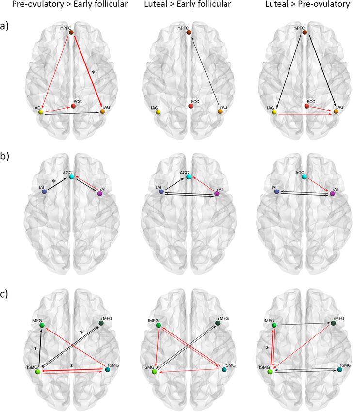
ARTICLE                                                             COMMUNICATIONS BIOLOGY | https://doi.org/10.1038/s42003-021-02447-w

P
       hysiological fluctuations of ovarian hormones (i.e., estradiol       phase, while the posterior DMN (PCC) was most flexible around
       and progesterone) affect the nervous system at multiple             ovulation, followed by an increased stability after the estradiol’s
       levels1. Animal research has broadly evidenced the rapid            peak13. Within the SN, higher activity and connectivity have been
changes exerted by estradiol and progesterone on the neuronal              consistently reported during the luteal cycle phase and related to
excitatory/inhibitory balance2, synaptogenesis3, myelination and           higher progesterone levels, both at rest and during tasks35,36.
re-myelination4. These effects result in synaptic connectivity             Finally, for the ECN, increased task-based activation with higher
changes and therefore neural function5,6, and in rodents high              levels of estradiol and progesterone have been shown for frontal
levels of both hormones have been shown to improve                         areas. Specifically, increased right dorsolateral PFC activation has
cognition7,8. In ovariectomized rats, both estradiol and proges-           been reported during the luteal phase, over a range of tasks
terone treatment enhances novel object recognition, mechan-                including spatial and verbal working memory and verbal
istically linked to increased long-term potentiation and N-                fluency37–40. Relatedly, during resting state, increased eigenvector
methyl-D-aspartic acid receptors at hippocampal synapses for               centrality in bilateral dorsolateral PFC has been found in the
estradiol7, and to an increased density of the basal dendrites of          presence of higher progesterone levels41.
hippocampal neurons for progesterone8. More importantly, as                   Regarding inter-network connectivity, previous findings
evidenced in hormonal replacement studies in rodents9 and non-             mainly report changes in the DMN-SN connectivity, pointing out
human primates10–12 at least some of the reported neuroactive              an increase during the luteal phase (however, not without
effects are dependent on a cyclic variation of hormone levels, as          inconsistencies, see ref. 33). Specifically, ACC and amygdala
opposed to a continuous schedule of treatment. Although in                 (nodes of the SN) increased their connectivity with the precuneus
humans such multilevel relationships between molecular                     (posterior DMN) during the luteal phase compared to early
mechanisms and neural functions are more difficult to determine,            follicular42. Likewise, when treated with progesterone, increased
new results hint that neural reorganization attributed to the              connectivity between several nodes of the SN-DMN has been
hormones also appears abolished when the endogenous fluctua-                reported35. This enhanced inter-connectivity between SN and
tion of ovarian hormones is disrupted13–15. This emphasizes the            DMN, alongside the increased intrinsic connectivity in the SN
importance of studying menstrual cycle-related neural changes in           and decreased intrinsic connectivity in the DMN, sets a very
healthy subjects, which still remains underinvestigated. Although          unique scenario for the luteal phase. Considering that both net-
sparse, acute structural changes related to hormonal fluctuations           works involved (specially ACC and mPFC) are also implicated in
have already been reported for both grey16,17 and white matter18.          self-referential43 and affective experience28,44, this distinctive
Accordingly, connectivity patterns vary in women depending on              connectivity pattern could elicit the misattribution of salient sti-
the hormonal status, as shown by resting-state functional MRI              muli and dysfunctional appraisal, leading to anxiety and
(fMRI)19–21.                                                               depressive symptoms26. Consequently, the coupling dynamics
   Amongst the most common approaches to resting-state func-               between and within the SN and DMN have been proposed to
tional MRI is the assessment of intrinsic connectivity networks            underlie the window of vulnerability for cycle-related affective
(ICNs). These sets of brain networks with temporally correlated            disorders during the luteal phase36,45. More importantly, the
activity at rest relate to task-based BOLD activation patterns22,23        precise role of the ECN, responsible for the regulation of the SN
and are consistent across healthy subjects24,25. Three large-scale         and the DMN, has not yet been defined. Weis et al.46 observed
systems derived by independent component analyses (ICA) arise              decreased connectivity during the pre-ovulatory phase between
as ‘core’ neurocognitive networks, essential for cognitive                 the DMN and the left MFG (part of the ECN), while Petersen
functions26. First, the default mode network (DMN), which                  et al.34 observed decreased connectivity during the luteal phase
includes the precuneus/posterior cingulate cortex (PCC), medial            between the ECN and the ACC (part of the SN), both compared
prefrontal cortex (mPFC) and bilateral angular gyrus (AG), is              to early follicular. These changes could imply a difference in
characterized by increased activity during the resting state and           functional integration of cognitive and affective processes, but up
decreased activity during goal-directed tasks27,28. Second, the            to now a cohesive model for understanding the global mechan-
salience network (SN), which comprises bilateral anterior insula           isms in which the healthy female brain adapts to hormonal
(AI) and dorsal anterior cingulate cortex (ACC), is specialized in         changes remains elusive.
identifying and mapping relevant inputs, such as emotional                    Despite being a useful resource to address brain organization,
stimuli29,30. Third, the executive control network (ECN), is               the assessment of resting-state functional connectivity23,25,28,47
composed of bilateral middle frontal gyri (MFG) and bilateral              does not allow to infer the directionality of coupling between
supramarginal gyri (SMG)30. This fronto-parietal system is                 these multiple distributed systems. Effective connectivity meth-
usually lateralized23,24 and responsible for higher cognitive con-         ods, on the other hand, constitute the best approach to investigate
trol functions, such as working memory or directed attention,              the complex within and between ICN relationships and the causal
once the relevant stimuli are detected25,30.                               influence that one node exerts over another. Specifically, model
   Although some studies have reported no menstrual cycle-                 inversion with spectral dynamic causal modelling (spDCM)
related effects on resting-state functional connectivity31,32, all of      estimates hidden neural states from the observed Blood Oxygen
these ICNs are susceptible to modulation by ovarian hormones.              Level Dependent (BOLD) signal—specifically, the cross-spectral
Specifically, within the DMN, intrinsic resting-state connectivity          density of BOLD signals from different brain regions. It assumes
has been shown to increase before ovulation16,33 and decrease              that spontaneous fluctuations in the signal during resting state
during the luteal phase to both left and right AG21,34. More               reflect the endogenous neural activity48. By parameterising the
recently, a single-subject study showed that the peak in estradiol         hidden coupling among the neuronal populations, one can gen-
right before ovulation had a key role in temporarily reorganizing          erate (complex) cross spectra among observed responses49–51. In
the functional coupling of ICNs, especially within-network con-            addition, spDCM is especially efficient to invert large DCMs of
nectivity of DMN and dorsal attention network14. Further ana-              resting-state fMRI51, which makes it feasible to assess large-scale
lyses in this dataset showed that during this time window several          between and within networks’ organization.
brain nodes increased their flexibility, defined as a measure of                To the best of our knowledge, no study has yet investigated the
how often each node changes its affiliation among different net-            directed connectivity across the different menstrual cycle phases
works or functional modules. Specifically, limbic and subcortical           and related to ovarian hormonal levels. In this work, we aim to
nodes were more flexible around ovulation and the mid-luteal                delineate the menstrual cycle-related changes in effective

2                        COMMUNICATIONS BIOLOGY | (2021)4:954 | https://doi.org/10.1038/s42003-021-02447-w | www.nature.com/commsbio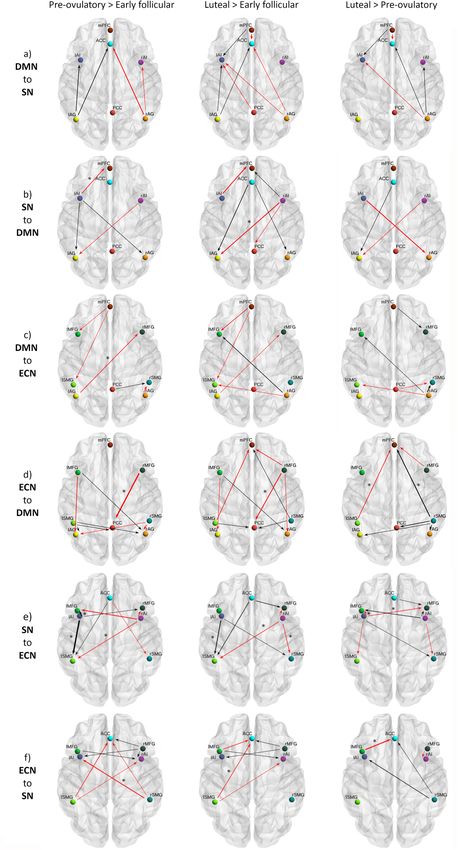
COMMUNICATIONS BIOLOGY | https://doi.org/10.1038/s42003-021-02447-w                                                                                         ARTICLE

 Table 1 Demographic data and hormone levels during each cycle phase.

 Sample (n = 58) Age (years)                   APM (IQ)            Cycle length (days) Cycle day of assesment (days) Estradiol (pg/ml) Progesterone (pg/ml)
 EF                       25.36 ± 0.56         110.31 ± 1.21       28.22 ± 0.31                   3.72 ± 0.20                     0.81 ± 0.05         65.22 ± 5.3
 P-O                                                                                             12.02 ± 0.32                      1.11 ± 0.07***     86.98 ± 8.02**
 M-L                                                                                             21.32 ± 0.47                     0.97 ± 0.06**      199.06 ± 18.28***

 Values are presented as mean ± standard error of the mean (M ± SEM) for the final sample of n = 58.
 EF early follicular, P-O pre-ovulatory, M-L mid-luteal.
 For hormone levels, ** corresponds to p < 0.01, and *** corresponds to p < 0.001, compared to early follicular.

connectivity within and between DMN, SN and ECN, and                                                   Within-network effective connectivity. DMN: In general, effec-
identify the specific direction of previously reported                                                  tive connectivity within anterior and posterior nodes in the DMN
effects16,34–36,42,52. For example, it is yet not defined whether the                                   was highest during early follicular and lowest during the pre-
enhanced inter-connectivity of the SN and DMN during the                                               ovulatory phase (Supplementary Data file 1, Fig. 2a, −0.14 < Ep <
luteal cycle phase42 originates from the SN or the DMN. Likewise,                                      0.11, Pp > 75%). Specifically, effective connectivity from mPFC to
it is unclear whether the downregulation of the SN during the                                          right AG (Ep = −0.14, Pp = 96%) decreased from early follicular
luteal phase is related to an increase in the directed connectivity                                    to the pre-ovulatory phase, negatively related to estradiol levels
from the ECN as proposed by the triple network model26. In                                             (Ep = −0.06, Pp = 95%). This negative impact of higher estradiol
addition, it also remains unexplored how the increased activation                                      levels was reversed by progesterone positive impact (Ep = 0.12,
of the right MFG relates to increased afferent connectivity, given                                     Pp = 100%), resulting in an interactive effect of both hormones
that the BOLD-response rather reflects the input to a neuronal                                          (Ep = −0.06, Pp = 96%) (Supplementary Data file 1, Fig. 2a).
population53. As several psychiatric and neurological disorders                                           SN: Within the SN bidirectional connectivity between left and
share an aberrant intrinsic organization of the aforementioned                                         right AI was strongest during the mid-luteal phase (Supplemen-
three core networks26, we consider it of the utmost importance to                                      tary Data file 1, Fig. 2b, 0.07 < Ep < 0.09, Pp > 75%). While
characterize their non-pathological directed organization in                                           effective connectivity of the right AI to the ACC decreased during
healthy women. In order to do so, we applied state-of-the-art                                          the pre-ovulatory and mid-luteal phase compared to early
effective connectivity analyses to a previously published long-                                        follicular, effective connectivity from the left AI to the ACC
itudinal resting state-fMRI data set acquired during early folli-                                      followed the opposite pattern (Ep = 0.12, Pp = 95%). Effective
cular, pre-ovulatory and mid-luteal cycle phase in a large sample                                      connectivity from the left AI to ACC was further positively
of healthy young women21. Finally, we aimed to identify, whether                                       related to the increase in estradiol levels (Ep = 0.08, Pp = 100%),
changes in specific directed connections were able to predict                                           and negatively related to progesterone levels (Ep = −0.06, Pp =
which cycle phase a woman was in.                                                                      98%)(Supplementary Data file 1, Fig. 2b).
                                                                                                          ECN: Within the ECN, in general, connectivity between
                                                                                                       homotopic areas was weakest during the pre-ovulatory phase
Results
                                                                                                       (Supplementary Data file 1, Fig. 2c, −0.15 < Ep < 0.15, Pp > 75%).
Demographic and hormonal data. For the final sample and as
                                                                                                       Specifically, effective connectivity from right to left SMG
expected, both estradiol and progesterone levels significantly
                                                                                                       decreased from early follicular to the pre-ovulatory phase
changed within subject across the menstrual cycle (F2, 114 =
                                                                                                       (Ep = −0.15, Pp = 100%), and was negatively related to estradiol
29.02, p < 0.001; F2, 114 = 33.49, p < 0.001, for estradiol and
                                                                                                       levels (Ep = −0.09, Pp = 100%) (Supplementary Data file 1,
progesterone respectively) (Table 1). For estradiol, levels were
                                                                                                       Fig. 2c). Conversely, from right MFG to left SMG effective
significantly higher during the pre-ovulatory phase compared to
                                                                                                       connectivity increased from early follicular to pre-ovulatory phase
early follicular (SE = 0.09, t = 7.61, p < 0.001) and mid-luteal
                                                                                                       (Ep = 0.10, Pp = 98%), positively related to estradiol levels (Ep =
phase (SE = 0.10, t = −3.01, p < 0.01), as well as during the mid-
                                                                                                       0.09, Pp = 100%). The positive impact of estradiol was reduced in
luteal phase compared to early follicular (SE = 0.10, t = 3.49, p <
                                                                                                       the presence of high progesterone levels, as indicated by a
0.01). Progesterone levels were significantly lower during early
                                                                                                       negative interaction effect (Ep = −0.04, Pp = 99%) (Supplemen-
follicular compared to the pre-ovulatory and the mid-luteal phase
                                                                                                       tary Data file 1). In general, connectivity between the left-
(SE = 0.06, t = 3.19, p < 0.01; SE = 0.15, t = 8.12, p < 0.001,
                                                                                                       lateralized nodes was the strongest right before ovulation (Fig. 2c).
respectively), and higher during the mid-luteal compared to the
                                                                                                       Specifically, effective connectivity from the left SMG to the left
pre-ovulatory phase (SE = 0.13, t = 7.82, p < 0.001).
                                                                                                       MFG increased from early follicular to the pre-ovulatory phase
                                                                                                       (Ep = 0.15, Pp = 100%), and decreased again during the mid-
Spectral dynamic causal modelling and parametric empirical                                             luteal phase (Ep = −0.12, Pp = 99%). For this connection, higher
bayes. Results are displayed in Fig. 1d, e, 2 (within-network) and                                     progesterone (Ep = 0.05, Pp = 99%) and the combined effect of
3 (between-network) and reported in Supplementary Data file 1.                                          estradiol and progesterone (Ep = 0.05, Pp = 100%) had a positive
Only parameters showing a ‘positive evidence’54 (more than 75%                                         impact.
posterior probability of having diverged from its prior expectation
of zero) are reported. Those connections surviving a 95%
threshold are further indicated in the figures and results section                                      Between-network effective connectivity. DMN—SN: Effective
alongside each parameter’s estimated mean (Ep) and posterior                                           connectivity between the DMN and SN across the menstrual
probability (Pp). Given that all parameters contributed to the                                         cycle was characterized by a lateralized pattern. From left AG to
model, figures including all parameters regardless of threshold                                         ipsilateral AI and ACC was stronger during the high-hormone
can be found in the supplementary material (SI, Fig. S2 cycle                                          phases, while from right AG to ipsilateral AI and ACC, con-
phase results, and Fig. S3 hormonal results). Most of the changes                                      nectivity was weakest during the pre-ovulatory phase. Further-
in cycle phase were backed up by hormonal correlations (com-                                           more, during the mid-luteal phase, connectivity from the mPFC
pare Supplementary Data file 1), but only those surviving a 95%                                         to left AI was the strongest, whereas to the ACC, the lowest
threshold are reported in the text.                                                                    (Supplementary Data file 1, Fig. 3a, −0.12 < Ep < 0.09, Pp > 75%).

COMMUNICATIONS BIOLOGY | (2021)4:954 | https://doi.org/10.1038/s42003-021-02447-w | www.nature.com/commsbio                                                               3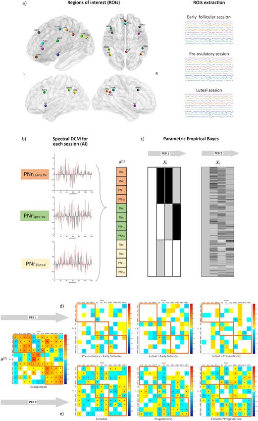
ARTICLE                                                            COMMUNICATIONS BIOLOGY | https://doi.org/10.1038/s42003-021-02447-w

In general, effective connectivity from the SN to bilateral AG was        −0.17, Pp = 100%) (Supplementary Data file 1, Fig. 3b). The
strongest during the high-hormone phases, except for those                effective connectivity from the SN to the anterior DMN followed
connections originating in the right hemisphere. Specifically,             an opposite lateralization, being the strongest during the early
from right AI to left AG it decreased from early follicular to the        follicular phase from the left AI to mPFC (Ep = −0.11, Pp =
mid-luteal phase (Ep = −0.11, Pp = 95%) and was negatively                95%), and negatively related to estradiol (Ep = −0.05, Pp = 97%).
correlated to estradiol (Ep = −0.01, Pp = 100%), progesterone             The negative impact of higher estradiol levels was partially
(Ep = −0.05, Pp = 98%) and their combinatory effect (Ep =                 reversed in the presence of high progesterone levels as indicated

4                       COMMUNICATIONS BIOLOGY | (2021)4:954 | https://doi.org/10.1038/s42003-021-02447-w | www.nature.com/commsbio
COMMUNICATIONS BIOLOGY | https://doi.org/10.1038/s42003-021-02447-w                                                                       ARTICLE

Fig. 1 Procedures for dynamic effective connectivity analysis. The regions of interest from each ICN used in the current study is shown in (a). The default
mode brain regions included the precuneus/posterior cingulate cortex (PCC), medial prefrontal cortex (mPFC) and bilateral angular gyrus (AG); the
salience network comprised bilateral anterior insula (AI) and anterior cingulate cortex (ACC), and the executive control network was composed of bilateral
middle frontal gyri (MFG) and bilateral supramarginal gyri (SMG). Each participant had three sessions locked to their menstrual cycle: during early
follicular, pre-ovulatory and mid-luteal phase. b A spectral DCM (spDCM) of 121 parameters was estimated for each session of every participant in a group
DCM (θ1). c For the group-level analysis, Parametric Empirical Bayesian analysis (PEB) was used to investigate the cycle phase and hormonal levels group
effects. This is a general linear model of the connectivity parameters. Shown are the design matrix X1 for cycle phase and X2 for hormonal levels, where
lighter colours indicate higher values. d Estimated parameters for PEB 1 (cycle phases). e Estimated parameters for PEB 2 (hormonal levels). For (d) and (e)
connections surpassing a posterior probability of 95% are indicated with an asterisk. The columns are the outgoing connections, the rows are the incoming
connections, ordered as: PCC, lAG, rAG, mPFC, lAI, rAI, ACC, lSMG, rSMG, lMFG, and rMFG. Hot colours indicate positive parameter estimates and cold
colours negative.

by a positive interaction effect (Ep = 0.10, Pp = 100%) (Supple-               combinatory effect of estradiol and progesterone (Ep = −0.12,
mentary Data file 1).                                                           Pp = 100%)(Supplementary Data file 1).
   DMN—ECN: In general, effective connectivity from the DMN                       On the other hand, from the right AI, effective connectivity to
to the ECN was left-lateralized during early follicular, whereas               posterior ECN decreased right before ovulation (Ep = −0.09, Pp
right-lateralized during the mid-luteal phase, and the lowest right            = 96% to the left SMG), and during the mid-luteal phase (Ep =
before ovulation (Supplementary Data file 1, Fig. 3c, −0.10 < Ep <              −0.11, Pp = 97% to the left and Ep = −0.11, Pp=95% to the right
0.11, Pp > 75%). From the left AG to right MFG, effective                      SMG) and was related to higher estradiol (left SMG: Ep = −0.13,
connectivity decreased from early follicular to the pre-ovulatory              Pp = 100%; right SMG: Ep = −0.07, Pp = 99%), lower progester-
phase (Ep = −0.11, Pp = 97%) and related negatively to proges-                 one (left SMG: Ep = −0.06, Pp = 100%; right SMG: Ep = −0.11,
terone levels (Ep = −0.10, Pp = 100%). This effect was further                 Pp = 100%) and their combinatory effect (right SMG: Ep = 0.09,
enhanced in the presence of high estradiol levels (Ep = −0.12,                 Pp = 100%) (Supplementary Data file 1, Fig. 3e).
Pp = 100%) (Supplementary Data file 1, Fig. 3c). In turn, effective                Effective connectivity from the frontal ECN to the SN was in
connectivity from the posterior nodes of the ECN to the posterior              general increased during the high-hormone phases (0.04 < Ep <
DMN increased from early follicular to the mid-luteal phase                    0.08, Pp > 75%), except from the left MFG to the ACC, which
(Supplementary Data file 1, Fig. 3d, 0.06 < Ep < 0.10, Pp > 75%).               decreased from the pre-ovulatory to the mid-luteal phase (Ep =
During this phase, effective connectivity to the mPFC, was the                 −0.15, Pp = 97%), and was negatively related to progesterone
highest from the right SMG (Ep = 0.11, Pp = 95%), whereas the                  levels (Ep = −0.15, Pp = 100%), and positively to estradiol (Ep =
lowest from the left SMG (Ep = −0.10, Pp = 96%). Accordingly,                  0.15, Pp = 100%) (Supplementary Data file 1, Fig. 3f).
these effects were positively (Ep = 0.14, Pp = 100%) and nega-                    Effective connectivity from bilateral SMG to ACC and each
tively related to progesterone levels (Ep = −0.05, Pp = 99%), but              contralateral AI decreased from early follicular to the pre-
reversed in the presence of high estradiol (right SMG: Ep = −0.11,             ovulatory phase. Specifically, from the right SMG to the left AI,
Pp = 100%, left SMG: Ep = 0.10, Pp = 100%) (Supplementary                      effective connectivity decreased right before ovulation (Ep =
Data file 1). Conversely, during early follicular, connectivity was             −0.11, Pp = 96%), and related negatively to estradiol levels (Ep =
the strongest from frontal ECN to the DMN (Fig. 3.d). Specifically,             −0.10, Pp = 100%). The negative impact of higher estradiol levels
from the right MFG to PCC, effective connectivity decreased                    was reversed by progesterone’s positive impact (Ep = 0.06, Pp =
during the pre-ovulatory (Ep = −0.17, Pp=99%) and the mid-                     100%), as indicated by a significant interaction effect (Ep = 0.08,
luteal phase (Ep = −0.13, Pp = 97%), and related negatively to                 Pp = 100%) (Supplementary Data file 1). During the mid-luteal
estradiol levels (Ep = −0.08, Pp = 100%) (Supplementary Data                   phase, only those connections originating in the right hemisphere
file 1).                                                                        increased again in connectivity strength, while connections
   SN—ECN: Effective connectivity changes from the SN to the                   originating from the left hemisphere stayed decreased (Ep =
ECN were also strongly lateralized. In general, during the early               −0.10, Pp = 95% from left SMG to ACC) and were negatively
follicular phase effective connectivity was the strongest from the             related to progesterone levels (Ep = −0.10, Pp = 100%). The
right AI while lowest from the left AI, whereas during the pre-                latter effect was partially reversed in the presence of high
ovulatory phase the opposite pattern was observed (Supplemen-                  estradiol, as indicated by a significant interaction effect (Ep =
tary Data file 1, Fig. 3e, −0.14 < Ep < 0.20, Pp > 75%).                        0.12, Pp = 100%) (Supplementary Data file 1, Fig. 3f).
   On one hand, from the left AI to the left SMG effective
connectivity was the lowest during early follicular compared to the
pre-ovulatory (Ep = 0.20, Pp = 100%), and mid-luteal phases (Ep                Leave-one-out cross-validation (LOOCV). We assessed whether
= 0.13, Pp = 99%), and positively related to estradiol levels (Ep =            the individual cycle phase could be predicted based on the
0.11, Pp = 100%) (Supplementary Data file 1). Likewise, effective               modulation of effective connectivity between those areas that
connectivity from the left AI to the left MFG also increased from              survived a threshold of posterior probability >0.99 (‘very strong
the early follicular to the pre-ovulatory phase (Ep = 0.12, Pp =               evidence’54) in the previous analyses. Those directed connections
96%). During the mid-luteal phase, effective connectivity from the             were from left AI and right SMG to left SMG, from left SMG to
left AI to frontal ECN decreased again (left MFG: Ep = −0.15, Pp               left MFG, and from right MFG to PCC. The Pearson’s correlation
= 99%; right MFG: Ep = −0.10, Pp = 96%). Accordingly, both                     coefficient between the actual cycle phase in the left-out-subject’s
connections were negatively correlated to progesterone levels (left            design matrix (early follicular, pre-ovulatory or mid-luteal) and
MFG: Ep = −0.18, Pp = 100%; right MFG: Ep = −0.20, Pp =                        the predicted cycle phase based on the left-out-subject’s con-
100%). Likewise, higher estradiol levels had a negative impact on              nectivity was rdf:172 = 0.21, p = 0.003 (Fig. 4). Thus, the differ-
the connectivity from the left AI to the left MFG (Ep = −0.10, Pp              ence across the menstrual cycle in effective connectivity between
= 100%), while the combinatory effect with progesterone was                    these areas was sufficiently large to predict the left-out subject’s
positive (Ep = 0.10, Pp = 100%). From the left AI to the right                 cycle phase above chance, although there is still a lot of variability
MFG, connectivity was further negatively affected by the                       to be explained.

COMMUNICATIONS BIOLOGY | (2021)4:954 | https://doi.org/10.1038/s42003-021-02447-w | www.nature.com/commsbio                                               5
ARTICLE                                                                COMMUNICATIONS BIOLOGY | https://doi.org/10.1038/s42003-021-02447-w

Fig. 2 Cycle phase differences in within-network effective connectivity. a DMN; b SN; c ECN. Only connections with a posterior probability >0.75 are
displayed. Connections surpassing a posterior probability of 95% are indicated with an asterisk. The results reflect connection strengths as a difference
between the indicated cycle phases. The differential connection strengths are depicted by the width of the arrow. Black arrows reflect positive values and
red arrows reflect negative values for those connections which showed differences in the former than in the latter indicated cycle phase.

Discussion                                                                    connectivity related to the endogenous hormone fluctuations.
Research on the brain organization of naturally cycling women is              Therefore, we used spectral DCM to characterize the temporal
of the utmost importance for understanding the neurobiological                dynamics of brain connectivity in a triple network model across
underpinnings of cognitive and emotional effects of ovarian                   the natural menstrual cycle. Overall, some distinct patterns arose
hormones. However, to the best of our knowledge no prior stu-                 in each cycle phase and distinctively for each network, which will
dies have longitudinally assessed the resting-state effective                 be discussed in detail in the following paragraphs and are

6                           COMMUNICATIONS BIOLOGY | (2021)4:954 | https://doi.org/10.1038/s42003-021-02447-w | www.nature.com/commsbio
COMMUNICATIONS BIOLOGY | https://doi.org/10.1038/s42003-021-02447-w                                                                     ARTICLE

Fig. 3 Cycle phase differences in between-network effective connectivity. a From the DMN to the SN; b From the SN to the DMN; c From the DMN to the
ECN; d From the ECN to the DMN; e From the SN to the ECN; f From the ECN to the SN. Only connections with a posterior probability >0.75 are displayed.
Connections surpassing a posterior probability of 95% are indicated with an asterisk. The results reflect connection strengths as a difference between the
indicated cycle phases. The differential connection strengths are depicted by the width of the arrow. Black arrows reflect positive values and red arrows
reflect negative values for those connections which showed differences in the former than in the latter indicated cycle phase.

COMMUNICATIONS BIOLOGY | (2021)4:954 | https://doi.org/10.1038/s42003-021-02447-w | www.nature.com/commsbio                                            7
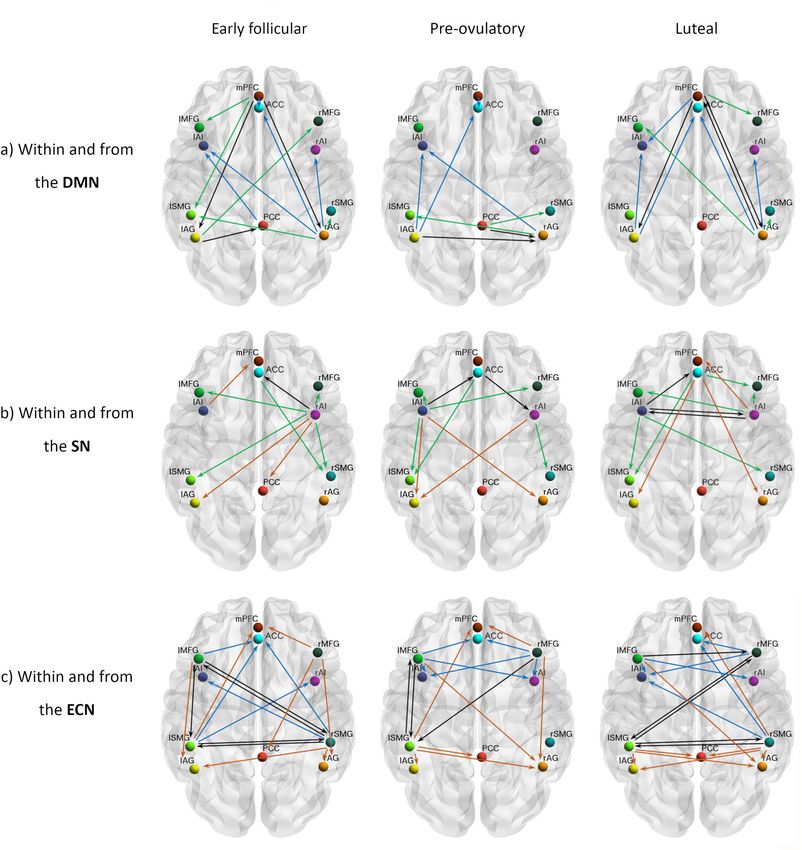
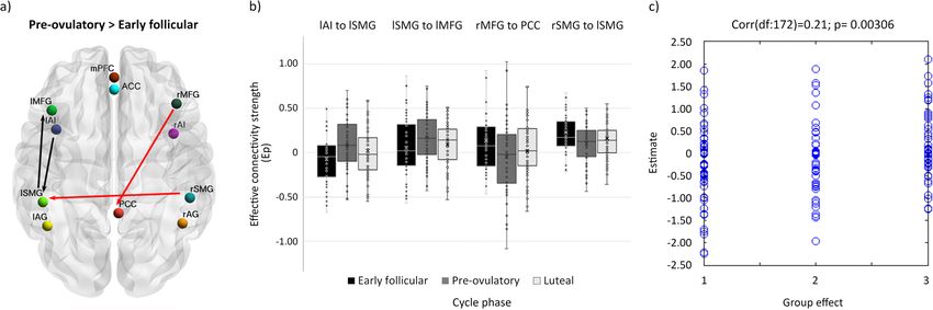
ARTICLE                                                                 COMMUNICATIONS BIOLOGY | https://doi.org/10.1038/s42003-021-02447-w

Fig. 4 Cycle phase differences in effective connectivity within and between intrinsic connectivity networks DMN, SN and ECN with a posterior
probability >0.99. a Differential connectivity strength between the indicated cycle phases is depicted by the width of the arrow. Black arrows reflect
positive values and red arrows reflect negative values for those connections which showed differences in the former than in the latter indicated cycle phase.
b Box plot showing parameter estimates per cycle phase for each connection, n = 174. c Out-of-samples correlation scatter plot from the LOOCV analysis
displaying the correlation between the actual cycle phase in the left-out-subject’s design matrix (early follicular, pre-ovulatory or mid-luteal) and the
predicted cycle phase based on the left-out-subject’s connectivity (rdf:172 = 0.21, p = 0.003). Source data can be found in Supplementary Data file 2 and 3,
respectively.

depicted in Fig. 5. In summary, during the early follicular phase,             between different brain networks56, but also as the most flexible
characterized by low levels of estradiol and progesterone, we                  node around the ovulatory window13,14. In Mueller et al. study13,
observed increased right lateralization of efferent connectivity               around ovulation, the PCC increased its functional connectivity
from the SN and DMN, increased integration within the DMN                      to other nodes, including prefrontal areas related to the DMN,
and between DMN and ECN, and a higher recruitment of the SN                    not included in our model. The present results corroborate
by the parietal ECN. Then, right before ovulation, the later-                  estradiol’s suggested role as a facilitator for this function13,14, and
alization shifted, as the left insula increased its efferent con-              extend previous work by adding the direction in which its levels
nectivity in response to heightened estradiol levels. It recruited             affect the PCC connectivity, being mostly the efferent connections
the fronto-parietal network, which caused the right MFG to                     to the other networks the ones positively related to estradiol levels
decouple from PCC. In exchange, the PCC engaged to bilateral                   (Fig. 1e).
AG, decoupling the DMN into anterior/posterior parts. Finally,                    The hypothesized increased connectivity between the DMN
during the mid-luteal phase, the SN increased the connectivity                 and SN36 was only corroborated partially, given the anterior/
within its own network, recruiting the right hemisphere again,                 posterior and lateralized pattern, and probably reflects an unba-
and affecting differentially the other networks depending on the               lanced mechanism rather than its organization in the non-
lateralization. Now the right insula recruited the frontal areas of            pathological brain. Women with a history of affective side effects
the other networks. In turn, frontal nodes of ECN maintained the               of the pill showed enhanced ACC-PCC connectivity both during
enhanced connectivity to the SN, while its posterior nodes                     treatment and the mid-luteal phase compared to menses (not pill-
increased their connectivity to the posterior DMN. In general,                 active)42. Furthermore, the over recruitment of the ACC into the
after ovulation, lateralization decreased as the homotopic regions             DMN and enhanced connectivity with the mPFC has been
of the ECN and SN were more connected to each other.                           characterized as unique for major depression symptoms and
   First, the present findings demonstrate an anterior-posterior                could reflect an inability to attend salient relevant external
modulation not only within the DMN but also in the connectivity                stimuli26. Conversely, in our model we observed a reduced con-
between the DMN and the other networks. The global integration                 nectivity from mPFC to ACC during the mid-luteal phase, related
of the DMN was highest during menses, with enhanced con-                       to higher progesterone levels, while increased mPFC-PCC cou-
nections from and within the entire DMN. During this phase,                    pling to parietal areas, involved in external information proces-
frontal ECN, particularly the left MFG, was most coupled with                  sing (Fig. 1e, Fig. 5). Thus, we suggest that these coupling-
the DMN, in line with Weis et al.46. Right before ovulation, the               decoupling effects respond to a compensatory mechanism in
bilateral AG disconnected from frontal areas and engaged to the                healthy women, and its absence could underlie the vulnerability
PCC and to each other, increasing the connectivity within the                  to affective symptoms described during the luteal phase36,57.
posterior DMN. After ovulation, the AG switched again their                       Second, these results suggest that the insula’s role as a switcher
connectivity to anterior areas disengaging from the PCC, and                   between networks may also respond to hormonal factors.
connections from the parietal ECN increased, especially to the                 Increased reactivity of the SN, crucial for detecting emotional
posterior part of the DMN. The increased connectivity between                  saliency, along with the deregulated coupling to frontoparietal
bilateral AG and anteromedial areas during the mid-luteal phase                systems has been proposed to underlie the vulnerability to
accompanied a decreased integrity of the posterior DMN, as the                 develop affective disorders, in general26, and particularly during
PCC decoupled from the DMN and was recruited by the parietal                   the luteal phase36,45,57. As expected, we found an increased within
ECN. Remarkably, these findings suggest that the decomposition                  SN connectivity during the mid-luteal phase related to higher
of DMN into anterior and posterior components, often described                 hormone levels, reflected in the enhanced inter-hemispheric
in ICA-analyses24,55 at least in women, respond to hormonal                    connectivity between the insulae (Figs. 1e, 5). Increased resting-
factors. Relatedly, the PCC has been already identified not only as             state functional connectivity between the insular cortices has
a highly interconnected node that may functionally switch                      already been related to higher estradiol levels in girls with

8                           COMMUNICATIONS BIOLOGY | (2021)4:954 | https://doi.org/10.1038/s42003-021-02447-w | www.nature.com/commsbio
COMMUNICATIONS BIOLOGY | https://doi.org/10.1038/s42003-021-02447-w                                                                 ARTICLE

Fig. 5 Summary of cycle-related differences in within and between-network effective connectivity. Each row depicts the connections observed to be
enhanced in each cycle phase compared to the others within and from (a) DMN; (b) SN; and (c) ECN. Only connections with a posterior probability >0.75
are displayed. Within-network connections are depicted in black and the efferent connectivity to the DMN in brown, to the SN in blue, and to the ECN
in green.

precocious puberty58 and higher flexibility of the limbic system               aspect of the insula’s function related to its role in responding to
concurrent to increased estradiol and progesterone levels, effect             changes in the endogenous hormonal milieu. Indeed, previous
absent when the menstrual cycle was disrupted13. As a core node               studies have already related the insula’s morphology and resting
strongly and reciprocally connected to widespread cortical and                state connectivity to women’s hormonal status in a lateralized
subcortical areas59, the insular cortex orchestrates interoception,           pattern65,66. In our sample, the left insula had higher connectivity
cognition and emotion, contributing to emotional awareness,                   to the anterior DMN during early follicular, while the right insula
learning and memory processes59–61. This unique position allows               was more strongly connected to the posterior DMN and ECN.
the AI to engage to the ECN as it disengages from the DMN,                    Then, during the pre-ovulatory phase related to the increased
acting as switcher between networks62–64.                                     estradiol levels, this pattern reversed and the left insula increased
   The present results not only corroborate these previous findings            its connectivity to posterior DMN, ACC and ECN, as the right
in causal networks’ dynamics62–64, but also suggest a further                 insula decreased it (Supplementary Data file 1). Although the

COMMUNICATIONS BIOLOGY | (2021)4:954 | https://doi.org/10.1038/s42003-021-02447-w | www.nature.com/commsbio                                        9
ARTICLE                                                             COMMUNICATIONS BIOLOGY | https://doi.org/10.1038/s42003-021-02447-w

bilateral AI are involved in the response to all emotional stimuli, a      but also according to an anterior/posterior specialization. Note-
left-hemisphere dominance for positive stimuli67 and empathy68             worthy, all four connections with the most confident effects
has been suggested, relating the lateralized pattern of insula cou-        included in the cross-validation analysis, involved at least one
pling and decoupling to the ECN to menstrual cycle-dependent               node from the ECN, reflecting the key role of this network in
changes in emotion processing69. During the mid-luteal phase               menstrual cycle-related changes and SN/DMN coupling
both insulae decoupled from the PCC as they increased the con-             dynamics.
nectivity to each other. In addition, the left insula maintained the          Fourth, across the menstrual cycle, two distinctive patterns
connectivity with parietal ECN while the right insula replaced its         regarding changes in lateralization could be distinguished. On the
counterpart recruiting again the frontal ECN. Accordingly, these           one hand, a right-lateralized pattern during early follicular, which
findings support the idea that the enhanced salience detection              changes to the left hemisphere during pre-ovulatory, and
during the mid-luteal phase could be buffered by coupling with             recruiting the right hemisphere again during the mid-luteal
frontal ECN. In fact, the increased connectivity between ACC and           phase. This is the case for connectivity from SMG to posterior
right MFG during the mid-luteal phase may reflect an enhanced               DMN, AG to SN, or the connectivity from the insula (broadly
top-downregulation in healthy women.                                       speaking). In addition, we observed an increased connectivity
   Third, the ECN showed both a lateralized and anterior-                  between homotopic regions of the SN and ECN during the mid-
posterior hormonal modulation, in line with previous neu-                  luteal phase. Therefore, in most cases, this time window was
roendocrine research. Converging evidence from animal and                  characterized by a decreased asymmetry, which has been pre-
human research relates estradiol to improved prefrontal-                   viously reported for different methods including behavioural76,
dependent functions, especially in verbal tasks (for a review, see         transcranial magnetic stimulation77 and fMRI studies40,78 and
ref. 5). In women, frontal areas such as the IFG and MFG have              suggested to reflect a reduced transcallosal inhibition79–81. On the
shown enhanced activity during the pre-ovulatory phase and                 other hand, a more symmetrical pattern during early follicular
related to increased estradiol levels for implicit memory70 and            and pre-ovulatory phase while right lateralized during the mid-
verbal encoding71. We recently found increased right frontros-             luteal phase is observed for the connectivity from ECN to the SN,
triatal functional connectivity during resting state, related to           in which the anterior-posterior modulation makes the scenario
higher estradiol levels during the pre-ovulatory phase21. The              more complex. Furthermore, and as discussed previously, the
increased afferent connectivity we observed from the left hemi-            right MFG also constitutes an exception to the lateralization
sphere to bilateral MFG right before ovulation could reflect                observed across the menstrual cycle: its connectivity to the SN
underlying structural changes, but in humans, the relationship             was the lowest during early follicular and increased as the hor-
between molecular mechanisms and neural functions are difficult             mone levels increased. These differential patterns could explain
to determine. Nevertheless, evidence from animal studies strongly          why the findings involving hemispheric asymmetries are some-
suggests the involvement of oestrogen-dependent synaptogenesis             time controversial and depend on the specific task and cognitive
in the PFC (in non-human primates3 and rodents72).                         system involved82.
   Our model shows a shift of lateralization across the cycle                 In summary, this triple network dynamic model corroborates
phases with the most evident change being the recruitment of               the plasticity of the brain in response to the acute ovarian hor-
bilateral MFG from the left insula versus the right insula during          mone fluctuations along the natural menstrual cycle. We have
early follicular and the mid-luteal phase. In turn, the increased          shown how both lateralization and anterior-posterior effective
top-down engagement to SN from frontal ECN was maintained                  connectivity patterns within and between networks depend on the
during the high-hormone phases, and positively related to both             endogenous hormonal status in healthy young women. Further-
estradiol and progesterone levels. As expected, the right MFG              more, when using those parameters with the largest posterior
increased its afferent connectivity during the mid-luteal phase,           probability, we found an above-chance prediction of the left-out
including from left MFG, which could explain the increased                 subjects’ cycle phase, although this should be interpreted with
executive-dependent activation previously observed37–40.                   caution, given the small effect size and therefore low percentage
Enhanced global network connectivity have been reported before             of the variance explained. Remarkably, all these connections
in dorsolateral PFC, associated with increased levels of                   reflect and summarize the effects that have been already dis-
progesterone41. Furthermore, overactivation of PFC15 and dif-              cussed: increased engagement of the DMN with the ECN during
ferential insular activity73 during high executive functions have          the early follicular phase, enhanced left frontoparietal con-
been described for premenstrual dysphoric disorder (PMDD).                 nectivity right before ovulation, increased frontoparietal recruit-
Specifically, in patients, the left insula was found less active than       ment by the left insula in response to enhanced estradiol levels as
in healthy controls before ovulation, while more active during the         well as interhemispheric decoupling in executive parietal areas,
mid-luteal phase compared to both controls and pre-ovulatory               partially reversed after ovulation. The impact of the surge of
patients73. If, as our results suggest, the balanced bottom-up/top-        estradiol levels around ovulation in brain connectivity and nodes
downregulation entails a shift in the lateralization of the dorso-         flexibility13,14 does not come as a surprise, since the neu-
lateral PFC afferent connections from the SN, its absence or               roendocrine feedback loops triggered by it are quite unique in the
deregulation could underlie some symptomatology.                           human physiology, and the biological relevance of ovulation is
   After ovulation, parietal areas coupled to the posterior DMN            undeniable. Structural changes between the early follicular and
and to each other. Related to higher progesterone levels (Sup-             the pre-ovulatory phase have been previously shown to accurately
plementary Data file 1), the right SMG increased the efferent               classify cycle phase using a machine learning approach, and
connectivity to every node of the other networks but to right              related to estradiol levels (BrainAGE83). A more detailed dis-
insula, which similarly decreased its connectivity to right SMG. A         cussion of the functional role of each of the connections included
rightward asymmetry in SMG-insula connectivity has been found              in the LOOCV can be found in the Supplementary Note 1.
to be stronger in females than in males, and related to suscept-              The present work also corroborates a differential effect of each
ibility to chronic pain disorders74. In addition, the right SMG is         hormone depending on the brain region and network dynamics,
particularly involved in shifting attention to salient stimuli75. The      which remarks the relevance of widening the focus to large-scale
shift in connectivity between the right insula and posterior ECN           systems interaction, rather than the activity of localized brain
to anterior ECN after ovulation may reflect the dynamic inte-               areas. In fact, the cycle-related effects on connectivity from dif-
gration of bottom-up/top-down processes, not only lateralized              ferent nodes of a network to other networks suggest that ICA, the

10                       COMMUNICATIONS BIOLOGY | (2021)4:954 | https://doi.org/10.1038/s42003-021-02447-w | www.nature.com/commsbio
COMMUNICATIONS BIOLOGY | https://doi.org/10.1038/s42003-021-02447-w                                                                                                  ARTICLE

most used approach up to now, might be too coarse to capture                              Table 2 Group level volume of interest coordinates.
the hormonal effects since the time-courses of various areas are
averaged together and the results depend on the areas included in
                                                                                          Region              MNI coordinates                                        Network
the ICN. Although we are still far away to comprehend the full
scope of the ovarian hormones effects on the human brain cou-                                                 X                     Y                Z
pling dynamics, this proposed causal model of menstrual cycle-                            PCC                 3                     −55              31              DMN
related changes may improve our understanding of the under-                               lAG                 −45                   −64              31              DMN
lying neuroendocrine interactions. At least to some extent, the                           rAG                 46                    −63              34              DMN
neural substrate needs the cyclic ovarian hormones fluctuation to                          mPFC                0                     53               −14             DMN
undergo this brain reorganization, as evidenced by animal12,84                            lAI                 −39                   14               −5              SN
and human research13,14,85. Therefore, results on healthy natu-                           rAI                 40                    11               −5              SN
rally cycling women may have further implications for those                               ACC                 0                     35               19              SN
menstrual cycle-related disorders where this pattern of brain                             lSMG                −48                   −49              37              ECN
coupling dynamics has been suggested to be impaired, such as                              rSMG                51                    −46              37              ECN
                                                                                          lMFG                −42                   17               31              ECN
premenstrual syndrome (PMS), PMDD or dysmenorrhoea19,86.
                                                                                          rMFG                42                    20               34              ECN
Moreover, some factors in other neurological and psychiatric
disorders, where the triple model network is impaired26, have                             l left, r right, PCC precuneus/posterior cingulate cortex, AG angular gyrus, mPFC medial
                                                                                          prefrontal cortex, DMN default mode network, AI anterior insula, ACC anterior cingulate cortex,
been previously related to hormonal levels, like the incidence of                         SN salience network, MFG middle frontal gyrus, SMG supramarginal gyrus, ECN executive
epileptic seizures87 addiction patterns and drug sensitivity88, as                        control network.
well as the sensitivity and risk to develop affective disorders89,90,
Alzheimer’s disease91 or Schizophrenia92,93. Therefore, some of
                                                                                         2250 ms, flip angle 70°, slice thickness 3.0 mm, matrix 192 × 192, FOV 192 mm, in-
the key findings here described, correspondent to healthy                                 plane resolution 2.6 × 2.6 mm). Participants were instructed to close their eyes,
mechanisms, may be targeted in future clinical neuroendocrine                            relax and let their mind flow. For the structural images we acquired a T1-weighted
studies.                                                                                 3D MPRAGE sequence of 5 min 58 sec (160 sagital slices, slice thickness = 1 mm,
                                                                                         TE 291 ms, TR 2300 ms, TI delay 900 ms, FA 9°, FOV 256 × 256 mm).

Methods
Participants. Seventy-eight healthy young women participated in 1 of 2 functional        Preprocessing. For the preprocessing, the first six images of each session were
imaging studies21. Main inclusion criteria were an age range of 18–35 years, and a       discarded, and functional images were despiked using 3d-despiking as imple-
regular menstrual cycle (MC) of 21–35 days with a variability between cycles of          mented in AFNI (afni.nimh.nih.gov). The despiked images were then pre-
ARTICLE COMMUNICATIONS BIOLOGY | https://doi.org/10.1038/s42003-021-02447-w
For the second-level analyses, the parameters (effective connectivity strengths) 7. Vedder, L. C., Smith, C. C., Flannigan, A. E. & McMahon, L. L. Estradiol-
were estimated in a Parametric Empirical Bayes (PEB) framework as described in induced increase in novel object recognition requires hippocampal NR2B-
Zeidman et al.104 The second-level model captured between-session and between- containing NMDA receptors. Hippocampus 23, 108–115 (2013).
subject effects, with a general linear model (GLM) to capture effects of interest and 8. Barreto-Cordero, L. M. et al. Cyclic changes and actions of progesterone and
a covariance component model to capture random effects (implemented in the allopregnanolone on cognition and hippocampal basal (stratum oriens)
Parametric Empirical Bayes framework in the SPM software, see ref. 104). dendritic spines of female rats. Behav. Brain Res. https://doi.org/10.1016/j.
An advantage of this fully Bayesian approach is that models with different bbr.2019.112355 (2020).
regressors in their group-level design matrix can be compared in terms of their log 9. Markowska, A. L. & Savonenko, A. V. Effectiveness of estrogen replacement in
model evidence or marginal likelihood (thereby identifying the model that offers restoration of cognitive function after long-term estrogen withdrawal in aging
the best trade-off between accuracy and complexity). In a preliminary analysis, we rats. J. Neurosci. 22, 10985–10995 (2002).
evaluated whether including dummy regressors for each subject in addition to 10. Hao, J. et al. Estrogen alters spine number and morphology in prefrontal
regressors for each phase of the menstrual cycle (thereby forming a within-subject cortex of aged female rhesus monkeys. J. Neurosci. https://doi.org/10.1523/
repeated measures ANOVA) would increase the model evidence, relative to a JNEUROSCI.3440-05.2006 (2006).
model that only included regressors for each phase of the menstrual cycle. We 11. Hao, J. et al. Interactive effects of age and estrogen on cognition and
found that the evidence decreased when including subject-specific regressors (i.e., pyramidal neurons in monkey prefrontal cortex. Proc. Natl Acad. Sci. USA
the added complexity outweighed any increase in accuracy). Therefore, we did not
104, 11465–11470 (2007).
include subject-specific regressors in the final regression model.
12. Ohm, D. T. et al. Clinically relevant hormone treatments fail to induce
To compute the difference in effective connectivity between the three different
spinogenesis in prefrontal cortex of aged female rhesus monkeys. J. Neurosci.
phases, the final regression model included three regressors: first, pre-ovulatory
https://doi.org/10.1523/JNEUROSCI.1881-12.2012 (2012).
versus early follicular; second, mid-luteal versus early follicular; and third, mid-
luteal versus pre-ovulatory (Fig. 1d). The PEB analysis returned estimated effect 13. Mueller, J. M. et al. Dynamic community detection reveals transient
sizes (PEB.Ep), in addition to the posterior probability (PEB.Pp) for each effect reorganization of functional brain networks across a female menstrual cycle.
having diverged from its prior expectation of zero. In Bayesian analysis there is no Netw. Neurosci. 5, 125–144 (2021).
need for further thresholding—there is simply the probability for each effect, with 14. Pritschet, L. et al. Functional reorganization of brain networks across the
no concept of ‘significance’. Nevertheless, it can be helpful to apply thresholding in human menstrual cycle. Neuroimage 220, 117091 (2020).
order to focus on the most probable effects. Here, we opted to use a commonly 15. Baller, E. B. et al. Abnormalities of dorsolateral prefrontal function in women
adopted definition of ‘positive evidence’54, by thresholding our effects at 75% with premenstrual dysphoric disorder: a multimodal neuroimaging study. Am.
posterior probability (Fig. 1). Only parameters surviving this threshold are reported J. Psychiatry 170, 305–314 (2013).
in the results section and summarized in Supplementary Data file 1. 16. Lisofsky, N. et al. Hippocampal volume and functional connectivity changes
We further assessed the hormonal modulation of the connections with a second during the female menstrual cycle. Neuroimage 118, 154–162 (2015).
PEB analysis including scaled estradiol and progesterone levels, and their 17. Pletzer, B., Harris, T. A. & Hidalgo-Lopez, E. Subcortical structural changes
interaction as regressors (Fig. 1e). Likewise, results in Supplementary Data file 1 along the menstrual cycle: beyond the hippocampus. Sci. Rep. 8, 16042 (2018).
were thresholded to only include parameters that had a 75% posterior probability. 18. Barth, C. et al. In-vivo dynamics of the human hippocampus across the
Those connections surviving a 95% threshold for both PEB1 and PEB2 are further menstrual cycle. Sci. Rep. https://doi.org/10.1038/srep32833 (2016).
indicated in the figures and results section alongside each parameter’s mean and 19. Toffoletto, S., Lanzenberger, R., Gingnell, M., Sundström-Poromaa, I. &
posterior probability (Fig. 1d, e). Nevertheless, given that all parameters Comasco, E. Emotional and cognitive functional imaging of estrogen and
contributed to the model, we provide figures listing all parameters regardless of progesterone effects in the female human brain: a systematic review.
threshold in the supplementary information (SI, Figs. S2 and S3). Psychoneuroendocrinology 50, 28–52 (2014).
Finally, for leave-one-out cross-validation (LOOCV), there is a strong dilution- 20. Sacher, J., Neumann, J., Okon-Singer, H., Gotowiec, S. & Villringer, A. Sexual
of-evidence effect, where parameters with small effect sizes can markedly reduce dimorphism in the human brain: evidence from neuroimaging. Magn. Reson.
predictive accuracy. It is important, therefore, to first perform feature selection, Imaging 31, 366–375 (2013).
retaining only the largest or most probable parameters. Therefore, we only assessed 21. Hidalgo-Lopez, E. et al. Human menstrual cycle variation in subcortical
the predictive accuracy of those parameters with 99% probability of being non- functional brain connectivity: a multimodal analysis approach. Brain Struct.
zero. In order to do so we used a leave-one-out scheme (spm_dcm_loo.m) as Funct. 225, 591–605 (2020).
described in Friston et al.50 and tested whether the effect on these particular 22. Biswal, B., Zerrin Yetkin, F., Haughton, V. M. & Hyde, J. S. Functional
connections could predict the cycle phase of participants. connectivity in the motor cortex of resting human brain using echo???planar
mri. Magn. Reson. Med. 34, 537–541 (1995).
Reporting summary. Further information on research design is available in the Nature 23. Laird, A. R. et al. Behavioral interpretations of intrinsic connectivity networks.
Research Reporting Summary linked to this article. J. Cogn. Neurosci. 23, 4022–4037 (2011).
24. Damoiseaux, J. S. et al. Consistent resting-state networks across healthy
subjects. Proc. Natl Acad. Sci. USA 103, 13848–13853 (2006).
Data availability 25. Fox, M. D. & Raichle, M. E. Spontaneous fluctuations in brain activity
Data and scripts are openly available online at http://webapps.ccns.sbg.ac.at/OpenData/ observed with functional magnetic resonance imaging. Nat. Rev. Neurosci. 8,
and OSF, https://osf.io/23d7x/105. MR-images are available upon request from the first 700–711 (2007).
author. A summary of the results is provided in Supplementary Data file 1 and source 26. Menon, V. Large-scale brain networks and psychopathology: a unifying triple
data for Fig. 4b and c are provided in Supplementary Data file 2 and 3, respectively. network model. Trends Cogn. Sci. 15, 483–506 (2011).
27. Raichle, M. E. et al. A default mode of brain function. Proc. Natl Acad. Sci.
Received: 10 September 2020; Accepted: 1 July 2021; USA 98, 676–682 (2001).
28. Greicius, M. D., Krasnow, B., Reiss, A. L. & Menon, V. Functional connectivity
in the resting brain: a network analysis of the default mode hypothesis. Proc.
Natl Acad. Sci. USA 100, 253–258 (2003).
29. Dosenbach, N. U. F. et al. Distinct brain networks for adaptive and stable task
control in humans. Proc. Natl Acad. Sci. USA 104, 11073–11078 (2007).
30. Seeley, W. W. et al. Dissociable intrinsic connectivity networks for salience
References processing and executive control. J. Neurosci. 27, 2349–2356 (2007).
1. McEwen, B. S. & Milner, T. A. Understanding the broad influence of sex
31. Hjelmervik, H., Hausmann, M., Osnes, B., Westerhausen, R. & Specht, K.
hormones and sex differences in the brain. J. Neurosci. Res. 95, 24–39 (2017).
Resting states are resting traits—An fMRI study of sex differences and
2. Clemens, A. M. et al. Estrus-cycle regulation of cortical inhibition. Curr. Biol.
menstrual cycle effects in resting state cognitive control networks. PLoS ONE
29, 605–615.e6 (2019).
9, 32–36 (2014).
3. Tang, Y. et al. Estrogen replacement increases spinophilin-immunoreactive
32. De Bondt, T. et al. Stability of resting state networks in the female brain during
spine number in the prefrontal cortex of female rhesus monkeys. Cereb. Cortex
hormonal changes and their relation to premenstrual symptoms. Brain Res.
https://doi.org/10.1093/cercor/bhg121 (2004).
1624, 275–285 (2015).
4. Schumacher, M. et al. Revisiting the roles of progesterone and
33. Pletzer, B., Crone, J. S., Kronbichler, M. & Kerschbaum, H. Menstrual cycle
allopregnanolone in the nervous system: Resurgence of the progesterone
and hormonal contraceptive-dependent changes in intrinsic connectivity of
receptors. Prog. Neurobiol. 113, 6–39 (2014).
resting-state brain networks correspond to behavioral changes due to
5. Luine, V. N. Estradiol and cognitive function: Past, present and future. Horm.
hormonal status. Brain Connect 6, 572–585 (2016).
Behav. 66, 602–618 (2014).
34. Petersen, N., Kilpatrick, L. A., Goharzad, A. & Cahill, L. Oral contraceptive pill
6. Frick, K. M. Molecular mechanisms underlying the memory-enhancing effects
use and menstrual cycle phase are associated with altered resting state
of estradiol. Horm. Behav. 74, 4–18 (2015).
functional connectivity. Neuroimage 90, 24–32 (2014).
12 COMMUNICATIONS BIOLOGY | (2021)4:954 | https://doi.org/10.1038/s42003-021-02447-w | www.nature.com/commsbioYou can also read