Perineuronal net degradation rescues CA2 plasticity in Rett syndrome model mice
←
→
Page content transcription
If your browser does not render page correctly, please read the page content below
Perineuronal net degradation rescues CA2 plasticity in Rett
syndrome model mice
Kelly E. Carstens, … , Georgia M. Alexander, Serena M. Dudek
J Clin Invest. 2021. https://doi.org/10.1172/JCI137221.

 Research   In-Press Preview Development      Neuroscience

Perineuronal nets (PNNs), a specialized form of extracellular matrix, are abnormal in the human brain of Rett syndrome
(RTT). We previously reported that PNNs function to restrict synaptic plasticity in hippocampal area CA2, which is
unusually resistant to long-term potentiation (LTP) and has been linked to social learning in mice. Here we reported that
PNNs appear elevated in area CA2 of a human RTT hippocampus and that PNNs develop precociously and remain
elevated in area CA2 of a mouse model of RTT (Mecp2-null). Further, we provided evidence that LTP could be induced at
CA2 synapses prior to PNN maturation (postnatal day 8-11) in wildtype mice and that this window of plasticity was
prematurely restricted at CA2 synapses in Mecp2-null mice. Degrading PNNs in Mecp2-null hippocampus was sufficient
to rescue the premature disruption of CA2 plasticity. We identified several molecular targets that were altered in the
developing Mecp2-null hippocampus that may explain aberrant PNNs and CA2 plasticity, and we discovered that CA2
PNNs are negatively regulated by neuronal activity. Collectively, our findings demonstrated that CA2 PNN development is
regulated by Mecp2 and identified a novel window of hippocampal plasticity that is disrupted in a mouse model of RTT.

Find the latest version:
https://jci.me/137221/pdf
Title: Perineuronal net degradation rescues CA2 plasticity in Rett syndrome model mice

Kelly E. Carstens1, 2 , Daniel J. Lustberg1, 3, Emma Shaughnessy1, 4, Katharine E. McCann1, 3, Georgia M.
Alexander1, and Serena M. Dudek1, 5

1
  Neurobiology Laboratory, National Institute of Environmental Health Sciences, National Institutes of
Health, 111 TW Alexander Dr., MD F2-04, Research Triangle Park, NC 27709, USA
2
  Current address:
Center for Computational Toxicology and Exposure, Environmental Protection Agency, Research
Triangle Park, NC 27709, USA
3
  Current address: Department of Human Genetics, Emory University, Atlanta, GA 30322, USA
4
  Current address: Neuroscience Institute, Georgia State University, Atlanta, GA 30303, USA
5
  Corresponding author: 984-287-3513, dudek@niehs.nih.gov

Conflict of interest statement: The authors have declared that no conflict of interest exists.

Number of pages: 30
Number of figures: 5
Number of tables: 1
Words- Abstract: 197
Words- Introduction: 891
Words- Results: 2921
Words- Discussion: 2109
Words- Figure Legends and methods: 1496+160= 1656
Words- References: 3064
Total number of words (Abstract, Introduction, Materials & Methods, Results, Discussion, References
and Figure Legends): 10,839

                                                                                                            1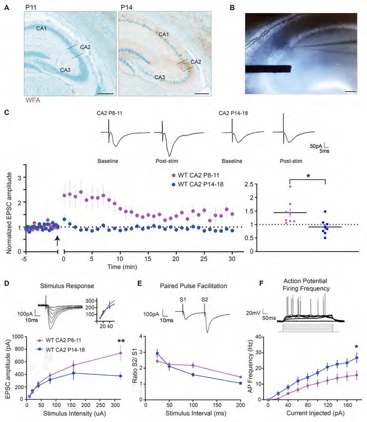
Abstract:

        Perineuronal nets (PNNs), a specialized form of extracellular matrix, are abnormal in the human

brain of Rett syndrome (RTT). We previously reported that PNNs function to restrict synaptic plasticity

in hippocampal area CA2, which is unusually resistant to long-term potentiation (LTP) and has been

linked to social learning in mice. Here we reported that PNNs appear elevated in area CA2 of a human

RTT hippocampus and that PNNs develop precociously and remain elevated in area CA2 of a mouse

model of RTT (Mecp2-null). Further, we provided evidence that LTP could be induced at CA2 synapses

prior to PNN maturation (postnatal day 8-11) in wildtype mice and that this window of plasticity was

prematurely restricted at CA2 synapses in Mecp2-null mice. Degrading PNNs in Mecp2-null

hippocampus was sufficient to rescue the premature disruption of CA2 plasticity. We identified several

molecular targets that were altered in the developing Mecp2-null hippocampus that may explain aberrant

PNNs and CA2 plasticity, and we discovered that CA2 PNNs are negatively regulated by neuronal

activity. Collectively, our findings demonstrated that CA2 PNN development is regulated by Mecp2 and

identified a novel window of hippocampal plasticity that is disrupted in a mouse model of RTT.

                                                                                                          2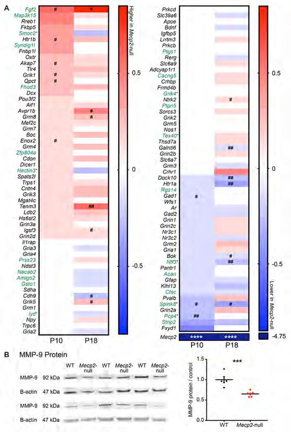
Introduction

        Rett syndrome (RTT) is a neurodevelopmental disorder caused by a loss-of-function mutation in

the gene methyl-CpG-binding protein 2 (MECP2), affecting about 1 in 10,000 girls worldwide (1, 2). The

MECP2 gene is located on the X-chromosome and MeCP2 functions as a transcriptional regulator, both

activating and repressing various target genes (3, 4). A defining characteristic of RTT symptomology is

apparently normal development in the first year of life, followed by a rapid, profound regression in

cognitive, motor, and social function. Some common symptoms of RTT include loss of coordination and

language skills, development of stereotypic hand movements, severe autonomic dysfunction, and

recurrent seizures (5-10). It is also typical for girls with RTT to exhibit autistic-like behaviors such as

social avoidance (11, 12). Early studies of postmortem brain tissue from RTT individuals reveal a reduced

number of dendritic spines and presynaptic markers, and rodent studies reveal deficits in neurogenesis,

synaptic plasticity, and experience-dependent synapse remodeling (1, 13-16). Although symptoms

develop approximately one year into postnatal life in humans, abnormalities in the RTT mouse brain

appear to develop prior to the presentation of symptoms (17-24). Much research has focused on

understanding the initiation and progression of RTT and how MECP2 mutation may disrupt critical

periods of early learning and memory.

        Among the pathologies that have been observed in the RTT brain are aberrant perineuronal nets

(PNNs), a specialized extracellular matrix (ECM) that typically deposit around inhibitory neurons in the

brain and have been implicated in several psychiatric and neurological disorders (25-29). In the normal

brain, PNNs first appear during postnatal development and gradually increase until they are fully mature

in adulthood (29-32). Their development is modulated by early-life experience (33-36) and mature PNNs

can be altered by neuronal activity, such as seizure (37-39). PNNs have been implicated in a wide variety

of functions, with recent focus on their role in limiting plasticity during critical windows of development

(40-44). PNN pathology was first identified in motor cortices of postmortem human RTT tissue, where

PNN-positive neurons were both more numerous and more intensely stained in layers III and V of motor

cortex compared to control cases (27). Similarly, in a mouse model of RTT, PNNs associated with

                                                                                                              3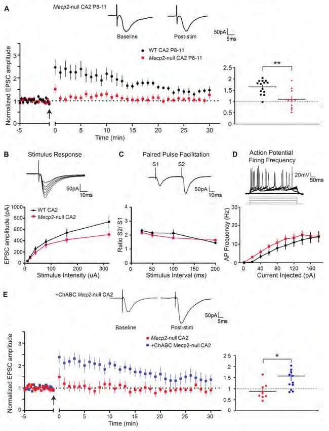
inhibitory neurons in the visual cortex were found to mature precociously and to be associated with an

accelerated onset and closure of critical period plasticity (45-47). These findings reveal a link between the

loss of Mecp2 and PNN pathology in several different brain regions and a possible link between PNN

pathology and impaired critical windows of plasticity. To this end, we sought to investigate PNNs in the

hippocampus of a mouse model of RTT (genetic knockout of the Mecp2 gene; Mecp2-null), a region

associated with many learning and memory impairments exhibited in RTT.

        Several transgenic and knockout mouse models of RTT have been generated that mimic human

RTT clinical symptomology and exhibit both neurobiological and behavioral impairments (14, 48, 49).

These mice exhibit reductions in dendritic spine number, impairments in excitatory and inhibitory

transmission, aberrant neurogenesis, and disorganization of axonal fibers (18, 50-52). Although, RTT

syndrome typically affects young girls, Mecp2-null male mice are often used as a model of RTT for

several reasons: 1) a homozygous knockout of Mecp2, an X-linked gene, is uncommon in the Mecp2-null

female (Mecp2-/-) because Mecp2-null males are unable to breed, while the heterozygous Mecp2-null

(Mecp2-/x) results in a partial knockout and mosaic expression of Mecp2 in the brain (53); 2)

heterozygous females, which we use for breeding in this study, do not present Rett-like symptomology

during early postnatal development but instead present delayed and variable phenotypic progression; 3)

the onset of pathology in the Mecp2-null males occurs in early postnatal development, a delay which

generally mimics the apparently normal early development and subsequent decline around 10-14 months

of age that is characteristic of individuals with RTT; and 4) in general, the severity of phenotypic

impairments observed in the Mecp2-null males more closely model the presentation of clinical features

relative to the heterozygous female. Whether neurological dysfunction develops prior to the onset of RTT

symptoms remains unclear, but several rodent studies suggest that abnormalities are present at pre-

symptomatic ages (19, 22, 54, 55). Understanding when and how neurological dysfunction develops in

the absence of Mecp2 will be critical for the ultimate goal of identifying early windows of intervention in

RTT individuals.

                                                                                                            4
In this study, we aimed to determine whether PNN pathology was present in the hippocampus of
a mouse model of RTT during early development. We focused specifically on hippocampal area CA2, a
population of excitatory pyramidal neurons that distinctly express PNNs in the hippocampus of both
mouse and human (29, 56-59). Unlike in neighboring hippocampal subregions, CA2 synapses are
resistant to the induction of plasticity at synapses in the stratum radiatum (60). The function of plasticity
resistance in CA2 remains to be established, although recent work points to a role for CA2 in social
learning and behavior (61-65). We previously identified PNNs as a negative regulator of plasticity at CA2
synapses of acute hippocampal slices (29, 66). Given that MeCP2 is a known regulator of critical period
plasticity in the developing brain and that PNNs are heavily implicated in regulating critical windows of
plasticity, we aimed to determine whether Mecp2 deletion resulted in PNN pathology in the developing
hippocampus, particularly in area CA2 where PNNs are observed around pyramidal neurons.
Results:
PNNs are increased in human RTT CA2 and develop prematurely in CA2 of a mouse model of RTT
PNNs function as at least one brake on synaptic plasticity in CA2 (29). Interestingly, previous
studies have found that PNN structural complexity is increased in motor cortex of individuals with RTT
and in visual cortex of Mecp2-null mice (27, 47). To determine whether PNNs may be altered in the
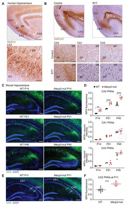
human RTT hippocampus, we stained postmortem human tissue from a RTT individual (NIH
NeuroBioBank) with an antibody against the PNN component, HAPLN1 link protein (58). First we
identified CA2 pyramidal cells in human hippocampus with the CA2 marker, RGS14 (67) (Figure 1A).
We next validated that PNNs localize to human area CA2 in control tissue. We found that HAPLN1 stain
is concentrated in area CA2 (Figure 1B) and that the HAPLN1 stain was darker in hippocampal tissue
from the RTT individual than in tissue from an age-matched healthy individual (Figure 1B). Higher
magnification images revealed dense localization of HAPLN1 stain to stratum pyramidale (SP) and
stratum radiatum (SR) layers, with the highest intensity of staining being in the SR of the RTT individual.
5Given the variability of the quality of human tissue, we next sought to determine whether PNN deposition
may be similarly altered in CA2 in a mouse model of RTT in which we could more precisely control
staining conditions.
Clinical features of RTT typically present after the first year of life and rapidly increase in
severity over the next several years (68). We therefore investigated PNN deposition in the hippocampus
over the course of postnatal development in a mouse model of RTT wherein Mecp2 is deleted (Mecp2-
null). Staining for mature PNNs with the marker wisteria floribunda agglutinin (WFA) first appears in
hippocampal area CA2 around postnatal day (P)14 in control mice and increases in intensity up to
adulthood (29). We therefore quantified WFA staining in tissue from Mecp2-null animals at P14, P21 and
P45 and found that staining for WFA was significantly greater in area CA2 in Mecp2-null mice compared
with wildtype (WT) littermates at each of these ages (Figure 1C two-way ANOVA, Bonferroni post hoc
test, **P< 0.005, ***P 0.05,
Figure 1D). We next looked at an age prior to the normal maturation of PNNs at P14 observed in WT
mice to determine whether CA2 PNNs may develop precociously in RTT. We found that PNN staining
was more intense at P11 in Mecp2-null mice compared with WT littermates (***P=0.0005, two-tailed
unpaired t-test, normalized to WT littermate, Figure 1E), indicating that PNNs develop prematurely in
CA2 of RTT mice.
MeCP2 is expressed in multiple cell types throughout the brain, including inhibitory interneurons,
astrocytes, and microglia, which have been implicated in the behavioral deficits observed in RTT model
mice (69-72). We therefore targeted the deletion of Mecp2 to CA2 pyramidal neurons to investigate the
cell-autonomous effect of the loss of Mecp2 in CA2 pyramidal neurons. We crossed a line of mice
expressing Cre recombinase in CA2 pyramidal neurons (Amigo2-Cre (73)) with a floxed-Mecp2 mouse.
This targeted deletion resulted in a similar increase in WFA staining in CA2 as observed in the Mecp2-
mice, and not in PNN+ neurons in neighboring CA1 and CA3 regions (Supplemental Figure 1).
6Potentiation at CA2 stratum radiatum (SR) synapses can be induced in early postnatal
development, before PNN maturation
To determine whether this premature development of PNNs had effects on synaptic plasticity we
first needed to characterize plasticity atour LTP induction protocol (15-30 µA), see inset. Paired-pulse facilitation (PPF) did not differ between
age groups (two-tailed unpaired t-test, P>0.05, n= 14 for P8-11 and n=7 for P14-18, Figure 2E) and AP
firing frequency of CA2 neurons was greater at P14-18 compared with P8-11 over a range of injected
currents (*P= 0.0185 at 180 pA of injected current, n= 20 and 13 respectively, two-way ANOVA,
Bonferroni post hoc test, Figure 2F). Note that the stimulus-response and PPF experiments were
performed without GABA blockers due to epileptiform activity during recordings at P8-11. Because we
found that PNNs develop prematurely in the Mecp2-null mice, we next investigated how the loss of
Mecp2, a known regulator of critical period plasticity, impacts this newly discovered window of CA2
synaptic plasticity.
Synaptic potentiation is prematurely restricted at CA2 SR synapses in a mouse model of RTT
We next tested whether the precocious development of PNNs alters LTP induction in Mecp2-null
CA2. First, we replicated the finding that plasticity occurs in CA2 at P8-11 in a separate cohort of male
WT littermates (Figure 3A). Consistent with the finding that global loss of Mecp2 accelerates the closure
of critical period plasticity in the visual cortex (45, 47, 74), we found that potentiation in CA2 is
prematurely restricted at P8-11 SR synapses in Mecp2-null compared to WT littermates (1.10 ± 0.2
baseline versus 1.72 ± 0.1 baseline in WT at 18-20 minutes, n=13 and 14, respectively, **P= 0.0031,
two-tailed unpaired t-test, Figure 3A). Baseline synaptic transmission, as assessed with a stimulus-
response curve, was not different at P8-11 CA2 synapses of Mecp2-null compared to WT littermates (P>
0.05 at 320 pA current stimulation, two-way ANOVA repeated measures, Sidak’s multiple comparison
test, Figure 3B). In addition, neither PPF nor AP firing frequency differed significantly between Mecp2-
null and WT littermates at P8-11 (P>0.05, two-way ANOVA, Figure 3C, D). Intrinsic properties did not
differ in a way that could explain the premature restriction of plasticity in CA2 (Table 1, P>0.05, unpaired
t-test). Although we did not perform these experiments in the presence of GABA blockers due to
epileptiform activity, we did perform stimulus-repsonse and PPF experiments at P14-18 in the presence of
the GABAA receptor blocker, bicuculline, and found no overall difference between Mecp2-null and WT
8littermates in excitatory response size or PPF (P>0.05, two-way ANOVA repeated measures, Sidak’s
multiple comparison test, Supplemental Figure 2).
PNN degradation restores plasticity at CA2 Mecp2-null synapses
Because we found an increase in both PNNs and another plasticity-restricting protein, RGS14, in
RTT CA2 at P10 (Supplemental Figure 3A), we sought to determine whether the precocious increase in
PNNs alone was sufficient to explain the premature restriction of LTP in RTT CA2. In this experiment,
we tested whether degrading PNNs with the exogenous enzyme chondroitinase (ChABC) in acutely
prepared hippocampal slices was sufficient to enable LTP induction in RTT CA2 at P8-11. We previously
reported that degrading PNNs with 0.05 U/ml ChABC for ≥2 hours is sufficient to degrade PNNs in CA2
in slices (see Figure 4 and methods in reference (29)). Following the same protocol, we found that
degradation of PNNs in P8-11 Mecp2-null hippocampal slices ‘restored’ the capacity for potentiation in
CA2 (1.57 ± 0.2 baseline versus 0.90 ± 0.1 baseline in untreated Mecp2-null at 18-20 minutes, n=14 and
10 respectively, *P= 0.035, two-tailed unpaired t-test, Figure 3E). Intrinsic properties did not change in a
way that would explain the restoration of LTP at CA2 synapses with ChABC treatment (Table 1). Taken
together, these data demonstrate that degradation of the aberrantly increased PNNs in RTT CA2 appears
sufficient to restore synaptic plasticity in CA2 at young postnatal ages. To better understand what might
be driving the precocious increase of PNNs in RTT CA2, and because seizure activity is comorbid with
RTT, we next investigated the role of aberrant neuronal activity as a potential mechanism upregulating
PNNs in CA2 (38, 75).
CA2 PNNs are inversely regulated by neuronal activity in vivo
PNN development is dependent on early-life experience and neuronal activity, shown in several
sensory brain regions and across several species (32, 33, 35, 75-77). For example, visual deprivation from
birth delays the deposition of PNNs and attenuates overall expression levels in the visual cortex (34).
Accordingly, we hypothesized that a precocious upregulation of PNNs in RTT CA2 is driven by increased
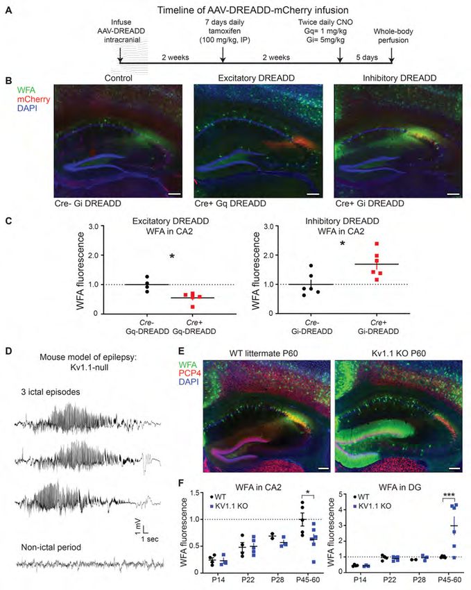
9hippocampal activity (78). To investigate this, we directly manipulated CA2 neuronal activity using a
chemogenetic approach (Designer Receptors Exclusively Activated by Designer Drugs (DREADD)). To
target CA2 pyramidal neurons, we infused an AAV vector encoding Cre-dependent Gq- (excitatory) or
Gi-coupled (inhibitory) DREADDs into CA2 of adult mice expressing Cre recombinase in CA2 neurons
(79). In a previous study from our lab, we found that clozapine N-oxide (CNO) increased the firing rate of
CA2/ proximal CA1 pyramidal neurons in mice expressing excitatory DREADDs (hM3Dq) in CA2 (79).
Similarly CNO decreased CA2 responses in mice expressing the inhibitory DREADD (hM4Di), with
effects lasting up to 24 hours post DREADD activation (79). In this study, we either chronically increased
or chronically decreased CA2 activity for 5 days and quantified WFA fluorescence in the hippocampus
(Figure 4A). Contrary to our hypothesis, we found that increased neuronal activity in the Gq DREADD-
expressing animals significantly decreased WFA staining in CA2 compared to Cre- controls (*P= 0.0111,
unpaired two-tailed t-test, Figure 4B and C). Conversely, we found the opposite in the Gi DREADD-
expressing animals: WFA staining was significantly increased in CA2 compared to Cre- controls (*P=
0.0179, unpaired two-tailed t-test). We also found that WFA staining in inhibitory neurons in areas CA1
and CA3, which did not express the DREADDs, was unchanged in Gi DREADD- or Gq DREADD-
expressing mice (Supplemental Figure 3B, P > 0.05). We did, however, observe a significant increase in
WFA fluorescence in overlying primary somatosensory cortex in Gq DREADD mice, suggesting that
altered CA2 activity has extrahippocampal effects (Supplemental Figure 3B, **P=0.0012, one-way
ANOVA, Tukey’s post hoc). Overall, these data demonstrate that PNNs in CA2 are cell-autonomously
regulated by aberrant neuronal activity and suggests that PNNs in CA2 are negatively regulated by
activity. Of note, neuronal activity in these experiments was manipulated in adulthood, leaving open the
possibility that aberrant changes in activity at even younger ages may still explain abnormal PNN
maturation.
In order to investigate the effects of aberrant activity on PNNs earlier in postnatal development,
we next studied the effects of pathological activity using a mouse model of epilepsy. The development of
seizures in childhood and persistent epilepsy is a common and devastating comorbidity of RTT (28, 80,
1081). Previous studies have shown that PNNs are altered by seizure activity in human and rodent (25, 28,
37, 38, 81), but a direct relationship between PNNs and abnormal neuronal activity remains unclear. Here,
we quantified WFA fluorescence in the hippocampus of a mouse model of epilepsy, the Kv1.1-null
mouse, a genetic knockout of the potassium channel Kv1.1 (Kcna1) (82). The Kv1.1-null mice exhibit
spontaneous and recurrent seizures beginning at P21 (82). During a one-hour recording of a Kv1.1 null
mouse (P40), we recorded 3 episodes of ictal activity (Figure 4D), each lasting 25-30 seconds, followed
by non-ictal activity for approximately 15-20 minutes, indicating that these mice display frequent,
recurrent seizure activity. We found that PNN staining was decreased in CA2 by P45-60 in the Kv1.1-null
mice but was unchanged at younger ages, after the initial onset of seizures (P22 and P28) (Figure 4E, F).
Interestingly, we found the opposite effect in the dentate gyrus, where PNNs were dramatically increased
at P45-60, similar to what we recently found in a different model of epilepsy in Angelman syndrome
model mice (25). Overall, PNNs appear to be regulated by aberrant activity in vivo in CA2, however,
contrary to our predictions, we found that CA2 PNNs were negatively regulated by activity. Because
activity has been reported to be abnormally increased in Mecp2-null mice (78), our findings do not readily
explain the upregulation of PNNs. However, these data may be interpreted to indicate hypo-excitability in
CA2. To gain further insight into the early PNN upregulation in postnatal development of RTT, we next
examined potential molecular mechanisms in these Mecp2-null mice.
Loss of Mecp2 alters the molecular profile of the developing hippocampus
MeCP2 plays an important role in activity-dependent transcriptional regulation in postnatal
development and loss of MeCP2 results in misregulation of numerous neuronal transcripts important for
activity-dependent plasticity (4, 83-85). We sought to identify molecular changes that may explain the
dysregulation of PNNs and plasticity in RTT CA2. We compared gene expression profiles by age and by
genotype, P10 versus P18, and WT versus Mecp2-null, respectively, using a custom designed code set
representing an array of genes specific to the different CA subregions and others of interest (NanoString,
https://www.nanostring.com) (86). At P10, when plasticity is prematurely restricted at CA2 SR synapses
11in the Mecp2-null hippocampus, CA2-enriched genes Pcp4 and Spink8 were significantly decreased in
RTT compared with WT littermates (Figure 5A, unadjusted P=0.00730 and 0.0487, respectively). The
inhibitory marker Gad-1, an enzyme important for the production of GABA, was also significantly
decreased at P10 in Mecp2-null compared with WT (unadjusted P=0.0332), indicating a possible loss of
inhibitory transmission in RTT hippocampus at P10. Expression of several genes were increased at P10 in
Mecp2-null hippocampus compared with WT, including the CA2-enriched gene Fgf2 (unadjusted P=
0.0101), a protein that has been implicated in the normal development of the hippocampus and relies on
ECM binding for growth factor signaling (87-90). Of note, Fgf2 is one of the few genes that is
significantly increased at both P10 and P18 (adjusted P=0.0146). Interestingly, mRNA for the CA2
plasticity-regulating genes, Rgs14 and Acan (aggrecan), were not increased despite the increase in protein
levels at P11 (Supplemental Figure 3). This difference may suggest a role for MeCP2 in regulating
translation of these transcripts. Finally, an accelerated maturation of parvalbumin (PV)+ inhibitory
neurons reportedly plays an important role in the upregulation of PNNs in RTT visual cortex and
regulating critical period plasticity and (45, 47). However, we report here no significant difference in
expression of the gene encoding PV (Parv), but note that the pattern of fold-change differences
comparing Mecp2-null and WT flips with age; Parv is lower in Mecp2-null at P10 and is higher in
Mecp2-null hippocampus at P18 (P>0.05). We also found that PV staining is decreased in the Mecp2-/y
mice compared to controls at P14 (data not shown, n=4; P=0.042), suggesting that any accelerated
maturation of PV circuitry may not be occurring until after the second postnatal week in the RTT
hippocampus.
Lastly, we sought to investigate the role of an important molecular regulator of PNNs, ECM-
degrading proteases, which were not in our Nanostring codeset. PNNs are dynamically regulated by
endogenous ECM-degrading enzymes, such as the matrix metallopeptidases (MMPs). Interestingly
MMP-9 has emerged as a critical regulator of neural circuit and plasticity development during early
postnatal development (91-93). MMP-9 expression is dysregulated in a number of neurological disorders,
such as schizophrenia and Fragile X syndrome, which are also characterized by social and cognitive
12deficits (91, 93-96). We hypothesized that an upregulation of PNNs in RTT CA2 may be explained by a
loss of the ECM-degrading enzyme MMP-9. Indeed, we found that MMP-9 protein was significantly
decreased in Mecp2-null hippocampus compared with WT littermates (P50-60) (Figure 5B). Taken
together, decreased MMP-9 may be one possible mechanism by which PNNs are increased in CA2. We
also found that several inhibitory neuron transcripts are decreased at P10 in RTT hippocampus,
suggesting that enhanced inhibition is an unlikely candidate to explain the upregulation of PNNs and the
accelerated loss of CA2 plasticity. Finally, we identified aberrant expression of several CA2-enriched
genes in the RTT hippocampus that may also be of interest for future study, such as Fgf2, a growth factor
that relies on ECM binding for signaling and plays a role in hippocampal development and learning.
Discussion:
A major challenge in understanding the pathogenesis of RTT syndrome is identifying key
molecular targets and determining how and when they cause synaptic dysfunction. Abnormalities in the
deposition of PNNs, a specialized ECM in the brain, were discovered in the motor cortex of postmortem
RTT individuals and in a mouse model of RTT (27, 45, 97). In this study, we report on the aberrant
expression of PNNs in hippocampal area CA2 in RTT. First, we demonstrate what appears to be an
increase in PNNs in a RTT individual compared to an age- and gender-matched healthy individual, noting
the limitations of data interpretations from only two individuals. Consistent with this finding was that we
similarly found an increase and a precocious maturation of PNNs in CA2 of a RTT model mouse.
Because PNNs function to limit synaptic plasticity in CA2 (29), we investigated how the accelerated
maturation of PNNs may alter CA2 plasticity. We identified a novel period of plasticity at CA2 synapses,
prior to the maturation of PNNs and identified PNNs as a causal mechanism underlying the premature
closure of CA2 plasticity in the RTT mouse. Together these data demonstrate that the timing of plasticity
restriction in CA2 maps onto the development of PNNs. We explored mechanisms to explain the aberrant
upregulation of PNNs and conclude that the sustained upregulation of PNNs in RTT could be explained
13by the downregulation of the endogenous PNN-degrading protease MMP-9 in the RTT hippocampus and
not by an accelerated maturation of inhibitory circuitry or by increases in neuronal activity. Importantly,
we found that the enzymatic degradation of PNNs was sufficient to rescue the plasticity deficit in RTT
CA2, identifying a potential therapeutic approach to rescue the functional consequences of PNN
overexpression in disease.
In this study, we used male hemizygous Mecp2-deficient mice. Although heterozygous female
Mecp2+/- mice may be a more clinically relevant model of Rett Syndrome, given that most affected
individuals are girls, the male Mecp2-/y mice presents several advantages (discussed by (53)), particularly
because heterozygous females add the experimental challenges of delayed and variable phenotypic
progression and cellular mosaicism due to X-linked inactivation (98-100). In addition, female
heterozygous mice are asymptomatic for the first four months of life, in contrast to hemizygous male
mice which exhibit overt phenotypes as early as 3 weeks of age. Male and female mice however do share
a subset of phenotypes such as reduced dendritic complexity, spine density and soma size in cortical
regions (17, 52, 101-103). Moreover, several behavioral phenotypes such as disrupted contextual fear
learning, anxiety-like behavior, inability to complete hippocampal-dependent tasks, and social deficits,
but depend on the behavioral test, mouse model and age tested (99) (104-106). Thus, in many ways,
although the male mice are more severely affected than females, they do provide certain advantages for
study.
PNNs and a critical period for synaptic plasticity in CA2
PNN maturation closely tracks the closing of critical windows of plasticity in several different
brain regions and are functionally implicated in inhibiting plasticity in the developing brain (30). For
example, in the mouse visual system, PNNs first appear around inhibitory neurons after eye opening
(P15) and are fully matured by P30, tracking the closure of critical period plasticity (45). In area CA2, we
found that plasticity is expressed prior to PNN maturation in CA2 (P8-11) and that degradation of PNNs
enables CA2 plasticity (29), providing strong evidence that PNNs play a critical role in inhibiting a
14window of plasticity in CA2. Given that PNN maturation is known to be experience-dependent (29, 32,
33, 35, 76, 77, 107, 108), we hypothesize that PNN deposition in CA2 is indicative of a developmentally-
regulated critical period related to hippocampal function. CA2 expresses receptors for social
neuropeptides and has been implicated in social behavior (61, 65, 109-112), and accordingly, a recent
study found that PNN degradation in CA2 disrupts social memory in the adult mouse (65). We propose
that this early-life window of social learning may be associated with contextual fear learning in infancy
(113, 114), or alternatively, maternal-infant bonding (115, 116).
RTT individuals often develop seemingly normal for the first 6-18 months of age with symptoms
presenting as a regression of function after the first year of life (68, 117). Whether abnormalities are
present prior to the onset of clinical features remains unclear. Several studies have identified differences
in synaptic function in the RTT mouse model prior to the development of behavioral impairment (19, 22,
54, 55). Our data are consistent with these studies and suggest that molecular and synaptic changes are
present prior to the development of behavioral deficits. Based on studies of human and rodent brain
development, P7-10 in mouse is approximately equivalent to 35-40 weeks gestation in human and P10-20
is similar to human infancy. Thus, our observations on CA2 plasticity and PNN maturation in the Mecp2-
null mouse suggest that abnormalities in the hippocampus could occur during the window of seemingly
normal development in humans. Given CA2’s possible role in social behavior during development (116),
abnormal PNN development in CA2 may be key in understanding the onset of social deficits that are
common in RTT and is a hallmark in children with autism (2, 118, 119).
PNN regulation by neuronal activity
PNN development is activity-dependent and is modulated by normal experience in early-life in
several brain regions, including motor (120) and visual (32, 36, 76) systems. In general, sensory
deprivation from birth delays and attenuates PNN maturation. This raises the question: could pathological
increases in hippocampal neuronal activity in RTT (78) explain the premature development of PNNs in
RTT CA2? Approximately 70% of RTT children develop partial and generalized convulsive seizures by
15the age of 7 years (1, 121). Moreover, the Mecp2-null hippocampus is reportedly hyper-excitable and
more susceptible to seizure (78). We addressed this question in our study by characterizing PNNs in an
epilepsy mouse model, in which seizures develop post-weaning and increases in severity with age (82).
Surprisingly, we did not see changes in PNN deposition in the hippocampus until much later (>P45), after
several weeks of recurrent seizure, and the direction of change was opposite of what we expected (PNNs
were reduced in CA2). We also observed a robust increase in dentate gyrus PNNs, resembling the effects
of seizure activity in a mouse model of another neurodevelopmental disorder, Angelman syndrome (25),
suggesting that PNN upregulation in the DG may be a marker of frequent seizures in neurodevelopmental
disorders. Consistent with our finding that seizure activity can decrease PNNs in CA2, we found that
pharmacological manipulation of neuronal activity with DREADDs inversely regulates PNN deposition.
Together, these findings indicate that aberrant neuronal activity indeed alters PNNs in the hippocampus.
However, the pathological increases in activity do not appear to directly explain the persistent
overexpression of PNNs in RTT CA2, and instead might suggest hypoactivity of CA2 neurons in RTT,
which could lead to increases in excitability elsewhere in the hippocampus (122). Lastly, the age at which
neuronal activity is altered may affect PNN regulation. We found PNNs decrease with seizures beginning
after P21, whereas another study reported an acute seizure at P10 increased PNNs in the hippocampus
(37). Further experiments will be necessary to determine the impact of aberrant neuronal activity on PNN
maturation early in development versus that in late postnatal life and adulthood.
Mechanisms underlying PNN upregulation in RTT CA2
Critical period plasticity is reportedly disrupted in several models of autism and in RTT mouse
models (123). For example, Krishnan, et al. discovered that ocular dominance plasticity onset and closure
were accelerated and PNNs developed precociously in area V1 of the Mecp2-null mouse (45). The
accelerated time course of the critical period is attributed to the precocious maturation of PV inhibitory
interneurons (45, 47, 74). Although we did not directly test for enhanced GABA transmission at P8-11
CA2 synapses, we did find a decrease in several genes expressed in inhibitory neurons at P10 in the
16Mecp2-null mouse compared to WT littermates. We speculate that the maturation of inhibitory circuitry
may instead be delayed in the RTT hippocampus, based on our finding that Parv expression is lower in
the Mecp2-null hippocampus at P8-11 but higher in the Mecp2-null hippocampus by P18 compared to
control. Of note, a recent study found that the developmental shift in GABA is altered to a more
depolarized state in Mecp2-/y CA3 pyramidal neurons at two weeks postnatal, providing evidence of a
reduced GABAergic inhibitory tone in the hippocampus, as well as additional evidence for the presence
of hippocampal impairments in early postnatal development, prior to overt signs of disease (54).
Together, our findings provide evidence against a role for an accelerated maturation of inhibitory circuitry
as an explanation for PNN upregulation/early closure of plasticity in RTT, and instead point to a
mechanism involving CA2 pyramidal neurons.
To determine whether the increase in PNNs in RTT CA2 is dependent on a global loss of Mecp2
in the hippocampus, perhaps by way of glial cells or inhibitory network properties (50, 124), or is
regulated in a cell-autonomous manner, we deleted Mecp2 from CA2 pyramidal neurons. Similar to our
findings in the global Mecp2-null mouse, we found a greater intensity of WFA fluorescence in CA2
compared with three different controls, but were not different around PNN+ neurons in CA1, CA3 or
overlying somatosensory cortex. These data support the idea that Mecp2 is likely acting cell-
autonomously in pyramidal neurons to upregulate PNNs in CA2. We therefore explored the expression
profile of CA2-enriched genes in the Mecp2-null hippocampus. We found the CA2-enriched gene Pcp4
was lower in the Mecp2-null hippocampus at P10. PCP4 is a known calcium modulator and therefore
could be implicated in CA2’s plasticity resistant phenotype; however its role is currently unclear (125,
126). The CA2-enriched gene Fgf2 was higher at both P10 and P18 in the Mecp2-null hippocampus.
Interestingly, binding of the growth factor FGF-2 to its receptor requires the ECM for functional signaling
(87, 127, 128). Whether changes in Fgf2 expression are a cause or effect of PNN upregulation is unclear,
but several studies suggest that FGF-2 may regulate ECM expression (129, 130). Moreover, FGF-2
signaling may ultimately be an important target for understanding hippocampal-dependent learning
deficits exhibited in RTT syndrome (88, 89).
17Finally, we explored one of the endogenous ECM-degrading enzymes, a matrix metalloproteinase
(MMP), as a mechanism by which the loss of Mecp2 may upregulate PNNs and disrupt CA2 plasticity.
MMPs function to post-translationally modify PNNs throughout the brain and are critical for normal
development and remodeling of synapses (131). Moreover, MMPs are activity-regulated in that increased
neuronal activity elevates MMP expression in response to both pathological activity and learning (132-
136). In particular, MMP-9 is an important regulator of structural and critical period plasticity, and has
been implicated in neurodevelopmental disorders (91). Postnatal reduction of MMP-9 levels promotes
PNN formation (131) and genetic knock-out of MMP-9 is sufficient to rescue behavioral deficits related
to reduced PNNs in Fragile X syndrome (93, 137). Consistent with the hypothesis that MMP-9 reduction
promotes PNN formation, we found that MMP-9 protein levels were reduced in samples from Mecp2-null
hippocampus, suggestive of a role of MMP regulation downstream of MeCP2.
Conclusions & Implications
Taken together, these data demonstrate the existence of a novel window of plasticity in hippocampal area
CA2 that is not observed in a mouse model of RTT. We report that PNNs appear to be abnormally high
and prematurely upregulated in CA2 of a RTT individual and in a mouse model of RTT. We identify
several molecular changes related to plasticity and PNN regulation in the RTT hippocampus, such as
reduced levels of the protease MMP-9. We identify PNNs as a target mechanism behind the disruption of
CA2 plasticity in the RTT mouse and demonstrate that enzymatic degradation of PNNs rescues this
window of CA2 plasticity in vitro. We propose that targeting aberrant PNNs in CA2 may ultimately shed
light on CA2-associated behavioral function, such as social abnormalities common in Mecp2 mouse
models and often children with RTT syndrome (68, 138-140), including social recognition memory and
aggression, which have been shown to be regulated by CA2 activity in mice (141). Interestingly, one
study found that MeCP2 deletion from hypothalamic neurons was sufficient to produce several behavioral
impairments similar to those linked to CA2 function such as social and aggressive behaviors (142). This
is particularly relevant to CA2 circuitry, as it is a major target of oxytocin- and vasopressin- expressing
18neurons in the paraventricular nucleus of the hypothalamus and regulate both social memory and social aggression (109, 112, 143). Thus, future study of hippocampal area CA2 could provide further insights into to the brain circuits underlying symptoms observed with Rett Syndrome, and may prove useful for development of treatment strategies. Methods Detailed experimental methods are outlined in the Supplemental Material. Statistics No specific method was used to randomize subject mice and were chosen arbitrarily for experiments. The experimenter was blinded to Mecp2 genotype and/or treatment group in all experiments and analyses. Statistical analyses were performed using GraphPad Prism 8 software (Version 8.2.1). Comparisons between groups were analyzed using two-tailed unpaired t-tests and multi-group comparisons were analyzed using 2-way ANOVA with post-hoc tests noted by experiments (Bonferroni/Sidak’s/Tukey’s) for pairwise comparisons. Analyses were corrected for repeated measures when appropriate. P-value and confidence level of
Author contributions: KEC, DJL, ES, and GMA performed experiments. KEC and KEM analyzed the

data. KEC, DJL, GMA, and SMD designed and coordinated the investigations. KEC wrote the manuscript

with input from the other authors.

Acknowledgements: This research was supported by the Intramural Research Program of the National

Institute of Environmental Health Sciences, National Institutes of Health (Z01 ES100221) and an award

from Rettsyndrome.org. We thank the NIEHS Molecular Genomics Core Laboratory, Fluorescence

Microscopy and Imaging Center, Heather Jensen in the Histology Core, Eli Ney at the Image Analysis

Group, and the NIEHS animal care staff for their support. The authors declare no competing financial

interests.

References

1.       Armstrong DD. Neuropathology of Rett syndrome. J Child Neurol. 2005;20(9):747-53.
2.       Percy AK, et al. Rett syndrome diagnostic criteria: lessons from the Natural History Study. Ann
         Neurol. 2010;68(6):951-5.
3.       Ebert DH, and Greenberg ME. Activity-dependent neuronal signalling and autism spectrum
         disorder. Nature. 2013;493(7432):327-37.
4.       Chahrour M, et al. MeCP2, a key contributor to neurological disease, activates and represses
         transcription. Science. 2008;320(5880):1224-9.
5.       Percy AK, et al. Rett syndrome: discrimination of typical and variant forms. Brain Dev.
         1987;9(5):458-61.
6.       Percy AK, et al. Rett syndrome: initial experience with an emerging clinical entity. Brain Dev.
         1985;7(3):300-4.
7.       Schultz RJ, et al. The pattern of growth failure in Rett syndrome. Am J Dis Child.
         1993;147(6):633-7.
8.       Ellison KA, et al. Examination of X chromosome markers in Rett syndrome: exclusion mapping
         with a novel variation on multilocus linkage analysis. Am J Hum Genet. 1992;50(2):278-87.
9.       Percy AK, et al. Rett syndrome: qualitative and quantitative differentiation from autism. J Child
         Neurol. 1988;3 Suppl:S65-7.

                                                                                                         20
10. Gilby KL, and O'Brien TJ. Epilepsy, autism, and neurodevelopment: kindling a shared
vulnerability? Epilepsy Behav. 2013;26(3):370-4.
11. Djukic A, and McDermott MV. Social preferences in Rett syndrome. Pediatr Neurol.
2012;46(4):240-2.
12. Neul JL. The relationship of Rett syndrome and MECP2 disorders to autism. Dialogues Clin
Neurosci. 2012;14(3):253-62.
13. Lyst MJ, and Bird A. Rett syndrome: a complex disorder with simple roots. Nat Rev Genet.
2015;16(5):261-75.
14. Boggio EM, et al. Synaptic determinants of Rett syndrome. Front Synaptic Neurosci. 2010;2:28.
15. Belichenko PV, et al. Rett syndrome: 3-D confocal microscopy of cortical pyramidal dendrites
and afferents. Neuroreport. 1994;5(12):1509-13.
16. Noutel J, et al. xperience-dependent retinogeniculate synapse remodeling is abnormal in MeCP2-
deficient mice. Neuron. 2011;70(1):35-42.
17. Fukuda T, et al. Delayed maturation of neuronal architecture and synaptogenesis in cerebral
cortex of Mecp2-deficient mice. J Neuropathol Exp Neurol. 2005;64(6):537-44.
18. Asaka Y, et al. Hippocampal synaptic plasticity is impaired in the Mecp2-null mouse model of
Rett syndrome. Neurobiol Dis. 2006;21(1):217-27.
19. Medrihan L, et al. Early defects of GABAergic synapses in the brain stem of a MeCP2 mouse
model of Rett syndrome. J Neurophysiol. 2008;99(1):112-21.
20. Belichenko NP, et al. Evidence for both neuronal cell autonomous and nonautonomous effects of
methyl-CpG-binding protein 2 in the cerebral cortex of female mice with Mecp2 mutation.
Neurobiol Dis. 2009;34(1):71-7.
21. Tropea D, et al. Partial reversal of Rett Syndrome-like symptoms in MeCP2 mutant mice. Proc
Natl Acad Sci USA. 2009;106(6):2029-34.
22. Zhang ZW, et al. MeCP2 is required for normal development of GABAergic circuits in the
thalamus. J Neurophysiol. 2010;103(5):2470-81.
23. Gantz SC, et al. Loss of Mecp2 in substantia nigra dopamine neurons compromises the
nigrostriatal pathway. J Neurosci. 2011;31(35):12629-37.
24. Smrt RD, et al. Mecp2 deficiency leads to delayed maturation and altered gene expression in
hippocampal neurons. Neurobiol Dis. 2007;27(1):77-89.
25. Gu B, et al. Ube3a reinstatement mitigates epileptogenesis in Angelman syndrome model mice. J
Clin Invest. 2019;129(1)163-8.
26. Pantazopoulos H, et al. Extracellular matrix-glial abnormalities in the amygdala and entorhinal
cortex of subjects diagnosed with schizophrenia. Arch Gen Psychiatry. 2010;67(2):155-66.
27. Belichenko PV, et al. Morphological study of neocortical areas in Rett syndrome. Acta
Neuropathol. 1997;93(1):50-61.
28. McRae PA, and Porter BE. The perineuronal net component of the extracellular matrix in
plasticity and epilepsy. Neurochem Int. 2012;61(7):963-72.
29. Carstens KE, et al. Perineuronal Nets Suppress Plasticity of Excitatory Synapses on CA2
Pyramidal Neurons. J Neurosci. 2016;36(23):6312-20.
30. Sorg BA, et al. Casting a Wide Net: Role of Perineuronal Nets in Neural Plasticity. J Neurosci.
2016;36(45):11459-68.
2131. Hockfield S, et al. Expression of neural proteoglycans correlates with the acquisition of mature
neuronal properties in the mammalian brain. Cold Spring Harb Symp Quant Biol. 1990;55:505-
14.
32. Kind PC, et al. Effects of early periods of monocular deprivation and reverse lid suture on the
development of Cat-301 immunoreactivity in the dorsal lateral geniculate nucleus (dLGN) of the
cat. J Comp Neurol. 1995;359(4):523-36.
33. Sur M, et al. Expression of a surface-associated antigen on Y-cells in the cat lateral geniculate
nucleus is regulated by visual experience. J Neurosci. 1988;8(3):874-82.
34. Kind PC, et al. The development and activity-dependent expression of aggrecan in the cat visual
cortex. Cereb Cortex. 2013;23(2):349-60.
35. McRae PA, et al. Sensory deprivation alters aggrecan and perineuronal net expression in the
mouse barrel cortex. J Neurosci. 2007;27(20):5405-13.
36. Lander C, et al. A family of activity-dependent neuronal cell-surface chondroitin sulfate
proteoglycans in cat visual cortex. J Neurosci. 1997;17(6):1928-39.
37. McRae PA, et al. Aggrecan expression, a component of the inhibitory interneuron perineuronal
net, is altered following an early-life seizure. Neurobiol Dis. 2010;39(3):439-48.
38. McRae PA, et al. Persistent decrease in multiple components of the perineuronal net following
status epilepticus. Eur J Neurosci. 2012;36(11):3471-82.
39. Okamoto M, et al. Kainic acid-induced convulsions cause prolonged changes in the chondroitin
sulfate proteoglycans neurocan and phosphacan in the limbic structures. Exp Neurol.
2003;184(1):179-95.
40. Pizzorusso T, et al. Reactivation of ocular dominance plasticity in the adult visual cortex. Science.
2002;298(5596):1248-51.
41. Romberg C, et al. Depletion of perineuronal nets enhances recognition memory and long-term
depression in the perirhinal cortex. J Neurosci. 2013;33(16):7057-65.
42. Lensjo KK, et al. Removal of Perineuronal Nets Unlocks Juvenile Plasticity Through Network
Mechanisms of Decreased Inhibition and Increased Gamma Activity. J Neurosci.
2017;37(5):1269-83.
43. Fawcett JW, et al. The roles of perineuronal nets and the perinodal extracellular matrix in
neuronal function. Nat Rev Neurosci. 2019.
44. Carulli D, et al. Animals lacking link protein have attenuated perineuronal nets and persistent
plasticity. Brain. 2010;133(Pt 8):2331-47.
45. Krishnan K, et al. MeCP2 regulates the timing of critical period plasticity that shapes functional
connectivity in primary visual cortex. Proc Natl Acad Sci USA. 2015;112(34):E4782-91.
46. Sigal YM, et al. Structural maturation of cortical perineuronal nets and their perforating synapses
revealed by superresolution imaging. Proc Natl Acad Sci USA. 2019;116(14):7071-6.
47. Patrizi A, et al. Accelerated Hyper-Maturation of Parvalbumin Circuits in the Absence of MeCP2.
Cereb Cortex. 2020;30(1)256-68.
48. Guy J, et al. A mouse Mecp2-null mutation causes neurological symptoms that mimic Rett
syndrome. Nat Genet. 2001;27(3):322-6.
49. Pelka GJ, et al. Mecp2 deficiency is associated with learning and cognitive deficits and altered
gene activity in the hippocampal region of mice. Brain. 2006;129(Pt 4):887-98.
50. Chao HT, et al. Dysfunction in GABA signalling mediates autism-like stereotypies and Rett
syndrome phenotypes. Nature. 2010;468(7321):263-9.
2251. Chao HT, et al. MeCP2 controls excitatory synaptic strength by regulating glutamatergic synapse
number. Neuron. 2007;56(1):58-65.
52. Belichenko PV, et al. Widespread changes in dendritic and axonal morphology in Mecp2-mutant
mouse models of Rett syndrome: evidence for disruption of neuronal networks. J Comp Neurol.
2009;514(3):240-58.
53. Ribeiro MC, and MacDonald JL. Sex differences in Mecp2-mutant Rett syndrome model mice
and the impact of cellular mosaicism in phenotype development. Brain Res. 2020;1729:146644.
54. Lozovaya N, et al. Early alterations in a mouse model of Rett syndrome: the GABA
developmental shift is abolished at birth. Sci Rep. 2019;9(1):9276.
55. Cobolli Gigli C, et al. Lack of Methyl-CpG Binding Protein 2 (MeCP2) Affects Cell Fate
Refinement During Embryonic Cortical Development. Cereb Cortex. 2018;28(5):1846-56.
56. Celio MR. Perineuronal nets of extracellular matrix around parvalbumin-containing neurons of
the hippocampus. Hippocampus. 1993;3 Spec No:55-60.
57. Bruckner G, et al. Region and lamina-specific distribution of extracellular matrix proteoglycans,
hyaluronan and tenascin-R in the mouse hippocampal formation. J Chem Neuroanat.
2003;26(1):37-50.
58. Lendvai D, et al. Perisynaptic aggrecan-based extracellular matrix coats in the human lateral
geniculate body devoid of perineuronal nets. J Neurosci Res. 2012;90(2):376-87.
59. Noguchi A, et al. Juvenile Hippocampal CA2 Region Expresses Aggrecan. Front Neuroanat.
2017;11:41.
60. Zhao M, et al. Synaptic plasticity (and the lack thereof) in hippocampal CA2 neurons. J Neurosci.
2007;27(44):12025-32.
61. Hitti FL, and Siegelbaum SA. The hippocampal CA2 region is essential for social memory.
Nature. 2014;508(7494):88-92.
62. Piskorowski RA, et al. Age-Dependent Specific Changes in Area CA2 of the Hippocampus and
Social Memory Deficit in a Mouse Model of the 22q11.2 Deletion Syndrome. Neuron.
2016;89(1):163-76.
63. Dudek SM, et al. Rediscovering area CA2: unique properties and functions. Nat Rev Neurosci.
2016;17(2):89-102.
64. Leroy F, et al. Input-Timing-Dependent Plasticity in the Hippocampal CA2 Region and Its
Potential Role in Social Memory. Neuron. 2017;95(5):1089-102.e5.
65. Dominguez S, et al. Maturation of PNN and ErbB4 Signaling in Area CA2 during Adolescence
Underlies the Emergence of PV Interneuron Plasticity and Social Memory. Cell Rep.
2019;29(5):1099-112.e4.
66. Carstens KE, and Dudek SM. Regulation of synaptic plasticity in hippocampal area CA2. Curr
Opin Neurobiol. 2019;54:194-9.
67. Squires KE, et al. Regulator of G protein signaling 14 (RGS14) is expressed pre- and
postsynaptically in neurons of hippocampus, basal ganglia, and amygdala of monkey and human
brain. Brain Struct Funct. 2018;223(1):233-53.
68. Chahrour M, and Zoghbi HY. The story of Rett syndrome: from clinic to neurobiology. Neuron.
2007;56(3):422-37.
69. Ballas N, et al. Non-cell autonomous influence of MeCP2-deficient glia on neuronal dendritic
morphology. Nat Neurosci. 2009;12(3):311-7.
2370. Rakela B, et al. Astrocytic modulation of excitatory synaptic signaling in a mouse model of Rett
syndrome. eLife. 2018;7.
71. Lioy DT, et al. A role for glia in the progression of Rett's syndrome. Nature.
2011;475(7357):497-500.
72. Derecki NC, et al. Wild-type microglia arrest pathology in a mouse model of Rett syndrome.
Nature. 2012;484(7392):105-9.
73. Alexander G, et al. CA2 neuronal activity controls hippocampal oscillations and social Behavior.
bioRxiv. 2017.
74. Durand S, et al. NMDA receptor regulation prevents regression of visual cortical function in the
absence of Mecp2. Neuron. 2012;76(6):1078-90.
75. Dityatev A, et al. Activity-dependent formation and functions of chondroitin sulfate-rich
extracellular matrix of perineuronal nets. Dev Neurobiol. 2007;67(5):570-88.
76. Guimaraes A, et al. Molecular and morphological changes in the cat lateral geniculate nucleus
and visual cortex induced by visual deprivation are revealed by monoclonal antibodies Cat-304
and Cat-301. J Neurosci. 1990;10(9):3014-24.
77. Ueno H, et al. Sensory experience-dependent formation of perineuronal nets and expression of
Cat-315 immunoreactive components in the mouse somatosensory cortex. Neuroscience.
2017;355:161-74.
78. Calfa G, et al. Network hyperexcitability in hippocampal slices from Mecp2 mutant mice
revealed by voltage-sensitive dye imaging. J Neurophysiol. 2011;105(4):1768-84.
79. Alexander GM, et al. CA2 neuronal activity controls hippocampal low gamma and ripple
oscillations. eLife. 2018;7.
80. Rankin-Gee EK, et al. Perineuronal net degradation in epilepsy. Epilepsia. 2015;56(7):1124-33.
81. Dityatev A. Remodeling of extracellular matrix and epileptogenesis. Epilepsia. 2010;51 Suppl
3:61-5.
82. Smart SL, et al. Deletion of the K(V)1.1 potassium channel causes epilepsy in mice. Neuron.
1998;20(4):809-19.
83. Ogier M, et al. Brain-derived neurotrophic factor expression and respiratory function improve
after ampakine treatment in a mouse model of Rett syndrome. J Neurosci. 2007;27(40):10912-7.
84. Samaco RC, et al. Crh and Oprm1 mediate anxiety-related behavior and social approach in a
mouse model of MECP2 duplication syndrome. Nat Genet. 2012;44(2):206-11.
85. Horvath PM, and Monteggia LM. MeCP2 as an Activator of Gene Expression. Trends Neurosci.
2018;41(2):72-4.
86. McCann KE, et al. Novel role for mineralocorticoid receptors in control of a neuronal phenotype.
Mol Psychiatry. 2021;26(1):350-64.
87. Rapraeger AC, et al. Requirement of heparan sulfate for bFGF-mediated fibroblast growth and
myoblast differentiation. Science (New York, NY). 1991;252(5013):1705-8.
88. Stevens HE, et al. Learning and memory depend on fibroblast growth factor receptor 2
functioning in hippocampus. Biol Psychiatry. 2012;71(12):1090-8.
89. Kang W, and Hebert JM. FGF Signaling Is Necessary for Neurogenesis in Young Mice and
Sufficient to Reverse Its Decline in Old Mice. J Neurosci. 2015;35(28):10217-23.
90. Bland ST, et al. Expression of fibroblast growth factor-2 and brain-derived neurotrophic factor
mRNA in the medial prefrontal cortex and hippocampus after uncontrollable or controllable
stress. Neuroscience. 2007;144(4):1219-28.
2491. Reinhard SM, et al. A delicate balance: role of MMP-9 in brain development and
pathophysiology of neurodevelopmental disorders. Front Cell Neurosci. 2015;9:280.
92. Wang XB, et al. Extracellular proteolysis by matrix metalloproteinase-9 drives dendritic spine
enlargement and long-term potentiation coordinately. Proc Natl Acad Sci USA.
2008;105(49):19520-5.
93. Wen TH, et al. Genetic Reduction of Matrix Metalloproteinase-9 Promotes Formation of
Perineuronal Nets Around Parvalbumin-Expressing Interneurons and Normalizes Auditory
Cortex Responses in Developing Fmr1 Knock-Out Mice. Cereb Cortex. 2017:1-14.
94. Rybakowski JK, et al. Functional polymorphism of the matrix metalloproteinase-9 (MMP-9) gene
in schizophrenia. Schizophr Res. 2009;109(1-3):90-3.
95. Kim GW, et al. The role of MMP-9 in integrin-mediated hippocampal cell death after
pilocarpine-induced status epilepticus. Neurobiol Dis. 2009;36(1):169-80.
96. Dziembowska M, et al. High MMP-9 activity levels in fragile X syndrome are lowered by
minocycline. Am J Med Genet. Part A. 2013;161a(8):1897-903.
97. Krishnan K, et al. MECP2 regulates cortical plasticity underlying a learned behaviour in adult
female mice. Nat Commun. 2017;8:14077.
98. Katz DM, et al. Preclinical research in Rett syndrome: setting the foundation for translational
success. Dis Model Mech. 2012;5(6):733-45.
99. Stearns NA, et al. Behavioral and anatomical abnormalities in Mecp2 mutant mice: a model for
Rett syndrome. Neuroscience. 2007;146(3):907-21.
100. Shahbazian MD, et al. Balanced X chromosome inactivation patterns in the Rett syndrome brain.
Am J Med Genet. 2002;111(2):164-8.
101. Kishi N, and Macklis JD. MECP2 is progressively expressed in post-migratory neurons and is
involved in neuronal maturation rather than cell fate decisions. Mol Cell Neurosci.
2004;27(3):306-21.
102. Rietveld L, et al. Genotype-specific effects of Mecp2 loss-of-function on morphology of Layer V
pyramidal neurons in heterozygous female Rett syndrome model mice. Front Cell Neurosci.
2015;9:145.
103. Wang IT, et al. Neuronal morphology in MeCP2 mouse models is intrinsically variable and
depends on age, cell type, and Mecp2 mutation. Neurobiol Dis. 2013;58:3-12.
104. McGill BE, et al. Enhanced anxiety and stress-induced corticosterone release are associated with
increased Crh expression in a mouse model of Rett syndrome. Proc Natl Acad Sci USA.
2006;103(48):18267-72.
105. Meng X, et al. Manipulations of MeCP2 in glutamatergic neurons highlight their contributions to
Rett and other neurological disorders. eLife. 2016;5.
106. Samaco RC, et al. Female Mecp2(+/-) mice display robust behavioral deficits on two different
genetic backgrounds providing a framework for pre-clinical studies. Hum Mol Genet.
2013;22(1):96-109.
107. Balmer TS, et al. Modulation of perineuronal nets and parvalbumin with developmental song
learning. J Neurosci. 2009;29(41):12878-85.
108. Ye Q, and Miao QL. Experience-dependent development of perineuronal nets and chondroitin
sulfate proteoglycan receptors in mouse visual cortex. Matrix Biol. 2013;32(6):352-63.
109. Smith AS, et al. Targeted activation of the hippocampal CA2 area strongly enhances social
memory. Mol Psychiatry. 2016.
25You can also read