Photochemical & Photobiological Sciences - RSC Publishing
←
→
Page content transcription
If your browser does not render page correctly, please read the page content below
Volume 16 Number 3 March 2017 Pages 273–446

Photochemical &
Photobiological
Sciences
An international journal
rsc.li/pps

Themed issue: The health benefits of UV radiation exposure
through vitamin D production or non-vitamin D pathways
                           ISSN 1474-905X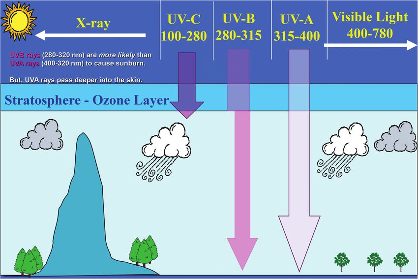
Photochemical &
Photobiological Sciences
View Article Online
PERSPECTIVE View Journal | View Issue
UVR: sun, lamps, pigmentation and vitamin D
This article is licensed under a Creative Commons Attribution 3.0 Unported Licence.
Open Access Article. Published on 25 October 2016. Downloaded on 7/11/2021 2:44:46 AM.
Cite this: Photochem. Photobiol. Sci., C. M. Lerche,* P. A. Philipsen and H. C. Wulf
2017, 16, 291
Exposure to ultraviolet radiation (UVR) has important and significant consequences on human health.
Received 28th July 2016, Recently, there has been renewed interest in the beneficial effects of UVR. This perspective gives an intro-
Accepted 18th October 2016
duction to the solar spectrum, UV lamps, UV dosimetry, skin pigment and vitamin D. The health benefits
DOI: 10.1039/c6pp00277c of UVR exposure through vitamin D production or non-vitamin D pathways will be discussed in this
rsc.li/pps themed issue in the following articles.
Solar spectrum Weather, ozone layer and air pollution
The quality and quantity of UVR change as the sun’s rays pass
Solar radiation includes ultraviolet radiation (UVR) through the atmosphere. Ozone and pollution particles reduce
100–400 nm, visible radiation (400–780 nm) and infrared radi- the UVR reaching the earth’s surfaces because they absorb,
ation (>780 nm) (Fig. 1).1 The wavelengths of UVR defined by scatter, and reflect the wavelengths. In the stratosphere (10–50
the International Commission on Illumination (CIE) are UV-C km above sea level) UVR is absorbed by ozone and scattered by
(100–280 nm), UV-B (280–315 nm) and UV-A (315–400 nm). molecules such as N2 and O2.1 In the troposphere (0 to 10 km
The UV component of terrestrial radiation from the midday above sea level) UVR is further absorbed by ozone, NO2, and
sun comprises about 95% UV-A and 5% UV-B; UV-C and most SO2 and scattered by particulates (e.g., soot) and clouds.3
of UV-B are removed from extraterrestrial radiation by strato- Clouds are either water or ice droplets, which are very weak
spheric ozone.1 The CIE nomenclature is not always followed absorbers of UVR. Clouds scatter both UV-B and UV-A to the
rigorously and some authors introduce slight variations; for same extent.3 The amount of UVR ultimately reaching the
example, distinguishing between UV-B and UV-A at 320 nm surface of the earth is called ambient UVR.
rather than 315 nm.1 However, these 5 nm can constitute an
important difference when calculating UVR doses. For Elevation, time of day, season and latitude
instance, if the 315 nm border is used, the solar irradiance
The angle between the sun and the local vertical is called the
consists of 2.11% UV-B (measured in Copenhagen in June)
solar zenith angle.3 The elevation of the sun above the horizon
and 4.34% if the 320 nm border is used. UV-B has shorter and
is called the solar elevation. Both the spectrum (quality) and
more energetic wavelengths than UV-A and is more effective in
intensity (quantity) of ambient UVR vary with the solar
inducing various biological effects. Despite the lower photon
elevation.3 The solar elevation depends on the time of day, day
energy, UV-A can also induce biological effects and penetrates
of year and geographical location (latitude and longitude).3
more deeply into biological tissue than UV-B. The depth of
The UVR irradiance at ground level increases closer to the
UVR penetration into the skin is dependent on the wavelength.
equator due to a shorter passage through the atmosphere.1
The longer the wavelength, the deeper the penetration.2
During the day the sun’s elevation reaches its maximum
around noon (90°) at the equator.1 This also means that
approximately 50–60% of the diurnal ambient UVR is ir-
Ambient exposure radiated in the 4-hour period around local noon.3 During the
year the sun’s elevation is the highest around the summer sol-
The intensity of solar UVR at the earth’s surface is influenced stice. Fig. 2 shows the number of standard erythema doses
by not only clouds, the ozone layer, air pollution and surface (SED) per hour during the day measured in July, April and
reflections but also the solar height, which is dependent on December in Copenhagen, Denmark, 55.7°N, 12.5°E. An up to
latitude, elevation, time of day, and season of the year.1 In the 5-fold decrease in erythema-effective UV-B may be seen when
following we will describe the factors influencing the intensity moving from the tropics to the most northern parts of
of solar UVR in detail. Europe.1,4 Fig. 3a shows non-erythema-weighted solar spectra
in Denmark, Spain and at the equator. However, when the
Department of Dermatology, D92, Bispebjerg Hospital, University of Copenhagen, spectra are erythema-weighted (Fig. 3b) the difference in the
Copenhagen, Denmark. E-mail: catharina.margrethe.lerche@regionh.dk UV-B area becomes clear. Not only is the overall level of
This journal is © The Royal Society of Chemistry and Owner Societies 2017 Photochem. Photobiol. Sci., 2017, 16, 291–301 | 291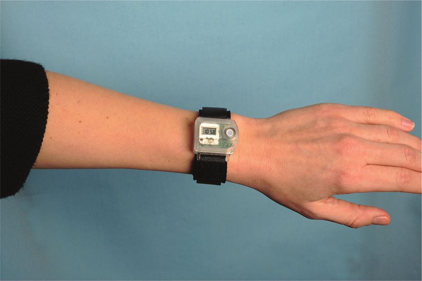
View Article Online

                                                                                                  Perspective                                                                                         Photochemical & Photobiological Sciences
            This article is licensed under a Creative Commons Attribution 3.0 Unported Licence.
Open Access Article. Published on 25 October 2016. Downloaded on 7/11/2021 2:44:46 AM.

                                                                                                  Fig. 1 Ultraviolet radiation (UVR) is part of the electromagnetic spectrum. The CIE bands are: UV-C (100–280 nm), UV-B (280–315 nm) and UV-A
                                                                                                  (315–400 nm). However, in photobiology a distinction is often made between UV-B and UV-A at 320 rather than 315 nm. Visible light is the region
                                                                                                  between 400 nm and 780 nm. The atmosphere attenuates these types of UV differently, thus affecting the levels of UV-A, UV-B, and UV-C at ground
                                                                                                  level on the earth.

                                                                                                                                                                           One standard erythema dose (SED) equals to 100 J m−2
                                                                                                                                                                           erythema-weighted and since there are 3600 seconds in an
                                                                                                                                                                           hour, the solar intensity in number of SED per hour can be cal-
                                                                                                                                                                           culated as erythema-weighted solar intensity multiplied by 36
                                                                                                                                                                           (SED per hour × m2 W−1). Even though the UV index is ∼11%
                                                                                                                                                                           higher than SED per hour, for practical purposes we can
                                                                                                                                                                           assume UV index = SED per hour.5

                                                                                                                                                                           Surface reflections
                                                                                                                                                                           UVR is reflected by surfaces such as snow, sand, concrete and
                                                                                                                                                                           water. Reflection of UVR from most ground surfaces is nor-
                                                                                                                                                                           mally less than 10%.1,6 The main exceptions are snow, which
                                                                                                                                                                           can reflect up to 90% although reflectance of about 30–50% is
                                                                                                                                                                           probably more typical and white sand, which reflects about
                                                                                                                                                                           10–30%.1,3,6 It is often believed that calm water reflects UVR
                                                                                                  Fig. 2 Number of standard erythema doses (SED) per hour during the       but the figure is actually only about 5%, although up to 20% is
                                                                                                  day and solar elevation angle measured in July, April and January in     reflected from choppy water.1,3,6 UVR goes easily through
                                                                                                  Copenhagen, Denmark.                                                     water, therefore bathing either in the sea or in open-air pools
                                                                                                                                                                           offers little protection against sunburn.3 Fig. 4 shows how
                                                                                                                                                                           much UVR one can receive in different outdoor situations. The
                                                                                                  ambient UVR greater at the equator and in Spain than in                  ambient UVR is 100% when there is no shade and no reflec-
                                                                                                  Denmark, but the spectra also contain a greater proportion of            tions. Ambient UVR is made up of both UVR directly from the
                                                                                                  UV-B than in Denmark, which explains why there is a UV index             sun and UVR diffused by scattering in the atmosphere. The
                                                                                                  nearly identical to 9 (SED per hour) in Spain and of 6 in                diffused ambient UVR results in “sun in the shade” meaning
                                                                                                  Denmark during summer. The UV index is dimensionless and                 that you can still receive about 50% of the ambient UVR if you
                                                                                                  defined as the erythema-weighted UV intensity divided by                 are situated in the periphery of the shade from a tree com-
                                                                                                  25 mW m−2, or more simply the erythema-weighted solar                    pared with less than 15% of the ambient UVR at the centre of
                                                                                                  intensity, measured in W m−2, multiplied by 40 (m2 W−1).1                the tree’s shadow (Fig. 4). Nevertheless, it is also possible to

                                                                                                  292 | Photochem. Photobiol. Sci., 2017, 16, 291–301                           This journal is © The Royal Society of Chemistry and Owner Societies 2017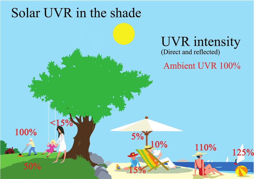
View Article Online
Photochemical & Photobiological Sciences Perspective
describe sun behaviour in detail in the next section. It is poss-
ible to measure personal UVR received by using chemical or
electronic UVR dosimeters.3 Chemical dosimeters, for example
polysulfone films, have been used in a number of dosimetric
studies in the past.3 However, electronic dosimeters can give
much more detailed information. One such dosimeter is the
SunSaver (Fig. 5) developed by the Department of
Dermatology, Bispebjerg Hospital, Copenhagen.8 An example
of data obtained from the SunSaver is shown in Fig. 6. This
This article is licensed under a Creative Commons Attribution 3.0 Unported Licence.
figure also illustrates the large difference in personal UVR
Open Access Article. Published on 25 October 2016. Downloaded on 7/11/2021 2:44:46 AM.
exposure depending on an individual’s behaviour.
Behavior
Studies have shown different behaviour patterns in the sun
depending on nationality and culture. In northern latitudes
sunshine is sparse and temperatures are low, especially in
winter, and this influences people’s behaviour when the sun
gains power in spring. The difference between nationalities
has been shown in a study in which Danish sun-seekers on a
short holiday were outdoors significantly longer, received sig-
nificantly higher percentages of ambient UVR, and received
greater accumulated UVR doses than Spanish sun-seekers.9
The Danish sun-seekers received an average of 9.4 SED per
day, while the highest daily dose received by one participant
was 32 SED and the lowest dose was 2 SED received by another
participant on the same day, so substantial differences can be
Fig. 3 (a) Solar UVR spectra from the equator, Spain and Denmark. (b) observed on the same day at the same location. Another study
The difference in UV-index (calculated by the area under the curve investigating the personal UVR exposure of farming families in
divided by 25 mW m−2) is visualized when the spectra are erythema- four European countries indicated a pronounced sun-seeking
weighted. Not only is the overall level of ambient UVR greater at the
attitude among farmers, their partners and children at higher
equator and in Spain but the spectra also contain a greater proportion of
SED than that in Denmark. latitudes.10 The difference among cultures is shown in a study
in which people of South Asian origin resident in the UK are
compared with the indigenous UK population. Despite the fact
receive more than 100% when reflections from choppy water that both groups spent the same time outside, the indigenous
or sand occur (Fig. 4). UK population received significantly more solar UVR during
summer than the UK South Asians due to the latter’s sun-
avoiding behaviour.11 A skin cancer diagnosis also influences
Personal exposure sun behaviour, but only for a short period of time. Newly diag-
nosed malignant melanoma patients exhibit cautious sun be-
The UVR dose an individual receives is also influenced by haviour the first summer after their diagnosis.12 Afterwards,
various parameters: the ambient UVR intensity, the exposure unfortunately, they tend to resume a careless attitude regard-
duration, the exposure geometry,6 UVR protection and the ing daily UVR dose, days with body exposure, holidays and
individual’s behaviour.7 days abroad, whereas controls maintain a stable UVR exposure
The ambient UVR intensity and exposure duration are self- dose. Hence it is concluded that patients with CMM do not
explanatory terms. Exposure geometry means that the amount maintain the initial cautious sun behavior they exhibit the first
of UVR received on different body sites depends on their orien- summer after CMM diagnosis.13 Healthy people also exhibit
tation towards the sun and any obstacles shielding or reflect- the same sun exposure habits over the years.14 Sun exposure
ing the UVR e.g. the urban canyon, mountains, hills and trees. behaviour is probably as difficult to change as other habits.
Personal UVR exposure is of course also dependent on the use
of sun protection like, sunscreen, shade and clothing.
Individual behaviour is also important. UVR exposure received Treatment with solar UV and UV lamps
within the same time interval cannot be assumed to be a con-
stant fraction of the ambient UVR exposure because humans Some thousand years ago solar UVR was already being used to
react differently to the sun. This is so important that we shall treat different skin conditions. At that time, the importance of
This journal is © The Royal Society of Chemistry and Owner Societies 2017 Photochem. Photobiol. Sci., 2017, 16, 291–301 | 293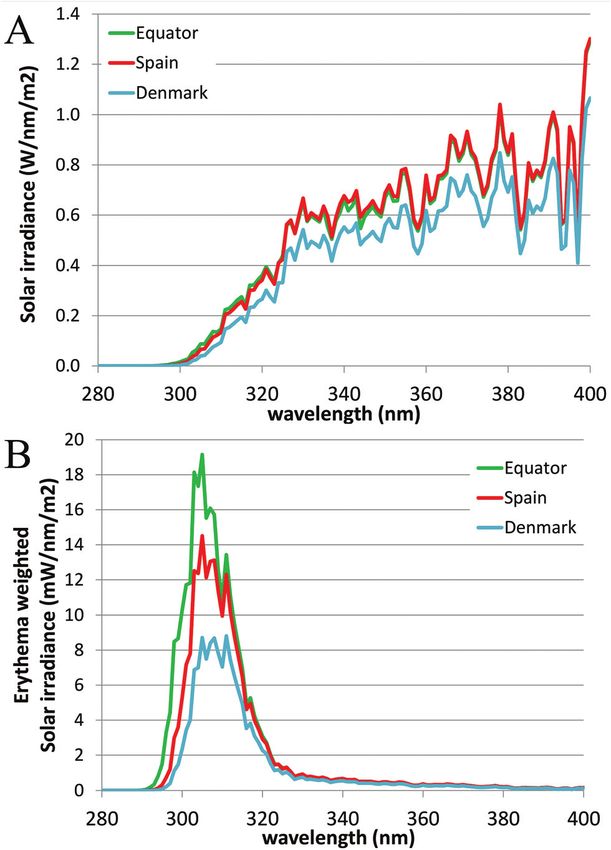
View Article Online

                                                                                                  Perspective                                                                                          Photochemical & Photobiological Sciences
            This article is licensed under a Creative Commons Attribution 3.0 Unported Licence.
Open Access Article. Published on 25 October 2016. Downloaded on 7/11/2021 2:44:46 AM.

                                                                                                  Fig. 4 This figure shows the % of ambient UVR possible to be received in different outdoor situations. 100% ambient UVR consists of direct and
                                                                                                  diffuse light from the sun without reflections.

                                                                                                  Fig. 5 An example of an electronic dosimeter called the “SunSaver”
                                                                                                  developed by the Department of Dermatology, Bispebjerg Hospital,
                                                                                                  Copenhagen.8 An example of the data obtained from the SunSaver is
                                                                                                  shown in Fig. 6. The sensor can measure every 8th second and store an     Fig. 6 The mean daily distribution of ambient UVR expressed as stan-
                                                                                                  average of the last 75 measurements every 10 minutes along with the       dard erythema dose (SED) per hour. The green graph illustrates the large
                                                                                                  time. The measurement range of the dosimeter is 0.1 SED per hour to       influence of behaviour on personal exposure.
                                                                                                  23 SED per hour and it is battery-driven. The dosimeter can run for 145
                                                                                                  days without maintenance, and the data can be transferred to a
                                                                                                  computer.

                                                                                                                                                                            lengths above 340 nm transmitted by Finsen’s lens systems
                                                                                                                                                                            that were responsible. Fluorescence measurements on
                                                                                                  UVR was not known, as UV rays were not discovered before the              M. tuberculosis showed that the therapeutic effect of Finsen
                                                                                                  1800s. Just before the year 1900 Niels Ryberg Finsen intro-               light therapy was probably due to photodynamic therapy with
                                                                                                  duced light therapy in medicine, first using focused sunlight,            coproporphyrin III produced by Mycobacterium tuberculosis.16
                                                                                                  later using a carbon arc lamp in treating lupus vulgaris, a skin          The mercury vapour lamp was invented in 1906.17,18 These
                                                                                                  condition caused by Mycobacterium tuberculosis.15 It was                  lamp types were used for phototherapy until the development
                                                                                                  believed that UVR was responsible for the effect of treatment              of the modern UV lamps in the 1960s. The Goeckerman proto-
                                                                                                  and not until 2005 was it discovered that it was the wave-                col was introduced in 1925 in the US where psoriasis and

                                                                                                  294 | Photochem. Photobiol. Sci., 2017, 16, 291–301                            This journal is © The Royal Society of Chemistry and Owner Societies 2017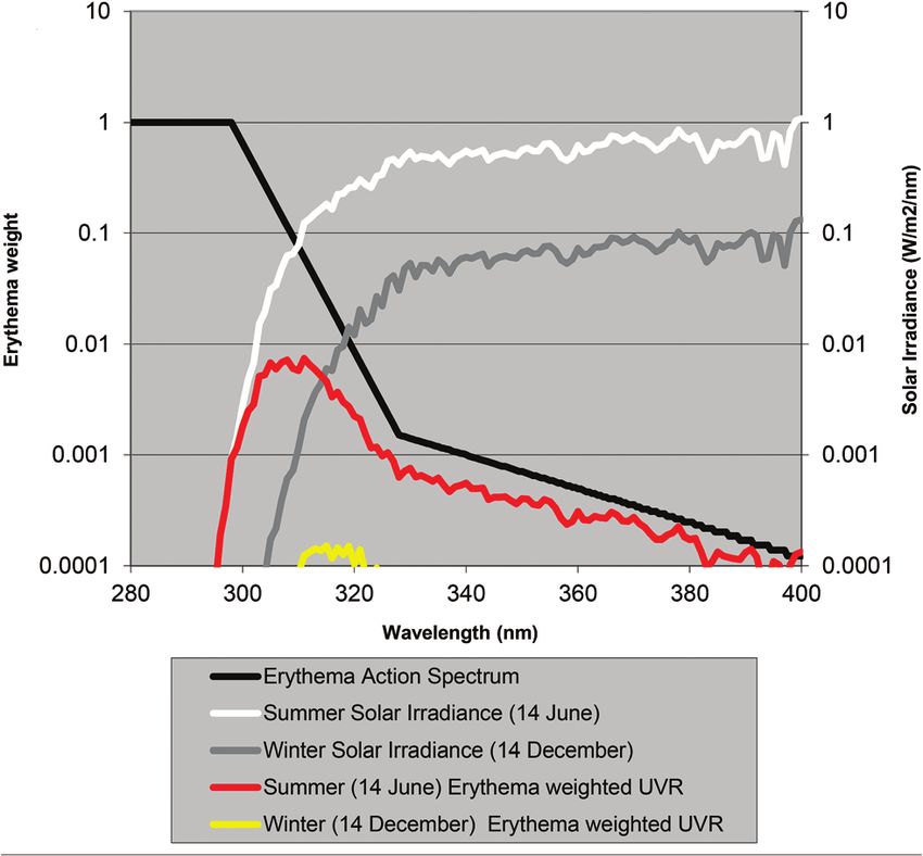
View Article Online
Photochemical & Photobiological Sciences Perspective
Today phototherapy is still used to treat common skin con-
ditions such as psoriasis, atopic eczema, other forms of der-
matitis, generalised itching, pityriasis lichenoides, cutaneous
T cell lymphoma, lichen planus, vitiligo and other less
common conditions.
Tanning devices
This article is licensed under a Creative Commons Attribution 3.0 Unported Licence.
Besides phototherapy artificial UVR sources are also used for
Open Access Article. Published on 25 October 2016. Downloaded on 7/11/2021 2:44:46 AM.
cosmetic purposes. Commercial sunbeds were developed in
the 1970s and came into widespread use in the 1990s. In 2006
the European Union (EU) Scientific Committee on Consumer
Fig. 7 Emission spectra of light sources used in treatment of skin dis- Products published an opinion stating that UV tanning equip-
eases. The dashed vertical line represents the border (315 nm) between
ment should not exceed an erythema-weighted irradiance level
UV-A and UV-B.
of 0.3 W m−2 or 11 SED per hour (see definition in the follow-
ing section), and that equipment above this level would be
considered unsafe in tanning devices used for cosmetic
atopic eczema were treated with coal tar combined with purposes.
UV-B.19 In the 1970s PUVA treatment was invented also for con- A recent review on UVR from indoor tanning devices con-
ditions like psoriasis.20 PUVA treatment is a combination of cludes that in 8 out of 12 studies the erythema-weighted irradi-
the chemical psoralen and UV-A (PUVA).21,22 Full spectrum ance is above the European limit of 0.3 W m−2.32 In 10 out of
UV-A (315–400 nm) induces crosslinks efficiently and therefore 12 studies the erythema-weighted irradiance was higher than
lamp types used for PUVA are often Cleo (Fig. 7) or a very that of tropical sunlight (23°S and 23°N) with substantial vari-
similar TL09.23 PUVA is generally indicated for chronic plaque ations between the devices.32 Also in 12 out of 13 studies UV-A
psoriasis and atopic eczema if UV-B has not been effective. irradiance from tanning devices was higher than that from
Failure to respond adequately to UV-B does not predict failure natural sunlight (Melbourne, Australia, 37°S and Crete,
of response to PUVA. PUVA is favoured over UV-B for some indi- Greece, 35°N).32 However, these studies did not discuss the
cations, such as mycosis fungoides beyond the patch stage, duration of exposure (UVR doses) received but only the
adult pityriasis rubra pilaris, pustular psoriasis, and hand and emitted irradiance. Another recent study shows that 52
foot eczema. More than 200 PUVA treatments are linked to tanning devices used regularly in Spanish facilities revealed a
increased risk of squamous cell carcinomas.24 high variation in erythema-weighted irradiance, not the least
Today solar UVR is still used in the treatment of various in UV type 4 devices (29% of the sample), for which medical
skin disorders, e.g. at the Blue Lagoon, Iceland and also the advice is required.33
Dead Sea and other Mediterranean areas, where patients with
chronic skin diseases such as psoriasis are exposed to solar
UVR.25–27 However, treatment with UVR from UV lamps is pre- UV dosimetry, erythema, photo skin
ferable; different UVR sources are used for different treatments type and pigment protection factor
and guidelines regarding the clinical use of phototherapy have
been developed.28 Broadband UV-B is mainly used for radi- Normally the effect of irradiation from UV lamps has been
ation between 280 and 350 nm (Philips TL12) or from about measured as physical irradiance (W m−2) or as a dose (J m−2).
290 to 370 nm (Waldman UV6) (Fig. 6). Both types of lamps However, for the skin it is much better to use a biologically
have proved effective in treating mild to moderate psoriasis erythema-weighted dose called the standard erythema dose
and eczema. The narrow-band UV-B (TL01) lamp, in which just (SED) because erythema is the most common endpoint in UVR
a narrow band around 311 nm is emitted, was developed for treatment of the skin.34 SED is calculated from the measured
use in phototherapy as an alternative to a broad-band UV-B spectral irradiance of the lamp (W m−2 nm−1) multiplied by
source and to photo chemotherapy. The narrow-band lamp the CIE erythema action spectrum and integrated over the
has been proved to be particularly effective at clearing psoria- whole spectrum35 (black line in Fig. 8). An action spectrum,
sis but it has also been acknowledged that the TL01 lamp is for a particular biological effect, expresses the effectiveness of
probably 2–3 times more carcinogenic per minimum erythema radiation at each wavelength as a fraction of the effectiveness
dose than broadband UV-B.29 On the other hand, the cumulat- at a certain standard wavelength.36 Each biological effect of
ive dose required in therapy is less than when using broad- UVR such as erythema, pigmentation, vitamin D synthesis, etc.
band UV-B sources.29,30 Narrow-band UV-B is also more ery- has its own action spectrum. An action spectrum can predict
throgenic (by a factor of 1.6) than expected from the CIE the effectiveness of an exposure to a certain spectral output
erythema spectrum,31 and therefore adjustment of the UVR (lamp). The biological effect on the skin will be identical for
dose is needed in therapy if doses are given in SED. equal effective doses e.g. erythemal doses. The erythema effect
This journal is © The Royal Society of Chemistry and Owner Societies 2017 Photochem. Photobiol. Sci., 2017, 16, 291–301 | 295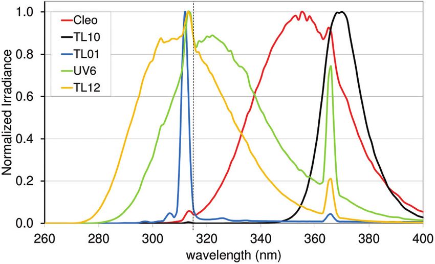
View Article Online
Perspective Photochemical & Photobiological Sciences
siderable variation of MED within different white skin photo-
types.40 Earlier the term MED was used widely as a ‘measure’
of erythemal radiation. This is a difficult measure to be used
because MED is not a standard measure of anything and it is
dependent on the individual’s sensitivity to UVR and other
variables such as optical and radiometric characteristics of the
source; determinants of the exposure such as dose increment
and field size; nature of the skin such as pigmentation, pre-
vious light exposure and anatomical site; and observational
This article is licensed under a Creative Commons Attribution 3.0 Unported Licence.
factors such as definition of the endpoint, time of reading
Open Access Article. Published on 25 October 2016. Downloaded on 7/11/2021 2:44:46 AM.
after exposure and ambient illumination.3 In 1988, Fitzpatrick
published a system for classifying skin phototypes.41 The
system is based on self-assessment of the skin’s erythema
response and the ability to tan after a defined dose in the
spring/early summer rather than the degree of pigmentation
because pigmentation is difficult to measure. However, the
system is strongly dependent on pigmentation. It is based on a
simple 6-grade classification system with 4 grades for
Caucasians and 2 grades for brown- and black-skinned people.
Fig. 8 The CIE erythema action spectrum and solar UVR irradiance Individuals complete a short questionnaire on their ability to
measured during summer and winter in Denmark. Not only is the overall get sunburned and the tendency to tan after the first un-
level of ambient UVR greater during summer, but the spectrum also
protected solar exposure in the spring/early summer around
contains a greater proportion of UV-B than that in winter.
noon. However, as an alternative to the subjective self-reported
Fitzpatrick skin type method it is possible to use a more objec-
tive and reliable measure of sun sensitivity expressed as the
pigment protection factor (PPF) measured by a skin reflectance
of solar UVR can be seen in Fig. 3b and this figure illustrates meter.37 The PPF score expresses the number of SEDs needed
why 320 nm is often used as the border between UV-B and to elicit just perceptible erythema on unexposed buttocks but
UV-A within dermatology and skin photobiology. The thickness can also be used on other anatomical sites. The meter has a
of the stratum corneum, cytokines and genetic factors such as measuring scale of 1.0–25.0 (1.0 is the PPF of the palest poss-
pigmentation and responsiveness of the vascular epithelium ible person while 25.0 corresponds to the darkest black
determines how many SEDs can be tolerated without the skin skin).42
being burnt.37 One SED is equivalent to an erythemal effective
radiant exposure of 100 J m−2.34 The ambient exposure on a
summer day with a clear sky in Europe is approximately 30–40 Skin pigment
SED and an exposure dose of 4 SED would be expected to
produce moderate erythema on unacclimatised white skin, but The skin provides a barrier to harmful environmental effects
minimal or no erythema on previously exposed skin.3 An and consists of three layers: the epidermis (top layer), the
average dose received on a winter holiday on Tenerife is 9.4 dermis (middle layer) and the subcutaneous layer. The epider-
SED per day for Danes and only 5.1 SED for Spaniards.9 Both mis consists mainly of epithelial keratinocytes and melano-
doses are higher than the normally tolerated minimal cytes, and some antigen presenting Langerhans cells and
erythema doses (MED), so without sun protection sunburn will Merkel cells. Melanocytes reside in the lower part of the epi-
occur. To induce erythema, the erythemal sensitivity of the dermis and are responsible for the synthesis of melanin
skin to UVR is very wavelength dependent; at 300 nm a 100 within specialised membrane-bound organelles, termed melano-
times lower physical dose is needed than that at 320 nm. In somes, and the subsequent transfer of the melanosomes to
Fig. 8 the solar UVR irradiance is depicted for summer and the surrounding keratinocytes.43 There are two types of
winter in Denmark. Not only is the overall level of ambient melanin, eumelanin (brown-black) and pheomelanin (red-
UVR greater during summer, but the spectrum also contains a yellow).44–46 Eumelanin is a brown-black polymer of dihydroxyi-
greater proportion of UV-B than that in winter. The erythemal ndole carboxylic acids and their reduced forms.44
sensitivity of the skin is also dependent on the part of the Pheomelanin is a red-brown polymer of benzothiazine units
body exposed.38 The trunk, head and neck are more sensitive largely responsible for red hair and freckles. Besides eu- and
than the extremities38 probably because the latter have thicker pheomelanin pigmentation can also be divided into constitu-
skin.39 tive pigmentation (the pigmentation you are born with) and
Individual erythema response can be assessed by determin- facultative pigmentation (the additional pigmentation
ing MED, which increases with the degree of pigmentation, obtained by UVR).44 It is beyond the scope of the present
and is not predictive of skin phototype because there is con- article to present the details about melanins and melanogen-
296 | Photochem. Photobiol. Sci., 2017, 16, 291–301 This journal is © The Royal Society of Chemistry and Owner Societies 2017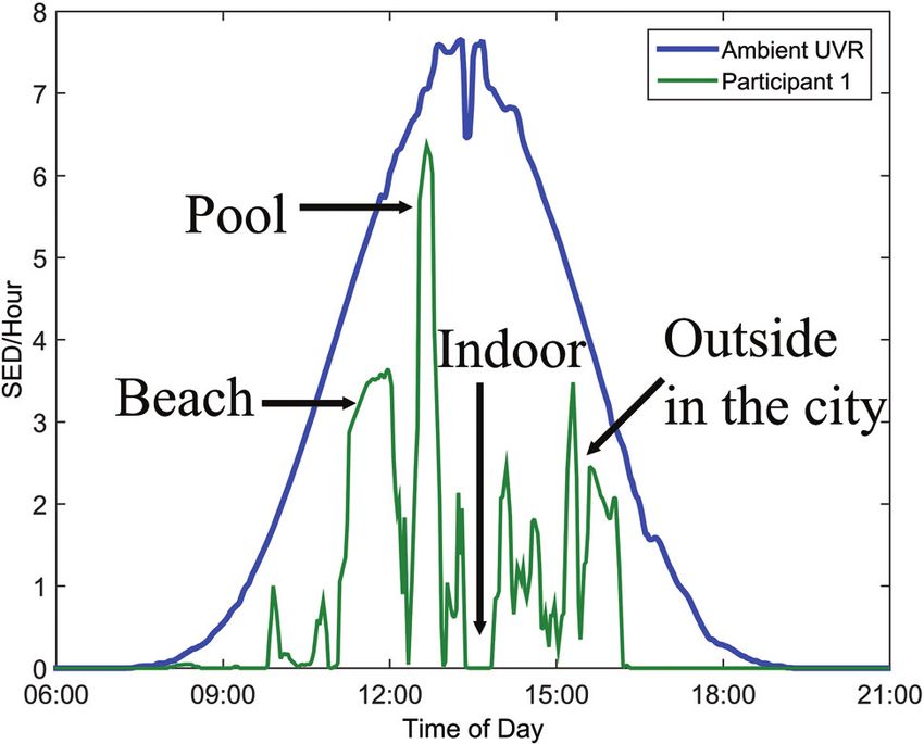
View Article Online
Photochemical & Photobiological Sciences Perspective
esis but these are described very well in two reviews by d’Ischia
et al.43,47
Within a few days of exposure to solar UVR delayed melano-
genesis (tanning) occurs. This process is dependent on skin
photo type and like erythema is primarily caused by UV-B.
UV-A and UV-B induce pigmentation differently. UV-A induces
immediate pigment darkening (within minutes after exposure)
and delayed tanning (within days after exposure) while UV-B
induces only delayed tanning. Immediate pigment darkening
This article is licensed under a Creative Commons Attribution 3.0 Unported Licence.
is a transitory darkening of the skin. Normally, the greater the
Open Access Article. Published on 25 October 2016. Downloaded on 7/11/2021 2:44:46 AM.
constitutive pigmentation, the greater the tendency to develop
immediate pigment darkening.48 An in vitro study has shown
that the cutaneous melanocyte carries an opsine receptor
tuned to a peak of about 360 nm, and that irradiation in the
UV-A band activates this receptor which causes immediate
pigment darkening.49 Darkening intensity is at its maximum
immediately after exposure and decreases rapidly within
1–2 hours.48,50 Delayed tanning is seen about 1–2 days after
Fig. 10 Considerable seasonal variation for skin pigmentation at UVR-
exposure and may persist for several months.48 It is caused by
exposed sites from 36 healthy volunteers. The blue “lines” are the daily
an augmented activity of the enzyme tyrosinase, which results SED during a year. The red line is the increase and decrease in PPF over
in a production of new melanin and more melanin granules.48 the year, adapted from ref. 53. Ambient UVR is measured in
Different action spectra for delayed tanning (melanogenesis) Copenhagen, Denmark (55.7°N, 12.5°E). All participants have Danish
in human skin have been published.48 Fig. 9 shows standard ancestors.
action spectra and proposed standards for pigmentation.48
The standards are based on the data from Parrish et al. deter-
mining the action spectrum in Caucasians for wavelengths
between 250 and 435 nm (ref. 51) and on Gange et al. who also erythema but the level of photo protection is equivalent to a
investigated the action spectrum for melanogenesis but only sunscreen with a sun protection factor (SPF) of 2–3.52 However,
at 3 different wavelengths and 8 and 24 hours after exposure. even though a SPF of 2–3 does not sound like much it actually
The action spectra for melanogenesis and erythema are means a reduction of the UVR dose of 50%. Fig. 10 shows a
very similar between 300 and 435 nm for fair untanned skin, considerable seasonal variation for skin pigmentation at UVR
which could mean that erythema and melanogenesis are exposed sites.53
initiated by the same photochemical events, i.e. the same Measurement of melanin levels in the skin has tradition-
chromophores.52 A UV-B tan is photo-protective against ally been via visual assessment performed by clinicians and
dermatologists, which is clearly a subjective method.54
Portable instruments that measure reflected light to inciden-
tal light non-invasively in vivo are increasingly being used to
quantify melanin.55,56 The term pigment protection factor
(PPF) is used (described in the previous section) and studies
have shown a positive correlation between PPF, phototype
and MED.42,57 Our research group and two other research
groups have used the quantification of the eumelanin
monomer pyrrole-2,3,5-tricarcoxylic acid based on only High
Pressure Liquid Chromatography (HPLC) or spectrophoto-
metric analyses but these methods have very low through-
put and high detection limits up to 2 µg.58 Recently,
Szekely-Klepser et al. (2005) developed a novel and objective
method for quantification of eumelanin from skin biopsies
by Liquid Chromatography-Mass Spectrometry (LC-MS).59
Immunohistological stainings of melanin are available such
as Fontana Masson but they cannot differentiate between eu-
and pheomelanin.
In addition to pigmentation, skin thickening especially of
Fig. 9 Standard action spectra and proposed standards for pigmenta-
tion. Adapted from ref. 48 with permission from the European Society
the stratum corneum is another UVR-protective mechanism
for Photobiology, the European Photochemistry Association, and The for avoiding further damage.60 UVR-induced skin adaptation/
Royal Society of Chemistry. hardening is very dependent on the spectrum of irradiance.61
This journal is © The Royal Society of Chemistry and Owner Societies 2017 Photochem. Photobiol. Sci., 2017, 16, 291–301 | 297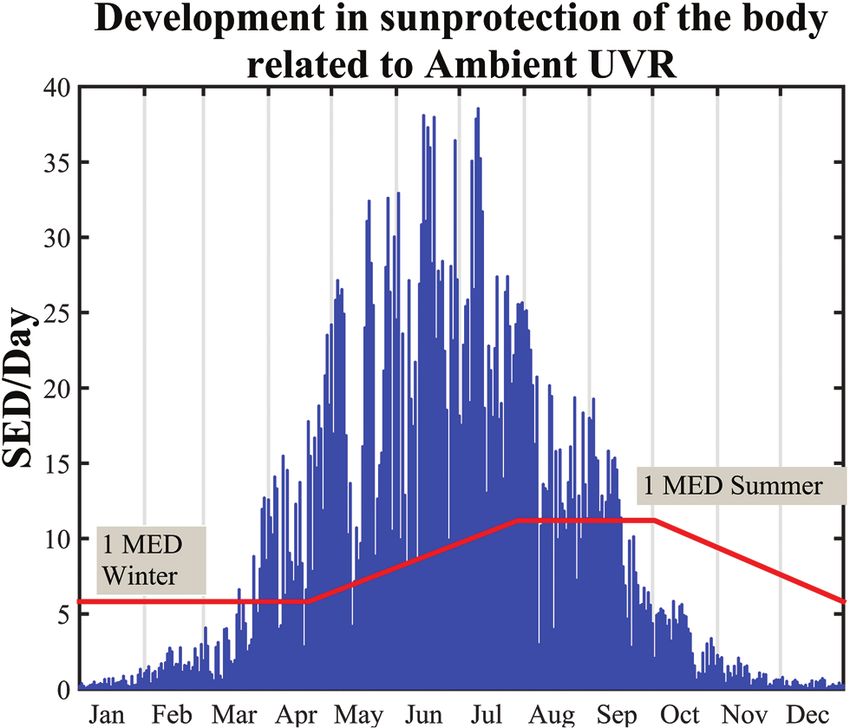
View Article Online
Perspective Photochemical & Photobiological Sciences
vitamin D concentrations are lower in humans with dark skin
than those in fair-skinned humans.77,78 However, the associ-
ation between skin pigmentation and the change in blood con-
centrations of 25-hydroxyvitamin D following experimental UV
irradiation is not fully elucidated. A recent review found seven
studies in which skin pigmentation influenced vitamin D pro-
duction and five studies in which skin pigmentation did not
influence vitamin D production.79 However, when compari-
sons of studies are performed, possible confounders like time
This article is licensed under a Creative Commons Attribution 3.0 Unported Licence.
of year, dietary supplementation habits and the 25-hydroxy-
Open Access Article. Published on 25 October 2016. Downloaded on 7/11/2021 2:44:46 AM.
vitamin D analysis method should be kept in mind.80 Different
analytical quantification methods are available, including
high-performance liquid chromatography (HPLC), and com-
Fig. 11 CIE Vitamin D action spectrum and CIE erythema spectrum. bined high-performance liquid chromatography and mass
spectrometry (LC-MS), radioimmunoassays, enzyme immuno-
assays, competitive protein-binding assays, automated chemi-
UV-B enriched sources induced much more epidermal thicken- luminescence protein-binding assays and chemiluminescence
ing of the skin than UV-A.61 immunoassays.81 However, the sensitivity and specificity of
these methods vary considerably. The LC-MS method is the
most precise and accurate and is the gold standard.80
Snellman et al. analysed blood samples for 25-hydroxyvitamin
Vitamin D D using three different methods and found high variability
between the different assays.81 Mean 25-hydroxyvitamin D
Exposure to UV-B initiates the synthesis of vitamin D in the
levels were the highest for the LC-MS technique (85 nmol L−1),
skin.62 There is a well-established relationship between the
intermediate for radioimmunoassay (70 nmol L−1) and the
vitamin D status and musculoskeletal health. However, the
lowest for chemiluminescence immunoassays (60 nmol L−1).81
association between vitamin D levels and other chronic dis-
Using the 50 nmol L−1 cutoff, 8% of the subjects were found to
eases is less clear. High levels of vitamin D have been linked to
be insufficient using by LC-MS, 22% by radioimmunoassay
a reduced risk of internal malignancies.63–69 The marker for
and 43% by chemiluminescence immunoassays.81
vitamin D status is 25-hydroxyvitamin D, a metabolite of
The health benefits of UVR exposure through vitamin D
vitamin D3. No internationally well-defined values for optimal
production or non-vitamin D pathways will be discussed in
serum 25-hydroxyvitamin D currently exist (reviewed in ref.
this themed issue in the following articles.
70). However, it is generally believed in Europe that values
>50 nmol l−1 (20 ng ml−1) are sufficient. 25-Hydroxyvitamin D
levels are often both defined in units of nmol L−1 and
ng ml−1 †. A single cutoff value has been debated and a recent Acknowledgements
study demonstrated a major inter-personal variation in the
25-hydroxyvitamin D response to UV-B as well as the maximal The work was funded by The Danish Council of Independent
UV-B-induced 25-hydroxyvitamin D level.71 Different action Research (Grant number 0602-01931B) and The Capital Region
spectra using rats, chickens and humans for the conversion of of Denmark (Grant number R135-A4663).
7-DHC to previtamin D3 by UVR exist (summarized in ref. 72).
All the results, irrespective of the method, assess the optimal
wavelength to be in the range 295–303 nm. Furthermore, the
production is very low or non-existing above 310 nm. This also References
includes a recently published action spectrum using ex vivo
1 WHO, IARC Monographs on the evaluation of carcinogenic
pig skin and UV light-emitting diodes (LED) to create narrow-
risk to humans, Solar and Ultraviolet Radiation, 1992, vol. 55,
band UVR.72 The mostly used action spectrum for the conver-
pp. 1–316.
sion of 7-DHC to previtamin D3 by UVR in human skin was
2 W. A. Bruls, H. Slaper, J. C. van der Leun and L. Berrens,
determined by MacLaughlin et al. in 1982.73 The same data
Transmission of human epidermis and stratum corneum
were further defined and extrapolated by the CIE in 200674
as a function of thickness in the ultraviolet and visible
(Fig. 11). The accuracy of the action spectrum has been ques-
wavelengths, Photochem. Photobiol., 1984, 40, 485–494.
tioned.75 An updated action spectrum for the conversion of
3 B. L. Diffey, Sources and measurement of ultraviolet radi-
7-DHC to previtamin D3 by UVR on human skin is under
ation, Methods, 2002, 28, 4–13.
development.76 Studies have shown that serum 25-hydroxy-
4 G. Seckmeyer, D. Pissulla, M. Glandorf, et al., Variability of
UV irradiance in Europe, Photochem. Photobiol., 2008, 84,
† The conversion for 25-hydroxyvitamin D is [nmol L−1] = 2.5 × [ng ml−1]. 172–179.
298 | Photochem. Photobiol. Sci., 2017, 16, 291–301 This journal is © The Royal Society of Chemistry and Owner Societies 2017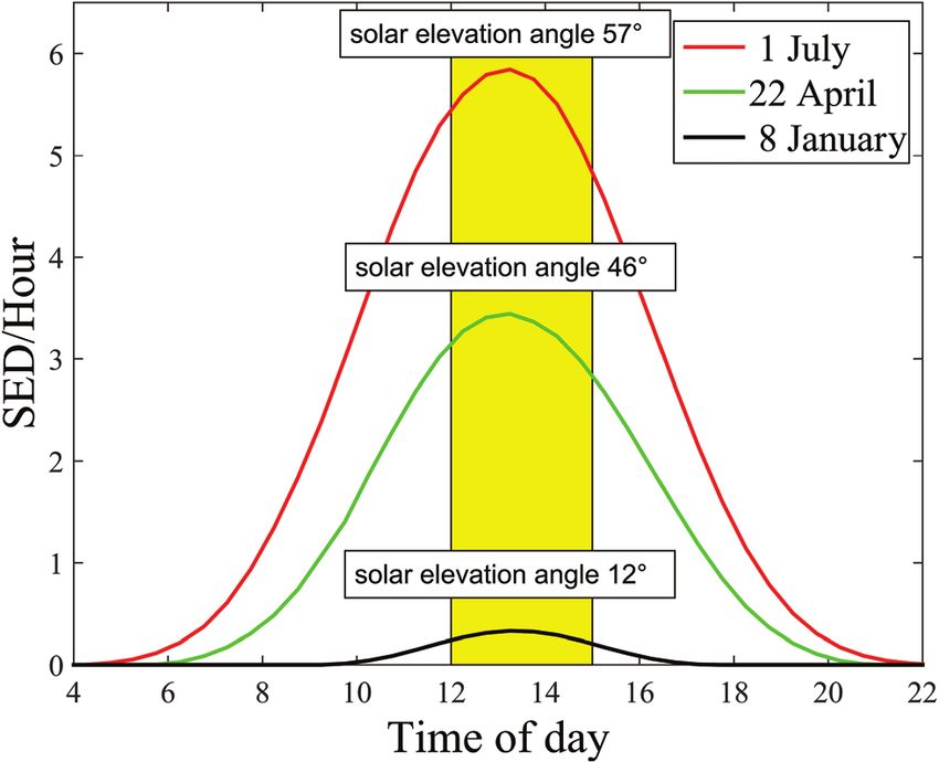
View Article Online
Photochemical & Photobiological Sciences Perspective
5 H. C. Wulf and P. Eriksen, UV index and its implications, the treatment of psoriasis: a cooperative clinical trial,
Ugeskr. Laeg., 2010, 172, 1277–1279. J. Invest. Dermatol., 1977, 68, 328–335.
6 N. Kromann, H. C. Wulf, P. Eriksen and H. Brodthagen, 21 J. A. Parrish, T. B. Fitzpatrick, L. Tanenbaum and
Relative ultraviolet spectral intensity of direct solar radi- M. A. Pathak, Photochemotherapy of psoriasis with oral
ation, sky radiation and surface reflections. Relative contri- methoxsalen and longwave ultraviolet light, N.
bution of natural sources to the outdoor UV irradiation of Engl. J. Med., 1974, 291, 1207–1211.
man, Photodermatology, 1986, 3, 73–82. 22 H. C. Wulf, Modified method for demonstrating sister
7 B. L. Diffey, C. J. Gibson, R. Haylock and A. F. McKinlay, chromatid exchange (SCE), Dan. Med. Bull., 1980, 27, 35–
Outdoor ultraviolet exposure of children and adolescents, 37.
This article is licensed under a Creative Commons Attribution 3.0 Unported Licence.
Br. J. Dermatol., 1996, 134, 1030–1034. 23 D. Bethea, B. Fullmer, S. Syed, et al., Psoralen photobiology
Open Access Article. Published on 25 October 2016. Downloaded on 7/11/2021 2:44:46 AM.
8 J. Heydenreich and H. C. Wulf, Miniature personal elec- and photochemotherapy: 50 years of science and medicine,
tronic UVR dosimeter with erythema response and time- J. Dermatol. Sci., 1999, 19, 78–88.
stamped readings in a wristwatch, Photochem. Photobiol., 24 R. S. Stern, L. A. Thibodeau, R. A. Kleinerman, J. A. Parrish
2005, 81, 1138–1144. and T. B. Fitzpatrick, Risk of cutaneous carcinoma in
9 B. Petersen, M. Triguero-Mas, B. Maier, et al., Sun behav- patients treated with oral methoxsalen photochemotherapy
iour and personal UVR exposure among Europeans on for psoriasis, N. Engl. J. Med., 1979, 300, 809–813.
short term holidays, J. Photochem. Photobiol., B, 2015, 151, 25 D. J. Abels and J. Kattan-Byron, Psoriasis treatment at the
264–269. Dead Sea: a natural selective ultraviolet phototherapy,
10 M. Bodekaer, G. I. Harrison, P. Philipsen, et al., Personal J. Am. Acad. Dermatol., 1985, 12, 639–643.
UVR exposure of farming families in four European 26 J. H. Eysteinsdottir, J. H. Olafsson, B. A. Agnarsson,
countries, J. Photochem. Photobiol., B, 2015, 153, 267–275. B. R. Luethviksson and B. Sigurgeirsson, Psoriasis treat-
11 R. Kift, J. L. Berry, A. Vail, M. T. Durkin, L. E. Rhodes and ment: faster and long-standing results after bathing in
A. R. Webb, Lifestyle factors including less cutaneous sun geothermal seawater. A randomized trial of three UVB
exposure contribute to starkly lower vitamin D levels in phototherapy regimens, Photodermatol., Photoimmunol.
U.K. South Asians compared with the white population, Photomed., 2014, 30, 25–34.
Br. J. Dermatol., 2013, 169, 1272–1278. 27 H. Matz, E. Orion and R. Wolf, Balneotherapy in dermato-
12 L. W. Idorn, P. Datta, J. Heydenreich, P. A. Philipsen and logy, Dermatol. Ther., 2003, 16, 132–140.
H. C. Wulf, Sun behaviour after cutaneous malignant mela- 28 H. Moseley, D. Allan, H. Amatiello, et al., Guidelines on the
noma: a study based on ultraviolet radiation measurements measurement of ultraviolet radiation levels in ultraviolet
and sun diary data, Br. J. Dermatol., 2013, 168, 367–373. phototherapy: report issued by the British Association of
13 L. W. Idorn, P. Datta, J. Heydenreich, P. A. Philipsen and Dermatologists and British Photodermatology Group 2015,
H. C. Wulf, A 3-year follow-up of sun behavior in patients Br. J. Dermatol., 2015, 173, 333–350.
with cutaneous malignant melanoma, JAMA Dermatol., 29 A. A. El-Ghorr and M. Norval, Biological effects of narrow-
2014, 150, 163–168. band (311 nm TL01) UVB irradiation: a review,
14 E. Thieden, J. Heydenreich, P. A. Philipsen and H. C. Wulf, J. Photochem. Photobiol., B, 1997, 38, 99–106.
People maintain their sun exposure behaviour in a 5-7-year 30 H. C. Wulf, A. B. Hansen and N. Bech-Thomsen,
follow-up study using personal electronic UVR dosimeters, Differences in narrow-band ultraviolet B and broad-spec-
Photochem. Photobiol. Sci., 2013, 12, 111–116. trum ultraviolet photocarcinogenesis in lightly pigmented
15 N. R. Finsen, Om anvendelse i medicinen af koncentrerede hairless mice, Photodermatol., Photoimmunol. Photomed.,
kemiske lysstråler, Gyldendalske Boghandels Forlag, 1896. 1994, 10, 192–197.
16 K. I. Moller, B. Kongshoj, P. A. Philipsen, V. O. Thomsen 31 A. B. Hansen, N. Bech-Thomsen and H. C. Wulf, Erythema
and H. C. Wulf, How Finsen’s light cured lupus vulgaris, after irradiation with ultraviolet B from Philips TL12 and
Photodermatol., Photoimmunol. Photomed., 2005, 21, 118– TL01 tubes, Photodermatol., Photoimmunol. Photomed.,
124. 1994, 10, 22–25.
17 E. Kromayer, Quecksilberwassenlampen zurBehandlung 32 L. T. Nilsen, M. Hannevik and M. B. Veierod, Ultraviolet
von Haut und Schleimhaut, Dtsch. Med. Wochenschr., 1906, exposure from indoor tanning devices: a systematic review,
48–51. Br. J. Dermatol., 2016, 174, 730–740.
18 K. I. Møller and H. C. Wulf, Early lamp developments for 33 Y. Sola, D. Baeza, M. Gomez and J. Lorente, Ultraviolet
phototherapy in dermatology, Forum for Nord Derm. Ven., spectral distribution and erythema-weighted irradiance
2005, 10, 20–24. from indoor tanning devices compared with solar radiation
19 L. E. Gibson and O. H. Perry, Goekerman Therapy, in exposures, J. Photochem. Photobiol., B, 2016, 161, 450–455.
Psoriasis, ed. H. H. Roenigk and H. I. Kaibach, Maarcel 34 B. L. Diffey, C. T. Jansen, F. Urbach and H. C. Wulf, The
Dekker Inc., New York-Basel-Hong Kong, 1998, pp. standard erythema dose: a new photobiological concept,
467–477. Photodermatol., Photoimmunol. Photomed., 1997, 13, 64–66.
20 J. W. Melski, L. Tanenbaum, J. A. Parrish, T. B. Fitzpatrick 35 CIE, Erythema reference action spectrum and standard
and H. L. Bleich, Oral methoxsalen photochemotherapy for erythema dose, CIE S 007/E-1998 (ISO 17166:1999),
This journal is © The Royal Society of Chemistry and Owner Societies 2017 Photochem. Photobiol. Sci., 2017, 16, 291–301 | 299View Article Online
Perspective Photochemical & Photobiological Sciences
Commission Internationale de l’ É clairage, Vienna, 1998, 53 J. Lock-Andersen and H. C. Wulf, Seasonal variation of
1999. skin pigmentation, Acta Derm.–Venereol., 1997, 77, 219–221.
36 V. Fioletov, J. B. Kerr and A. Fergusson, The UV index: defi- 54 K. Miyamoto, H. Takiwaki, G. G. Hillebrand and S. Arase,
nition, distribution and factors affecting it, Can. J. Public Utilization of a high-resolution digital imaging system for
Health, 2010, 101, I5–I9. the objective and quantitative assessment of hyperpigmen-
37 H. C. Wulf, Methods and an apparatus for determining an ted spots on the face, Skin Res. Technol., 2002, 8,
individual’s ability to stand ultraviolet radiation, US 73–77.
Patent4882598, 1986. 55 H. Takiwaki, Y. Miyaoka, N. Skrebova, H. Kohno and
38 R. L. Olson, R. M. Sayre and M. A. Everett, Effect of ana- S. Arase, Skin reflectance-spectra and colour-value depen-
This article is licensed under a Creative Commons Attribution 3.0 Unported Licence.
tomic location and time on ultraviolet erythema, Arch. dency on measuring-head aperture area in ordinary reflec-
Open Access Article. Published on 25 October 2016. Downloaded on 7/11/2021 2:44:46 AM.
Dermatol., 1966, 93, 211–215. tance spectrophotometry and tristimulus colourimetry,
39 J. Sandby-Moller, T. Poulsen and H. C. Wulf, Epidermal Skin Res. Technol., 2002, 8, 94–97.
thickness at different body sites: relationship to age, 56 G. Zonios, J. Bykowski and N. Kollias, Skin melanin, hemo-
gender, pigmentation, blood content, skin type and globin, and light scattering properties can be quantitatively
smoking habits, Acta Derm.–Venereol., 2003, 83, 410–413. assessed in vivo using diffuse reflectance spectroscopy,
40 G. I. Harrison and A. R. Young, Ultraviolet radiation-induced J. Invest. Dermatol., 2001, 117, 1452–1457.
erythema in human skin, Methods, 2002, 28, 14–19. 57 M. R. Tylman, J. Narbutt, M. Fracczak, A. Sysa-Jedrzejowska
41 T. B. Fitzpatrick, The validity and practicality of sun-reac- and A. Lesiak, Pigment protection factor as a predictor of
tive skin types I through VI, Arch. Dermatol., 1988, 124, skin photosensitivity–a Polish study, Acta Dermatovenerol.
869–871. Croat., 2015, 23, 23–27.
42 H. C. Wulf, P. A. Philipsen and M. H. Ravnbak, Minimal 58 B. Kongshoj, A. Thorleifsson and H. C. Wulf, Pheomelanin
erythema dose and minimal melanogenesis dose relate and eumelanin in human skin determined by high-per-
better to objectively measured skin type than to formance liquid chromatography and its relation to in vivo
Fitzpatricks skin type, Photodermatol., Photoimmunol. reflectance measurements, Photodermatol., Photoimmunol.
Photomed., 2010, 26, 280–284. Photomed., 2006, 22, 141–147.
43 M. d’Ischia, K. Wakamatsu, F. Cicoira, et al., Melanins and 59 G. Szekely-Klepser, K. Wade, D. Woolson, R. Brown,
melanogenesis: from pigment cells to human health and S. Fountain and E. Kindt, A validated LC/MS/MS method
technological applications, Pigm. Cell Melanoma Res., 2015, for the quantification of pyrrole-2,3,5-tricarboxylic acid
28, 520–544. (PTCA), a eumelanin specific biomarker, in human skin
44 S. Ito and K. Jimbow, Quantitative analysis of eumelanin punch biopsies, J. Chromatogr. B: Anal. Technol. Biomed.
and pheomelanin in hair and melanomas, J. Invest. Life Sci., 2005, 826, 31–40.
Dermatol., 1983, 80, 268–272. 60 M. Gniadecka, H. C. Wulf, N. N. Mortensen and
45 D. Parsad, K. Wakamatsu, A. J. Kanwar, B. Kumar and T. Poulsen, Photoprotection in vitiligo and normal skin. A
S. Ito, Eumelanin and phaeomelanin contents of de- quantitative assessment of the role of stratum corneum,
pigmented and repigmented skin in vitiligo patients, viable epidermis and pigmentation, Acta Derm.–Venereol.,
Br. J. Dermatol., 2003, 149, 624–626. 1996, 76, 429–432.
46 K. Wakamatsu and S. Ito, Advanced chemical methods in 61 N. Bech-Thomsen and H. C. Wulf, Photoprotection due to
melanin determination, Pigm. Cell Res, 2002, 15, 174–183. pigmentation and epidermal thickness after repeated
47 M. d’Ischia, K. Wakamatsu, A. Napolitano, et al., Melanins exposure to ultraviolet light and psoralen plus ultraviolet A
and melanogenesis: methods, standards, protocols, Pigm. therapy, Photodermatol., Photoimmunol. Photomed., 1996,
Cell Melanoma Res., 2013, 26, 616–633. 11, 213–218.
48 A. W. Schmalwieser, S. Wallisch and B. Diffey, A library of 62 R. Vieth, The role of vitamin D in the prevention of osteo-
action spectra for erythema and pigmentation, Photochem. porosis, Ann. Med., 2005, 37, 278–285.
Photobiol. Sci., 2012, 11, 251–268. 63 J. Ahn, U. Peters, D. Albanes, et al., Serum vitamin D con-
49 N. L. Wicks, J. W. Chan, J. A. Najera, J. M. Ciriello and centration and prostate cancer risk: a nested case-control
E. Oancea, UVA phototransduction drives early melanin study, J. Natl. Cancer Inst., 2008, 100, 796–804.
synthesis in human melanocytes, Curr. Biol., 2011, 21, 64 D. M. Freedman, A. C. Looker, S. C. Chang and
1906–1911. B. I. Graubard, Prospective study of serum vitamin D and
50 C. Routaboul, A. Denis and A. Vinche, Immediate pigment cancer mortality in the United States, J. Natl. Cancer Inst.,
darkening: description, kinetic and biological function, 2007, 99, 1594–1602.
Eur. J. Dermatol., 1999, 9, 95–99. 65 E. Giovannucci, The epidemiology of vitamin D and cancer
51 J. A. Parrish, K. F. Jaenicke and R. R. Anderson, Erythema incidence and mortality: a review (United States), Cancer,
and melanogenesis action spectra of normal human skin, Causes Control, Pap. Symp., 2005, 16, 83–95.
Photochem. Photobiol., 1982, 36, 187–191. 66 N. Khazai, S. E. Judd and V. Tangpricha, Calcium and
52 N. Agar and A. R. Young, Melanogenesis: a photoprotective vitamin D: skeletal and extraskeletal health, Curr.
response to DNA damage?, Mutat. Res., 2005, 571, 121–132. Rheumatol. Rep., 2008, 10, 110–117.
300 | Photochem. Photobiol. Sci., 2017, 16, 291–301 This journal is © The Royal Society of Chemistry and Owner Societies 2017View Article Online
Photochemical & Photobiological Sciences Perspective
67 L. Lu, J. Qiu, S. Liu and W. Luo, Vitamin D3 analogue 75 M. Norval, L. O. Bjorn and F. R. de Gruijl, Is the action
EB1089 inhibits the proliferation of human laryngeal squa- spectrum for the UV-induced production of previtamin D3
mous carcinoma cells via p57, Mol. Cancer Ther., 2008, 7, in human skin correct?, Photochem. Photobiol. Sci., 2010, 9,
1268–1274. 11–17.
68 H. G. Skinner, Vitamin D for the treatment and prevention 76 P. A. Philipsen, K. A. Morgan, G. Harrison, B. Petersen,
of pancreatic cancer, Cancer Biol. Ther., 2008, 7, 437– H. C. Wulf and A. R. Young, The CIE action spectrum for
439. pre-vitamin D requires adjustment to be valid in human
69 R. Z. Stolzenberg-Solomon, Vitamin D and pancreatic skin in vivo, European Society for Photobiology, 2015
cancer, Ann. Epidemiol., 2009, 19, 89–95. Congress, 2015.
This article is licensed under a Creative Commons Attribution 3.0 Unported Licence.
70 E. S. LeBlanc, B. Zakher, M. Daeges, M. Pappas and 77 S. S. Harris and B. Dawson-Hughes, Seasonal changes in
Open Access Article. Published on 25 October 2016. Downloaded on 7/11/2021 2:44:46 AM.
R. Chou, Screening for vitamin D deficiency: a systematic plasma 25-hydroxyvitamin D concentrations of young
review for the U.S. Preventive Services Task Force, Ann. American black and white women, Am. J. Clin. Nutr., 1998,
Intern. Med., 2015, 162, 109–122. 67, 1232–1236.
71 P. Datta, P. A. Philipsen, P. Olsen, et al., Major inter- 78 S. Nesby-O’Dell, K. S. Scanlon, M. E. Cogswell, et al.,
personal variation in the increase and maximal level of Hypovitaminosis D prevalence and determinants among
25-hydroxy vitamin D induced by UVB, Photochem. African American and white women of reproductive age:
Photobiol. Sci., 2016, 15, 536–545. third National Health and Nutrition Examination Survey,
72 L. L. Barnkob, A. Argyraki, P. M. Petersen and J. Jakobsen, 1988-1994, Am. J. Clin. Nutr., 2002, 76, 187–192.
Investigation of the effect of UV-LED exposure conditions 79 F. Xiang, R. Lucas, G. F. de and M. Norval, A systematic
on the production of vitamin D in pig skin, Food Chem., review of the influence of skin pigmentation on changes in
2016, 212, 386–391. the concentrations of vitamin D and 25-hydroxyvitamin D
73 J. A. MacLaughlin, R. R. Anderson and M. F. Holick, in plasma/serum following experimental UV irradiation,
Spectral character of sunlight modulates photosynthesis of Photochem. Photobiol. Sci., 2015, 14, 2138–2146.
previtamin D3 and its photoisomers in human skin, 80 H. C. Wulf, The relation between skin disorders and
Science, 1982, 216, 1001–1003. vitamin D, Br. J. Dermatol., 2012, 166, 471–472.
74 CIE, Action spectrum for production of previtamin D3 in 81 G. Snellman, H. Melhus, R. Gedeborg, et al., Determining
human skin, CIE Technical Report 174, Commission vitamin D status: a comparison between commercially
Internationale de l’ Éclairage, Vienna, 2006, 2006. available assays, PLoS One, 2010, 5, e11555.
This journal is © The Royal Society of Chemistry and Owner Societies 2017 Photochem. Photobiol. Sci., 2017, 16, 291–301 | 301You can also read