Potential novel biomarkers for chronic lung allograft dysfunction and azithromycin responsive allograft dysfunction - Nature
←
→
Page content transcription
If your browser does not render page correctly, please read the page content below
www.nature.com/scientificreports

                OPEN             Potential novel biomarkers
                                 for chronic lung allograft
                                 dysfunction and azithromycin
                                 responsive allograft dysfunction
                                 Cecilia Veraar1,8, Jonathan Kliman2,8, Alberto Benazzo2, Felicitas Oberndorfer3,
                                 Maria Laggner2, Philipp Hacker2, Thomas Raunegger2, Stefan Janik4, Peter Jaksch2,
                                 Walter Klepetko2, Hendrik J. Ankersmit2,5,6 & Bernhard Moser2,7*

                                 Chronic Lung Allograft Dysfunction (CLAD), manifesting as Bronchiolitis Obliterans Syndrome (BOS)
                                 or Restrictive Allograft Syndrome (RAS), is the main reason for adverse long-term outcome after
                                 Lung Transplantation (LTX). Until now, no specific biomarkers exist to differentiate between CLAD
                                 phenotypes. Therefore, we sought to find suitable cytokines to distinguish between BOS, RAS and
                                 Azithromycin Responsive Allograft Dysfunction (ARAD); and reveal potential similarities or differences
                                 to end-stage fibrotic diseases. We observed significantly increased Lipocalin-2 serum concentrations
                                 in RAS compared to BOS patients. In addition, in RAS patients immunohistochemistry revealed
                                 Lipocalin-2 expression in bronchial epithelium and alveolar walls. Patients with ARAD showed
                                 significantly lower Activin-A serum concentrations compared to Stable-LTX and BOS patients. Further,
                                 increased serum concentrations of Lipocalin-2 and Activin-A were predictors of worse freedom-
                                 from-CLAD in Stable-LTX patients. These biomarkers serve as promising serum biomarkers for CLAD
                                 prediction and seem suitable for implementation in clinical practice.

                                 Abbreviations
                                 ARAD	Azithromycin-responsive allograft dysfunction
                                 BAL	Bronchioalveolar lavage
                                 BOS	Bronchiolitis obliterans syndrome
                                 BO	Bronchiolitis obliterans
                                 CLAD	Chronic lung allograft dysfunction
                                 CF	Cystic fibrosis
                                 ELISA	Enzyme-linked immunosorbent assay
                                 FEV1	Forced expiratory volume in one second
                                 IPF	Idiopathic pulmonary fibrosis
                                 LTX	Lung transplantation
                                 MMP-9	Matrix metalloproteinase-9
                                 OS	Overall survival
                                 RAS	Restrictive allograft syndrome
                                 TIMP-1	Tissue inhibitors of metalloproteinase-1
                                 TLC	Total lung capacity

                                 1
                                  Division of Cardiac Thoracic Vascular Anaesthesia and Intensive Care Medicine, Department of Anaesthesiology,
                                 General Intensive Care and Pain Medicine, Medical University of Vienna, Vienna, Austria. 2Division of Surgery,
                                 Department of Thoracic Surgery, Medical University of Vienna, Vienna, Austria. 3Clinical Institute of Pathology,
                                 Medical University of Vienna, Vienna, Austria. 4Department of Otorhinolaryngology, Head and Neck Surgery,
                                 Medical University of Vienna, Vienna, Austria. 5Christian Doppler Laboratory for Diagnosis and Regeneration
                                 of Cardiac and Thoracic Diseases, Medical University of Vienna, Vienna, Austria. 6Head FFG Project “APOSEC”,
                                 FOLAB Surgery, Medical University of Vienna, Vienna, Austria. 7Division of Thoracic Surgery, Department of
                                 Surgery, Medical University of Vienna, Waehringer Guertel 18‑20, 1090 Vienna, Austria. 8These authors contributed
                                 equally: Cecilia Veraar and Jonathan Kliman. *email: bernhard.moser@meduniwien.ac.at

Scientific Reports |   (2021) 11:6799               | https://doi.org/10.1038/s41598-021-85949-1                                                1

                                                                                                                                           Vol.:(0123456789)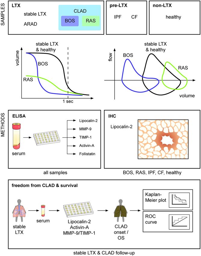
www.nature.com/scientificreports/
Lung Transplantation (LTX) is performed as the last therapeutic option for end-stage pulmonary fibrotic diseases,
including Idiopathic Pulmonary Fibrosis (IPF) and Cystic Fibrosis (CF). Five- and ten-year survival rates of
adults after primary LTX are 55–60% and 34%, respectively. Despite gradually improving perioperative survival
rates, long-term outcomes remained almost unaffected. Chronic Lung Allograft Dysfunction (CLAD) remains
the main limiting factor for reduced life expectancy after LTX occurring in approximately 50% of transplant
recipients within 5 years after primary L TX1,2.
There are two predominant CLAD phenotypes: bronchiolitis obliterans syndrome (BOS) and Restrictive Allo-
graft Syndrome (RAS)3. Bronchiolitis Obliterans (BO) after LTX manifests as narrowing or complete occlusion of
the small airways. As the histopathological diagnosis of BO is hard to obtain, BOS was introduced as the clinical
correlate of BO, characterized by an irreversible airway o bstruction2. RAS, contributing to 25–35% of CLAD has
an even worse prognosis than BOS with an average life expectancy of 540 versus 1200 days. RAS is histologically
characterised by diffuse alveolar damage and extensive fibrosis of the alveolar i nterstitium4,5. Before CLAD can
be diagnosed, allograft-related (e.g. persistent acute rejection, Azithromycin Responsive Allograft Dysfunction
(ARAD), infection/colonization, anastomotic stricture) and extra-allograft-related (e.g. pleural disease, dia-
phragmatic dysfunction) confounding factors leading to non-chronic rejection with Forced Expiratory Volume
in one second ( FEV1) decline have to be excluded6.
IPF is the most lethal fibrotic disease of the lung. Healthy tissue is replaced by altered extracellular matrix
deposition and alveolar architecture is destroyed, leading to decreased lung compliance, disrupted gas exchange,
and ultimately respiratory failure and d eath7. CF is a genetic multi-systemic disorder affecting lungs, pancreas,
liver and i ntestine8. Mutations of the Cystic Fibrosis Transmembrane Conductance Regulator (CFTR) gene cause
impaired mucociliary clearance leading to mucus plugging, opportunistic infections, chronic inflammation,
oxidative stress and airway r emodeling9. Although the etiologies of IPF, CF and CLAD significantly differ, the
pathophysiology of these diseases show overlapping characteristics, including epithelial cell injury, increased
production and deposition of extracellular matrix, immune cell activation, and fibroblast proliferation10.
In human lungs Lipocalin-2 is expressed in small quantities under healthy conditions and is up-regulated
during pulmonary disease, infection and lung injury11. In CF patients Lipocalin-2 was increased in bronchial
secretions and thereby reflecting neutrophilic inflammation12. In low concentrations Lipocalin-2 is involved
in innate immunity by acting as a siderophore, causing iron depletion to prevent bacterial growth and exhibits
anti-inflammatory effects on m acrophages13.
The imbalance between Matrix Metalloproteinases (MMPs) and Tissue Inhibitors of Metalloprotein-
ases (TIMPs) might be an important trigger for fibrogenic processes. Elevated MMP-9 concentrations and
MMP-9/TIMP-1 ratios in serum and Bronchioalveolar Lavage (BAL) were associated with CLAD, IPF and CF14.
Activin-A belongs to the transforming growth factor-β superfamily and is produced by immune, epithelial
and endothelial cells. Activin-A promotes fibrotic processes by stimulating fibroblast proliferation, differentia-
tion and inducing regulators of fibrosis15. Follistatin is an activin-binding protein that inhibits the activity of
Activin-A. Thereby, Follistatin is capable of reducing inflammation, fibrosis and m ortality16.
So far the diagnosis of CLAD remains limited to clinical parameters reflecting permanent loss of lung func-
tion. We aimed to identify novel biomarkers for early CLAD diagnosis, prior to emerging irreversible lung injury
and refine distinguishing biomarkers for ARAD and CLAD phenotypes: BOS and RAS, associated with different
prognosis and thereby enabling early interventions that could modify or even avoid the inevitable degradation
of pulmonary function.
The aim of the study was to shed light on the cytokine pathophysiology of patients with fibrotic diseases,
including LTX patients with CLAD (BOS and RAS) and ARAD; and patients with end-stage pulmonary fibrosis
(IPF and CF).
The loss of pulmonary function in a patient after LTX will prompt a series of diagnostic investigations. The
first clinical question that arises is about reversibility of the observed pulmonary function loss. After a thorough
workup excluding extra-CLAD causes of pulmonary function loss (e.g. silent aspiration), patients not respond-
ing to azithromycin are only identified after 3 months of unsuccessful azithromycin treatment. Thus the first
objective of this study was to identify biomarkers able to distinguish patients with ARAD from those with CLAD
and to investigate whether treatment responses to azithromycin are predictable via cytokines. After ARAD is
excluded clinicians want to distinguish the different forms of CLAD: BOS and RAS. So, our second objective
was to identify biomarkers that can help to distinguish BOS from RAS. We believe that different biomarkers
could be a helpful additive diagnostic tool in decision-making at certain crossroads (ARAD vs. CLAD or BOS
vs. RAS) in the context of clinical workup and follow-up. Apart from these clinical decision-making algorithms
in LTX patients our third objective was of a purely academic interest contributing to better understanding of
possible similarities or differences between the pathophysiology of CLAD and end-stage fibrotic diseases such
as CF and IPF. Regarding prognosis, we sought to determine biomarkers capable of predicting freedom-from-
CLAD. We therefore sought to investigate the outcome of CLAD in the context of biomarkers (outcome: overall
survival (OS)/re-LTX).
Results
Cytokine serum concentrations of patients with Stable-LTX, ARAD, CLAD (BOS and RAS), end-stage IPF and
CF as well as healthy volunteers were summarized in Table 1. Intra-assay and inter-assay Coefficients of Vari-
ability (CV) can be found in the Supplementary Table 1. The lowest intra-assay %CV was at 1.0 for Lipocalin-2,
the highest %CV was at 3.4 for Folliastatin. The lowest inter-assay %CV was calculated for MMP-9, while the
highest inter-assay %CV was measured for TIMP-1.
Scientific Reports | (2021) 11:6799 | https://doi.org/10.1038/s41598-021-85949-1 2
Vol:.(1234567890)
www.nature.com/scientificreports/

                                                                                        MMP-9/TIMP-1                                                  Activin-A/FST
                       LCN-2 (ng/ml)         MMP-9 (ng/ml)        TIMP-1 (ng/ml)        ratio             Activin-A (pg/ml)     FST (pg/ml)           ratio
Healthy (n = 63)       344.6 [126.2–389.3]   559.9 [304.0–1013.0] 44.4 [33.6–106.8]     11.2 [4.9–28.2]   362.8 [304.6–544.5]   207.8 [141.3–438.2]   1.6 [0.7–3.3]
Stable-LTX (n = 63)    365.7 [313.5–510.9]   511.0 [321.7–1246.4] 134.4 [60.1–319.2]    4.5 [1.8–10.6]    369.0 [196.9–505.5]   333.1 [129.5–505.5]   1.1 [0.4–3.5]
ARAD (n = 22)          368.4 [276.8–485.5]   490.4 [315.9–1314.8] 93.0 [40.6–213.9]     6.8 [1.9–17.3]    118.1 [72.6–343.0]    363.8 [180.1–721.9]   0.4 [0.1–0.9]
CLAD (n = 41)          388.6 [310.4–602.8]   908.0 [467.4–1552.8] 105.9 [57.6–319.7]    7.5 [2.5–17.7]    371.1 [205.1–994.5]   297.1 [232.6–942.3]   1.1 [0.5–2.7]
BOS (n = 30)           367.0 [284.1–584.2]   879.4 [537.7–1619.9] 93.7 [49.6–209.8]     8.1 [2.8–22.6]    343.9 [212.3–759.1]   268.0 [182.0–1023.7] 1.0 [0.6–2.3]
                                             1073.2 [289.6–
RAS (n = 11)           518.4 [444.8–768.2]                        199.4 [103.3–486.9]   2.7 [2.1–9.6]     883.8 [84.7–2295.4]   387.8 [295.4–903.0]   1.7 [0.3–6.6]
                                             1546.9]
                                             1634.7 [1115.2–
IPF (n = 23)           321.6 [162.0–379.2]                        121.0 [50.9–434.0]    11.8 [3.0–42.3]   327 [212.8–636.4]     210.5 [177.7–257.7]   1.4 [0.7–3.6]
                                             2429.4]
                                             2186.7 [1526.9–
CF (n = 21)            366.3 [282.3–592.8]                        220.8 [62.6–505.1]    8.6 [3.8–39.0]    377 [212.8–636.4]     207.6 [153.8–308.2]   1.7 [0.8–3.8]
                                             3791.5]

                                       Table 1.  Comparison of cytokine serum concentrations. Serum cytokine concentrations of healthy volunteers,
                                       Stable-LTX, ARAD, CLAD (RAS and BOS) and patients with end-stage pulmonary fibrosis (IPF and CF) were
                                       measured by immunoassays and reported as median [25–75% Percentile]. n number of individuals, LCN-
                                       2 Lipocalin-2, MMP-9 Matrix Metalloproteinase-9, TIMP-1 Tissue Inhibitor of Matrix Metalloproteinase,
                                       FST Follistatin, Stable-LTX transplanted patients free from CLAD, ARAD azithromycin-responsive allograft
                                       dysfunction, CLAD chronic lung allograft dysfunction, BOS bronchiolitis obliterans syndrome. RAS restrictive
                                       allograft syndrome, IPF idiopathic pulmonary fibrosis, CF cystic fibrosis.

                                       Lipocalin‑2 serum concentrations differentiate RAS from BOS, ARAD, and Stable‑LTX. There
                                       were no differences in Lipocalin-2 serum concentrations in CLAD compared to ARAD patients (p = 0.224).
                                       However, RAS patients showed significantly higher serum concentrations of Lipocalin-2 compared to BOS
                                       (p = 0.049), ARAD (p = 0.002) and Stable-LTX patients (p = 0.009). By comparison, BOS and Stable-LTX dis-
                                       played similar Lipocalin-2 serum concentrations (p = 0.868). Lipocalin-2 serum concentrations of all patient
                                       groups: LTX patients (RAS, BOS, ARAD, stable-LTX) as well as patients with end stage pulmonary fibrosis
                                       (CF and IPF) were depicted in Fig. 1A. Performing multiple testing between BOS, RAS, ARAD and stable LTX
                                       revealed no statistically significant difference in Lipocalin-2 serum concentrations (p = 0.082; Supplementary
                                       Fig. 1A).

                                       MMP‑9 is increased in BOS compared to Stable‑LTX. There were no differences in MMP-9 and
                                       TIMP-1 serum concentrations in CLAD compared to ARAD patients (p = 0.086) and (p = 0.276). However,
                                       MMP-9 serum concentrations were significantly higher in BOS compared to Stable-LTX patients (p = 0.042).
                                       TIMP-1 (p = 0.052) serum concentrations and the MMP-9/TIMP-1 ratio (p = 0.176) did not display significant
                                       differences among groups, respectively. MMP-9 and TIMP-1 serum concentrations of all patient groups: LTX
                                       patients (RAS, BOS, ARAD, stable-LTX) as well as patients with end stage pulmonary fibrosis (CF and IPF) were
                                       depicted in Fig. 1B,C. Performing multiple testing between BOS, RAS, ARAD and stable LTX revealed no statis-
                                       tically significant difference in MMP-9 and TIMP-1 serum concentrations (p = 0.188, p = 0.147; Supplementary
                                       Fig. 1B,C).

                                       Decreased Activin‑A in ARAD compared to Stable‑LTX and BOS.                    Activin-A serum concentrations
                                       were significantly decreased in ARAD patients compared to CLAD (p = 0.004), BOS (p = 0.004) and Stable-LTX
                                       (p = 0.002) patients. There was no difference among patients with RAS and ARAD (p = 0.305), RAS and BOS
                                       (p = 0.410), RAS and Stable-LTX (p = 0.166) or Stable-LTX and BOS patients (p = 0.601), respectively. Activin-
                                       A/Follistatin ratio was significantly decreased in ARAD compared to CLAD (p = 0.017) and BOS (p = 0.017)
                                       patients, respectively. Activin-A serum concentrations of all patient groups: LTX patients (RAS, BOS, ARAD,
                                       stable-LTX) as well as patients with end stage pulmonary fibrosis (CF and IPF) were depicted in Fig. 1D. Per-
                                       forming multiple testing between BOS, RAS, ARAD and stable LTX revealed significant differences in Activin-A
                                       serum concentrations (p = 0.008; Supplementary Fig. 1D).

                                       Low serum concentrations of Activin‑A in ARAD patients. Eighty-three percent of patients with
                                       suspected BOS (41 out of 49) and 50% (6 out of 12) with suspected RAS were treated with azithromycin. While
                                       a treatment response to azithromycin was evident in 19 (46%) patients with suspected BOS, only one (16%)
                                       patient with suspected RAS was responsive to therapy. Patients with ARAD displayed reduced Activin-A serum
                                       concentrations (p = 0.001) and Activin-A/Follistatin ratio (p = 0.011) compared to patients not responding
                                       to azithromycin therapy. Activin-A serum concentrations (cut-off = 350 ng/ml) predicted responsiveness to
                                       azithromycin therapy (sensitivity = 0.772, specificity = 0.500, positive predictive value = 0.531 and negative pre-
                                       dictive value = 0.750).

                                       Increased serum cytokine expression in end‑stage pulmonary fibrotic diseases. IPF patients
                                       showed significantly elevated serum concentrations of MMP-9 (p < 0.001) and TIMP-1 (p = 0.002) compared to
                                       healthy volunteers, respectively (Fig. 2A,B). Similar to IPF, we detected higher serum concentrations of MMP-9

Scientific Reports |      (2021) 11:6799 |                     https://doi.org/10.1038/s41598-021-85949-1                                                                  3

                                                                                                                                                                      Vol.:(0123456789)
www.nature.com/scientificreports/
Figure 1. Differential expression of Lipocalin-2, Activin-A and MMP-9. Statistically significant differences for
Lipcalin-2, MMP-9 and Activin-A are depicted in (A,B) and (D) where applicable. There were no statistically
significant differences for TIMP-1 serum concentrations between all groups (C). Mann–Whitney U testing was
performed between two groups each. The graphical depiction of all six groups together was chosen for better
overview. The following p-values ware not corrected for multiple testing. *p < 0.05; **p < 0.01; ***p < 0.001 RAS
Restrictive allograft syndrome, BOS bronchiolitis obliterans syndrome, ARAD azithromycin-responsive allograft
dysfunction, MMP-9 matrix metalloproteinase-9, TIMP-1 tissue inhibitor of matrix metalloproteinase.
(p = 0.002) and TIMP-1 (p = 0.008) in CF patients (Fig. 2C,D). Furthermore, CF patients showed higher Lipoca-
lin-2 (p = 0.007; Fig. 2E) and lower Follistatin (p = 0.024) serum concentrations than healthy controls.
Qualitative and quantitative differences in cytokine expression between end‑stage pulmo-
nary fibrosis and CLAD. Patients with IPF had significantly lower Lipocalin-2 (p = 0.011), Follistatin
(p = 0.002) and MMP-9 (p = 0.007) serum concentrations compared to CLAD patients. CLAD subtype analy-
sis revealed significantly higher Lipocalin-2, Follistatin and MMP-9 serum concentrations in RAS (p = 0.003,
p = 0.001, p = 0.049) and BOS (p = 0.029, p = 0.033, p = 0.013) compared to IPF patients, respectively. In com-
parison to ARAD patients, IPF patients had decreased Follistatin (p = 0.035) but increased MMP-9 (p = 0.002),
Activin-A (p = 0.004) serum concentrations and Activin-A/Follistatin ratio (p = 0.035). Further, IPF patients
displayed significantly decreased Follistatin (p = 0.038), however increased MMP-9 (p < 0.001) serum concentra-
tions compared to Stable-LTX patients. Patients with CF had significantly increased MMP-9 compared to CLAD
Scientific Reports | (2021) 11:6799 | https://doi.org/10.1038/s41598-021-85949-1 4
Vol:.(1234567890)www.nature.com/scientificreports/
Figure 2. Increased expression of serum cytokines in end stage fibrotic diseases. IPF patients displayed higher serum concentrations
of MMP-9 (A) and TIMP-1 (B) compared to healthy controls. CF patients also showed higher serum concentrations of MMP-9 (C)
and TIMP-1 (D). Furthermore, Lipocalin-2 serum concentrations were elevated in CF patients compared to healthy volunteers (E).
IPF Idiopathic pulmonary fibrosis, MMP-9 matrix metalloproteinase-9, TIMP-1 tissue inhibitors of metalloproteinase-1, CF cystic
fibrosis, RAS restrictive allograft syndrome, BOS bronchiolitis obliterans syndrome.
Scientific Reports | (2021) 11:6799 | https://doi.org/10.1038/s41598-021-85949-1 5
Vol.:(0123456789)www.nature.com/scientificreports/
Scientific Reports | (2021) 11:6799 | https://doi.org/10.1038/s41598-021-85949-1 6
Vol:.(1234567890)www.nature.com/scientificreports/
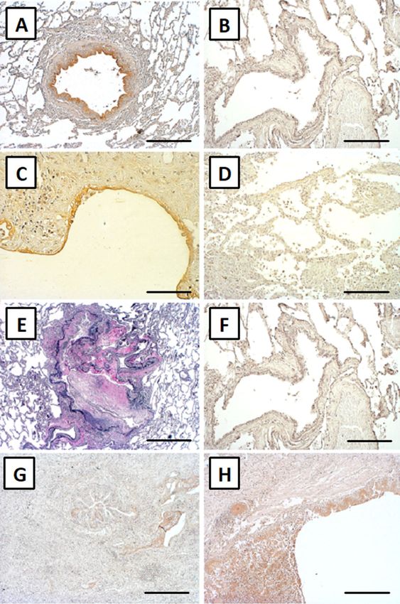
◂Figure 3. Increased pulmonary expression of Lipocalin-2 in RAS. Immunohistochemistry: Pulmonary
Lipocalin-2 expression in bronchial walls of a RAS patient. [(A) scale bar: 400 µm]; in contrast, absent
expression of Lipocalin-2 in bronchial walls of a healthy control lung [(B) scale bar: 400 µm]. Lipocalin-2
expression in alveolar pneumocytes of a RAS patient [(C) scale bar: 80 µm]. No Lipocalin-2 expression in
alveolar pneumocytes of a BO patient [(D) scale bar: 80 µm]. Elastica-van-Gieson staining detecting BO lesions
[(E) scale bar: 400 µm]. Adjacent slide to (E) showing no increased expression of Lipocalin-2 within a BO
lesion [(F) scale bar: 400 µm]. Discrete Lipocalin-2 expression in bronchial walls of an IPF patient [(G) scale
bar: 160 µm] and a CF patient [(H) scale bar: 400 µm]. RAS Restrictive allograft syndrome, BO bronchiolitis
obliterans, CF cystic fibrosis, IPF idiopathic pulmonary fibrosis.
Bronchial epithelium Alveolar walls Lung parenchyma BO lesions
Y/N (%) Y/N (%) Y/N (%) Y/N (%)
BOS (n = 11) 11/0 (100) 0/11 (0) 0/11 (0)
RAS (n = 6) 6/0 (100) 4/2 (66,6) 0/6 (0)
IPF (n = 10) 10/0 (100) 2/8 (20) 0/10(0)
CF (n = 10) 9/1 (90) 0/10 (0) 0/10 (0)
Control (n = 9) 5/4 (55,5) 0/9 (0) 0/9 (0)
Table 2. Semi-quantitative analysis of pulmonary Lipocalin-2 expression. Immunohistochemistry for
Lipocalin-2 was performed on lung specimens of patients with CLAD (RAS and BOS), end stage pulmonary
fibrosis (IPF and CF) and healthy controls. Antibody reactivity in BO lesions of BOS patients, bronchial
epithelium, alveolar walls and lung parenchyma was assessed. Y positive antibody staining, N negative
antibody staining, BO bronchiolitis obliterans, BOS bronchiolitis obliterans syndrome, RAS restrictive allograft
syndrome, IPF idiopathic pulmonary fibrosis CF cystic fibrosis.
(p = 0.001), BOS (p = 0.001) and RAS (p = 0.001). Serum concentrations in CF patients were significantly lower
for Follistatin (p = 0.033), but increased for Activin-A (p = 0.004), MMP-9 (p = 0.001), and Activin-A/Follistatin
ratio (p = 0.001) compared to ARAD patients. Comparing CF with Stable-LTX patients MMP-9 serum concen-
trations (p = 0.001) and the Activin-A/Follistatin ratio (p = 0.049) were significantly increased in CF patients,
respectively.
Lipocalin‑2 is expressed in bronchial epithelium and alveolar pneumocytes in RAS. We
detected Lipocalin-2 expression in the bronchial epithelium of 11 BO (100%), 6 RAS (100%), 10 IPF (100%)
and 9 CF (90%) patients, but only in 5 (55.5%) healthy lung specimens by immunohistochemistry. In the lung
parenchyma, only small foci of reactive alveolar pneumocyte hyperplasia showed cytoplasmic staining, which
was exclusively found in 4 RAS (66.6%) and 2 IPF (20%) patients (Fig. 3 and Table 2).
Serum cytokines are associated with CLAD onset and survival. We investigated the correlation
of cytokine serum concentrations and clinical outcome, including future onset of CLAD and OS or re-trans-
plantation. The median time between serum sampling and onset of CLAD was at 62.2 (range 84.7) months
and for overall survival or re-transplantation at 62.3 (range 85.2) months. Eight Stable-LTX patients (12.7%)
developed CLAD during follow-up. Of those patients, three underwent extracorporeal photopheresis (37.5%)
for 5.8–30.0 months (mean = 14.6 months). Of 63 Stable-LTX patients, 11 died during follow up (17.4%). Among
them, four died from later developed CLAD (36.4%; 3 BOS and 1 RAS), five died from infections (45.5%), one
from cerebral bleeding (9.1%) and one patient died from unknown causes (9.1%).
High serum concentrations of Lipocalin-2 were significantly associated with earlier onset of CLAD (Cut-
off = 350.4 ng/ml, sensitivity = 0.875, specificity = 0.418, p = 0.022) and adverse OS (Cut-off = 354.1 ng/ml, sen-
sitivity = 0.800, specificity = 0.604, p = 0.014) (Fig. 4A,B). Higher Activin-A serum concentrations were signifi-
cantly associated with onset of CLAD (Cut-off = 217 ng/ml, sensitivity = 1.000, specificity = 0.364, p = 0.038)
(Fig. 4C). With regard to CLAD phenotypes, higher Activin-A serum concentrations (Cut-off = 347 ng/ml,
sensitivity = 0.800, specificity = 0.564, p = 0.044) (Fig. 4D) and higher MMP-9/TIMP-1 ratios (Cut-off = 4.1, sen-
sitivity = 1.000, specificity = 0.509, p = 0.032) (Fig. 4E) predicted onset BOS, but not RAS patients.
Discussion
While milestones in the perioperative management of LTX were achieved, CLAD, manifesting as BOS or RAS
remains the main reason for adverse long-term outcome. The underlying pathomechanisms of the CLAD pheno-
types are barely understood and an accurate diagnosis is often difficult to obtain. Biomarkers aiding in diagnosis
and identifying pathophysiological pathways might spark new developments in CLAD management.
Our data identified Lipocalin-2 as a potentially useful biomarker to distinguish RAS from BOS and Stable-
LTX. Lipocalin-2 is constitutively expressed and stored in neutrophilic granules making it a possible biomarker
for neutrophil a ctivity17. However, previous studies did not show differences in neutrophil counts in lungs
of RAS compared to BOS patients18. Thus, whether elevated levels of circulating Lipocalin-2 in RAS patients
Scientific Reports | (2021) 11:6799 | https://doi.org/10.1038/s41598-021-85949-1 7
Vol.:(0123456789)www.nature.com/scientificreports/
Figure 4. Lipocalin-2 serum concentrations in Stable-LTX predict future onset of CLAD and BOS. Stable-LTX patients with high
Lipocalin-2 serum concentrations were significantly more likely to develop CLAD during follow-up compared to those with low
Lipocalin-2 concentrations (A). Higher Lipocalin-2 serum concentrations in Stable-LTX patients resulted in worse OS during
follow-up (B). High Activin-A serum concentrations were also associated with higher CLAD onset rates in Stable-LTX patients (C).
Higher serum concentrations of Activin-A where significantly associated with the onset of CLAD subtype BOS (D). High MMP-9/
TIMP-1 ratios translated to higher BOS onset rates compared to low MMP-9/TIMP-1 ratios in Stable-LTX patients (E). LTX Lung
transplantation, CLAD chronic lung allograft dysfunction, RAS restrictive allograft syndrome, BOS bronchiolitis obliterans syndrome,
MMP-9 matrix metalloproteinase-9, TIMP-1 tissue inhibitors of metalloproteinase-1.
Scientific Reports | (2021) 11:6799 | https://doi.org/10.1038/s41598-021-85949-1 8
Vol:.(1234567890)www.nature.com/scientificreports/
Figure 5. Graphical depiction of the study design. Serum samples were selected of LTX patients diagnosed
with CLAD (BOS (n = 30) and RAS (n = 11)), ARAD (n = 22) and Stable-LTX (n = 56), patients with end-
stage pulmonary disease [IPF (n = 31) and CF (n = 15)] listed for LTX and healthy volunteers (n = 63). CLAD
phenotypes, Stable-LTX and ARAD patients were diagnosed via spirometry. Prototypical spirometric volume
changes over time and flow volume loops of stable lung transplanted patients/healthy people and patients with
BOS and RAS are depicted. Enzyme-linked immunosorbent assays were employed for measuring cytokine
serum concentrations of Lipocalin-2, MMP-9, TIMP-1, Activin-A and Follistatin. MMP-9/TIMP-1 and
Activin-A/Follistatin ratios were calculated. Immunohistochemical stainings were performed in specimens of 20
patients who underwent re-transplantation for either BOS (n = 11) or RAS (n = 9), 20 patients who underwent
primary LTX for either IPF (n = 10) or CF (n = 10), and 10 patients who served as healthy controls. Further,
the correlation of cytokine serum concentrations and clinical outcome, including OS, future onset of CLAD
and re-transplantation was analysed by performing Kaplan–Meier analysis. LTX Lung transplantation, CLAD
chronic lung allograft dysfunction, BOS bronchiolitis obliterans syndrome, RAS restrictive allograft syndrome,
ARAD azithromycin-reversible allograft dysfunction, IPF idiopathic pulmonary fibrosis, CF cystic fibrosis,
MMP-9 matrix metalloproteinase-9, TIMP-1 tissue inhibitors of metalloproteinase-1.
Scientific Reports | (2021) 11:6799 | https://doi.org/10.1038/s41598-021-85949-1 9
Vol.:(0123456789)www.nature.com/scientificreports/
Stable-LTX
(n = 63) ARAD (n = 22) CLAD (n = 41) BOS (n = 30) RAS (n = 11) IPF (n = 23) CF (n = 21)
Age mean ± SD 50.6 ± 14.3 50.4 ± 14.3 51.0 ± 13.5 49.8 ± 13.2 55.9 ± 14.0 51.7 ± 11.1 31.0 ± 11.7
Gender F/M ratio 0.966 0.953 0.906 1.042 0.500 0.381 1.286
Diagnosis prior to TX
COPD n (%) 37 (58.7) 9 (40.9) 18 (43.9) 14 (46.6) 6 (54.5) – –
IPF n (%) 8 (12.6) 7 (31.8) 8 (19.5) 6 (20.0) 2 (18.8) 23 (100.0) 21 (100.0)
IPAH n (%) 4 (6.3) 1 (4.5) 4 (9.7) 3 (10.0) 1 (9.0) – –
CF n (%) 8 (12.6) 5 (22.7) 4 (9.7) 4 (13.3) – – –
Others n (%) 5 (7.9) – 5 (12.1) 3 (10.0) 2 (18.8) – –
Type of TX
DLUTX n (%) 52 (82.5) 20 (90) 33 (80.4) 24 (80.0) 9 (81.8) – –
SLUTX n (%) 10 (15) 2 (9.0) 7 (17.0) 5 (16.6) 2 (18.1) – –
HLTX n (%) 1 (1.5) 0 (0.0) 1 (2.4) 1 (3.3) – – –
CMV risk
D+/R− n (%) 15 (23.8) 5 (22.7) 9 (21.1) 8 (26.6) 1 (9.0) – –
D+/R+ n (%) 30 (47.6) 10 (45.5) 19 (46.3) 15 (50.0) 4 (36.3) – –
D−/R+ n (%) 12 (19.0) 4 (18.1) 7 (17.0) 4 (13.3) 3 (27.2) – –
D−/R− n (%) 5 (1.5) 3 (15.0) 6 (14.6) 3 (10.0) 3 (27.2) – –
Table 3. Demographic and clinical data. Stable LTX Lung transplantation, ARAD sa, CLAD chronic lung
allograft dysfunction, BOS bronchiolitis obliterans syndrome, RAS restrictive allograft syndrome, IPF
idiopathic pulmonary fibrosis, CF cystic fibrosis, n number of individuals, SD standard deviation;
stem from increased neutrophil activity remains elusive. A possible source of serum Lipocalin-2 might be
secreted Lipocalin-2 expressed in the bronchial epithelium of all BOS and RAS patients (as detected by our
immunohistochemistry).
In the case of increased serum Lipocalin-2 in end-stage CF, neutrophils are a plausible source. The airways
of adult CF patients are chronically besieged by neutrophilic inflammation and circulating Lipocalin-2 was
reported to be elevated in acute exacerbations and Pseudomonas aeruginosa infections of CF p atients19. It is
strongly and selectively induced in bronchial and alveolar cells of inflamed lungs by Interleukin-1β20. We found
Lipocalin-2 expression in epithelial cells of 90% of all CF patients. In line with our findings in IPF are correla-
tions of Lipocalin-2 BAL levels with BAL neutrophilia and forced vital capacity, as well as no elevation of plasma
Lipocalin-2. Further, Lipocalin-2 was expressed in airway epithelial cells that covered the honeycomb cysts and
in bronchioles of IPF p atients21.
Lipocalin-2 is known to build complexes with MMP-9, thereby stabilizing MMP-9 activity22. Airway remod-
eling is promoted by active secretion of MMP-9 by bronchial epithelial cells triggered by T-cells23 In accordance
to these findings, several studies linked elevated MMP-9 concentrations and MMP-9/TIMP-1 ratios in serum
and BAL to CLAD, IPF and C F24,25. We detected higher MMP-9 serum concentrations (but not MMP-9/TIMP-1
ratio) in BOS patients compared to Stable-LTX patients and lower MMP-9/TIMP-1 ratios in CLAD compared
to CF and IPF patients, suggesting a different importance of protease-antiprotease imbalance in pathogenesis.
Divergent study results concerning cytokine profiles may pertain to different immunosuppressive therapy regi-
mens. In previous studies, TIMP-1 was shown to be up-regulated in human pulmonary fi brosis26. Our study
supported these results since TIMP-1 serum concentrations were higher in end-stage CF and IPF compared to
healthy volunteers, but not compared to Stable-LTX or CLAD patients.
In a study on patients after LTX Activin-A serum concentrations decreased from postoperative week two to
week twelve, while Follistatin serum concentrations remained unchanged until they increased 24 weeks post-
LTX. Patients with primary graft dysfunction expressed lower serum Follistatin and higher Activin-A/follistatin
ratios27. Our study was neither performed in the perioperative setting nor during a dramatic event such as pri-
mary graft dysfunction. We discovered Activin-A as a promising biomarker to distinguish ARAD patients from
Stable-LTX and BOS patients, however not from RAS. Activin-A served as a predictive molecule to measure the
responsiveness to azithromycin therapy. Low Activin-A concentrations in ARAD patients are in line with previ-
ous studies showing that Activin-A promotes inflammation at lower concentrations mediated by resting mono-
cytes/macrophages, but inhibits inflammatory activity as concentrations i ncrease16. Azithromycin was shown to
exhibit inhibitory effects on neutrophils, such as diminishing neutrophil extracellular traps r elease28. Activin-A
serum concentrations were not elevated in IPF and CF patients. Interestingly, Activin-A serum concentrations
were reported to correlate inversely with lung function and nutritional status in CF patients29. In a CF mouse
model with CF-like lung disease, Follistatin administration reduced Activin-A levels, mucus hypersecretion, and
airway neutrophilia. ln CF patients, decreased serum concentrations of Follistatin and elevated Activin-A resulted
in an increased Activin-A/Follistatin r atio29. While we detected decreased serum concentrations of Follistatin,
we did not observe elevated Activin-A concentrations and Activin-A/Follistatin ratios in end-stage CF patients.
These different results might stem from different CF disease severity: the aforementioned study sampled blood
from stable CF patients during outpatient visits, while our study investigated patients with end-stage pulmonary
Scientific Reports | (2021) 11:6799 | https://doi.org/10.1038/s41598-021-85949-1 10
Vol:.(1234567890)www.nature.com/scientificreports/
CF immediately before LTX. In our study, neither Activin-A serum concentrations nor Activin-A/Follistatin
ratios were elevated in end-stage IPF patients.
Our study has several limitations due to the retrospective analysis of prospectively collected blood samples,
the single center experience and the limited sample size. Large-scale studies are warranted for further biomarker
evaluation in CLAD, ARAD, CF and IPF patients. We are not suggesting that our absolute serum concentra-
tion values can be used to make any judgments about patient’s diagnosis. While comparative results during one
experiment could be repeated in separate ELISA tests. The commercially available ELISA tests were only sold
for research use and not for diagnostic purposes. Since cut-offs were chosen via Youden Index out of our study
results, sensitivity and specificity must be interpreted with caution. Early recognition of different CLAD pheno-
types is essential for developing new therapies and improving outcomes. This study identified promising new
biomarkers for better characterization and classification of CLAD phenotypes in addition to well-established
diagnostic methods including spirometry and radiological imaging. Lipocalin-2 might serve as a potential diag-
nostic or even therapeutic target for RAS patients. Activin-A seems helpful to identify ARAD patients. Further
prospective studies will be required confirming our biomarker results. The statistically significant difference in
Lipocalin-2 serum concentrations between BOS and RAS deserves further investigations; as does Activin-A in
ARAD patients by employing further methods including immunostaining.
Materials and methods
This single-centre explorative cohort study was performed at the institutional Division of Thoracic Surgery.
All methods were carried out in accordance with relevant guidelines and regulations in the manuscript. Ethi-
cal approval was obtained from the institutional Ethics Committee of the medical university of vienna (EC-
No:846/2010). All patients provided written informed consent prior to participation in the study. A graphical
depiction of the study design is shown in Fig. 5.
Definition of CLAD phenotypes and ARAD. CLAD was defined as persistent pulmonary function
decline (FEV1 ≥ 20%) calculated from baseline (the best two F
EV1 measurements after LTX performed more
than three weeks apart) after exclusion of non-CLAD causes by pulmonary function testing, bronchoscopy
including transbronchial/endobronchial biopsies and BAL, blood work and radiological imaging. Patients suf-
fering from suspected CLAD were treated with azithromycin 250–500 mg three times per week (irrespective of
BAL neutrophilia) for 3 months. Patients with an FEV1 improvement greater than 10% were classified as ARAD
and not included in the analysis of CLAD. CLAD was further subdivided into RAS (concomitant drop in Total
Lung Capacitiy (TLC) ≥ 10% compared to baseline levels and persistent opacities on chest imaging) and BOS (no
restrictive pulmonary function pattern)6.
Patient cohort. Demographic and clinical data were depicted in Table 3. For serum analysis, we enrolled
119 patients undergoing LTX. Among them, 41 patients were diagnosed with CLAD and dichotomized into BOS
(n = 30) and RAS (n = 11) phenotypes. The included BOS patients displayed the following disease severity: BOS1
(n = 5), BOS2 (n = 13) and BOS3 (n = 12). Ten out of 11 RAS patients had a mixed phenotype.
Patients with ARAD (n = 22) were separately analysed and not included in the CLAD cohort. Sixty-three LTX
patients with stable pulmonary function were included in this study (Stable-LTX). Blood samples of patients
with stable-LTX were drawn at clinical routine check-up including surveillance bronchoscopy. Furthermore
we enrolled patients with end-stage IPF (n = 31) and CF (n = 15) listed for LTX. Serum samples of 63 healthy
volunteers served as a control group. None of the healthy volunteers suffered from acute or chronic diseases,
such as asthma, chronic obstructive pulmonary disease, acute or chronic kidney diseases, autoimmune diseases
or any cancer type. Furthermore, none of the healthy volunteers took medical drugs for at least three months
before the blood draw.
Blood samples were aliquoted after centrifugation with 2851×g for 15 min at 4 °C and stored in 2 ml cryotubes
at − 80 °C in until further analysis.
CLAD, Stable-LTX, ARAD and control groups were matched by gender and age. There were no statistically
significant gender differences in any of the study groups (CLAD, comprising BOS and RAS, ARAD, stable-LTX
and healthy volunteers). Mean age of the study groups ranged from 49.8 years in BOS to 51.7 years in IPF patients.
There were no statistically significant age differences between ARAD, BOS, RAS, Stable-LTX and IPF patients.
However, CF patients were significantly younger (mean age of 31.0 years) compared to patients with ARAD,
BOS, RAS, stable LTX and IPF.
Standard immunosuppressive treatments. Fifty patients (42.0%) received induction therapy with
either alemtuzumab (n = 34, 68.0%), anti-thymocyte globulin (n = 15, 30.0%) or daclizumab (n = 1, 2.0%).
Maintenance therapy was performed in all patients with tacrolimus, mycophenolat-mofetil (only in the non-
alemtuzumab group) and corticosteroids. All patients undergoing LTX received postoperative anti-infectious
prophylaxis therapy with piperazillin/tazobactam, a lifelong pneumocystis prophylaxis with trimethoprim-
sulfamethoxazole, prophylactic inhalation therapy with amphotericin B and gentamicin; and cytomegalovirus
(CMV) prophylaxis including CMV hyperimmunoglobulines, together with valganciclovir.
Measurement of cytokine serum concentrations. Enzyme-Linked Immunosorbent Assays (ELISA)
were employed for measuring cytokine serum concentrations according to the manufacturer’s protocol, using
commercially available DuoSet ELISA kits: Human Lipocalin-2/NGAL, Human MMP-9, Human TIMP-1,
Human Activin-A, Human Follistatin (R&D Systems, Minneapolis, MN 55413, USA). Accordingly, MMP-9/
TIMP-1 and Activin-A/Follistatin ratios were calculated. Serum samples were diluted with reagent diluent for
Scientific Reports | (2021) 11:6799 | https://doi.org/10.1038/s41598-021-85949-1 11
Vol.:(0123456789)www.nature.com/scientificreports/
the measurements of Lipocalin-2 and TIMP-1 (500-fold), MMP-9 (5000-fold), and Follistatin (twofold) but not
Activin-A (no dilution).
Immunohistochemistry. Immunohistochemistry was performed with specimens of 50 patients allo-
cated to the following subgroups: 20 patients who underwent re-transplantation for either BOS (n = 11) or RAS
(n = 9), 20 patients who underwent primary LTX for either IPF (n = 10) or CF (n = 10) and 10 patients who
served as healthy controls. Patients of the latter group underwent lobectomy for peripherally located lung cancer
(pT1pN0M0). The tumour was located at least five centimetres from the investigated lung area and patients
showed no evidence for advanced parenchymal lung diseases.
Immunohistochemical staining was performed on formaldehyde-fixed, paraffin-embedded tissue specimens
according to routine p rotocols30. Anti-Lipocalin-2/NGAL antibody ab115324 (Abcam, Cambridge, UK) and
anti-goat IgG secondary antibodies (Vector Laboratories, Burlingame, CA, USA) were used for staining. Omis-
sion of the primary antibody served for negative control staining. Results were assessed by two independent
investigators, including one specialist on pulmonary histopathology (O.F.). Antibody staining of alveolar lining
cells, bronchial epithelium and the interstitium were evaluated on each slide. In case of interobserver discrepan-
cies, results were discussed until consensus was achieved. BOS lesions were identified by Elastica van Gieson
staining on adjacent slides.
Statistical methods. Statistical data analysis was performed using SPSS software (Version 20; IBM SPSS
Inc., Chicago, IL, USA). Graphical methods (histograms) were employed to test normality. Data were reported
as median (range) for non-normal distributions. Mann–Whitney U test was used to compare two independent
groups. Kruskal–Wallis rank test was used to perform multiple testing followed by Dunn’s multiple comparison
tests. P-values < 0.05 were considered statistically significant. For follow-up analysis, cytokine serum concentra-
tions were dichotomized into low and high subgroups by using ROC-Curves for calculating Youden-Indices.
We used the best cut-offs for maximizing sensitivity at the expense of specificity in order to create a test that is
maximally sensitive. Accordingly, we performed Kaplan–Meier survival analysis and used log-rank analysis for
outcome assessment. For the analysis of the prognostic value of serum cytokines, the date of serum withdrawal
was taken as the starting point for follow-up analysis. We analysed Stable-LTX patients for freedom from CLAD
and OS. In order to express the precision and repeatability of our ELISA experiments we calculated the intra-
assay and inter-assay CV. Therefore, we performed following calculation % CV = SD of plate means ÷ mean of
plate means × 100. GraphPad Prism 5 (GraphPad Software, La Jolla, CA, USA) was used for data visualization.
Boxplots were designed as follows: box: 1st to 3rd quartile, bar: median, whiskers: percentile 5–95, outliers: all
shown as dots.
Data availability
All data generated or analysed during this study are included in this published article.
Received: 24 August 2020; Accepted: 3 March 2021
References
1. Verleden, G. M., Raghu, G., Meyer, K. C., Glanville, A. R. & Corris, P. A new classification system for chronic lung allograft dys-
function. J. Heart Lung Transplant. 33(2), 127–133 (2014).
2. Meyer, K. C. et al. An international ISHLT/ATS/ERS clinical practice guideline: Diagnosis and management of bronchiolitis
obliterans syndrome. Eur. Respir. J. 44(6), 1479–1503 (2014).
3. Nishikawa, T., Inomata, S., Igarashi, M., Goyagi, T. & Naito, H. Plasma lidocaine concentrations during epidural blockade with
isoflurane or halothane anesthesia. Anesth. Analg. 75(6), 885–888 (1992).
4. Sato, M. et al. Restrictive allograft syndrome (RAS): A novel form of chronic lung allograft dysfunction. J. Heart Lung Transplant.
30(7), 735–742 (2011).
5. Sato, M., Hwang, D. M., Waddell, T. K., Singer, L. G. & Keshavjee, S. Progression pattern of restrictive allograft syndrome after
lung transplantation. J. Heart Lung Transplant. 32(1), 23–30 (2013).
6. Verleden, G. M. et al. Current views on chronic rejection after lung transplantation. Transpl. Int. 28(10), 1131–1139 (2015).
7. Fernandez, I. E. & Eickelberg, O. New cellular and molecular mechanisms of lung injury and fibrosis in idiopathic pulmonary
fibrosis. Lancet 380(9842), 680–688 (2012).
8. Walshaw, M. J. Cystic fibrosis: Diagnosis and management—NICE guideline 78. Paediatr. Respir. Rev. 31, 12–14 (2019).
9. Castellani, S., Di Gioia, S., di Toma, L. & Conese, M. Human cellular models for the investigation of lung inflammation and mucus
production in cystic fibrosis. Anal. Cell Pathol. (Amst). 2018, 3839803 (2018).
10. Fernandez, I. E., Heinzelmann, K., Verleden, S. & Eickelberg, O. Characteristic patterns in the fibrotic lung. Comparing idiopathic
pulmonary fibrosis with chronic lung allograft dysfunction. Ann. Am. Thorac. Soc. 12(Suppl 1), S34-41 (2015).
11. Dittrich, A. M., Meyer, H. A. & Hamelmann, E. The role of lipocalins in airway disease. Clin. Exp. Allergy. 43(5), 503–511 (2013).
12. Redl, B., Wojnar, P., Ellemunter, H. & Feichtinger, H. Identification of a lipocalin in mucosal glands of the human tracheobronchial
tree and its enhanced secretion in cystic fibrosis. Lab. Investig. 78(9), 1121–1129 (1998).
13. Flo, T. H. et al. Lipocalin 2 mediates an innate immune response to bacterial infection by sequestrating iron. Nature 432(7019),
917–921 (2004).
14. Gueders, M. M., Foidart, J. M., Noel, A. & Cataldo, D. D. Matrix metalloproteinases (MMPs) and tissue inhibitors of MMPs in the
respiratory tract: Potential implications in asthma and other lung diseases. Eur. J. Pharmacol. 533(1–3), 133–144 (2006).
15. Karagiannidis, C. et al. Activin A is an acute allergen-responsive cytokine and provides a link to TGF-beta-mediated airway
remodeling in asthma. J. Allergy Clin. Immunol. 117(1), 111–118 (2006).
16. Hedger, M. P. & de Kretser, D. M. The activins and their binding protein, follistatin-Diagnostic and therapeutic targets in inflam-
matory disease and fibrosis. Cytokine Growth Factor Rev. 24(3), 285–295 (2013).
17. Cruz, D. N., Gaiao, S., Maisel, A., Ronco, C. & Devarajan, P. Neutrophil gelatinase-associated lipocalin as a biomarker of cardio-
vascular disease: A systematic review. Clin. Chem. Lab. Med. 50(9), 1533–1545 (2012).
Scientific Reports | (2021) 11:6799 | https://doi.org/10.1038/s41598-021-85949-1 12
Vol:.(1234567890)www.nature.com/scientificreports/
18. Vandermeulen, E. et al. Immunological diversity in phenotypes of chronic lung allograft dysfunction: A comprehensive immu-
nohistochemical analysis. Transpl. Int. 30(2), 134–143 (2017).
19. Zughaier, S. M., Tangpricha, V., Leong, T., Stecenko, A. A. & McCarty, N. A. Peripheral monocytes derived from patients with cystic
fibrosis and healthy donors secrete NGAL in response to Pseudomonas aeruginosa infection. J. Investig. Med. 61(6), 1018–1025
(2013).
20. Cowland, J. B., Sorensen, O. E., Sehested, M. & Borregaard, N. Neutrophil gelatinase-associated lipocalin is up-regulated in human
epithelial cells by IL-1 beta, but not by TNF-alpha. J. Immunol. 171(12), 6630–6639 (2003).
21. Ikezoe, K. et al. Neutrophil gelatinase-associated lipocalin in idiopathic pulmonary fibrosis. Eur. Respir. J. 43(6), 1807–1809 (2014).
22. Bouchet, S. & Bauvois, B. Neutrophil gelatinase-associated lipocalin (NGAL), pro-matrix metalloproteinase-9 (pro-MMP-9) and
their complex pro-MMP-9/NGAL in leukaemias. Cancers (Basel). 6(2), 796–812 (2014).
23. Pain, M. et al. T cells promote bronchial epithelial cell secretion of matrix metalloproteinase-9 via a C-C chemokine receptor type
2 pathway: Implications for chronic lung allograft dysfunction. Am. J. Transplant. 17(6), 1502–1514 (2017).
24. Kennedy, V. E., Todd, J. L. & Palmer, S. M. Bronchoalveolar lavage as a tool to predict, diagnose and understand bronchiolitis
obliterans syndrome. Am. J. Transplant. 13(3), 552–561 (2013).
25. Heijink, I. H. et al. Metalloproteinase profiling in lung transplant recipients with good outcome and bronchiolitis obliterans syn-
drome. Transplantation 99(9), 1946–1952 (2015).
26. Lagente, V. & Boichot, E. Role of matrix metalloproteinases in the inflammatory process of respiratory diseases. J. Mol. Cell Cardiol.
48(3), 440–444 (2010).
27. Westall, G. P. et al. Activin biology after lung transplantation. Transplant Direct. 3(6), e159 (2017).
28. Bystrzycka, W. et al. Azithromycin and chloramphenicol diminish neutrophil extracellular traps (NETs) release. Int. J. Mol. Sci.
18(12), 2666 (2017).
29. Hardy, C. L. et al. The activin A antagonist follistatin inhibits cystic fibrosis-like lung inflammation and pathology. Immunol. Cell
Biol. 93(6), 567–574 (2015).
30. Janik, S. et al. HSP27 and 70 expression in thymic epithelial tumors and benign thymic alterations: Diagnostic, prognostic and
physiologic implications. Sci. Rep. 6, 24267 (2016).
Author contributions
V.C. and K.J. contributed equaly to conception, design, laboratory work, patient recruitment, data collection,
data analysis and writing. M.B. contributed to conception, design, laboratory work, patient recruitment, data
collection, supervision, writing and critical revision. B.A. contributed to data collection and critical revision.
O.F., H.P., R.T. and J.S. contributed to laboratory work, data collection and critical revision. L.M. contributed to
design and critical revision. J.P., A.H.J. and K.W. contributed to critical revision. All authors read and approved
the final manuscript.
Funding
This work was supported by the institutional surgical research laboratories ARGE Moser (FOLAB Chirurgie)
and FFG-Grant “APOSEC” (#852748; 2015–2018).
Competing interests
The authors declare no competing interests.
Additional information
Supplementary Information The online version contains supplementary material available at https://doi.org/
10.1038/s41598-021-85949-1.
Correspondence and requests for materials should be addressed to B.M.
Reprints and permissions information is available at www.nature.com/reprints.
Publisher’s note Springer Nature remains neutral with regard to jurisdictional claims in published maps and
institutional affiliations.
Open Access This article is licensed under a Creative Commons Attribution 4.0 International
License, which permits use, sharing, adaptation, distribution and reproduction in any medium or
format, as long as you give appropriate credit to the original author(s) and the source, provide a link to the
Creative Commons licence, and indicate if changes were made. The images or other third party material in this
article are included in the article’s Creative Commons licence, unless indicated otherwise in a credit line to the
material. If material is not included in the article’s Creative Commons licence and your intended use is not
permitted by statutory regulation or exceeds the permitted use, you will need to obtain permission directly from
the copyright holder. To view a copy of this licence, visit http://creativecommons.org/licenses/by/4.0/.
© The Author(s) 2021
Scientific Reports | (2021) 11:6799 | https://doi.org/10.1038/s41598-021-85949-1 13
Vol.:(0123456789)You can also read