Preparation and Neutralization Efficacy of Novel Jellyfish Antivenoms against Cyanea nozakii Toxins
←
→
Page content transcription
If your browser does not render page correctly, please read the page content below
Article

Preparation and Neutralization Efficacy of Novel Jellyfish
Antivenoms against Cyanea nozakii Toxins
Rongfeng Li 1,2,*, Huahua Yu 1,2, Aoyu Li 1, Chunlin Yu 1,3 and Pengcheng Li 1,2,*

                                            1 Key Laboratory of Experimental Marine Biology, Center for Ocean Mega-Science, Institute of Oceanology,
                                              Chinese Academy of Sciences, Qingdao 266071, China; yuhuahua@qdio.ac.cn (H.Y.);
                                              aoyulee1227@163.com (A.L.); Yuchunlin17@mails.ucas.ac.cn (C.Y.)
                                            2 Laboratory for Marine Drugs and Bioproducts, Pilot National Laboratory for Marine Science and

                                              Technology (Qingdao), No. 1 Wenhai Road, Qingdao 266237, China
                                            3 College of Earth and Planetary Sciences, University of Chinese Academy of Sciences, Beijing 100049, China

                                            * Correspondence: rongfengli@qdio.ac.cn (R.L.); pcli@qdio.ac.cn (P.L.);
                                              Tel.: +86-532-82898512 (R.L.); +86-532-82898707 (P.L.)

                                            Abstract: Jellyfish stings are a common issue globally, particularly in coastal areas in the summer.
                                            Victims can suffer pain, itching, swelling, shock, and even death. Usually, hot water, vinegar, or
                                            alumen is used to treat the normal symptoms of a jellyfish sting. However, a specific antivenom
                                            may be an effective treatment to deal with severe jellyfish stings. Cyanea nozakii often reach a diam-
                                            eter of 60 cm and are responsible for hundreds of thousands of stings per year in coastal Chinese
                                            waters. However, there has been no specific C. nozakii antivenom until now, and so the development
                                            of this antivenom is very important. Herein, we collected C. nozakii antisera from tentacle extract
                                            venom immunized rabbits and purified the immunoglobulin (IgG) fraction antivenom (AntiC-
                                            nTXs). Subsequently, two complete procedures to produce a refined F(ab’)2 type of antivenom
                                            (F(ab’)2-AntiCnTXs) and Fab type of antivenom (Fab-AntiCnTXs) by multiple optimizations and
                                            purification were established. The neutralization efficacy of these three types of antivenoms was
Citation: Li, R.; Yu, H.; Li, A.; Yu, C.;   compared and analyzed in vitro and in vivo, and the results showed that all types of antibodies
Li, P. Preparation and Neutralization       displayed some neutralization effect on the lethality of C. nozakii venom toxins, with the neutraliza-
Efficacy Analysis of Novel Jellyfish        tion efficacy as follows: F(ab’)2-AntiCnTXs ≥ AntiCnTXs > Fab-AntiCnTXs. This study describes the
Antivenoms against Cyanea nozakii           preparation of novel C. nozakii jellyfish antivenom preparations towards the goal of developing a
Toxins. Toxins 2021, 13, 165.               new, effective treatment for jellyfish stings.
https://doi.org/10.3390/
toxins13020165                              Keywords: jellyfish; Cyanea nozakii; antivenom; AntiCnTXs; F(ab’)2-AntiCnTXs; Fab-AntiCnTXs

Received: 4 January 2021
                                            Key Contribution: Jellyfish Cyanea nozakii F(ab’)2 type antivenom F(ab’)2-AntiCnTXs is more effec-
Accepted: 18 February 2021
                                            tive than IgG type antivenom AntiCnTXs and much more effective than Fab type antivenom Fab-
Published: 21 February 2021
                                            AntiCnTXs.

Publisher’s Note: MDPI stays neu-
tral with regard to jurisdictional
claims in published maps and institu-
tional affiliations.                        1. Introduction
                                                  Venomous animal bites or stings pose a major threat to human beings. Snake bites,
                                            spider stings, scorpion stings, jellyfish stings, etc. cause many deaths every year [1]. Jelly-
                                            fish stings are a common issue globally in coastal areas in the summer. Victims can suffer
Copyright: © 2021 by the authors. Li-       pain, itching, swelling, shock, and even death [2,3]. Usually, a hot water compress, vine-
censee MDPI, Basel, Switzerland.            gar, alumen solution, or seawater rinsing are used as first aid to alleviate pain or prevent
This article is an open access article
                                            further discharge of the unfired nematocysts remaining on the skin in the case of a mild
distributed under the terms and con-
                                            jellyfish sting. However, some treatments, such as seawater rinsing, have actually been
ditions of the Creative Commons At-
                                            proven to increase the venom load [4–6]. For severe jellyfish stings, a more effective treat-
tribution (CC BY) license (http://crea-
                                            ment is needed. Zinc gluconate inhibited potassium efflux and prolonged survival time
tivecommons.org/licenses/by/4.0/).
                                            in mice and MβCD, while HPβCD suppressed tissue necrosis and pain in mice after box

Toxins 2021, 13, 165. https://doi.org/10.3390/toxins13020165                                                              www.mdpi.com/journal/toxins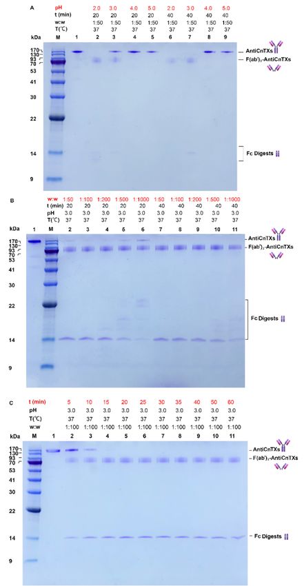
Toxins 2021, 13, 165                                                                                                      2 of 16

                                     jellyfish envenomation [7,8]. Moreover, a specific antivenom may also be an additional
                                     therapeutic approach to deal with severe jellyfish stings. Cyanea nozakii jellyfish often
                                     reach a diameter of 60 cm and are responsible for hundreds of thousands of stings per
                                     year in coastal Chinese waters. Unfortunately, many deaths from jellyfish stings have been
                                     reported in China in recent years. However, there has been no effective method to treat
                                     severe Cyanea nozakii stings; dexamethasone, aspirin, and antihistamines cannot stop vic-
                                     tims’ systemic symptoms in clinic [9]. Therefore, the development of a C. nozakii anti-
                                     venom is urgent.
                                           In general,antivenom has effectively neutralized venom toxins and saved thousands
                                     of lives since the 19th century [10,11]. Historically, whole antiserum was used to neutral-
                                     ize the toxins. However, this contains not only antitoxins but also many other proteins,
                                     which may cause some potential side effects after injection into the body. In 1937, the γ
                                     globulin (immunoglobulin G, IgG) was discovered to be the antitoxin in the antiserum
                                     [12–15]. The purified IgG without other serum proteins was then used as a second-gener-
                                     ation antitoxin. IgG is composed of two light chains and two heavy chains connected by
                                     disulfide bonds and contains a fragment of antigen-binding domain (Fab) and fragment
                                     crystallized domain (Fc) [16]. As most antiserums are produced by animals such as horses
                                     and rabbits, the heterogenous Fc domain may cause an immunological reaction in the
                                     body, and many serum sicknesses have also been reported after injection of antivenom,
                                     including a previously sheep-sourced and IgG type of box jellyfish antivenom [17–19].
                                     Therefore, the removal of the Fc fragment from IgG not only preserves the function of
                                     antigen-binding but also decreases the potential serum sickness of the heterogenous Fc
                                     domain.
                                           Two types of Fc fragment that remove IgG, F(ab’)2, and Fab are available. Both F(ab’)2
                                     and Fab types of antivenom have been successfully used in snakebite treatment. F(ab’)2
                                     antivenom has two Fab domains; is very similar to the whole IgG in structure; and can
                                     form multivalent immunocomplexes with toxin antigens, such as IgG, and then be cleaned
                                     by phagocytic cells. Fab antivenom only has one Fab domain with a smaller molecular
                                     weight and can be easily distributed to the whole body. However, Fab antivenom cannot
                                     work as F(ab’)2 or IgG antivenom. Currently, commercial snake, scorpion, spider, stone-
                                     fish IgG, F(ab’)2, and Fab antivenom are available for emergency treatment (Table 1).

                              Table 1. Species and types of some commercial venomous animals’ antivenoms.

    Animal                   Species Neutralized               Antivenom Name         Manufacturer          Source      Type

                                                                                     Instituto Clodo-
                                  Bitis arietans
                                                                                    miro Picado, Uni-
                                 Echis ocellatus               EchiTab-Plus-ICP                             Horse        IgG
                                                                                    versidad de Costa
                                 Naja nigricollis
                                                                                           Rica
                               Bungarus fasciatus,
                             Bungarus multicinctus,
                       Agkistrodon acutus, Vipera russelli                           Shanghai Serum
    Snakes             siamensis, Trimeresurus stejnegeri,           +3C              Bio-technology        Horse       F(ab’)2
                         Trimeresurus mucrosquamatus,                                Co., LTD, China
                        Agkistrodon halys, Naja naja atra,
                              Ophiophagus Hannah
                       Bitis arietans, Bitis gabonica, Echis
                                                                                     Instituto Bioclon
                        leucogaster, Echis ocellatus, Echis
                                                               ANTI-VIPMYN          S.A. de C.V, Mex-       Horse       F(ab’)2
                       Pyramidum, Dendroaspis polylepis,
                                                                                            ico
                       Dendroaspis viridis, Naja haje, Naja
Toxins 2021, 13, 165                                                                                                       3 of 16

                       melanoleuca, Naja nigricollis, Naja
                                    pallida

                       Bitis arietans, Bitis gabonica, Echis
                        leucogaster, Echis ocellatus, Den-
                                                                                      Sanofi-Pasteur,
                         droaspis polylepis, Dendroaspis        FAV-Afrique                                  Horse      F(ab’)2
                                                                                         France
                       jamesoni, Dendroaspis viridis, Naja
                               haje, Naja nigricollis

                                                                                    BTG International,
                             Crotalinae subfamily                  CroFab                                    Sheep        Fab
                                                                                        Inc. USA

                                                                                     Accredo Health
                           Centruroides sculpturatus              Anascorp                                   Horse      F(ab’)2
                                                                                     Group, Inc. USA

                            Androctonus crassicauda
   Scorpions
                              Androctonus aeneas
                                                                                    VINS Bioproducts
                             Androctonus australis                  VINS                                     Horse      F(ab’)2
                                                                                     Limited, India
                            Scorpiomarus palmatus
                               Bathus occitanus
    Jellyfish                   Chironex fleckeri                                                            Sheep        IgG
                       Red back spiders Latrodectus has-                             Commonwealth
                                                                                     Serum Laborato-         Horse        IgG
    Spiders                         selti                            CSL
                                                                                    ries, Limited, Aus-
                              Funnel web spider                                                             Rabbit        IgG
                                                                                            tralia
   Stonefish                   Synanceia trachynis                                                           Horse        IgG

                                          However, all of these commercial antivenoms can lead to side effects, such as head-
                                     ache, fever, nausea, swollen glands, chest tightness, pounding heartbeats, or trouble
                                     breathing. So, it is hard to say which kind of antivenom is better, especially for the enven-
                                     omation of different animals. To date, only one jellyfish antivenom, the Commonwealth
                                     Serum Laboratories™ (CSL) box jellyfish antivenom, has been available for the treatment
                                     of box jellyfish stings worldwide, but this antivenom does not reliable to prevent death; it
                                     in fact lessens the survival time in mice [7,20]. Additionally, the venom components are
                                     very different between box jellyfish Chironex fleckeri and Cyanea nozakii [21–24], which
                                     means that CSL box jellyfish antivenom is not suitable to deal with a C. nozakii jellyfish
                                     sting. Furthermore, no evidence shows that C. fleckeri antivenom has any efficacy in
                                     nonchirodropid box jellyfish stings [25,26].
                                          In the present study, we prepared a C. nozakii antivenom in rabbits by immunizing
                                     rabbits with an extract preparation of tentacle venom comprising venom toxins and other
                                     tentacle components. IgG (AntiCnTXs) was isolated by Protein A resin from the antise-
                                     rum. Subsequently, AntiCnTXs were refined to F(ab’)2 type of antivenom (F(ab’)2-AntiC-
                                     nTXs) by pepsin and Fab type of antivenom (Fab-AntiCnTXs) by papain, respectively. We
                                     then compared their neutralization efficacy on CnTXs both in vitro and in vivo. All kinds
                                     of antivenom showed some neutralization effect on the lethality of CnTXs in an in vivo
                                     experiment, and the neutralization efficacy was as follows: F(ab’)2-AntiCnTXs ≥ AntiC-
                                     nTXs > Fab-AntiCnTXs. Furthermore, our study provides important information for the
                                     preparation of different antivenoms for the treatment of C. nozakii jellyfish stings in the
                                     future.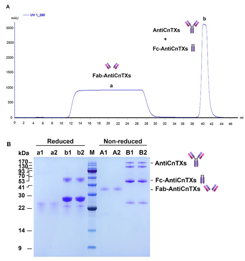
Toxins 2021, 13, 165 4 of 16
2. Results
2.1. Affinity Purification of AntiCnTXs from Antiserum
The crude serum contains many blood proteins and immunoglobulins. A Protein A
column can separate IgG of AntiCnTXs from other protein with very high affinity (Figure
1). Most other proteins do not bind the resin and wash out in the flow-through fraction.
However, AntiCnTXs can bind to the resin and are eluted with elution buffer B. The SDS-
PAGE profile (Figure 1B) indicates that the purity of AntiCnTXs is very high, and there is
only one protein band with a molecular weight of ~150 kDa under the nonreduced condi-
tion. IgG is composed of two heavy chains and two light chains; however, the disulfide
bonds in the IgG are broken under the reduced condition and then generate two separate
heavy chains and two separate light chains.
Figure 1. Purification of IgG-AntiCnTXs from the antiserum. (A) Protein A affinity purification of AntiCnTXs from anti-
serum. (B) SDS-PAGE analysis of CnTXs and the fractions from protein A affinity purification. M, protein markers; CnTXs,
the antigen used for preparation of antivenom; S, antiserum under the nonreduced condition; A1 and A2, fractions of peak
A under the nonreduced condition; B1 and B2, fractions of peak B under the nonreduced condition; s, antiserum under
the reduced condition; a1 and a2, fractions of peak A under the reduced condition; b1 and b2, fractions of peak B under
the reduced condition.
2.2. Preparation of F(ab’)2 Fragment of AntiCnTXs
2.2.1. Optimization of Pepsin Digestion of AntiCnTXs
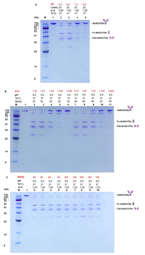
The reaction system is very important to the pepsin digestion of AntiCnTXs. pH is
among the most critical factors for the enzyme. As seen in Figure 2, the SDS-PAGE profile
shows the screening of optimal reaction conditions for the pepsin digestion of AntiCnTXs.
In Figure 2A, all the AntiCnTXs can be digested by pepsin at pH 2.0 in 20 min and about
half at pH 3.0, and pepsin can digest all the AntiCnTXs at pH 3.0 in 40 min. However,
almost none is digested at pH 4.0 or 5.0, even after 40 min. So, pepsin activity is highest at
pH 2.0. Given the extreme condition at pH 2.0 for AntiCnTXs, pH 3.0 is not only much
milder but also very effective for AntiCnTXs digestion. Therefore, pH 3.0 is more suitable
for the pepsin digestion of AntiCnTXs. Figure 2B shows the pepsin to AntiCnTXs ratio for
digestion, and the SDS-PAGE profile displays that Wpepin:WAntiCnTXs = 1:50 to 1:200 is very
effective for pepsin to cleave AntiCnTXs into F(ab’)2-AntiCnTXs and Fc digests. The reac-
tion time assay shows that AntiCnTXs can be almost fully digested by pepsin in 15 min at
37 °C at a ratio of 1:100 (Figure 2C). So, to ensure that the AntiCnTXs are totally digested
by pepsin, the reaction conditions for pepsin digestion of AntiCnTXs are pH 3.0,
Wpepin:WAntiCnTXs = 1:100, and 30 min for the following preparation of F(ab’)2-AntiCnTXs.
Toxins 2021, 13, 165                                                                                               5 of 16

                       Figure 2. SDS-PAGE analysis of the optimal conditions screen for the pepsin digestion of AntiC-
                       nTXs. (A) The effect of pH on the pepsin digestion of AntiCnTXs; (B) the effect of w:w (pepsin:An-
                       tiCnTXs) on the pepsin digestion of AntiCnTXs; (C) the effect of reaction time on the pepsin diges-
                       tion of AntiCnTXs. The details of digestion conditions are listed for each lane.

                       2.2.2. Purification of F(ab’)2-AntiCnTXs
                            AntiCnTXs digests were purified by size exclusion chromatography HiLoad Super-
                       dex 200 16/60. Figure 3A shows that only two protein peaks were achieved in the chroma-
                       togram, and the SDS-PAGE profile of the two peaks displays very good purity in Figure
                       3B. The molecular weight of F(ab’)2-AntiCnTXs was about 90 kDa the under nonreduced
                       condition; however, a ~26 kDa band was observed under the reduced condition because
                       the disulfide bond was broken.
Toxins 2021, 13, 165 6 of 16
Figure 3. Purification of F(ab’)2-AntiCnTXs from the pepsin digests of AntiCnTXs. (A) Size exclu-
sion chromatography purification of F(ab’)2-AntiCnTXs from the pepsin digests of AntiCnTXs. (B)
SDS-PAGE analysis of the fractions from size exclusion chromatography purification. A1 and A2,
peak a under the nonreduced condition; B1 and B2, peak b under the nonreduced condition; M,
protein markers; a1 and a2, peak a under the reduced condition; b1 and b2, peak b under the re-
duced condition.
2.3. Preparation of Fab Fragment of AntiCnTXs
2.3.1. Optimization of Papain Digestion of AntiCnTXs
Reaction condition is very important to the papain digestion of AntiCnTXs. pH 5.0
or 6.0 is much more effective than pH 7.0 or 8.0 (Figure 4A) for papain digestion. The SDS-
PAGE profile displays that WPapain:WAntiCnTXs = 1:10‒1:20 is more effective for pepsin to
cleave AntiCnTXs into Fab-AntiCnTXs and Fc digests (Figure 4B). Figure 4C shows that
papain is not very effective at digesting AntiCnTXs; they are not totally digested even
after 180 min. The reaction conditions for papain digestion of AntiCnTXs are pH 6.0,
Wpapain:WAntiCnTXs = 1:200, and 60 min for the following preparation of Fab-AntiCnTXs.Toxins 2021, 13, 165 7 of 16
Figure 4. SDS-PAGE analysis of the optimal conditions screen for the papain digestion of AntiC-
nTXs. (A) The effect of pH on the papain digestion of AntiCnTXs; (B) the effect of w:w (papain:
AntiCnTXs) on the papain digestion of AntiCnTXs; (C) the effect of reaction time on the papain
digestion of AntiCnTXs. The details of digestion conditions are listed in each lane.
2.3.2. Purification of Fab-AntiCnTXs
As the papain digests of AntiCnTXs contain Fab-AntiCnTXs, Fc-AntiCnTXs, and
some undigested AntiCnTXs, a Protein A column was used to separate Fab-AntiCnTXs
from other proteins. Figure 5A shows the chromatogram of the purification. The SDS-
PAGE profile indicates that the purity of Fab-AntiCnTXs is very good in Figure 5B. The
molecular weight of Fab-AntiCnTXs was about 36 kDa under nonreduced conditions;
however, the disulfide bond between two Fab-AntiCnTXs was broken by βME under re-
duced conditions.Toxins 2021, 13, 165 8 of 16
Figure 5. Purification of Fab-AntiCnTXs from the papain digests of AntiCnTXs. (A) Protein A af-
finity purification of Fab-AntiCnTXs from the papain digests of AntiCnTXs. (B) SDS-PAGE analy-
sis of the fractions from Protein A affinity purification of Fab-AntiCnTXs. A1 and A2, peak a un-
der the nonreduced condition; B1 and B2, peak b under the nonreduced condition; M, protein
markers; a1 and a2, peak a under the reduced condition; b1 and b2, peak b under the reduced con-
dition.
2.4. Neutralization Assay of the Antivenoms
The efficacy of these antibodies and IgG fragments to neutralize venom toxins was
evaluated using in vivo and in vitro assays. The in vivo assay results show that all the
mice died within 8 h after intraperitoneal injection of CnTXs and 40% died within 40 min.
However, the mice in the antibody-neutralized groups died much later than those in the
CnTXs group (Figure 6A). Moreover, 20% of mice survived in both the AntiCnTXs and
F(ab’)2-AntiCnTXs groups. However, all the mice in the Fab-AntiCnTXs group died
within 8 h after injection (Figure 6A), which indicated that the neutralization of Fab-An-
tiCnTXs was less effective than that of AntiCnTXs or F(ab’)2-AntiCnTXs. PLA2, hemolytic,
and metalloprotease activity are among the most obvious toxicities of CnTXs in vitro. All
kinds of antibodies, AntiCnTXs, F(ab’)2-AntiCnTXs, and Fab-AntiCnTXs, significantly in-
hibited the hemolytic activity of CnTXs (Figure 6B). However, no inhibitory effect was
observed in the PLA2 and metalloprotease activity assay. In contrast, it could promote
PLA2 and metalloprotease activity in vitro (Figure 6C,D).Toxins 2021, 13, 165 9 of 16
Figure 6. Neutralization assay of the antivenoms against the toxicities of CnTXs. (A) Neutralization assay of AntiCnTXs,
F(ab’)2-AntiCnTXs, and Fab-AntiCnTXs against the lethality of CnTXs in vivo. Control, injection of dialysis buffer; CnTXs,
injection of CnTXs; F(ab’)2-AntiCnTXs: injection of F(ab’)2-AntiCnTXs neutralized CnTXs; Fab-AntiCnTXs, injection of
Fab-AntiCnTXs neutralized CnTXs, n = 10. (B) Neutralization assay of antivenoms against the hemolytic activity of CnTXs
in vitro; (C) neutralization assay of antivenoms against the metalloprotease activity of CnTXs in vitro; (D) neutralization
assay of antivenoms against the PLA2 activity of CnTXs in vitro; *p < 0.05, **p < 0.03, ***p < 0.0003, ****p < 0.0001, n = 3.
2.5. LC-MS/MS and GO Analysis of Antivenom
The LC-MS/MS analysis of CnTXs antiserum identified 130 proteins in total (Table
S1). The CnTXs antiserum contains many immune molecules, including IgG, a membrane
attack complex, to resist the invasion of CnTXs (Figure 7). All the IgGs are very similar in
the structure, such as the Y shape and Fc domain. However, the Fab domains of those
IgGs are quite different from each other, so they can bind to different antigens and act as
protein inhibitors. So, in the molecular function analysis, many protein inhibitors and an-
tigen-binding proteins were identified, and that is why the antibody can neutralize the
antigen CnTXs.Toxins 2021, 13, 165 10 of 16
Figure 7. LC-MS/MS and GO analysis of antiserum. All the identified proteins were summarized
in three categories: cellular component, biological process, and molecular function.
3. Discussion
Antivenom is a good way to treat venomous animal bites or stings, and so is highly
recommended as first aid by the World Health Organization. Terrestrial venomous ani-
mals, such as snakes, scorpions, spiders, and bees, pose a threat to human beings. Anti-
venoms have already been well studied and developed over many years to treat bites and
stings [27–29]. Snake antivenom is the most successful example, as it has been widely used
for centuries and has saved hundreds of thousands of lives all over the world. There are
also many venomous marine animals, including sea snakes, jellyfish, stonefish, blue-
ringed octopus, cone snails, pufferfish, and ciguatoxin-containing fishes. However, only
certain sea snakes, the box jellyfish Chironex fleckeri, and stonefish have antivenom to date
[26,30], and many people have died from a lack of effective medicine, such as antivenom;
deaths have also occurred, despite the administration of antivenoms sometimes. So, the
development of marine antivenom is very important.
Antivenom is composed of many antibodies for the neutralization of animal toxins.
Nowadays, both traditional IgG antivenom and F(ab’)2 or Fab type of refined antivenoms
are produced by antivenom manufacturers (Table 1). However, it is difficult to balance
the efficacy of IgG and its side effects. The best way is to analyze the neutralization effec-
tiveness of all three types of antivenom for different animal antivenoms. Therefore, in the
current study, we prepared a CnTXs jellyfish antivenom in rabbits and then purified
CnTXs antibodies from the antiserum and made refined antibodies. Usually, pepsin and
papain are used to produce F(ab’)2 and Fab types of antivenom, respectively [31–36]. So,
we optimized multiple reaction conditions for the digestion of CnTXs by pepsin or papain
and finally produced two types of refined CnTXs antibodies, F(ab’)2-AntiCnTXs and Fab-
AntiCnTXs. However, F(ab’)2-AntiCnTXs digestion by pepsin was much faster and sim-
pler than Fab-AntiCnTXs prepared by papain. Pepsin could also digest almost 100% of
AntiCnTXs in 30 min with a ratio of 1:100. However, papain only digested about 90% of
AntiCnTXs in 3 h with 5-fold more enzyme than pepsin. So, pepsin is much more effective
than papain at removing the Fc domain from IgG AntiCnTXs. Furthermore, the in vivo
neutralization efficacy of Fab-AntiCnTXs is much worse than that of AntiCnTXs or F(ab’)
2-AntiCnTXs. This is because Fab-AntiCnTXs could not form multivalentToxins 2021, 13, 165 11 of 16
immunocomplexes with toxins, but AntiCnTXs and F(ab’)2-AntiCnTXs could. This may
be why most commercial antivenoms are of the IgG or F(ab’)2 type instead of the Fab type.
Our previous study showed that CnTXs are composed of many types of toxins, in-
cluding phospholipase A2, metalloproteinase, serine protease inhibitor, plancitoxin-1, and
alpha-latrocrustotoxin-Lt1a [21], and metalloproteinase might be the key lethal toxin in
the venom of Cyanea nozakii [37]. In the present study, the LC-MS/MS analysis of the
CnTXs antiserum identified many proteinase inhibitors, which indicated that the Fab do-
main of some AntiCnTXs is homologous with proteinase inhibitors in structure and may
inhibit the proteinases in the venom. So, in the neutralization assay, the mice that were
treated with F(ab’) 2-AntiCnTXs or AntiCnTXs preincubated with the tentacle venom ex-
tract died much later than the mice injected with the tentacle venom extract alone, and
20% of mice survived in both groups. No commercial antivenom is a guaranteed lifesaver.
However, the survival rate of F(ab’) 2-AntiCnTXs and AntiCnTXs treatment is still not
very high. The low proportion of lethal toxins in CnTXs may be among the reasons for
this. CnTXs, used for the preparation of antivenom, are complex mixtures that contain
many other nontoxic proteins and do not represent the real toxins injected in an authentic
sting. It is hard to extract pure jellyfish venom toxins, as with snakes and spiders. Jellyfish
toxins are in the tentacle nematocyst. The sonication, glass bead disruption, or high-pres-
sure cell rupture of isolated nematocysts is often used for the extraction of jellyfish toxins,
and the whole extract is then used as jellyfish toxin for the research [7,38–40]. Therefore,
these “jellyfish toxins” are composed of toxins and many other nontoxic nematocyst pro-
teins. The nontoxins’ antibodies in the antivenom may affect the neutralization efficacy.
Moreover, the antivenom is a mixture of rabbit immunoglobulins, which are heterologous
proteins to humans and may also be recognized by the human immune system as anti-
gens. Although real jellyfish toxins’ antibodies could neutralize these toxicities, other
nontoxins’ antibodies in the antivenom may become toxic to the body. The antivenoms
we produced in this study may not be suitable for the treatment of real jellyfish stings,
and further studies will be needed to improve the efficacy and safety of antivenom, in-
cluding collecting and using pure jellyfish venom [7,41] or purified lethal toxins as antigen
to prepare antivenom to neutralize the jellyfish toxins and minimize potential side effects.
4. Conclusions
In the current study, a C. nozakii jellyfish antivenom was prepared in rabbits and re-
fined AntiCnTXs into F(ab’)2-AntiCnTXs by pepsin and Fab-AntiCnTXs by papain, re-
spectively. The neutralization efficacy of these three types of antivenom was compared
and analyzed both in vitro and in vivo. The results showed that the neutralization effect
on the lethality of CnTXs was as follows: F(ab’)2-AntiCnTXs ≥ AntiCnTXs > Fab-AntiC-
nTXs. Future research on more effective C. nozakii jellyfish antivenom still needs to be
performed, using purified toxins as antigens. Moreover, an animal model will also be set
up using live tentacles to model an authentic sting to assess the efficacy of C. nozakii anti-
venom. This study not only provides useful information on the preparation of a C. nozakii
jellyfish antivenom but also offers new insights to produce other marine antivenoms in
the future.
5. Materials and Methods
5.1. Jellyfish Specimen Collection and Toxin Preparation
Cyanea nozakii specimens were collected from the coast of Qingdao, China, in 2019.
The fresh tentacles were cut from the body and stored at −80 °C. The frozen tentacles were
autolyzed at 4 °C for 12–24 h, and the undissolved samples were removed with a plankton
net. Finally, the autolyzed solution was centrifuged at 10,000× g for 15 min at 4 °C, and the
supernatant containing jellyfish toxins was used as C. nozakii toxins (CnTXs).Toxins 2021, 13, 165 12 of 16
5.2. Animal Immunization and Antiserum Preparation
Firstly, the lethality of CnTXs was tested to make sure that it contained jellyfish toxins
to produce their antibodies. Subsequently, the toxicity of CnTXs was attenuated so that
the rabbits were not killed in the immunization process. The attenuation of CnTXs was as
follows: 40% formaldehyde was added to CnTXs at a ratio of 1:50 and then incubated at
37 °C for a week; 40% formaldehyde was added again into the mixture at a ratio of 1:200
with incubation at 37 °C for another week. Subsequently, it was dialyzed against 20 mM
PBS, pH 7.0, to remove the formaldehyde. Finally, after centrifugation, the supernatant
was filtered with a 0.22 µm filter and kept in a −80 °C freezer.
Attenuated CnTXs (0.68 mg, 0.43 mg/mL), together with complete Freund’s adjuvant,
was injected into three healthy New Zealand white rabbits (~2 kg) as the first immuniza-
tion. The second immunization was performed three weeks later using 0.34 mg attenuated
CnTXs and incomplete Freund’s adjuvant, and the third and fourth immunizations were
performed two weeks after the previous immunization, using 0.34 mg attenuated CnTXs
and incomplete Freund’s adjuvant. The final immunization was completed three weeks
after the fourth immunization. A titer test of the antiserum was performed after the fourth
and fifth immunizations. Briefly, the titer of the antiserum was evaluated using the ELISA
method. The antigen of attenuated CnTXs was coated in a microtiter palate with a coating
buffer (50 mm, pH 9.6, Na2CO3) at 4 °C overnight. After removing the coating buffer, the
plate was washed with 0.05% Tween-20, 20 mM NaH2PO4, pH 7.4 (PBST) three times, fol-
lowed by blocking with 5% skim milk for 1 h. After another three washes with PBST, var-
ious diluted antiserums were added and incubated at 37 °C for 1 h, with three washes
after. Then, HRP-labeled Goat Antirabbit IgG (H+L) was used and incubated at 37 °C for
45 min. The plate was again washed three times with PBST. Subsequently, 3’3’5’5’-tetra-
methylbenidine dihydrochloride (TMB) substrate was added, and H2SO4 was used to stop
the reaction 15 min later. Finally, the absorbance was recorded at 450 nm.
Once the titer test of the antiserum was qualified, the whole blood was collected and
the antiserum was prepared by centrifugation at 4 °C and 3000 rpm for 15 min. Finally,
the antiserum was stored at −80 °C until further use. All animals in this experiment re-
ceived humane care, as approved by the Ethics Committee of the Institute of Oceanology,
Chinese Academy of Sciences; approval code: IOCAS/KLEMB/20180309; approval date: 9
March 2018.
5.3. Purification of CnTXs Antibody
The antiserum was diluted with binding buffer A (0.15M NaCl, pH 7.0, 20 mM
Na2HPO4) at a ratio of 1:1 (v:v) and purified with a fast protein liquid chromatogram sys-
tem ÄKTA pure (GE Healthcare, Chicago, IL, USA), equipped with a 1 mL Protein A pre-
packed column (GenScript, Piscataway, NJ, USA), and monitored at 280 nm. Buffer A was
used to wash and remove the unbound antiserum proteins, and Buffer B (pH = 3.0, 100
mM glycine) was used to elute the antibodies of CnTXs (AntiCnTXs). The elution was
immediately adjusted with 1 M Tris-HCl, pH8.5. The purity of flow-through and elution
were analyzed by SDS-PAGE under both nonreduced and reduced conditions.
5.4. Refinement of AntiCnTXs
5.4.1. F(ab’)2 Fragments of AntiCnTXs Preparation
The Optimum Screen of Pepsin Digestion of AntiCnTXs
The optimum pH for the pepsin digestion was conducted as follows: aliquots of An-
tiCnTXs were dialyzed at 4 °C overnight against dialysis buffers of pH 2.0, pH 3.0, pH 4.0,
pH 5.0, and 100 mM glycine, respectively. Equal pepsin was added into 100 µL AntiCnTXs
at different pH values at the same ratio of Wpepsin:WAntiCnTXs = 1:50, followed by incubation
at 37 °C for 20 and 40 min, with three replicates. Subsequently, the digested AntiCnTXs
were analyzed by SDS-PAGE under the nonreduced conditions. The optimum ratio of
Wpepsin: WAntiCnTXs for the digestion was determined as follows: pepsin was added
to the same AntiCnTXs to a final ratio of 1:50, 1:100, 1:200, 1:500, or 1:1000. The reactionToxins 2021, 13, 165 13 of 16
was carried out at pH 3.0, 37 °C for 20 min with three replicates. Finally, the digested
AntiCnTXs were analyzed by SDS-PAGE under the nonreduced conditions. The optimum
time for digestion was determined as follows: AntiCnTXs were digested by pepsin at a
ratio of 1:100 at pH 3.0 and 37 °C for 5, 10, 15, 20, 25, 30, 35, 40, 50, or 60 min, with three
replicates. The digests were immediately quenched at 95 °C for 5 min once time was up.
Finally, the digested AntiCnTXs were analyzed by SDS-PAGE under the nonreduced con-
ditions.
Purification of F(ab’)2-AntiCnTXs
AntiCnTXs digestion by pepsin was scaled up at pH 3.0, Wpepsin:WAntiCnTXs = 1:100 for
30 min. The digests were concentrated with concentrators (MWCO 10 kDa Millipore,
USA) at 6000× g, 4 °C, and then loaded onto a HiLoad Superdex 200 16/60 column (GE
Healthcare) with buffer A. The purity was analyzed by SDS-PAGE under both nonre-
duced and reduced conditions.
5.4.2. Fab Fragments of AntiCnTXs (Fab-AntiCnTXs) Preparation
The Optimum Screen of Papain Digestion of AntiCnTXs
The optimum pH for the papain digestion was determined as follows: aliquots of
AntiCnTXs were dialyzed at 4 °C overnight against dialysis buffers of pH 5.0, 100 mM
glycine, pH 6.0, pH 7.0, pH 8.0, and 20 mM PBS. The same amount of papain was added
into 100 µL AntiCnTXs at different pH values at the same ratio of Wpapain:WAntiCnTXs = 1:50,
followed by incubation at 37 °C for 20 min, with three replicates. Subsequently, the di-
gested AntiCnTXs were analyzed by SDS-PAGE under the nonreduced conditions. The
optimum ratio of Wpapain:WAntiCnTXs for the digestion was determined as follows: papain
was added to the same amount of AntiCnTXs to a final ratio of 1:10, 1:20, 1:50, 1:100, or
1:200. The reaction was carried out at pH 3.0, 37 °C for 20 and 40 min, with three replicates.
Finally, the digested AntiCnTXs were analyzed by SDS-PAGE under the nonreduced con-
ditions. The optimum time for the digestion was determined as follows: AntiCnTXs were
digested by papain at a ratio of 1:20 at pH 6.0, 37 °C for 20, 40, 60, 80, 100, 120, 140, 160, or
180 min, with three replicates. The digests were immediately quenched at 95 °C for 5 min
once time was up. Finally, the digested AntiCnTXs were analyzed by SDS-PAGE in non-
reduced conditions.
Purification of Fab-AntiCnTXs
AntiCnTXs digestion by pepsin was scaled up under the optimized conditions of pH
6.0, Wpapain:WAntiCnTXs = 1:20 for 60 min. The digests were again purified by a Protein A
column to remove the Fc fragments and undigested AntiCnTXs. The purity of Fab-AntiC-
nTXs was analyzed by SDS-PAGE under both nonreduced and reduced conditions.
5.5. Neutralization Assay of the Antivenoms
5.5.1. In Vivo Neutralization Assay of the Antivenom
SPF KM mice (18–20 g) were used for in vivo neutralization assay. Each group con-
tained 10 mice, five males and five females. AntiCnTXs, F(ab’)2-AntiCnTXs, Fab-AntiC-
nTXs, and CnTXs were dialyzed in a dialysis buffer (20 mM, Tris-HCl, pH 7.0, 0.15 M
NaCl) at 4 °C overnight. The concentration of each sample was measured using the Brad-
ford method [42]. A total of 700 µL mixture containing 330 µg CnTXs and 330 µg AntiC-
nTXs, 198 µg F(ab’)2-AntiCnTXs, or 86 µg Fab-AntiCnTXs was incubated to neutralize the
toxicity of CnTXs at 4 °C for 1 h. Then, 700 µL neutralized CnTXs and the same amount
of unnaturalized CnTXs were intraperitoneally injected into each mouse using a dialysis
buffer as a control. The mortality was recorded over the next 98 h. All animal experiments
in this study were approved by the Ethics Committee of the Institute of Oceanology, Chi-
nese Academy of Sciences.
5.5.2. In Vitro Hemolysis Activity Neutralization Assay
In vitro neutralization efficacy on the hemolysis activity of CnTXs was assayed using
a previous method with some modifications [37]. In brief, 25 µg CnTXs, 25 µg AntiCnTXs,Toxins 2021, 13, 165 14 of 16
15 µg F(ab’)2-AntiCnTXs, or 6.5 µg Fab-AntiCnTXs was incubated to neutralize the tox-
icity of CnTXs and then diluted to 100 µL with 0.9% NaCl. Then, 200 µL human erythro-
cyte suspended was then added and incubated at 37 °C for 30 min using an isotonic buffer
and Triton X-100 as the blank and positive control, respectively. After centrifugation at
3000 rpm for 10 min, the hemoglobin released in the supernatant was assayed at 405 nm.
The hemolysis rate was calculated as (A405(sample)-A405(blank))/(A405(Triton X-100)-
A405(blank)) × 100%. All the experiments were conducted with three replicates.
5.5.3. In Vitro Phospholipase A2 (PLA2) Activity Neutralization Assay
The neutralization efficacy of this antivenom on PLA2 activity was measured accord-
ing to a method described before [37]. Briefly, 25 µg CnTXs, 25 µg AntiCnTXs, 15 µg
F(ab’)2-AntiCnTXs, or 6.5 µg Fab-AntiCnTXs was incubated to neutralize the toxicity of
CnTXs, respectively, and then diluted to 250 µL with 50 mM Tris-HCl, 5 mM CaCl2, 100
mM NaCl, pH 8.0, followed by the addition of 25 µL, 1 mg/mL 4-nitro-3-octanoyloxyben-
zoic acid (NOBA); 50 mM Tris-HCl, 5 mM CaCl2, 100 mM NaCl, pH 8.0, and CnTXs were
used as controls. Subsequently, the plate was incubated at 37 °C for 1 h, and the absorb-
ance was measured at 405 nm. All the experiments were conducted with three replicates.
5.5.4. In Vitro Metalloproteinase Activity Neutralization Assay
The neutralization efficacy of this antivenom on metalloproteinase activity was as-
sayed according to a previous method [37]. Briefly, 25 µg CnTXs, 25 µg AntiCnTXs, 15 µg
F(ab’)2-AntiCnTXs, or 6.5 µg Fab-AntiCnTXs was incubated to neutralize the toxicity of
CnTXs; then diluted to 100 µL with 50 mM Tris-HCl, pH 8.8, 5 mM CaCl2, 150 mM NaCl;
followed by the addition of 100 µL of 5 mg/mL Azocasein and incubation at 37 °C for 90
min. The reactions were quenched by the addition of 200 µL of 0.5M trichloroacetic acid
and placed at room temperature for 30 min. The precipitate was removed by centrifuga-
tion at 10,000 rpm for 10 min. Finally, 150 µL supernatant was neutralized with 150 µL 0.5
M NaOH, and the absorbance was measured at 450 nm. All the experiments were con-
ducted with three replicates.
5.6. LC-MS/MS and GO Analysis of Antivenom
LC-MS/MS analysis of the antiserum was conducted according to a previous method
[21]. Briefly, all the antiserum proteins in the SDS-PAGE gel were cut off and then des-
tained by 25 mM NH4HCO3, 50% acetonitrile (ACN), followed by dehydration by 50%
and 100% ACN for 30 min, separately. Next, 10 mM DTT or 25 mM NH4HCO3 was used
at 57 °C for 1 h. Subsequently, the sample was treated at room temperature with 50 mM
iodoacetamide and 25 mM NH4HCO3 for 30 min, 25 mmol/L NH4HCO3 for 10 min, 10 mM
DTT and 25 mM NH4HCO3 for 30 min, and 50 mM iodoacetamide and 25 mM NH4HCO3
for 30 min in turn. The sample was rehydrated with 10 µL of 0.02 µg/µL trypsin, 25 mM
NH4HCO3, and 10% ACN for 30 min and then 20 µL cover solution for 16 h at 37 °C. The
sample was extracted with 50 µL, 5% TFA, and 67% ACN. Finally, the extracted peptides
and the supernatant of the gel were combined to dry.
The digested peptides were dissolved in 0.1% formic acid and 2% ACN and analyzed
by a C18 nanoLC trap column (100 µm × 3 cm, 3 µm, 150 Å) that was washed with 0.1% FA
and 2% ACN at 2 µL/min for 10 min, followed by a ChromXP (SCIEX, Framingham, MA,
USA) C18 column (75 µm × 15 cm, 3 µm 120 Å) using a gradient of 5–35% CAN, 0.1% FA
for 90 min. All the data were acquired from a Triple TOF 5600 system (SCIEX, Framing-
ham, MA, USA) with a Nanospray III source and a pulled quartz tip as the emitter. In-
strument parameters were set as ion spray voltage of 2.5 kV, curtain gas of 30 PSI, nebu-
lizer gas of 5 PSI, and an interface heater temperature of 150 °C; 250 ms survey scans were
employed for information-dependent acquisition (IDA) with a rolling collision energy set-
ting for all precursor ions. All proteins were matched according to both MS and MS/MS
spectra, with ≥95% confidence interval scores in the MASCOT V2.3 (Matrix Science, Inc.,
Boston, MA, USA) search engine in the database Oryctolagus cuniculus. All the identifiedToxins 2021, 13, 165 15 of 16
proteins were annotated in the nonredundant protein database GO (Nr, NCBI) based on
the biological process, cell component, and molecular function.
5.7. Statistical Analysis
All the results were expressed as mean ± SD. Statistically significant differences be-
tween groups were considered only when p < 0.05.
Supplementary Materials: The following are available online at www.mdpi.com/2072-
6651/13/2/165/s1, Table S1: All the proteins identified in the antiserum by LC-MS/MS.
Author Contributions: Conceptualization, R.L.; methodology, R.L.; validation, R.L. and
H.Y.; formal analysis, R.L.; investigation, R.L.; resources, R.L, A.L. and C.Y.; data curation,
R.L.; writing—original draft preparation, R.L.; writing—review and editing, R.L. and P.L.;
visualization, R.L.; supervision, P.L.; project administration, P.L.; funding acquisition,
R.L. All authors have read and agreed to the published version of the manuscript.
Funding: This research was funded by the National Natural Science Foundation of China (41876164,
41776163), Natural Science Foundation of Shandong Province (ZR2019QD012), and the National
Key Research and Development Program of China (2017YFE0111100-04).
Institutional Review Board Statement: The study was approved by the Ethics Committee of the
Institute of Oceanology, Chinese Academy of Sciences; approval code: IOCAS/KLEMB/20180309;
approval date: 9 March 2018.
Informed Consent Statement: Not applicable.
Data Availability Statement: The data presented in this study are available on request from the
corresponding author. The data are not publicly available due to animal ethical reasons.
Conflicts of Interest: The authors declare no conflict of interest.
References
1. Warrell, D.A. Venomous Bites, Stings, and Poisoning: An Update. Infect. Dis. Clin. North Am. 2019, 33, 17–38.
2. Mebs, D. Jellyfish sting injuries. Hautarzt 2014, 65, 873–878.
3. Burnett, J.W.; Calton, G.J.; Burnett, H.W. Jellyfish envenomation syndromes. J. Am. Acad. Dermatol. 1986, 14, 100–106.
4. Cegolon, L.; Heymann, W.C.; Lange, J.H.; Mastrangelo, G. Jellyfish Stings and Their Management: A Review. Mar. Drugs 2013,
11, 523–550.
5. Yanagihara, A.A.; Wilcox, C.L. Cubozoan Sting-Site Seawater Rinse, Scraping, and Ice Can Increase Venom Load: Upending
Current First Aid Recommendations. Toxins 2017, 9, 105.
6. Yanagihara, A.A.; Wilcox, C.; King, R.; Hurwitz, K.; Castelfranco, A.M. Experimental Assays to Assess the Efficacy of Vinegar
and Other Topical First-Aid Approaches on Cubozoan (Alatina alata) Tentacle Firing and Venom Toxicity. Toxins 2016, 8, 21.
7. Yanagihara, A.A.; Shohet, R.V. Cubozoan venom-induced acute cardiovascular collapse is caused by hyperkalemia and
prevented by zinc gluconate. PLoS ONE 2012, 7, e51368.
8. Lau, M.T.; Manion, J.; Littleboy, J.B.; Oyston, L.; Khuong, T.M.; Wang, Q.P.; Nguyen, D.T.; Hesselson, D.; Seymour, J.E.; Neely,
G.G. Molecular dissection of box jellyfish venom cytotoxicity highlights an effective venom antidote. Nat. Commun. 2019, 10,
1655.
9. Zhang, M.; Qin, S.; Li, M.; Chen, G. Investigation of coelenterare stings in north China sea. Acta Academiae Medicinae Qingdao
1993, 29, 263–268.
10. Calmette, A. The Treatment of Animals Poisoned with Snake Venom by the Injection of Antivenomous Serum. Br. Med. J. 1896,
2, 399–400.
11. Winkel, K.D.; Mirtschin, P.; Pearn, J. Twentieth century toxinology and antivenom development in Australia. Toxicon 2006, 48,
738–754.
12. Tiselius, A. Electrophoresis of serum globulin II. Electrophoretic analysis of normal and immune sera. Biochem. J. 1937, 31, 1464–
1477.
13. Tiselius, A. Electrophoresis of purified antibody preparations. J. Exp. Med. 1937, 65, 641–646.
14. Kabat, E.A.; Heidelberger, M. A quantitative theory of the precipitin reaction V. The reaction between crystalline horse serum
albumin and antibody formed in the rabbit. J. Exp. Med. 1937, 66, 229–250.
15. Heidelberger, M.; Kabat, E.A.; Shrivastava, D.L. A quantitative study of the cross reaction of types iii and VIII pneumococci in
horse and rabbit antisera. J. Exp. Med. 1937, 65, 487–496.
16. Huber, R.; Deisenhofer, J.; Colman, P.M.; Matsushima, M.; Palm, W. Crystallographic structure studies of an IgG molecule and
an Fc fragment. Nature 1976, 264, 415–420.Toxins 2021, 13, 165 16 of 16
17. Miller, M.K.; Whyte, I.M.; Dawson, A.H. Serum sickness from funnelweb spider antivenom. Med J. Aust. 1999, 171, 54.
18. Shim, J.S.; Kang, H.; Cho, Y.; Shin, H.; Lee, H. Adverse Reactions after Administration of Antivenom in Korea. Toxins 2020, 12,
507.
19. Ryan, N.M.; Downes, M.A.; Isbister, G.K. Clinical features of serum sickness after Australian snake antivenom. Toxicon 2015,
108, 181–183.
20. Andreosso, A.; Smout, M.J.; Seymour, J.E. Dose and time dependence of box jellyfish antivenom. J. Venom. Anim. Toxins Incl.
Trop. Dis. 2014, 20, 34.
21. Li, R.; Yu, H.; Yue, Y.; Liu, S.; Xing, R.; Chen, X.; Li, P. Combined proteomics and transcriptomics identifies sting-related toxins
of jellyfish Cyanea nozakii. J. Proteom. 2016, 148, 57–64.
22. Brinkman, D.L.; Aziz, A.; Loukas, A.; Potriquet, J.; Seymour, J.; Mulvenna, J. Venom Proteome of the Box Jellyfish Chironex
fleckeri. PLoS ONE 2012, 7, e47866.
23. Diane, L.; Brinkman, X.J.; Potriquet, J.; Kumar, D.; Dash, D.; Kvaskoff, D.; Mulvenna, J. Transcriptome and venom proteome of
the box jellyfish Chironex fleckeri. BMC Genom. 2015, 16, 407–421.
24. Daly, N.L.; Seymour, J.; Wilson, D. Exploring the therapeutic potential of jellyfish venom. Future Med. Chem. 2014, 6, 1715–1724.
25. Fenner, P.; Rodgers, D.; Williamson, J. Box jellyfish antivenom and “Irukandji” stings. Med J. Aust. 1986, 144, 665–666.
26. Currie, B.J. Marine Antivenoms. J. Toxicol. Clin. Toxicol. 2003, 41, 301–308.
27. Glaucia-Silva, F.; Torres-Rego, M.; Soares, K.S.R.; Damasceno, I.Z.; Tambourgi, D.V.; da Silva, A.A.; Fernandes-Pedrosa, M.D.
A biotechnological approach to immunotherapy: Antivenom against Crotalus durissus cascavella snake venom produced from
biodegradable nanoparticles. Int. J. Biol. Macromol. 2018, 120, 1917–1924.
28. Vaz de Melo, P.D.; Lima, S.d.A.; Araujo, P.; Santos, R.M.; Gonzalez, E.; Belo, A.A.; Machado-de-Avila, R.A.; Costal-Oliveira, F.;
Soccol, V.T.; Guerra-Duarte, C.; et al. Immunoprotection against lethal effects of Crotalus durissus snake venom elicited by
synthetic epitopes trapped in liposomes. Int. J. Biol. Macromol. 2020, 161, 299–307.
29. Fry, B.G. Snakebite: When the Human Touch Becomes a Bad Touch. Toxins 2018, 10, 24.
30. Winkel, K.D.; Hawdon, G.M.; Fenner, P.J.; Gershwin, L.A.; Collins, A.G.; Tibballs, J. Jellyfish antivenoms: Past, present, and
future. J. Toxicol. Toxin Rev. 2003, 22, 115–127.
31. Al-Abdulla, I.; Casewell, N.R.; Landon, J. Single-reagent one-step procedures for the purification of ovine IgG, F(ab')2 and Fab
antivenoms by caprylic acid. J. Immunol. Methods 2014, 402, 15–22.
32. El Amrani, M.; Donners, A.A.M.; Hack, C.E.; Huitema, A.D.R.; van Maarseveen, E.M. Six-step workflow for the quantification
of therapeutic monoclonal antibodies in biological matrices with liquid chromatography mass spectrometry—A tutorial. Anal.
Chim. Acta 2019, 1080, 22–34.
33. Taherian, A.; Fazilati, M.; Moghadam, A.T.; Tebyanian, H. Optimization of purification procedure for horse F(ab')2 antivenom
against Androctonus crassicauda (Scorpion) venom. Trop. J. Pharm. Res. 2018, 17, 409–414.
34. Morais, V.; Massaldi, H. Effect of pepsin digestion on the antivenom activity of equine immunoglobulins. Toxicon 2005, 46, 876–
882.
35. Jones, R.G.A.; Landon, J. Enhanced pepsin digestion: A novel process for purifying antibody F(ab ')2 fragments in high yield
from serum. J. Immunol. Methods 2002, 263, 57–74.
36. Ulmer, N.; Ristanovic, D.; Morbidelli, M. Process for Continuous Fab Production by Digestion of IgG. Biotechnol. J. 2019, 14,
1800677.
37. Li, R.; Yu, H.; Yue, Y.; Li, P. Combined Proteome and Toxicology Approach Reveals the Lethality of Venom Toxins from Jellyfish
Cyanea nozakii. J. Proteome Res. 2018, 17, 3904–3913.
38. Carrette, T.; Seymour, J. A rapid and repeatable method for venom extraction from Cubozoan nematocysts. Toxicon 2004, 44,
135–139.
39. Chung, J.J.; Ratnapala, L.A.; Cooke, I.M.; Yanagihara, A.A. Partial purification and characterization of a hemolysin (CAH1) from
Hawaiian box jellyfish (Carybdea alata) venom. Toxicon 2001, 39, 981–990.
40. Helmholz, H.; Ruhnau, C.; Schütt, C.; Prange, A. Comparative study on the cell toxicity and enzymatic activity of two northern
scyphozoan species Cyanea capillata (L.) and Cyanea lamarckii (Péron & Léslieur). Toxicon 2007, 50, 53–64.
41. Robinson, P.J.; Trim, S.A.; Trim, C.M. Non-invasive extraction of Cnidarian venom through the use of autotomised tentacles.
Anim. Technol. Welf. 2019, 18, 167–173.
42. Bradford, M.M. A rapid and sensitive method for the quantitation of microgram quantities of protein utilizing the principle of
protein-dye binding. Anal. Biochem. 1976, 72, 248–254.You can also read