Protein S-palmitoylation regulates different stages of meiosis in Schizosaccharomyces pombe
←
→
Page content transcription
If your browser does not render page correctly, please read the page content below
Published Online: 17 January, 2023 | Supp Info: http://doi.org/10.26508/lsa.202201755
Downloaded from life-science-alliance.org on 22 October, 2024

Research Article

Protein S-palmitoylation regulates different stages of
meiosis in Schizosaccharomyces pombe
Thanh-Vy Pham1,2 , Wan-Yi Hsiao1, Yi-Ting Wang1, Shu-Dan Yeh2 , Shao-Win Wang1

Posttranslational protein S-palmitoylation regulates the locali-                   lipidation, which will advance our understanding of their pathological
zation and function of its target proteins involved in diverse                     relevance, leading to strategies for targeting protein lipidation for
cellular processes including meiosis. In this study, we demon-                     therapeutic applications.
strate that S-palmitoylation mediated by Erf2-Erf4 and Akr1                            Fatty acylation is a type of protein lipidation with the at-
palmitoylacyltransferases is required at multiple meiotic stages                   tachment of saturated and unsaturated fatty acids to the glycine,
in the fission yeast Schizosaccharomyces pombe. We find that                         serine, lysine, or cysteine residues of proteins. Palmitoylation
S-palmitoylation by Erf2-Erf4 is required for Ras1 localization at                 and myristoylation represent the two most common forms of
the cell periphery to enrich at the cell conjugation site for mating               fatty acylation. Palmitoylation is a process involving the covalent
pheromone response. In the absence of Erf2 or Erf4, mutant cells                   attachment of the 16-carbon palmitate to proteins (Linder &
are sterile. A role of Akr1 S-palmitoylating the nuclear fusion                    Deschenes, 2007). Palmitoylation targets several protein resi-
protein Tht1 to function in karyogamy is identified. We demon-                      dues, including serine (O-palmitoylation) and cysteine (S-
strate that S-palmitoylation stabilizes and localizes Tht1 to ER,                  palmitoylation or N-palmitoylation when it occurs at the
interacting with Sey1 ER fusion GTPase for proper meiotic nuclear                  N-terminal of the protein). Both O- and N-palmitoylation appear
fusion. In akr1, tht1, or sey1 mutant, meiotic cells, haploid nuclei               to be irreversible. Conversely, S-palmitoylation is reversible, and
are unfused with subsequent chromosome segregation defects.                        S-palmitoylated proteins can undergo cycles of acylation and
Erf2-Erf4 has an additional substrate of the spore coat protein                    deacylation in response to upstream signals (Mumby, 1997; Chen
Isp3. In the absence of Erf2, Isp3 is mislocalized from the spore                  et al, 2018). In mammalian cells, S-palmitoylate is added by a
coat. Together, these results highlight the versatility of the cel-                family of 23 transmembrane zinc finger DHHC (Asp-His-His-
lular processes in which protein S-palmitoylation participates.                    Cys)–containing protein acyl transferases and is removed by
                                                                                   fatty acyl protein thioesterases (Malgapo & Linder, 2021). About
DOI 10.26508/lsa.202201755 | Received 6 October 2022 | Revised 6 January
2023 | Accepted 6 January 2023 | Published online 17 January 2023
                                                                                   10% of eukaryotic proteins are supposed to be S-palmitoylated,
                                                                                   identified by high-throughput screens (SwissPalm database)
                                                                                   (Blanc et al, 2015).
                                                                                       The reversible nature of S-palmitoylation enables fine-tuned
Introduction                                                                       regulation of protein function. More importantly, the proper
                                                                                   function of many membrane proteins such as surface receptors
Protein lipidation is a unique co- or posttranslational modification in             and signaling proteins requires palmitoylation-mediated par-
which lipid moieties are covalently attached to proteins. There are at             titioning into lipid rafts. The small GTPase Ras, which is involved
least six types of lipids including fatty acids, isoprenoids, sterols,             in cellular signal transduction, has been described in many
phospholipids, glycosylphosphatidylinositol anchors, and lipid-                    studies as a palmitoylated protein, and site-specific palmitoy-
derived electrophiles, which can be covalently attached to proteins                lation affects its trafficking and domain localization (Onken et al,
(Chen et al, 2018). Lipidation markedly increases the hydrophobicity of            2006). Given the vital role of signal transduction in cancer de-
proteins, leading to changes in their conformation, stability, membrane            velopment (mutations in RAS genes are associated with 30% of
association, localization, trafficking, and binding affinity to their co-            human cancer) (Gimple & Wang, 2019), a connection of protein
factors. Protein lipidation is involved in pathways that play critical roles       S-palmitoylation to cancer development is suggested. In keeping
in cell signaling to regulate protein functions linked to diseases such            with this notion, the expressions of some zDHHC genes are al-
as neurological disorders, metabolic diseases, and cancers (Yeste-                 tered in various cancer tissues (Ko & Dixon, 2018), but how these
Velasco et al, 2015; Zare˛ ba-Kozioł et al, 2018). It is important to un-          palmitoylacyltransferases participate in cancer development
derstand the functions and regulatory mechanisms of protein                        remains to be defined.

1                                                                                                           2
 Institute of Molecular and Genomic Medicine, National Health Research Institutes, Zhunan Town, Taiwan       Department of Life Sciences, National Central University,
Taoyuan, Taiwan

Correspondence: shaowinwang@nhri.edu.tw

© 2023 Pham et al.                                                                      https://doi.org/10.26508/lsa.202201755   vol 6 | no 4 | e202201755     1 of 12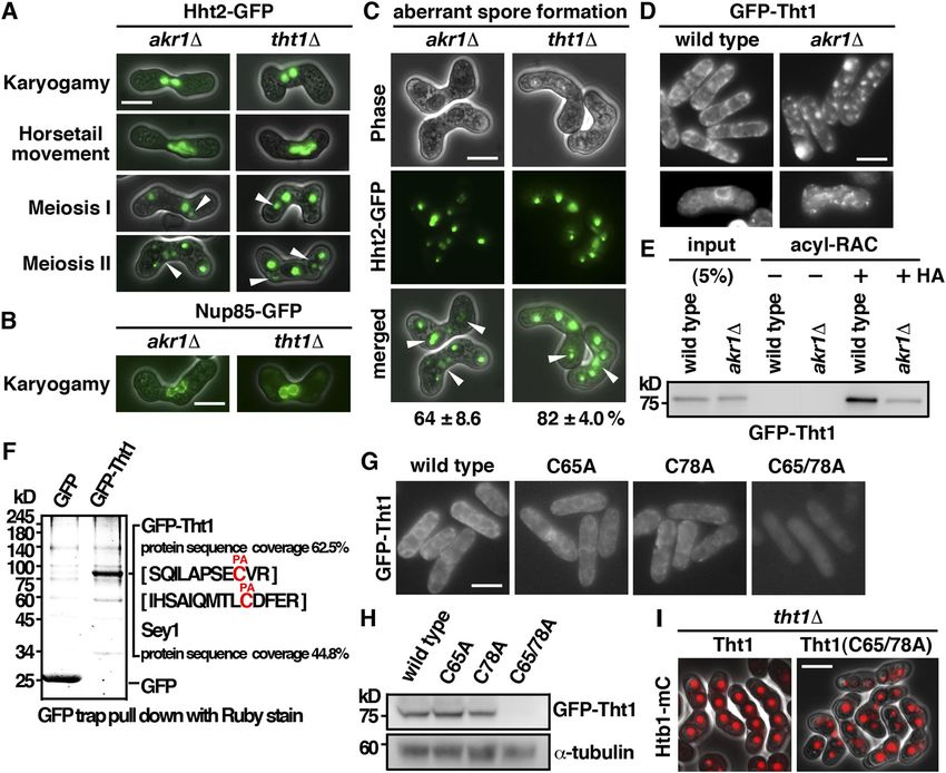
The association of S-palmitoylation with cellular differentiation to delete the whole ORF of these palmitoylacyltransferases except
in different model organisms has also been reported. In Arabi- for the essential swf1 gene. In agreement with the PomBase ge-
dopsis thaliana, male and female gametogenesis both require a nomic data (Harris et al, 2022), all mutants have been successfully
fully functional AtPAT21 palmitoylacyltransferase (Li et al, 2019). obtained as validated by PCR and were viable. During the con-
Xenopus palmitoylacyltransferase ZDHHC3 plays an important role struction of these mutants, we observed that some mutants caused
in oocyte maturation (Fang et al, 2016). The fission yeast Schizo- severe meiotic defects and were subjected to further phenotypical
saccharomyces pombe is a well-established model to probe the analysis using Hht2 (histone H3) GFP fusion protein as the nuclear
processes of sexual differentiation. Although a function of protein marker. As shown in Fig 1E, we found that although other mutants
S-palmitoylation in the fission yeast meiosis has been suggested proceeded through meiosis to generate spores after mating, mu-
(Onken et al, 2006; Zhang et al, 2013), a detailed characterization of tants of Erf2 and its cofactor Erf4 failed to generate any zygote for
the involvements of these palmitoylacyltransferases in meiosis has spore formation and were completely sterile homothallic (self-
not been described. In this study, we demonstrated that protein conjugation) or heterothallic with WT cells. In addition, aberrant
S-palmitoylation was required at different stages of meiosis in meiosis with unequal and inaccurate spore formation was also
S. pombe and the underlying molecular mechanisms were observed in the akr1 mutant. Together, these results suggested that
determined. protein S-palmitoylation has multiple functions in meiosis. Ex-
periments were performed to determine the underlining molecular
mechanisms.
Results Palmitoylation of Ras1 by Erf2-Erf4 is required for mating
pheromone response
The fission yeast palmitoylacyltransferases
The results described above suggested a function of Erf2-Erf4 in
There are five palmitoylacyltransferases Akr1, Erf2, Pfa3, Pfa5, and zygote formation. Ras1 and Rho3, two substrates of Erf2-Erf4, have
Swf1 encoded in the S. pombe genome (Zhang et al, 2013). Each been implicated to function in meiosis (Onken et al, 2006; Zhang
contains the conserved Asp-His-His-Cys (DHHC) motif in the en- et al, 2013). Experiments were performed to determine their roles in
zyme active site with two to three transmembrane domains (Politis meiosis. As shown in Fig 2A, similar to erf2 and erf4 mutants, the ras1
et al, 2005) (Fig 1A). Erf4 is an accessory protein of Erf2 (Salaun et al, mutant was defective in forming zygote. In contrast, although with
2020). Akr1 contains six additional ankyrin repeats for protein– reduced efficiency, rho3 mutant can still progress through meiosis,
protein interaction. As a first step toward the characterization of forming asci. These results suggested that Ras1, but not Rho3, was
these enzymes, experiments were performed to determine their the primary target of Erf2-Erf4 in meiosis and was explored further
cellular localization. Given the relatively low expression level of in the following experiments.
these proteins (Harris et al, 2022), we generated C-terminally tagged The Ras1-MAPK signaling pathway is intimately involved in
GFP fusion protein, ectopically expressed by the thiamine- controlling mating in S. pombe (Chang et al, 1994). Activation of Ras1
repressible nmt1 promoter. To avoid ectopic localization of over- is essential to transduce the pheromone signal. It has been recently
expressed proteins, we took advantage of the leaky nmt1 promoter demonstrated that during early mating, Ras1-GTP exhibits oscil-
to moderately express these proteins in the presence of a low latory polarization dynamics (Merlini et al, 2018). Experiments were
concentration of thiamine (Fig S1). In keeping with the idea of multi- performed to determine whether palmitoylation of Ras1 by Erf2-Erf4
domain membrane-integrated proteins, examination of living cells was required for these processes. As shown in Fig 2B, in mating S.
by fluorescence microscopy revealed distinct localization patterns pombe cells, GFP-Ras1 was enriched at the polarized projection,
of these proteins throughout the secretory pathway. Interestingly, marked by myo52-tdTomato, over a broader zone. In the absence of
some showed specific subcellular localization such as Pfa3 and Erf2, Ras1 was mislocalized from the cell periphery, forming
Pfa5 in the vacuole-like structures and Swf1 on the endoplasmic granule-like structures in the cytoplasm. In contrast, no significant
reticulum (ER: peripheral and nuclear membranes) (Fig 1B), change in cellular localization of GFP-Rho3 was found during early
whereas others appeared in more than one location. Akr1 proteins mating or in the absence of Erf2. Erf2 S-palmitoylated Ras1 at
were found on the ER (Fig 1C) and mobile dots resembling Golgi (Fig cysteine residue 215 (Onken et al, 2006). As in the erf2 mutant, Ras1
1D), and Erf2-Erf4 appeared in the cell periphery and Golgi. The with cysteine residue 215 replaced by alanine residue was mis-
localization of these proteins was essentially identical to those localized from the cell periphery, forming granule-like structures in
when cultured in the absence of thiamine expressed at the full the cytoplasm (Fig 2C), and, unlike the WT Ras1, failed to rescue the
strength of the nmt1 promoter (Fig S2). The different pattern of meiotic defect of the ras1 mutant that was revealed using Htb1
cellular localization implies that these enzymes regulate the (histone H2B) mono cherry fusion protein as the nuclear marker (Fig
S-palmitoylation of substrates involved in diverse cellular pro- 2D). Together, these results suggested that palmitoylation of Ras1 at
cesses. Except for Erf2-Erf4, the functionalities of these proteins in Cys215 by Erf2-Erf4 was required for mating pheromone response to
S. pombe have not been described. Experiments are under way to generate the zygote for spore formation. In addition to mating upon
identify their substrates to determine the functionality of these meiotic entry, Ras1 is also required for cell shape control during the
proteins. vegetative growth phase. However, we found that in contrast to the
To gain more insight into the biological functions of these en- pear-shaped vegetative ras1 cells, erf2 and erf4 mutants displayed
zymes in S. pombe, the one-step gene disruption method was used relatively normal cell morphology (Fig 2E). These results suggested
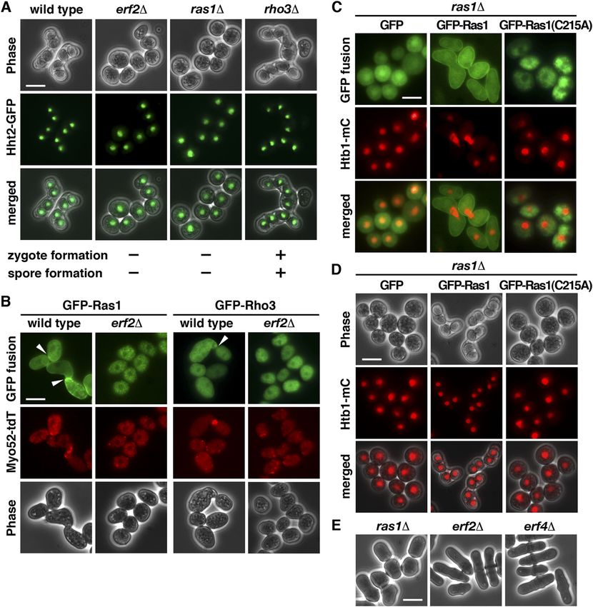
Meiotic roles of S-palmitoylation Pham et al. https://doi.org/10.26508/lsa.202201755 vol 6 | no 4 | e202201755 2 of 12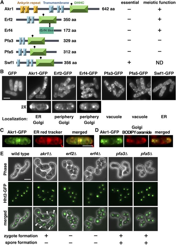
Figure 1. Fission yeast palmitoylacyltransferases.
(A) Schematic representation of the domain structures of the palmitoylacyltransferases in S. pombe. (B, C, D) Cellular localization of the fission yeast
palmitoylacyltransferases. GFP fusion proteins with (C) ER red tracker and (D) BODIPY ceramide for Golgi structure applied to the study. Scale bar, 5 μm. (E) Micrographs of
meiotic cells of the indicated homothallic strains expressing Hht2-GFP protein as the nuclear marker. Arrowheads indicate aberrant spores.

Meiotic roles of S-palmitoylation   Pham et al.                                           https://doi.org/10.26508/lsa.202201755     vol 6 | no 4 | e202201755       3 of 12
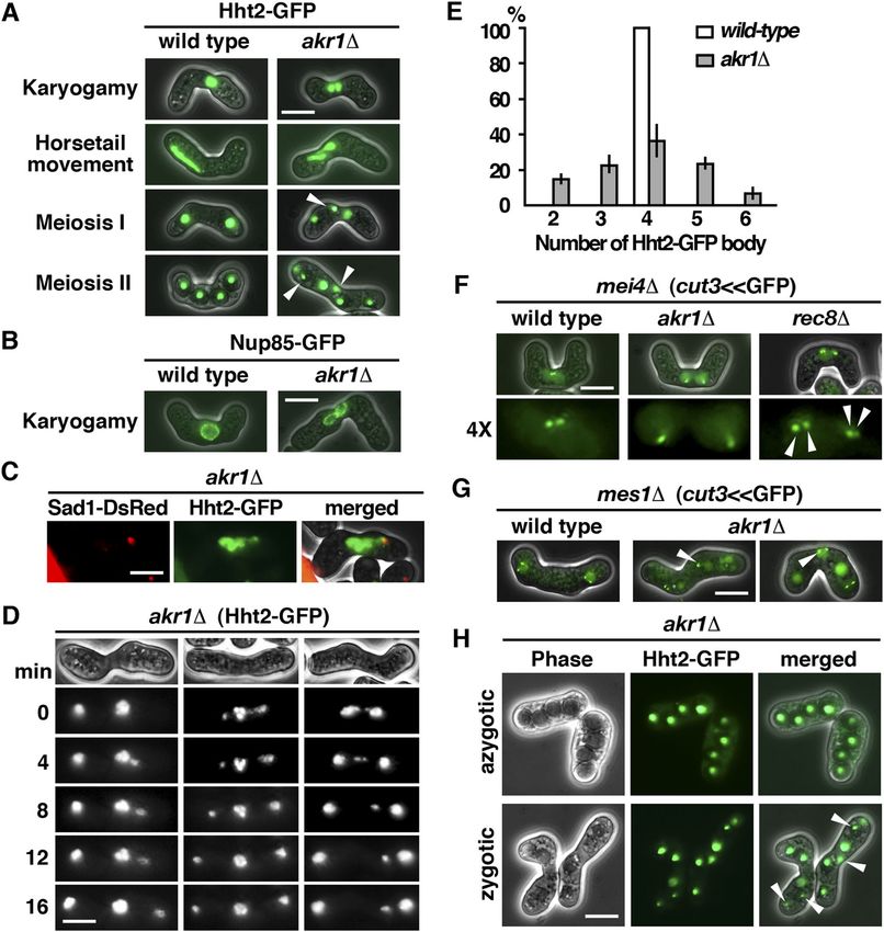
Figure 2. S-Palmitoylation of Ras1 by Erf2-Erf4 is required for mating pheromone response.
(A) Micrographs of meiotic cells of the indicated homothallic strains expressing Hht2-GFP protein as the nuclear marker. Scale bar, 5 μm. (B) Fluorescence micrographs
of the indicated homothallic strains expressing GFP-Ras1 or Rho3 (green) also expressing Myo52-TdTomato (red) proteins. Arrowheads indicate polarized projections.
(C) Fluorescence micrographs of ras1 strains transformed with a plasmid expressing WT GFP-Ras1 or C215A mutant (green) and also expression of Htb1-mCherry (red)
proteins. (D) Micrographs of homothallic ras1 cells transformed with a plasmid expressing WT Ras1 or C215A mutant. Htb1-mCherry proteins were used as the nuclear
marker. (E) Micrographs of vegetative cells of the indicated strains were shown.

that the requirement of Er2-Erf4 for Ras1 function seems only                        meiosis using homothallic strains with or without the mutation. As
during meiosis.                                                                      shown in Fig 3A, WT S. pombe cells usually immediately proceeded
                                                                                     to meiosis after nuclear fusion (karyogamy) in which cells with two
akr1 mutant is defective in meiotic nuclear fusion                                   conjugated nuclei only present transiently. In contrast, in the akr1
                                                                                     mutant, cells with two closely juxtaposed but not fused nuclei could
The mutant phenotype of akr1 suggested a function in meiosis. To                     be readily detected, suggesting that the unfused state was fairly
gain more insight into the role of Akr1 in meiosis, we performed a                   stable. This phenotype was further explored using Nup85, a
detailed phenotypical analysis of the defects at different stages of                 nucleoporin protein (Chen et al, 2004), tagged with GFP to reveal the

Meiotic roles of S-palmitoylation   Pham et al.                                       https://doi.org/10.26508/lsa.202201755   vol 6 | no 4 | e202201755       4 of 12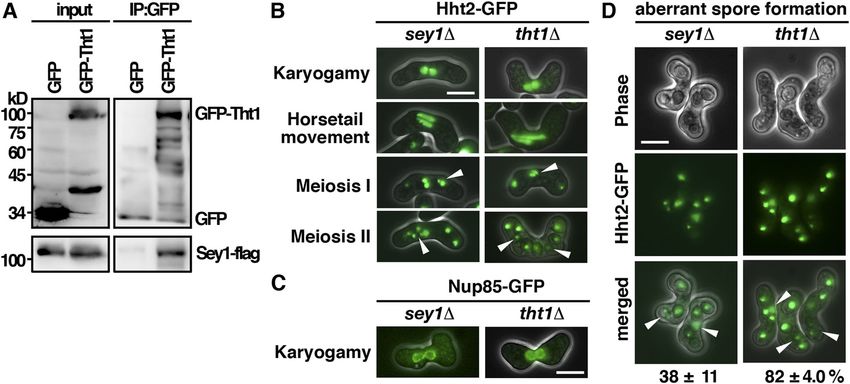
Figure 3. akr1 mutant is defective in meiotic nuclear fusion.
(A) Micrographs of meiotic cells of the indicated strains expressing Hht2-GFP protein as the nuclear marker. Scale bar, 5 μm. Arrowheads indicate segregation defects.
(B) Micrographs of meiotic cells of the indicated strains expressing Nup85-GFP protein as the nuclear envelop marker. (C) Micrographs of the tht1 cell expressing Hht2-GFP
and Sad1-DsRed protein as the spindle pole body marker. (D) Time-lapse imaging of Hht2-GFP was performed at 4-min intervals with the akr1 strain. (E) Quantification of
the sporulation defect in akr1 cells. The percentages of all asci containing different numbers of Hht2-GFP nuclear bodies were shown as indicated (error bars represent
one SD; the mean of three separate measurements was shown, n = 200). (F, G) Micrographs of meiotic cells of the indicated strains arrested at different stages of the
meiotic cycle (mei4 for prophase and mes1 for meiosis I) expressing cut3
support of this idea, a single SPB signal revealed by the SUN domain    S-palmitoylation of Tht1 by Akr1 is required for meiotic
Sad1-DsRed protein (Hagan & Yanagida, 1995) leading the twin            nuclear fusion
horsetails movement was observed in akr1 mutant (Fig 3C).
   At meiosis I, the WT cell diploid nucleus was segregated into two    The meiotic defects of the akr1 mutant including the unfused
nuclei and generated four equal spores in meiosis II. However, in       nuclei, twin horsetails movement, unequal chromosome seg-
the akr1 mutant, chromosomes were frequently not equally seg-           regation, and aberrant spore formation, which were suppressed
regated into daughters with more than two nuclei at meiosis I (Fig      in azygotic meiosis, resembled those of the tht1 mutant (Fig
3D) and generated unequal spores in meiosis II (Fig 3A). As a result,   4A–C) (Tange et al, 1998). Tht1 (twin horsetails protein 1) is a
up to 64% of spores bore with more or less than four spores (Fig 3E)    meiotic nuclear fusion protein reported but not validated in the
containing nuclei in various sizes (Hht2-GFP bodies) with reduced       SwissPalm—the S-palmitoylation database (Blanc et al, 2015).
spore viability (23% in the akr1 mutant compared with 98% in            Experiments were performed to determine whether Tht1 might
WT cells determined by tetra analysis).                                 be the target of Akr1 responsible for the meiotic defects ob-
   In S. pombe, the pairing of homologous chromosomes was               served. Tht1 was a meiosis-specific protein. To facilitate the
achieved during horsetail nuclear movement to establish a               characterization, we generated genomic N-terminally tagged
physical link for monopolar attachment to ensure accurate               GFP fusion Tht1 protein, ectopically expressed in exponentially
chromosome segregation at meiosis I. The chromosome seg-                growing cells by the thiamine-repressible nmt1 promoter.
regation defects of the akr1 mutant could be a consequence of           Similar to Akr1, Tht1 is an ER protein localized at the cell pe-
the defect in nuclear fusion in which chromosome homologues             ripheral and nuclear membrane (Fig 4D). In the absence of Akr1,
failed to pair and interact for proper chromosome segregation.          Tht1 was mislocalized from the ER as indicated by granule-like
Alternatively, sister chromatid cohesion could be defective in          structures observed in the log phase and in meiotic cells. These
the akr1 mutant. To distinguish between these two possibilities,        results suggested a role of Akr1 in regulating Tht1 function,
experiments were performed to determine whether akr1 cells              probably mediated by S-palmitoylation.
precociously separated their sister chromatid using homothallic            We further carried out the acyl-resin–assisted capture (acyl-
strains by monitoring GFP associated with the chromosome arm            RAC) experiments (Tewari et al, 2020) to determine the
at the cut3 locus (cut3
Figure 4. S-Palmitoylation of Tht1 by Akr1 is required for meiotic nuclear fusion.
(A) Micrographs of meiotic cells of the indicated strains expressing Hht2-GFP protein as the nuclear marker. Scale bar, 5 μm. Arrowheads indicate segregation defects.
(B) Micrographs of meiotic cells of the indicated strains expressing Nup85-GFP protein as the nuclear envelop marker. (C) Quantification of the sporulation defect in tht1
cells. The percentages of asci with unequal and inaccurate spore formation revealed by the numbers of Hht2-GFP nuclear bodies were expressed as means ± SD in
triplicate. (n = 200). (D) Micrographs of log-phase and meiotic cells of the indicated strains expressing GFP-Tht1 protein. (E) Samples from acyl-RAC experiments were
subjected to immunoblotting using an anti-GFP antibody to reveal proteins of interest. Specifically, purified palmitoyl-proteins can be seen enriched in the +HA samples.
10% of the input was loaded onto the gel. (F) GFP-Trap pulldown of GFP-Tht1 proteins resolved by SDS–PAGE was visualized by SYPRO Ruby staining. The identities of the
constituent proteins and S-palmitoylated peptides identified by MALDI-MS/MS analysis are indicated on the right. (G) Micrographs of cells expressing WT or mutant
GFP-Tht1 proteins. (H) Whole-cell protein extracts prepared from cells expressing WT or mutant GFP-Tht1 proteins were separated by SDS–PAGE and subjected to
immunoblotting using an anti-GFP antibody to reveal proteins of interest. Antibody against α-tubulin was used as the loading control. (I) Micrographs of homothallic tht1
cells transformed with a plasmid expressing WT Tht1 or C65/78A mutant. Htb1-mCherry proteins were used as the nuclear marker.

coverage rate suggested that Sey1 was a potential interacting                          reorganizing ER to facilitate nuclear fusion for faithful meiotic
protein of Tht1. In support of this idea, we found that Sey1 spe-                      chromosome segregation.
cifically interacted with Tht1 but not with the GFP control as
demonstrated by coimmunoprecipitation (Fig 5A). Furthermore,
meiotic defects similar to that of the tht1 mutant including the twin                  Discussion
horsetails movement, unfused nuclei, unequal chromosome seg-
regation, and aberrant spore formation were observed in the sey1                       Although changes in the global protein S-palmitoylation pattern
mutant (Fig 5B–D). However, the frequency of defective meiotic cells                   have been associated with different physiological states such as
was much lower in the sey1 mutant (38% as compared with 82% in                         cellular differentiation (Zhang & Hang, 2017) and pathological
the tht1 mutant), suggesting that in addition to Sey1, Tht1 required                   conditions (Zhang et al, 2021), the detailed molecular mechanisms
other proteins to facilitate chromosome segregation. Nevertheless,                     of how protein S-palmitoylation regulates these processes remained
the similar meiotic defects suggested a role of Tht1-Sey1 in                           to be defined.

Meiotic roles of S-palmitoylation   Pham et al.                                         https://doi.org/10.26508/lsa.202201755    vol 6 | no 4 | e202201755       7 of 12
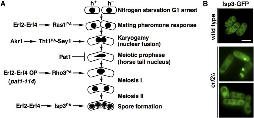
Figure 5. Tht1 interacted with Sey1 to function in the nuclear function.
(A) Coimmunoprecipitation was performed with extracts prepared from Sey1-flag–tagged strains expressing GFP-Tht1 or GFP control proteins. GFP-Trap affinity resin
was used to pull down GFP proteins. Immunoprecipitation was then analyzed by Western immunoblotting with antibodies against GFP and flag. Five percent of the input
was loaded onto the gel. (B) Micrographs of meiotic cells of the indicated strains expressing Hht2-GFP protein as the nuclear marker. Scale bar, 5 μm. Arrowheads indicate
segregation defects. (C) Micrographs of meiotic cells of the indicated strains expressing Nup85-GFP protein as the nuclear envelop marker. (D) Quantification of the
sporulation defect in sey1 cells. The percentages of asci with unequal and inaccurate spore formation revealed by the numbers of Hht2-GFP nuclear bodies were
expressed as means ± SD in triplicate. (n = 200).

Figure 6. S-palmitoylation is required at multiple stages of meiosis mediated by Erf2-Erf4 and Akr1 palmitoylacyltransferases in Schizosaccharomyces pombe.
(A) Schematic representation of the involvements of S-palmitoylation at different stages of meiosis. (B) Micrographs of pat1-induced haploid meiotic cells of the
indicated strains expressing Isp3-GFP. Scale bar, 5 μm.

   In an earlier study of protein S-palmitoylation in S. pombe, using                  Rho3 S-palmitoylated by Erf2 in the meiotic entry was suggested.
the synchronized meiosis induced from pat1-114 diploid cells,                          However, this work, performed primarily in azygotic diploid cells,
Zhang et al (2013) demonstrated that the global protein S-palmi-                       largely overlooked the early processes of cellular differentiation
toylation pattern was significantly altered and shaped by varying                       such as zygote formation (Fig 6A), and no detailed molecular
Erf2 palmitoylacyltransferase activity during meiosis. Meiotic entry                   mechanism was suggested.
was delayed in the erf2 mutant and increasing Erf2 palmitoyla-                            Here, we tracked cells throughout the natural sexual life cycle
cyltransferase activity trigged aberrant meiosis in sensitized cells.                  and performed a detailed phenotypic analysis of these palmitoy-
Multiple connate substrates of Erf2 were identified. A function of                      lacyltransferases mutants. We found that Ras1, but not Rho3, was

Meiotic roles of S-palmitoylation   Pham et al.                                         https://doi.org/10.26508/lsa.202201755    vol 6 | no 4 | e202201755       8 of 12
the primary target of Erf2-Erf4 in meiosis. Similar to the ras1 mutant,      and Yasushi Hiraoka. A complete list of the strains used in this
mutants of erf2 and erf4 were sterile and failed to form any zygote          study is given in Table 1. Conditions for growth, maintenance,
(Fig 2A). Compartment-specific signaling by Ras1 has been reported            and genetic manipulation of fission yeast were as described
(Chang et al, 1994; Onken et al, 2006). We further demonstrated that         previously (Hsiao et al, 2020). Except otherwise stated, strains
in the absence of Erf2-Erf4, Ras1 was mislocalized in the cytoplasm          were grown at 30°C in yeast extract or Edinburgh Minimal Me-
and failed to enrich at the cell conjugation site for mating pher-           dium (EMM2) with appropriate supplements. Where necessary,
omone response (Fig 2B). Thus, S-palmitoylation of Ras1 by Erf2-Erf4         gene expression from the nmt1 promoter was repressed by the
is intimately involved in controlling mating in S. pombe.                    addition of 15 mM thiamine to the culture medium. Homothallic
   In addition, a function of Akr1 in karyogamy was demonstrated.            strains were spotted onto a nitrogen-free medium to induce
The nuclear fusion protein Tht1 was identified as the target of Akr1          meiosis at room temperature.
responsible for the meiotic phenotype. S-palmitoylation is required
for Tht1 protein stability and cellular localization to ER to function
                                                                             Plasmid construction
for nuclear fusion. Furthermore, the ER fusion GTPase Sey1 was
identified as an interacting protein of Tht1 (Fig 5A). A role of Tht1-
                                                                             Plasmids for the expression of GFP-Ras1 and GFP-Tht1 (1,549–1,751)
Sey1 in reorganizing ER to facilitate nuclear fusion for faithful
                                                                             were constructed by PCR amplification of the corresponding ORF of
meiotic chromosome segregation was suggested. Experiments are
                                                                             cDNA derived from the genomic N-terminal GFP–tagged S. pombe
in progress to test this hypothesis.
                                                                             strains and subsequently ligated into plasmids derived from pREP1
   The results described above established the vital roles of protein
                                                                             at the multiple cloning sites. The Ras1 (C215A) and Tht1 (C65A and
S-palmitoylation at the early stage of meiosis (Fig 6A). The spore
                                                                             C78A) mutants were created using PCR-based site-directed mu-
coat protein Isp3 is another substrate of Erf2-Erf4 (Zhang et al, 2013).
                                                                             tagenesis and verified by DNA sequencing.
Using pat1-induced haploid meiosis to overcome the erf2
mutation–induced block in meiotic entry, we found that in the
absence of Erf2, Isp3 was mislocalized from the spore coat (Fig 6B).         Microscopy
These results suggested a role of protein S-palmitoylation in
regulating Isp3 function at the later stage of meiosis. Taken to-            Visualization of GFP-, mono cherry–, and DsRed-tagged proteins in
gether, our study demonstrated that protein S-palmitoylation is              living cells was performed at room temperature. Images acquired
required at both early and late stages of meiosis to ensure proper           from a Leica DM RA2 microscope equipped with a Leica DC 350F
cellular differentiation (Fig 6A). Furthermore, using comparative            camera were assembled using Adobe Photoshop. Visualization of
proteomics, 238 proteins were suggested to be preferentially                 Hht2-GFP in living cells embedded in 0.6% LMP agarose was per-
S-palmitoylated in meiosis by Erf2, the major meiotic palmitoyla-            formed at room temperature as previously described (Win et al,
cyltransferase in S. pombe (Zhang et al, 2013). These proteins are           2006).
involved in diverse cellular processes (Fig S3A). Among them,
mutants of 38% proteins (56 out of 146 characterized) were iden-
                                                                             Antibodies and immunoprecipitation
tified with meiotic phenotypes (mating, chromosome segregation,
or sporulation defects) in two genome-wide screens (Fig S3B and C)
                                                                             The antibody against GFP (ab290) was purchased from Abcam.
(Dudin et al, 2017; Blyth et al, 2018). Characterizing the role of protein
                                                                             The anti-Flag M2 antibody was from Sigma-Aldrich. Antibody
S-palmitoylation in the meiotic function of these proteins will
                                                                             against α-tubulin (Sigma-Aldrich) was used as a control. For
provide further insight into our understanding of the fundamental
                                                                             immunoprecipitation from yeast extracts, 2 × 10⁸ yeast cells were
mechanism coordinating cell differentiation in general. However,
                                                                             lysed in 200 ml NP-40 lysis buffer (6 mM Na2HPO4, 4 mM NaH2PO4,
even without addressing this point, the work alone and analysis
                                                                             1% NP-40, 150 mM NaCl, 2 mM EDTA, 50 mM NaF, 0.1 mM Na3VO4,
described here highlight the vital role of protein S-palmitoylation in
                                                                             1 mM PMSF, 1 mM DTT, complete protease inhibitor cocktail) by
meiosis at different stages with the versatility of the complicated
                                                                             vortexing with acid-washed glass beads. The lysate was clarified
regulatory network involved, which has significantly advanced our
                                                                             by centrifugation, and GFP fusion proteins were retrieved using
understanding of the biological function of protein S-palmitoyla-
                                                                             GFP-Trap-coupled agarose beads (ChromoTek) following the
tion in eukaryotes.
                                                                             manufacturer’s instructions.

                                                                             GFP protein identification
Materials and Methods
                                                                             The purified proteins were separated on SDS–PAGE gels and vi-
Fission yeast strains and methods                                            sualized by Sypro Ruby (Invitrogen). Protein bands were excised
                                                                             from gels, treated with trypsin, and subjected to analysis for protein
We constructed all strains according to standard procedures.                 identification using the Waters NanoAcquity UPLC-Synapt G2 HDM
Information of oligonucleotides for gene disruption or modifi-                instrument by the Core Facilities for Proteomics at National Health
cation can be obtained upon request. The original GFP-ras1                   Research Institutes. Data were processed for database searching
(Myo52-tdTomato), GFP-rho3, and histone GFP, mono cherry–                    using ProteinLynx Global Server against the Swiss-Prot database
tagged strains were gifts from Sophie G Martin, Reiko Sugiura,               with the Mascot program.

Meiotic roles of S-palmitoylation   Pham et al.                              https://doi.org/10.26508/lsa.202201755   vol 6 | no 4 | e202201755   9 of 12
Table 1. Schizosaccharomyces pombe strains used in this study.
Genotype Source
−
h pREP1-akr1-GFP leu1-32 This study
−
h pREP1-erf2-GFP leu1-32 This study
h− pREP1-erf4-GFP leu1-32 This study
−
h pREP1-pfa3-GFP leu1-32 This study
h− pREP1-pfa5-GFP leu1-32 This study
−
h pREP1-swf1-GFP leu1-32 This study
h90 hht2-GFP::ura4 leu1-32 ura4-D18 Yasushi Hiraoka
90
h hht2-GFP::ura4 akr1::hyh (hygromycin B phospho-transferase) ura4-D18 This study
h90 hht2-GFP::ura4 erf2::hyh ura4-D18 This study
90
h hht2-GFP::ura4 erf4::hyh ura4-D18 This study
90
h hht2-GFP::ura4 pfa3::hyh ura4-D18 This study
h90 hht2-GFP::ura4 pfa5::hyh ura4-D18 This study
90
h hht2-GFP::ura4 ras1::hyh ura4-D18 This study
h90 hht2-GFP::ura4 rho3::hyh ura4-D18 This study
90
h GFP-ras1 myo52-tdTomato::natMX Sophie G Martin
h90 GFP-ras1 myo52-tdTomato::natMX erf2::hyh This study
90
h GFP-rho3::LEU2 myo52-tdTomato::natMX leu1-32 Reiko Sugiura for GFP-rho3
h90 GFP-rho3::LEU2 myo52-tdTomato::natMX erf2::hyh leu1-32 This study
h90 htb1-mCherry:: natMX pREP1-GFP-ras1 leu1-32 Yasushi Hiraoka for htb1-mCherry
h90 htb1-mCherry::natMX pREP1-GFP-ras1(C215A) leu1-32 This study
90 r
h nup85-GFP::kan This study
90 r
h nup85-GFP::kan akr1::hyh This study
h90 hht2-GFP::ura4 sad1-DsRed::LEU2 akr1::kanr leu1-32 ura4-D18 This study
90
h mei4::ura4 cut3Acyl-RAC assay Blyth J, Makrantoni V, Barton RE, Spanos C, Rappsilber J, Marston AL (2018)
Genes important for schizosaccharomyces pombe meiosis identified
through a functional genomics screen. Genetics 208: 589–603.
The palmitoylation assay was conducted based on the acyl-RAC assay
doi:10.1534/genetics.117.300527
described previously (Tewari et al, 2020) with the following modifi-
Chang EC, Barr M, Wang Y, Jung V, Xu H-P, Wigler MH (1994) Cooperative
cations. Cell lysate extracted from 3 × 10⁹ S. pombe cells in ice-cold lysis interaction of s. Pombe proteins required for mating and
buffer (PBS, pH 7.4, 0.5 mM EDTA, protease inhibitor [PI], and 1 mM morphogenesis. Cell 79: 131–141. doi:10.1016/0092-8674(94)90406-5
phenylmethylsulfonyl fluoride) was incubated with 1% Triton X-100 and Chen XQ, Du X, Liu J, Balasubramanian MK, Balasundaram D (2004)
25 mM N-ethylmaleimide (NEM) for 1 h. The protein pellet was collected Identification of genes encoding putative nucleoporins and transport
by chloroform-methanol (CM) precipitation, which may be stored at factors in the fission yeast schizosaccharomyces pombe: A deletion
−80oC for up to 2 wk for the following assay. analysis. Yeast 21: 495–509. doi:10.1002/yea.1115
For acyl-RAC, the protein pellet was dissolved in a small volume Chen B, Sun Y, Niu J, Jarugumilli GK, Wu X (2018) Protein lipidation in cell
of 4% SDS buffer (4% SDS, 50 mM Tris–HCl, pH 7.4, 5 mM EDTA) signaling and diseases: Function, regulation, and therapeutic
opportunities. Cell Chem Biol 25: 817–831. doi:10.1016/
incubated at 37°C to improve dissolution before free cysteines
j.chembiol.2018.05.003
blocking with 20 mM N-ethylmaleimide (NEM) in LB buffer (150 mM
Chikashige Y, Tsutsumi C, Yamane M, Okamasa K, Haraguchi T, Hiraoka Y
NaCl, 50 mM Tris–HCl, pH 7.4, 5 mM EDTA) overnight. Three se-
(2006) Meiotic proteins bqt1 and bqt2 tether telomeres to form the
quential CM precipitations were processed to remove excessive bouquet arrangement of chromosomes. Cell 125: 59–69. doi:10.1016/
NEM, and the sample was resolved in LB buffer with 0.2% SDS before j.cell.2006.01.048
treatment with neutral 1 M hydroxylamine (HAM) to cleave the Dudin O, Merlini L, Bendezú FO, Groux R, Vincenzetti V, Martin SG (2017) A
thioester bound between cysteine residues and a fatty acid moiety. systematic screen for morphological abnormalities during fission
30 µl of thiopropyl resin (G-Biosciences) was added to every tube yeast sexual reproduction identifies a mechanism of actin aster
formation for cell fusion. PLoS Genet 13: e1006721. doi:10.1371/
and incubated for 1–2 h at RT. The beads were washed five times
journal.pgen.1006721
with washing buffer (LB buffer with 0.1% Triton X-100) to remove
Fang J, Wang H, Miao L, Kuang X, Ma W, Wang C, Zhang J, Xia G (2016)
unbound proteins, and the palmitoylated proteins were eluded in a
Involvement of protein acyltransferase ZDHHC3 in maintaining oocyte
SDS sample buffer with DTT detected by Western blotting. meiotic arrest in Xenopus laevis. Biol Reprod 95: 67. doi:10.1095/
biolreprod.116.138941
Gimple RC, Wang X (2019) Ras: Striking at the core of the oncogenic circuitry.
Supplementary Information Front Oncol 9: 965. doi:10.3389/fonc.2019.00965
Hagan I, Yanagida M (1995) The product of the spindle formation gene sad1+
Supplementary information is available at https://doi.org/10.26508/lsa. associates with the fission yeast spindle pole body and is essential for
202201755. viability. J Cell Biol 129: 1033–1047. doi:10.1083/jcb.129.4.1033
Harris MA, Rutherford KM, Hayles J, Lock A, Bähler J, Oliver SG, Mata J, Wood V
(2022) Fission stories: Using pombase to understand
Acknowledgements schizosaccharomyces pombe biology. Genetics 220: iyab222.
doi:10.1093/genetics/iyab222
We thank Y Herry Sun for funding support; Sophie G Martin, Reiko Sugiura, Horie S, Watanabe Y, Tanaka K, Nishiwaki S, Fujioka H, Abe H, Yamamoto M,
and Yasushi Hiraoka for yeast strains; and Mingzi M Zhang for critical review Shimoda C (1998) The schizosaccharomyces pombe mei4+ gene encodes
of the manuscript. This work was supported by the National Health Research a meiosis-specific transcription factor containing a forkhead DNA-
Institute (MG-110-PP-07). Taiwan. T-V Pham was supported by the NHRI-NCU binding domain. Mol Cell Biol 18: 2118–2129. doi:10.1128/mcb.18.4.2118
Graduate Student Program.
Hsiao WY, Wang YT, Wang SW (2020) Fission yeast puf2, a pumilio and fbf
Author Contributions family rna-binding protein, links stress granules to processing bodies.
Mol Cell Biol 40: e00589-19. doi:10.1128/mcb.00589-19
T-V Pham: investigation and writing—original draft, review, and Kimata Y, Trickey M, Izawa D, Gannon J, Yamamoto M, Yamano H (2008) A
editing. mutual inhibition between apc/c and its substrate mes1 required for
meiotic progression in fission yeast. Dev Cell 14: 446–454. doi:10.1016/
W-Y Hsiao: investigation.
j.devcel.2007.12.010
Y-T Wang: investigation.
Ko PJ, Dixon SJ (2018) Protein palmitoylation and cancer. EMBO Rep 19:
S-D Yeh: supervision and writing—review and editing. e46666. doi:10.15252/embr.201846666
S-W Wang: conceptualization, investigation, and writing—original
Li Y, Li H-J, Morgan C, Bomblies K, Yang W, Qi B (2019) Both male and female
draft, review, and editing. gametogenesis require a fully functional protein s-acyl transferase 21
in arabidopsis thaliana. Plant J 100: 754–767. doi:10.1111/tpj.14475
Conflict of Interest Statement
Linder ME, Deschenes RJ (2007) Palmitoylation: Policing protein stability and
traffic. Nat Rev Mol Cell Biol 8: 74–84. doi:10.1038/nrm2084
The authors declare that they have no conflict of interest.
Malgapo MIP, Linder ME (2021) Substrate recruitment by zdhhc protein
acyltransferases. Open Biol 11: 210026. doi:10.1098/rsob.210026
References Merlini L, Khalili B, Dudin O, Michon L, Vincenzetti V, Martin SG (2018) Inhibition
of ras activity coordinates cell fusion with cell–cell contact during yeast
Blanc M, David F, Abrami L, Migliozzi D, Armand F, Bürgi J, van der Goot FG mating. J Cell Biol 217: 1467–1483. doi:10.1083/jcb.201708195
(2015) Swisspalm: Protein palmitoylation database. F1000Res 4: 261. Mumby SM (1997) Reversible palmitoylation of signaling proteins. Curr Opin
doi:10.12688/f1000research.6464.1 Cell Biol 9: 148–154. doi:10.1016/S0955-0674(97)80056-7
Meiotic roles of S-palmitoylation Pham et al. https://doi.org/10.26508/lsa.202201755 vol 6 | no 4 | e202201755 11 of 12Onken B, Wiener H, Philips MR, Chang EC (2006) Compartmentalized signaling Yeste-Velasco M, Linder ME, Lu Y-J (2015) Protein s-palmitoylation and
of ras in fission yeast. Proc Natl Acad Sci U S A 103: 9045–9050. cancer. Biochim Biophys Acta 1856: 107–120. doi:10.1016/
doi:10.1073/pnas.0603318103 j.bbcan.2015.06.004
Politis EG, Roth AF, Davis NG (2005) Transmembrane topology of the protein Zare˛ ba-Kozioł M, Figiel I, Bartkowiak-Kaczmarek A, Włodarczyk J (2018)
palmitoyl transferase akr1. J Biol Chem 280: 10156–10163. doi:10.1074/ Insights into protein s-palmitoylation in synaptic plasticity and
jbc.M411946200
neurological disorders: Potential and limitations of methods for
Rogers JV, Arlow T, Inkellis ER, Koo TS, Rose MD (2013) Er-associated snares detection and analysis. Front Mol Neurosci 11: 175. doi:10.3389/
and sey1p mediate nuclear fusion at two distinct steps during yeast fnmol.2018.00175
mating. Mol Biol Cell 24: 3896–3908. doi:10.1091/mbc.E13-08-0441
Zhang MM, Hang HC (2017) Protein s-palmitoylation in cellular
Salaun C, Locatelli C, Zmuda F, Cabrera González J, Chamberlain LH (2020)
differentiation. Biochem Soc Trans 45: 275–285. doi:10.1042/
Accessory proteins of the zdhhc family of s-acylation enzymes. J Cell
bst20160236
Sci 133: jcs251819. doi:10.1242/jcs.251819
Tange Y, Horio T, Shimanuki M, Ding DQ, Hiraoka Y, Niwa O (1998) A novel fission Zhang MM, Wu P-YJ, Kelly FD, Nurse P, Hang HC (2013) Quantitative control of
yeast gene, tht1+, is required for the fusion of nuclear envelopes during protein s-palmitoylation regulates meiotic entry in fission yeast. PLoS
karyogamy. J Cell Biol 140: 247–258. doi:10.1083/jcb.140.2.247 Biol 11: e1001597. doi:10.1371/journal.pbio.1001597
Tewari R, West SJ, Shayahati B, Akimzhanov AM (2020) Detection of protein Zhang Y, Qin Z, Sun W, Chu F, Zhou F (2021) Function of protein
s-acylation using acyl-resin assisted capture. J Vis Exp. doi:10.3791/ s-palmitoylation in immunity and immune-related diseases. Front
61016 Immunol 12: 661202. doi:10.3389/fimmu.2021.661202
Watanabe Y, Nurse P (1999) Cohesin rec8 is required for reductional
chromosome segregation at meiosis. Nature 400: 461–464.
doi:10.1038/22774 License: This article is available under a Creative
Win TZ, Stevenson AL, Wang SW (2006) Fission yeast cid12 has dual functions Commons License (Attribution 4.0 International, as
in chromosome segregation and checkpoint control. Mol Cell Biol 26: described at https://creativecommons.org/
4435–4447. doi:10.1128/mcb.02205-05 licenses/by/4.0/).
Meiotic roles of S-palmitoylation Pham et al. https://doi.org/10.26508/lsa.202201755 vol 6 | no 4 | e202201755 12 of 12You can also read