Pseudogymnoascus destructans growth in wood, soil and guano substrates - Nature
←
→
Page content transcription
If your browser does not render page correctly, please read the page content below
www.nature.com/scientificreports
OPEN Pseudogymnoascus destructans
growth in wood, soil and guano
substrates
Jenny Urbina1*, Tara Chestnut2, Jennifer M. Allen1 & Taal Levi1
Understanding how a pathogen can grow on different substrates and how this growth impacts
its dispersal are critical to understanding the risks and control of emerging infectious diseases.
Pseudogymnoascus destructans (Pd) causes white-nose syndrome (WNS) in many bat species and
can persist in, and transmit from, the environment. We experimentally evaluated Pd growth on
common substrates to better understand mechanisms of pathogen persistence, transmission and
viability. We inoculated autoclaved guano, fresh guano, soil, and wood with live Pd fungus and
evaluated (1) whether Pd grows or persists on each (2) if spores of the fungus remain viable 4 months
after inoculation on each substrate, and (3) whether detection and quantitation of Pd on swabs is
sensitive to the choice to two commonly used DNA extraction kits. After inoculating each substrate
with 460,000 Pd spores, we collected ~ 0.20 g of guano and soil, and swabs from wood every 16 days
for 64 days to quantify pathogen load through time using real-time qPCR. We detected Pd on all
substrates over the course of the experiment. We observed a tenfold increase in pathogen loads
on autoclaved guano and persistence but not growth in fresh guano. Pathogen loads increased
marginally on wood but declined ~ 60-fold in soil. After four months, apparently viable spores were
harvested from all substrates but germination did not occur from fresh guano. We additionally found
that detection and quantitation of Pd from swabs of wood surfaces is sensitive to the DNA extraction
method. The commonly used PrepMan Ultra Reagent protocol yielded substantially less DNA than did
the QIAGEN DNeasy Blood and Tissue Kit. Notably the PrepMan Ultra Reagent failed to detect Pd in
many wood swabs that were detected by QIAGEN and were subsequently found to contain substantial
live conidia. Our results indicate that Pd can persist or even grow on common environmental
substrates with results dependent on whether microbial competitors have been eliminated. Although
we observed clear rapid declines in Pd on soil, viable spores were harvested four months after
inoculation. These results suggest that environmental substrates and guano can in general serve as
infectious environmental reservoirs due to long-term persistence, and even growth, of live Pd. This
should inform management interventions to sanitize or modify structures to reduce transmission risk
as well early detection rapid response (EDRR) planning.
Emerging fungal pathogens have become a major conservation issue for w ildlife1–3. Fungal pathogen transmis-
sion occurs through diverse pathways causing opportunistic infections such as aspergillosis4, cryptococcosis5
and mucormycosis6–8, and endemic infections with indirect transmission from the environment such as
blastomycosis9,10 and histoplasmosis11. Additionally, there are zoophilic pathogens with near-direct transmis-
sion such as the chytrid fungi Batrachochytrium dendrobatidis12 and Batrachochytrium salamandrivorans13 that
cause chytridiomycosis. These highly variable modes of transmission arise because fungi readily survive outside
of hosts and can persist, or even grow, on environmental substrates.
Although hosts are typically central to pathogen transmission, the epidemiological triangle reminds us of the
importance of the environment on transmission d ynamics14,15. In the traditional triad, the physical environment
can modify the response of both the pathogen and the host16,17. The presence and types of fomites can influence
dispersal of pathogens and disease dynamics18,19. Therefore, the role of substrates is significant in situations
where viable pathogens from an environmental substrate can re-infect hosts or remain as an environmental
reservoir20–22. Understanding the growth and survival of pathogens on environmental substrates is critical to
inform management interventions that might sanitize or modify structures to reduce transmission risk23,24.
1
Department of Fisheries and Wildlife, Oregon State University, 2820 SW Campus Way, Nash Hall, Corvallis,
OR 97331, USA. 2National Park Service, Mount Rainier National Park, Ashford, WA, USA. *email: jenny.gonzalez@
oregonstate.edu
Scientific Reports | (2021) 11:763 | https://doi.org/10.1038/s41598-020-80707-1 1
Vol.:(0123456789)
www.nature.com/scientificreports/
Obtaining a better insight about links among substrates, pathogen and hosts will improve our knowledge of
pathogen transmission and inform actions and strategies for disease mitigation.
One of the most important fungal pathogens for wildlife is Pseudogymnoascus destructans (Pd), which causes
white-nose syndrome (WNS) disease in bats. The first evidence for the emergence of this disease in North
America was reported in Albany, New York in 200625,26. By 2009 WNS had been reported from four species
including little brown bat (Myotis lucifugus), northern long-eared (M. septentrionalis), big brown (Eptesicus
fuscus) and tricolored bats (Perimyotis subflavus). Since then, WNS has been reported in nine additional species
within the genus Myotis. WNS causes mortality in bats by damaging skin and disrupting critical physiological
processes that can result in starvation or d ehydration27–29. This disease has been implicated in the unprecedented
decline of previously abundant, widely distributed, and healthy bat populations30–33, and it is estimated to have
killed more than 6 million bats34. To date, eight bat species and subspecies from the genera Corynorhinus (4),
Lasionycteris, Lasiurus, Tadarida and Myotis (http://www.whiten osesy ndrom
e.org, accessed April 13, 2020) have
been tested positive for Pd according to qPCR results, without diagnostic signs of the disease documented in
the tested individuals.
Pd can be transmitted directly among bats35 or indirectly via environmental substrates36. Indirect transmis-
sion of Pd is facilitated by long survival times37 and persistence outside of its optimal growth temperature38.
The prevalence of Pd in hibernacula remains stable or slightly increases through time after the initial detection
of the pathogen22. Pd is persistent in hibernacula in the United States while in Eurasian hibernacula the preva-
lence and pathogen loads decrease due to a seasonal decay of Pd during summer that leads to delayed infection,
shorter period of Pd growing on bats and low loads22. Although transmission of Pd has been linked to colony
size and c lustering32, the role played by particular substrates as environmental reservoirs on disease impacts is
only partially understood39.
We conducted an experimental study of Pd growth and survival on guano, soil, and plywood, which are typical
environmental substrates present in bat hibernacula or roosting locations. We assessed whether Pd would grow
on guano to determine whether guano could be an effective environmental reservoir for Pd and a transmission
pathway to susceptible bats. We hypothesized that guano might supply ample nutrients for growth, but this
growth might be inhibited by fecal bacteria. Therefore, we assessed Pd growth and viability on both non-sterile
and autoclaved guano. We tested Pd growth and persistence on plywood because this is a common substrate for
bats found in structures made by humans particularly artificial roost habitats such as bat houses. Based on unex-
pectedly low Pd concentrations detected in this substrate, we additionally compared the efficiency of two com-
monly used DNA extractions kits. Finally, we tested soil because it is a common substrate beneath hibernacula
and roost sites. We inoculated each substrate with a known amount of P. destructans conidia to answer whether
(1) Pd grows or persists on all the substrates, (2) there is a difference in the growth rates of Pd among different
substrates, and (3) whether conidia retrieved from each substrate were still viable after the 64-day experiment.
Methods
We characterized the growth of Pd in four substrates (1) autoclaved guano, (2) fresh guano, (3) soil and (4)
wood. We collected fresh guano of Myotis lucifugus from different piles in the attic of a storage barn in Corvallis,
Oregon, USA. Half of the guano collected was autoclaved for 60 min at 121 °C and at least 20 psi (Consolidated,
MA, USA) and the other half was not sterilized. We used commercial garden soil Proven Winners (all-purpose
type) as our soil substrate. As previous studies have already established the viability of Pd on sediments40,41, we
assessed the growth/decay rate in only non-autoclaved soil similar to field conditions. Finally, we autoclaved (to
control for the presence of other fungi and bacteria) square pieces of plywood (5.08 cm side, ~ 3 mm thick) as
our wood substrate to evaluate if Pd can persist on this substrate and to determine whether the substrate itself
was capable of supporting Pd.
We prepared the inoculum by resuspending, counting, and harvesting Pd conidia from cultured plates fol-
lowing standard m ethods35. Cultures of the Pd type strain (American Type Culture Collection ATCC MYA-4855)
were maintained for 30 days on Sabouraud Dextrose Agar (SDA) with gentamicin at 9 °C. We prepared a suspen-
sion by flooding plates with phosphate—buffered saline solution containing 0.5% Tween (PBST) and counted the
conidia using a hemocytometer. We inoculated four replicates of autoclaved guano, fresh guano, wood and soil
held in individual petri dishes by pipetting 1 ml of inoculum with a total estimated dose of 4.6 × 10⁵ Pd conidia
on top of 1.06 (± 0.0066) grams of substrate. A similar dose (5 × 10⁵) produced WNS infection and induced
behavioral changes in b ats35,42. We gently moved the samples to evenly distribute and spread the inoculum on
the guano and soil substrates. Inoculation of the plywood pieces was done by dividing the piece of wood into
five equal parts that were assigned a random order to be sampled. We inoculated each part of the plywood by
pipetting 5 drops of 40 μl per part (200 μl), so the total piece of wood had the same amount of conidia as the
other substrates (Fig. 1). We added one negative control replicate per substrate that was inoculated as previously
described with PBST without conidia. We used SDA plates with Pd growing as positive control.
We performed the first sampling event (time zero) immediately after adding the Pd inoculum to all the sub-
strates. We collected 0.18 g (± 0.05) grams of autoclaved guano, fresh guano and soil to quantify the amount of
Pd for each substrate replicate petri dish. For the wood sampling, sterile rayon swabs (MVE, Medical Wire, UK)
were rolled into the length of the plywood within a row allocated for each sampling event to avoid swabbing the
same portion of wood more than once.
We continued collecting samples from each substrate at four regular intervals every 16 days until day 64.
All petri dishes holding the substrates were kept in a refrigerator at 9 °C without particular light/dark cycle and
were organized randomly. For guano and soil, we collected an average mass of 0.23 (± 0.08) grams. Samples were
stored in vials at − 20 °C prior to DNA extraction for no longer than 2 weeks.
Scientific Reports | (2021) 11:763 | https://doi.org/10.1038/s41598-020-80707-1 2
Vol:.(1234567890)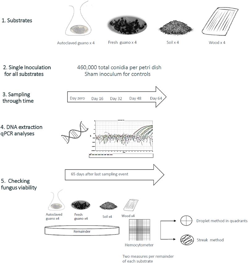
www.nature.com/scientificreports/
Figure 1. Graphic summary of the experimental design and methods we used during our experiments.
We performed DNA extractions from autoclaved guano, fresh guano, and soil using the DNeasy Powerlyzer
Powersoil Kit (QIAGEN, MD, US), which includes inhibitor removal s teps44. Replicate swab samples from ply-
wood were extracted using the PrepMan Ultra Sample Preparation Reagent (Applied Biosystems, Inc, Foster,
CA, USA) and DNeasy Blood and Tissue Kit (QIAGEN, MD, USA), which are commonly used for detection
of Pd from s wabs21,22,45,46. PrepMan Ultra Reagent is a common choice due to its low cost and ease of use. The
additional purification steps with the QIAGEN DNeasy Blood and Tissue Kit may reduce PCR inhibition but
is 3 times more expensive than PrepMan (PrepMan $0.735/sample, DNeasy $2.94/sample). We compared the
efficiency of these methods by evaluating the detection (presence/absence of pathogen) and yield of DNA after
the first sampling event through the end of the experiment. We began this comparison after the first sampling
event because initial results using the PrepMan Reagent returned less DNA than expected. All samples were
extracted individually after each sampling event and one extraction blank was included at each sampling event.
After DNA extraction, we quantified Pd using a probe-based qPCR assay targeting the intergenic spacer
region (IGS) of the f ungus47. Our reactions contained 12.5 μl of TaqMan Environmental MasterMix 2.0 (Life
Technologies, Carlsbad, CA), forward primer nu-IGS-0169-5′Gd and reverse primer nu-IGS-0235-3′Gd at a
final concentration of 400 nM, TaqMan FAM-labeled probe at a final concentration of 200 nM and 5 µl of DNA
template47. We used an ABI PRISM 7500 Fast real-time PCR system (Applied Biosystems, Foster City, CA)
with the following cycling conditions: initial activation 95 °C for 10 min; denaturation 95 °C for 15 s, annealing
and extension 60 °C for 60 s with a total of 40 cycles. We used a 4-point standard curve in triplicate from 10 to
10,000 fg of genomic DNA (ATCC MYA-4855™) to quantify the amount of DNA in each sample. We analyzed all
samples in triplicate and they were reported as positive if 2 out of the 3 wells amplified within 40 cycles.
Scientific Reports | (2021) 11:763 | https://doi.org/10.1038/s41598-020-80707-1 3
Vol.:(0123456789)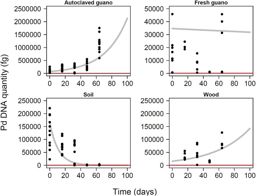
www.nature.com/scientificreports/
We harvested conidia from the remaining material from each substrate 51 days after the last sampling event,
which corresponds with 115 days post inoculation. We soaked the substrate material in 5 ml of PBST for 15 min.
We used a binocular compound microscope (40 × magnification, Model B120C, AmScope) to count the number
of conidia in two replicates of the resuspended substrate using a hemocytometer. To confirm conidia viability, we
plated 10ul of the harvested liquid using the droplet method in two SDA petri dishes (150 mm × 15 mm) divided
into four quadrants. We also used the streak method to plate two additional petri dishes for each sample using
a sterile cotton swab that was submerged in the harvesting liquid. We visually checked and photographed the
plates to assess whether harvested spores were viable two weeks later (i.e. 65 days after the last sampling event,
which corresponds with 129 days post inoculation).
We used standard protocols for decontamination of prepared suspensions of bleach and ethanol as established
by the National White-nose syndrome decontamination protocol v09.13.201843. All material was additionally
autoclaved before being discarded.
Data analysis. For each substrate, we evaluated evidence for Pd growth or decay using linear mixed models
to model log-10 Pd DNA quantity as a function of days since inoculation using each sampling event per petri
plate as random effect to account for the non-independent qPCR replicates nested within each DNA extraction.
We added a positive constant value (141.75 fg), the minimum amount of Pd detected, to handle loads of Pd
reported as zeros. We interpreted an increase in DNA concentration as evidence of Pd growth and stable levels
as evidence of Pd persistence. Detection of Pd DNA does not necessarily imply that Pd is alive and pathogenic39.
For each day corresponding to a sampling event (i.e. every 16 days since inoculation) we included data from 4
replicate inoculations each with 3 qPCR replicates per substrate type (Fig. 1). We additionally used the regres-
sion coefficients of this model to project estimated untransformed Pd DNA quantities through time. For these
analyses, we only included DNA quantified after extraction using QIAGEN kits (i.e. excluding DNA quantities
derived from the the PrepMan Ultra extraction kit used for wood swabs).
We compared the DNA yield from swabs of plywood extracted with the commonly used PrepMan Ultra and
Qiagen Blood Tissue Kits using a linear model for each sampling event with extraction method as a factor. This
result excludes data from the first sampling event that was extracted only with the PrepMan Ultra kit. Statistical
analyses were done in R (Version 3.4.4, R Core Team 2017). All procedures were approved by the Institutional
Biosafety Office (IBC) at Oregon State University (IBC tracking number 4218).
Results
Pseudogymnoascus destructans was detected in all substrates during all our sampling events (Fig. 2 and Table S1).
In autoclaved guano, the quantity of Pd detected increased over time (βautoclaved = 0.014; P = 0.003). There was a ten-
fold increase in the amount of Pd detected from the experimental setup to the last sampling event. Pd remained
stable in fresh guano (βfresh = − 0.0003; P = 0.007) but decreased substantially in soil (βsoil = − 0.03; P = 0.004). We
observed a significant increase on wood (βwood = 0.009; P = 0.009) despite the most Pd DNA detected during the
final sampling session. This effect was not stronger due to low Pd DNA detected during the third sampling event
for unknown reasons (Fig. 2). All controls were negative for Pd amplification.
Pseudogymnoascus destructans grew optimally in autoclaved guano. After 12 days, it was possible to see
mycelial mat on top of the autoclaved pellets but no growth was visible on fresh guano (Fig. 3). Although growth
through time was detected by qPCR in wood, there were no visible mycelia on top of this substrate during the
experiment. Similarly, in soil, there was no visually noticeable growth of the fungus.
The quantity of fungus detected in wood varied according to the extraction protocol used. For all sampling
events, the average amount of DNA detected was higher when using the Blood and Tissue Kit (QIAGEN). For
the first sampling event, we detected more DNA when using the QIAGEN Kit (β1 = 34,918.80, P < 0.00006) in
contrast to Prepman Ultra (16.45 fg). Similarly, the Pd quantities were higher for the second (β2 = 47,898.19,
P = 0.004) and third sampling event (β3 = 15,553, P < 0.00000023) after using the QIAGEN Kit in comparison
to Prepman Ultra (39.79 fg and 2.60 fg). During the last sampling event, the amount of Pd detected was high
in comparison to previous events after using Prepman Ultra (24,435 fg), however the QIAGEN Kit detected a
higher amount (β4 = 144,058, P = 0.02) (Fig. 4).
The projections for fungus growth extrapolated for 100 days post inoculation highlight the trajectory toward
high levels of Pd in autoclaved guano, stability in fresh guano, undetectable levels in soil, and growth in wood
(Fig. 5).
Counts of conidia 51 days after finishing the experiment were low for all the substrates. Wood had the highest
average number of conidia (mean 3.12 ± SD 1.64), followed by autoclaved guano (mean 2.75 ± SD 2.25), fresh
guano (mean 2.5 ± SD 3.62) and soil (mean 1.5 ± SD 1.69 (Fig. 6).
Pd grew in all four quadrants of SDA plates inoculated with 10 μl of the harvesting liquid obtained from wood,
soil and autoclaved guano. No growth was evident in plates inoculated with material from fresh guano (Fig. 7).
There was no growth of Pd in plates inoculated with the sham control.
Discussion
The increase of invasive fungal infections worldwide requires accurate and efficient identification of pathogens
and their viability. Detection of Pseudogymnoascus destructans through monitoring and surveillance is performed
by detecting DNA extracted from swabs or environmental samples using qPCR. However, samples can be qPCR
positive either because of extracellular D NA39, DNA from dead cells, or DNA from live fungus, but only live
fungus is infectious. Therefore, assessing the growth and viability of pathogens such as Pd on different substrates
can provide insight about the role of environmental substrates as reservoirs to inform management of potential
fomites implicated in the spread of the disease.
Scientific Reports | (2021) 11:763 | https://doi.org/10.1038/s41598-020-80707-1 4
Vol:.(1234567890)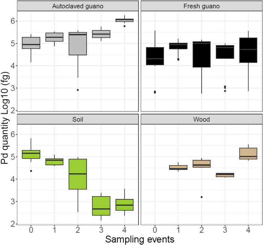
www.nature.com/scientificreports/
Figure 2. Pd fungus growth on different substrates over 64 days for five different sampling events (Day 0, 16, 32,
48 and 64). To facilitate comparison with the same extraction kit, we only present data for wood corresponding
to values obtained using the QIAGEN Blood and Tissue extraction Kit. We discovered substantially lower yield
with the Prepman Ultra Reagent after the first sampling event and initiated sampling with both Prepman and
QIAGEN kits for all subsequent sampling events.
We have demonstrated that live fungus can persist for long periods in the absence of a host and grow on
environmental substrates for over 2 months and retain infectious conidia for at least four months. The differential
growth and survival of this pathogen on the substrates we evaluated can be explained by the interplay between
medium composition, nutrient a vailability48, and microbial a ntagonists49–51. Pd grew substantially on autoclaved
guano as reflected by a tenfold increase in DNA quantity detected over the course of the study (Fig. 2) as well
as visible fungal growth (Fig. 3). The autoclaved guano depleted of microbial organisms presumably allowed
for substantial Pd growth by providing nutrients in a substrate without microbial competitors. In contrast, Pd
persisted but did not grow detectably in fresh guano over the full 64-day experiment and harvested spores did
not germinate. The diversity of bacterial communities in fresh guano includes Phyla such as Actinobacteria52
and members of the families Enterococcaceae, Baciillaceae and E nterobacteriaceae53,54. Actinobacteria play a
role in the decomposition of organic material. Enterococcus species are reported to inhibit hyphal morphogenesis
and virulence of f ungus55,56, while Bacillus and Enterobacter species are reported to control fungal growth by
synthesis of hydrolytic enzymes and production of antifungal compounds e.g., bacterial volatile c ompounds57,
and inhibiting sporulation58 respectively. As similar mechanisms could be affecting the growth of Pd in fresh
guano, we recommend that future studies of Pd viability evaluate microbial competition on Pd persistence and
transmission. These studies can benefit from the use of culture media types with a better resemblance to the
complex nutrient of the host38. We observed long-term persistence of Pd on sterile plywood. We sterilized ply-
wood to determine whether the woody substrate was a suitable medium for growth while controlling for extant
microbial communities that might be present. Such microbial communities on non-sterile plywood might also
inhibit Pd growth, but we expect this effect to be small relative to fresh guano. In contrast, DNA concentrations
of Pd in soil declined ~ 60-fold over the course of the experiment. This could be related to the presence of soil
fungi such as Mortierella spp. and Mucor spp., which are fast-growing and potentially able to outcompete P d48.
Scientific Reports | (2021) 11:763 | https://doi.org/10.1038/s41598-020-80707-1 5
Vol.:(0123456789)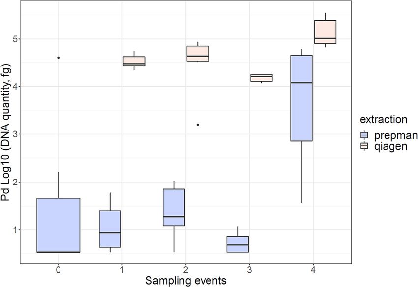
www.nature.com/scientificreports/
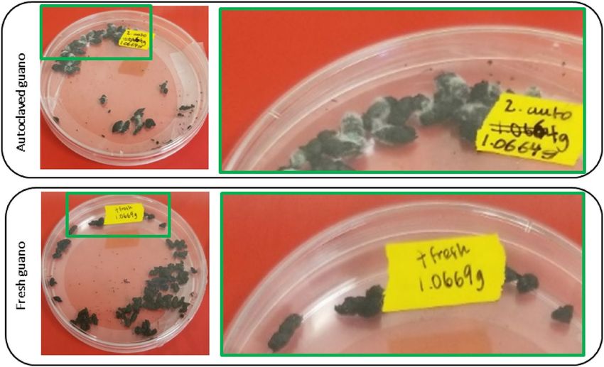
Figure 3. Plates of autoclaved guano and fresh guano pellets 48 days after inoculation. Top row shows visible
growth of Pseudogymnoascus destructans on autoclaved guano pellets compared to bottom row without visible
growth on fresh guano pellets.
Figure 4. Fungal loads recovered from plywood using either PrepMan Ultra Reagent or QIAGEN DNeasy
Blood and Tissue Kit for DNA extraction.
The presence of soil fungi growing on our SDA plates in addition to Pd provides some support for this hypothesis.
Future studies can benefit from evaluating soils found in roosting places and sediments from caves to understand
Pd growth, persistence and viability in these substrates.
Although we counted conidia from resuspension prepared from leftover material of each substrate, Pd was
only viable in culture for autoclaved guano, soil and wood (Fig. 7). This is unsurprising for autoclaved guano,
which exhibited visible Pd growth and a large increase in Pd DNA concentration (Figs. 2, 3). However, conidia
were collected from soil and readily germinated despite rapidly declining Pd DNA concentrations below the limit
of detection (Figs. 2, 7). We were also surprised to harvest the most conidia from wood and to observe the most
visible growth in culture (Figs. 6, 7). Although harvesting seemingly viable conidia from fresh guano, we did not
detect growth of Pd after inoculating SDA plates with these conidia. We rule out the number of viable conidia as
a potential explanation as the number obtained was similar between autoclaved (2.75 conidia) and fresh guano
(2.5 conidia). This lack of growth can potentially be explained by effects produced by bacteria action such as
production of volatile compounds that can reduce the growth from conidia and mycelial extension of Pd59,60.
Alternatively, fungal spores are able to enter into exogenous dormancy to later germinate and grow when the
effect of an unfavorable chemical or physical condition s tops61,62, which could explain these results. Therefore,
Scientific Reports | (2021) 11:763 | https://doi.org/10.1038/s41598-020-80707-1 6
Vol:.(1234567890)
www.nature.com/scientificreports/
Figure 5. Model projections for quantity of Pseudogymnoascus destructans during 100 days in four different
substrates with experimental data overlapped. Red line indicates the lowest amount of Pd detected during this
experiment.
Figure 6. Number of conidia counted per substrate 51 days after termination of the experiment to check
viability in SDA cultures (115 days post inoculation).
although we did not see germination, we cannot assume that conidia will not germinate at a later time when
conditions become more favorable. Recently, Pd was reported growing at higher temperatures (24 °C, 30 °C and
37 °C) outside of its optimal temperature growth38 exemplifying the ability of this pathogen to thrive in different
conditions. Also, Pd has been reported as viable 238 days after inoculation in sediments as flood debris with the
growth of the pathogen being influenced by geochemistry and the total amount of organic carbon a vailable41.
Physical and chemical characteristics of the evaluated substrates can directly or indirectly affect the growth or
persistence of Pd through time. Changes in relative humidity, nutrient content, pH or temperature can trigger
Scientific Reports | (2021) 11:763 | https://doi.org/10.1038/s41598-020-80707-1 7
Vol.:(0123456789)www.nature.com/scientificreports/
Figure 7. Growth of Pd in SDA plates 14 days after plating 10 μl of harvesting liquid obtained from leftovers of
the different substrates evaluated.
germination in ascomycete fungi63. The vegetative growth of Pd increase with high levels of relative humidity
while the production of conidia is not affected by relative h umidity64. Pd is able to grow in a broad pH range
between 5 and 11 with substrates as dead fish Poecilia sp., insect (Locusta migratoria), mushrooms (Lentinula
edodes) and demineralized exoskeletons of shrimp (Pleoticus muelleri) appropriate for the fungus to germinate
and grow65. Also, a range of chemical compounds like essential o ils66,67, volatile organic c ompounds60 can also
affect the growth and persistence of Pd.
The viability of Pd on environmental substrates suggests that many exposed substrates could be infectious to
bats. The viability of Pd on plywood could be an indication that human made structures could be more impor-
tant than bat guano for disease spread, particularly if the fungus invades and persists in the wood substrate.
Importantly, our results suggest that sampling wood surfaces with swabs and extract DNA using Prepman Ultra
will result in under-detection of Pd from buildings and structures such as attics and bat boxes. It is plausible
that hot summer temperatures could partially sterilize guano that may then serve as an efficient medium for Pd
growth in fall and winter. Researchers who come into contact with environmental substrates, including guano,
should take great care not to spread Pd to new environments or bats. Risk analysis for WNS in Australian bats has
accounted for this possibility with specific policy to implement controls to identify and decontaminate fomites
on environmental substrates to reduce the spread of Pd68. We recommend updating and clarifying existing
disinfection protocols to ensure effective methods for decontamination, including sites that are outside of caves
and enclosed spaces where guano may accumulate, such as in building attics and below a constructed bat house.
Evaluations of existing chemical treatments to determine what dose and substance constitutes an efficient treat-
ment as sporicidal (killing spores) or fungistatic (inhibition of fungal growth) will ensure complete decontamina-
tion and prevention of spreading Pd. We suggest additional experimental work to evaluate the effectiveness of
Scientific Reports | (2021) 11:763 | https://doi.org/10.1038/s41598-020-80707-1 8
Vol:.(1234567890)www.nature.com/scientificreports/
current decontamination protocols, so there are assurances that field and laboratory decontamination protocols
can minimize the risk of humans-cause spread of the pathogen.
Our experiment comparing DNA concentrations obtained through the two methods of extraction (PrepMan
Ultra Reagent and QIAGEN) has substantial implications for surveillance needing to sensitively detect Pd. Use
of the more affordable PrepMan extraction protocol is substantially more likely to lead to false negative results.
Surveillance in locations with unknown infection status should prioritize high extraction efficiency given that
Pd prevalence at these locations is expected to be low. This is also the case for surveillance conducted among
seasons to allow for the detection of minimum changes in pathogen load on environmental reservoirs of Pd. In
cases when infection is already high, both methods may work adequately for detection, but DNA concentrations
determined with PrepMan will be estimated with lower precision. To ensure the highest level of testing sensitiv-
ity, practitioners should consider using the QIAGEN protocol, especially in locations where Pd and WNS is not
yet known to occur and where a positive detection may result in a management action such as a cave closure.
This testing sensitivity could facilitate management actions including clearer communication of the presence
and spread of Pd, leading to more assertive action plans for bat conservation.
This research contributes important information to the understanding of the interaction between environ-
mental reservoirs and Pd, which has been previously overlooked, as research in WNS has focused mainly on the
pathogen-host relationship. This study demonstrates the utility of controlled experiments to better understand
how Pd viability varies among substrates, how persistence of Pd is potentially substrate specific, and how Pd can
persist through time in different environmental reservoirs. Understanding the role of environmental substrates
can help us to identify targets to apply management actions to prevent and control the indirect transmission of
WNS via fomites.
Data availability
Data are included as part of the supplementary information.
Received: 20 July 2020; Accepted: 24 December 2020
References
1. Fisher, M. C. et al. Emerging fungal threats to animal, plant and ecosystem health. Nature 484, 186–194 (2012).
2. Fisher, M. C., Gow, N. A. R. & Gurr, S. J. Tackling emerging fungal threats to animal health, food security and ecosystem resilience.
Philos. Trans. R. Soc. B Biol. Sci. 371, 20160332 (2016).
3. Ghosh, P. N., Fisher, M. C. & Bates, K. A. Diagnosing emerging fungal threats: A one health perspective. Front. Genet. 9, 376 (2018).
4. Seyedmousavi, S. et al. Aspergillus and aspergilloses in wild and domestic animals: A global health concern with parallels to human
disease. Med. Mycol. 53, 765–797 (2015).
5. Stephen, C., Lester, S., Black, W., Fyfe, M. & Raverty, S. Multispecies outbreak of cryptococcosis on southern Vancouver Island,
British Columbia. Can. Vet. J. 43, 792–794 (2002).
6. Speare, R., Thomas, A. D., O’Shea, P. & Shipton, W. A. Mucor amphibiorum in the toad, Bufo marinus Australia. J. Wildl. Dis. 30,
399–407 (1994).
7. Connolly, J. H. A review of mucormycosis in the platypus (Ornithorhynchus anatinus). Aust. J. Zool. 57, 235–244 (2009).
8. Gust, N. & Griffiths, J. Platypus mucormycosis and its conservation implications. Austral. Mycol. 28, 1–8 (2009).
9. Thiel, R. P., Mech, L. D., Ruth, G. R., Archer, J. R. & Kaufman, L. Blastomycosis in wild wolves. J. Wildl. Dis. 23, 321–323 (1987).
10. Storms, T. N., Victoria L. Clyde, Linda Munson & Edward C. Ramsay. Blastomycosis in nondomestic felids. J. Zool. Wildl. Med.
34, 231–238 (2003).
11. Guillot, J., Guérin, C. & Chermette, R. Histoplasmosis in Animals. in Emerging and Epizootic Fungal Infections in Animals (eds.
Seyedmousavi, S., de Hoog, G. S., Guillot, J. & Verweij, P. E.) 115–128 (Springer International Publishing, 2018). doi:https://doi.
org/10.1007/978-3-319-72093-7_5.
12. Scheele, B. C. et al. Amphibian fungal panzootic causes catastrophic and ongoing loss of biodiversity. Science 363, 1459 (2019).
13. Martel, A. et al. Batrachochytrium salamandrivorans sp. nov. causes lethal chytridiomycosis in amphibians. Proc. Natl. Acad. Sci.
USA 110, 15325 (2013).
14. Riley, S. Large-scale spatial-transmission models of infectious disease. Science 316, 1298 (2007).
15. Johnson, P. T. J., de Roode, J. C. & Fenton, A. Why infectious disease research needs community ecology. Science 349, 1259504
(2015).
16. Engering, A., Hogerwerf, L. & Slingenbergh, J. Pathogen–host–environment interplay and disease emergence. Emerg. Microbes
Infect. 2, 1–7 (2013).
17. Shikano, I. & Cory, J. S. Impact of environmental variation on host performance differs with pathogen identity: Implications for
host-pathogen interactions in a changing climate. Sci. Rep. 5, 15351 (2015).
18. Kraay, A. N. M. et al. Fomite-mediated transmission as a sufficient pathway: A comparative analysis across three viral pathogens.
BMC Infect. Dis. 18, 540 (2018).
19. Stephens, B. et al. Microbial exchange via fomites and implications for human health. Curr. Pollut. Rep. 5, 198–213 (2019).
20. Langwig, K. E. et al. Host and pathogen ecology drive the seasonal dynamics of a fungal disease, white-nose syndrome. Proc. Biol.
Sci. 282, (2015).
21. Huebschman, J. J. et al. Detection of Pseudogymnoascus destructans during Summer on Wisconsin Bats. J. Wildl. Dis. https://doi.
org/10.7589/2018-06-146 (2019).
22. Hoyt, J. R. et al. Environmental reservoir dynamics predict global infection patterns and population impacts for the fungal disease
white-nose syndrome. Proc. Natl. Acad. Sci. USA 117, 7255 (2020).
23. Foley, J., Clifford, D., Castle, K., Cryan, P. & Osfeld, R. S. Investigating and managing the rapid emergence of white nose syndrome,
a novel, fatal, infectious disease of hibernating bats. Conserv. Biol. 25, 223–231 (2011).
24. Blanco, C. M. & Garrie, J. Species specific effects of prescribed burns on bat occupancy in northwest Arkansas. For. Ecol. Manage.
460, 117890 (2020).
25. Gargas, A., Trest, M., Christensen, M., Volk, T. J. & Blehert, D. Geomyces destructans sp. nov. associated with bat white-nose
syndrome. Mycotaxon 108, 147–154 (2009).
26. Blehert, D. S. et al. Bat white-nose syndrome: An emerging fungal pathogen?. Science 323, 227 (2009).
27. Cryan, P. M. et al. Electrolyte depletion in white-nose syndrome bats. J. Wildl. Dis. 49, 398–402 (2013).
Scientific Reports | (2021) 11:763 | https://doi.org/10.1038/s41598-020-80707-1 9
Vol.:(0123456789)www.nature.com/scientificreports/
28. Warnecke, L. et al. Pathophysiology of white-nose syndrome in bats: A mechanistic model linking wing damage to mortality. Biol.
Lett. 9, 20130177 (2013).
29. Verant, M. L. et al. White-nose syndrome initiates a cascade of physiologic disturbances in the hibernating bat host. BMC Physiol.
14, 10 (2014).
30. Frick, W. F. et al. An emerging disease causes regional population collapse of a common North American bat species. Science 329,
679 (2010).
31. Turner, G. G., Reeder, D. M. & Coleman, J. T. H. A Five-year assessment of mortality and geographic spread of white-nose syndrome
in North American Bats, with a Look at the Future. Update of white-nose syndrome in bats. Bat Res. News 52, 13–27 (2011).
32. Langwig, K. E. et al. Sociality, density-dependence and microclimates determine the persistence of populations suffering from a
novel fungal disease, white-nose syndrome. Ecol. Lett. 15, 1050–1057 (2012).
33. Langwig, K. E. et al. Invasion dynamics of white-nose syndrome fungus, midwestern United States. Emerg. Infect. Dis. 21, 1023–
1026 (2015).
34. USFW. U.S. Fish and Wildlife Service. 2019. White-nose syndrome: The devastating disease of hibernating bats in North America.
Accessed 1 May 2020. https://www.whitenosesyndrome.org/mmedia-education/white-nose-syndrome-fact-sheet-june-2018.
(2019).
35. Lorch, J. M. et al. Experimental infection of bats with Geomyces destructans causes white-nose syndrome. Nature 480, 376 (2011).
36. Lorch, J. M. et al. Distribution and environmental persistence of the causative agent of white-nose syndrome, geomyces destructans,
in bat hibernacula of the Eastern United States. Appl. Environ. Microbiol. 79, 1293–1301 (2013).
37. Hoyt, J. R. et al. Long-term persistence of Pseudogymnoascus destructans, the Causative Agent of white-nose syndrome, in the
absence of bats. EcoHealth 12, 330–333 (2015).
38. Campbell, L. J., Walsh, D., Blehert, D. S. & Lorch, J. M. Long-term survival of Pseudogymnoascus destructans at elevated tem-
peratures. J. Wildl. Dis. 56, 278–287 (2020).
39. Urbina, J., Chestnut, T., Schwalm, D., Allen, J. & Levi, T. Experimental evaluation of genomic DNA degradation rates for the
pathogen Pseudogymnoascus destructans (Pd) in bat guano. PeerJ 8, e8141 (2020).
40. Lorch, J. M. et al. A culture-based survey of fungi in soil from bat hibernacula in the eastern United States and its implications for
detection of Geomyces destructans, the causal agent of bat white-nose syndrome. Mycologia 105, 237–252 (2013).
41. Reynolds, H. T., Ingersoll, T. & Barton, H. A. Modeling the environmental growth of Pseudogymnoascus destructans and its impact
on the White-nose syndrome epidemic. J. Wildl. Dis. 51, 318–331 (2015).
42. Warnecke, L. et al. Inoculation of bats with European Geomyces destructans supports the novel pathogen hypothesis for the origin
of white-nose syndrome. Proc. Natl. Acad. Sci. USA 109, 6999 (2012).
43. WNS Multiagency decontamination team. https://www.whitenosesyndrome.org/mmedia-education/united-states -national-white
-nose-syndrome-decontamination-protocol-april-2016-2. (2018).
44. Verant, M., Bohuski, E., Lorch, J. & Blehert, D. Optimized methods for total nucleic acid extraction and quantification of the bat
white-nose syndrome fungus, Pseudogymnoascus destructans, from swab and environmental samples. J. VET Diagn. Invest. 28,
110–118 (2016).
45. Rocke, T. E. et al. Virally-vectored vaccine candidates against white-nose syndrome induce anti-fungal immune response in little
brown bats (Myotis lucifugus). Sci. Rep. 9, 6788 (2019).
46. Zhelyazkova, V. L. et al. Screening and biosecurity for white-nose Fungus Pseudogymnoascus destructans (Ascomycota: Pseu-
deurotiaceae) in Hawai‘i. Pac. Sci. 73, 357–365 (2019).
47. Muller, L. K. et al. Bat white-nose syndrome: A real-time TaqMan polymerase chain reaction test targeting the intergenic spacer
region of Geomyces destructans. Mycologia 105, 253–259 (2013).
48. Vanderwolf, K. J., Malloch, D. & McAlpine, D. F. Detecting viable Pseudogymnoascus destructans (Ascomycota: Pseudeurotiaceae)
from walls of bat hibernacula: Effect of culture media. J. Cave Karst Stud. 78, 158 (2016).
49. Cheng, T. L. et al. Efficacy of a probiotic bacterium to treat bats affected by the disease white-nose syndrome. J. Appl. Ecol. 54,
701–708 (2017).
50. Micalizzi, E. W., Mack, J. N., White, G. P., Avis, T. J. & Smith, M. L. Microbial inhibitors of the fungus Pseudogymnoascus
destructans, the causal agent of white-nose syndrome in bats. PLoS ONE 12, e0179770 (2017).
51. Singh, A., Lasek-Nesselquist, E., Chaturvedi, V. & Chaturvedi, S. Trichoderma polysporum selectively inhibits white-nose syndrome
fungal pathogen Pseudogymnoascus destructans amidst soil microbes. Microbiome 6, 139 (2018).
52. De Mandal, S., Zothansanga, Panda, A. K., Bisht, S. S. & Senthil Kumar, N. First report of bacterial community from a Bat Guano
using Illumina next-generation sequencing. Genom. Data 4, 99–101. (2015).
53. Banskar, S., Bhute, S. S., Suryavanshi, M. V., Punekar, S. & Shouche, Y. S. Microbiome analysis reveals the abundance of bacterial
pathogens in Rousettus leschenaultii guano. Sci. Rep. 6, 36948 (2016).
54. Newman, M. M., Kloepper, L. N., Duncan, M., McInroy, J. A. & Kloepper, J. W. Variation in bat guano bacterial community
composition with depth. Front. Microbiol. 9, 914 (2018).
55. Cruz, M. R., Graham, C. E., Gagliano, B. C., Lorenz, M. C. & Garsin, D. A. Enterococcus faecalis inhibits hyphal morphogenesis
and virulence of Candida albicans. Infect. Immun. 81, 189 (2013).
56. Graham, C. E., Cruz, M. R., Garsin, D. A. & Lorenz, M. C. Enterococcus faecalis bacteriocin EntV inhibits hyphal morphogenesis,
biofilm formation, and virulence of Candida albicans. Proc. Natl. Acad. Sci. USA 114, 4507 (2017).
57. Khan, N. et al. Antifungal activity of bacillus species against fusarium and analysis of the potential mechanisms used in biocontrol.
Front. Microbiol. 9, 2363 (2018).
58. Kerr, J. R. Bacterial inhibition of fungal growth and pathogenicity. Microb. Ecol. Health Dis. 11, 129–142 (1999).
59. Wheatley, R. E. The consequences of volatile organic compound mediated bacterial and fungal interactions. Antonie Van Leeu-
wenhoek 81, 357–364 (2002).
60. Cornelison, C. T., Gabriel, K. T., Barlament, C. & Crow, S. A. Inhibition of pseudogymnoascus destructans growth from conidia
and mycelial extension by bacterially produced volatile organic compounds. Mycopathologia 177, 1–10 (2014).
61. Sussman, A. & Douthit, H. Dormancy in microbial spores. Annu. Rev. Plant Physiol. 24, 311–352 (1973).
62. Feofilova, E. P., Ivashechkin, A. A., Alekhin, A. I. & Sergeeva, Ya. E. Fungal spores: Dormancy, germination, chemical composition,
and role in biotechnology (review). Appl. Biochem. Microbiol. 48, 1–11 (2012).
63. Gasch, A. P. Comparative genomics of the environmental stress response in ascomycete fungi. Yeast 24, 961–976 (2007).
64. Marroquin, C. M., Lavine, J. O. & Windstam, S. T. Effect of humidity on development of pseudogymnoascus destructans, the
causal agent of bat white-nose syndrome. Northeastern Nat. 24, 54–64 (2017).
65. Raudabaugh, D. B. & Miller, A. N. Nutritional capability of and substrate suitability for pseudogymnoascus destructans, the causal
agent of bat white-nose syndrome. PLoS ONE 8, e78300 (2013).
66. Gabriel, K. T., Kartforosh, L., Crow, S. A. & Cornelison, C. T. Antimicrobial activity of essential oils against the fungal pathogens
ascosphaera apis and pseudogymnoascus destructans. Mycopathologia 183, 921–934 (2018).
67. Boire, N. et al. Potent inhibition of pseudogymnoascus destructans, the causative agent of white-nose syndrome in bats, by cold-
pressed, terpeneless valencia orange oil. PLoS ONE 11, e0148473 (2016).
68. Turbill, C. & Welbergen, J. A. Anticipating white-nose syndrome in the Southern Hemisphere: Widespread conditions favourable
to Pseudogymnoascus destructans pose a serious risk to Australia’s bat fauna. Austral. Ecol. 45, 89–96 (2020).
Scientific Reports | (2021) 11:763 | https://doi.org/10.1038/s41598-020-80707-1 10
Vol:.(1234567890)www.nature.com/scientificreports/
Acknowledgements
We are grateful to those who helped during this experiment: Maria Alejandra Reyna for participation during
sample collection and DNA extraction. Center for Genome Research and Biocomputing at Oregon State Uni-
versity provided space, and instruments for real time PCR.
Author contributions
J.U. conceived and designed the experiments, performed the experiments, analyzed the data, contributed rea-
gents/materials/analysis tools, prepared figures and/or tables, authored pictures and diagrams, authored and
reviewed drafts of the paper, approved the final draft. T.C. conceived and designed the experiments, contributed
reagents/materials, reviewed drafts of the paper, approved the final draft. J.A. contributed reagents/materials,
authored or reviewed drafts of the paper, approved the final draft. T.L. conceived and designed the experi-
ments, analyzed the data, contributed reagents/materials/analysis tools, authored or reviewed drafts of the paper,
approved the final draft.
Funding
This project was supported by a National Park Service grant to TC and TL. Any opinions, findings, and conclu-
sions or recommendations expressed in this publication are those of the author(s) and do not necessarily reflect
the views of the NPS.
Competing interests
The authors declare no competing interests.
Additional information
Supplementary Information The online version contains supplementary material available at https://doi.
org/10.1038/s41598-020-80707-1.
Correspondence and requests for materials should be addressed to J.U.
Reprints and permissions information is available at www.nature.com/reprints.
Publisher’s note Springer Nature remains neutral with regard to jurisdictional claims in published maps and
institutional affiliations.
Open Access This article is licensed under a Creative Commons Attribution 4.0 International
License, which permits use, sharing, adaptation, distribution and reproduction in any medium or
format, as long as you give appropriate credit to the original author(s) and the source, provide a link to the
Creative Commons licence, and indicate if changes were made. The images or other third party material in this
article are included in the article’s Creative Commons licence, unless indicated otherwise in a credit line to the
material. If material is not included in the article’s Creative Commons licence and your intended use is not
permitted by statutory regulation or exceeds the permitted use, you will need to obtain permission directly from
the copyright holder. To view a copy of this licence, visit http://creativecommons.org/licenses/by/4.0/.
© The Author(s) 2021
Scientific Reports | (2021) 11:763 | https://doi.org/10.1038/s41598-020-80707-1 11
Vol.:(0123456789)You can also read