Q/V SPECT CT in times of COVID 19: Changing the order to improve safety without sacrificing accuracy - Journal of Nuclear ...
←
→
Page content transcription
If your browser does not render page correctly, please read the page content below
Journal of Nuclear Medicine, published on May 7, 2021 as doi:10.2967/jnumed.120.261263
Q/V‐SPECT CT in times of COVID‐19: Changing the order to improve
safetywithout sacrificing accuracy
Wolfgang M. Schaefer MD, PhD (1), D. Knollmann MD (1), Philipp T. Meyer MD, PhD (2)
(1) Dept. of Nuclear Medicine,Kliniken Maria Hilf, Viersener Straße 450,
41063Mönchengladbach, Germany
(2) Department of Nuclear Medicine, Medical Center – University of Freiburg,
Faculty ofMedicine, University of Freiburg, Freiburg, Germany
Corresponding author
W. M. Schaefer, MD PhD (ORCID 0000-0002-9764-3138)
Dept. of Nuclear Medicine, Kliniken Maria
HilfViersener Straße 450
41063 Mönchengladbach, Germany
Phone +49 (0)2161 892-2430
Fax +49 (0)2161 892-2417
E-Mail: wolfgang.schaefer@mariahilf.de
The institutional ethics committee approved this case presentation and the
patientsigned a written informed consent.
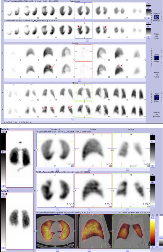
No potential conflicts of interest relevant to this article exist.TO THE EDITOR: Various nuclear medicine associations and colleagues (1-6) discussed whether a ventilation examination should be carried out at all when performing V/Q scans for diagnosis of pulmonary embolism in the SARS-CoV-2 pandemic situation. This consideration was prompted by the concern that a ventilation scan may be an aerosol-prone manoeuvre and, thus, carry a potential infection risk of the personnel. Usually the V/Q scan procedure starts with a ventilation scan (V), followed by a perfusion (Q) scan and eventually closed up with a low-dose CT if available. The sequence is traditionally chosen for a V/Q scan because it is easier to surpass the ventilation activity with the perfusion marker than vice versa. Instead of completely eliminating the ventilation scan resulting in specificity reduction (7) with all its negative consequences - like bleeding events due to unnecessary anticoagulation-, we suggest to modify the workflow in pandemic times to start routinely with the perfusion SPECT (somewhat lower administered activity than usual associated with increased acquisition time) and proceed with a low-dose CT if applicable. A ventilation SPECT (somewhat higher administered activity than usual associated with decreased acquisition time) is only performed when perfusion deficits are present that are not sufficiently explained by structural findings on low-dose CT. When the patient is SARS-CoV-2- positive or if there are COVID-like findings on low- dose CT, it remains the discretion of the physician whether a ventilation scan is performed under appropriate security measures. By doing so we can reduce the number of ventilation scans and avoid the aforementioned discussion held by various nuclear medicine associations and colleagues (1-6). Figure 1 shows a representative example of the proposed approach in an 80-year- old, recently bed-ridden lady, who was referred from an external hospital with dyspnoea, thoracic pain and increased D-dimer to rule out pulmonary embolism (PE). We startedby injecting about 45 MBq 99mTc-MAA and performed a perfusion SPECT/CT. Since we found relevant perfusion deficits and an unremarkable low- dose CT we subsequently also performed a ventilation SPECT with about 88 MBq 99mTc- Technegas (net ventilated activity, calculated from the projection data). The ventilation and perfusion studies show mismatch findings typical for pulmonary embolism (figure 1A). In combination with the normal low-dose CT examination (figure 1B), we were able to detect PE with the highest degree of certainty
according to Gutte et al (7).

Overall, we believe that the suggested routine reversal of the traditional workflow
helps to minimise aerosol-prone and potentially infectious manoeuvres without
compromising the accuracy of PE diagnostics.
References
  1. Zuckier LS, Moadel RM, Haramati LB, Freeman LM. Diagnostic evaluation
     of pulmonary embolism during the COVID-19 pandemic. J Nucl Med.
     2020;61:630-631.
  2. SNMMI Newsline: COVID-19 and ventilation/perfusion (V/Q) lung studies.
     J Nucl Med. 2020;61:23N-24N.
  3. https://www.sfmn.org/drive/CNP/CODIV-
     19/Recommandations_GT-            ExplorationsPulmonaires30-03-
     2020.pdf (Accessed on 01-21-2021)
  4. Buscombe JR, Notghi A, Croasdale J, et al. COVID-19: guidance for
     infection prevention and control in nuclear medicine. Nucl Med Commun.
     2020;41:499–504.
  5. Das JP, Yeh R, Schöder H. Clinical utility of perfusion (Q)-single-photon
     emission computed tomography (SPECT)/CT for diagnosing pulmonary
     embolus (PE) in COVID-19 patients with a moderate to high pre-test
     probabilityof PE. Eur J Nucl Med Mol Imaging. 2020;22:1–6.
  6. Vöö S, Dizdarevic S. Single photon emission computed tomography lung
     perfusion imaging during the COVID-19 pandemic: does nuclear medicine
     needto reconsider its guidelines? Nucl Med Commun. 2020;41:991-993.
  7. Gutte H, Mortensen J, Jensen CV, et al. Detection of pulmonary embolism
     with combined ventilation-perfusion SPECT and low-dose CT: head-to-head
     comparison with multidetector CT angiography. J Nucl Med. 2009;50:1987–
     1992.
Figure legend Figure 1. Q/V-SPECT CT of a 80-year-old lady. (A) Slice by slice comparison of ventilation and perfusion SPECT showing mismatch findings in both lungs (see arrows). Image noise is higher in the perfusion images due to the lower administered activity in the inverted workflow protocol. (B) Combining the SPECT data with the low dose CT proves pulmonary embolism in absence of structural alterations.
You can also read