Ready Species One: Exploring the Use of Augmented Reality to Enhance Systematic Biology with a Revision of Fijian Strumigenys Hymenoptera: ...
←
→
Page content transcription
If your browser does not render page correctly, please read the page content below
Insect Systematics and Diversity, (2019) 3(6): 6; 1–43
                                                                                                                   doi: 10.1093/isd/ixz005
                                                                                                                                 Research
Taxonomy

Ready Species One: Exploring the Use of Augmented
Reality to Enhance Systematic Biology with a Revision of
Fijian Strumigenys (Hymenoptera: Formicidae)
Eli M. Sarnat1, , Francisco Hita Garcia, Kenneth Dudley, Cong Liu, Georg Fischer, and
Evan P. Economo

                                                                                                                                                                            Downloaded from https://academic.oup.com/isd/article/3/6/6/5610803 by guest on 31 December 2021
Okinawa Institute of Science and Technology Graduate University, Biodiversity and Biocomplexity Unit1919-1 Tancha, Onna-son,
Okinawa 904-0495, Japan, and 1Corresponding author, e.sarnat@gmail.com

Subject Editor: István Mikó

Received 26 December, 2018; Editorial decision 13 May, 2019

Abstract
Advances in technology are rapidly changing the way people transmit, view, and interact with information.
These advances offer new opportunities for researchers to share scientific discoveries with each other and
the general public as never before. The field of revisionary biology has audiences confined to small groups
of specialists, but the core task of systematic biology—documenting the endless forms of nature—is particu-
larly well suited to capitalize on innovations in the realm of virtual, mixed and augmented reality. Interactive
three-dimensional (3D) digital models of biological specimens can help bridge barriers across scientific discip-
lines by circumventing technical jargon, and also promise to open exciting new vistas for public engagement.
Here, we explore the potential of augmented reality for communicating the discovery of new species. As a
test case, we revise a radiation of Strumigenys Smith (Hymenoptera: Formicidae) miniature trap-jaw ants in
Fiji. In addition to traditional revisionary elements, we present the augmented reality application ‘Insects3D’
built specifically for this study. The application runs on mobile devices and allows users to interact with X-ray
microtomography-derived 3D specimen models and visualize 3D geographic distribution maps. We recognize
23 species in Fiji, including 6 new species: S. anorak n. sp., S. artemis n. sp., S. avatar n. sp., S. gunter n. sp.,
S. oasis n. sp., and S. parzival n. sp. This study demonstrates the potential of leveraging 3D data and tech-
nology for a more interactive systematic biology, and the need for research programs to develop robust and
generalized tools to realize this potential.

Key words: augmented reality, specimens, biodiversity, microtomography, taxonomy

    For a bunch of hairless apes, we’ve actually managed to                            imaging technologies. The marriage of molecular data to traditional
    invent some pretty incredible things. ― Ernest Cline,                              morphology has made taxonomy a more robust science. Genetic ana-
    Ready Player One                                                                   lysis has proved indispensable for stabilizing higher level classification,
                                                                                       rooting out morphological homoplasy, and providing early scaffolds for
Taxonomy—the science of classifying life on earth—is foundational                      large taxonomic revisions (Padial et al. 2010, Schlick-Steiner et al. 2010).
to organismal biology. As elder statesmen of biodiversity science have                     Although taxonomy benefits from the integration of molecular
noted, ‘The goal of discovering, describing, and classifying the species               data, the discipline remains rooted in descriptive and analytical
of our planet assuredly qualifies as big science’ (Wheeler et al. 2004).               morphology. In this respect, the integration of new imaging tech-
During the turn of the 21st century, the scientific community began dis-               nologies has arguably had more impact on the scientific quality and
cussing both the biodiversity crisis and the taxonomic impediment: the                 clarity of new species descriptions than has the inclusion of gen-
pace of species extinction was accelerating even as taxonomic research                 etic data. Modern imaging is especially crucial for studying small
was losing prestige, resources, and talent (Pimm et al. 1995, Godfray                  invertebrates, which compose the majority of both described and
2002, Godfray and Knapp 2004, Wheeler 2004). Were taxonomists to                       undescribed species. As example, the broad adoption of focus
thrive in the molecular era, taxonomy would need to adapt.                             stacking technology during the past two decades has revolutionized
    Two crucial developments of taxonomy in the 21st century are the                   photography of arthropod specimens by eliminating depth of field
integration of molecular evidence and the adoption of new specimen                     constraints (Blagoderov et al. 2012, Brecko et al. 2014). In addition

© The Author(s) 2019. Published by Oxford University Press on behalf of Entomological Society of America.                                                               1
This is an Open Access article distributed under the terms of the Creative Commons Attribution Non-Commercial License (http://creativecommons.org/licenses/by-nc/4.0/),
which permits non-commercial re-use, distribution, and reproduction in any medium, provided the original work is properly cited. For commercial re-use, please contact
journals.permissions@oup.com
Version of Record, first published online November 12, 2019 with fixed content and layout in compliance with Art. 8.1.3.2 ICZN.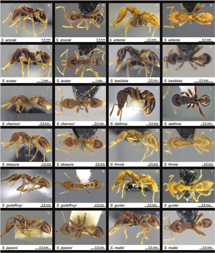
2                                                                                      Insect Systematics and Diversity, 2019, Vol. 3, No. 6

to enhancing the quality of new species descriptions, online image         Specifically, we demonstrate how mesh models generated from the
repositories of type specimens enable faster species descriptions by       morphological and geographic data of newly described Strumigenys
reducing time-intensive museum visits and specimen loans.                  Smith, 1860 (Hymenoptera: Formicidae) ants can be viewed and ma-
     The most compelling advances in biological specimen imaging           nipulated in three dimensions using commonplace mobile devices. We
are arguably coalescing around three-dimensional (3D) imaging              present methods and results for all stages of AR deployment, including
and processing technologies capable of generating virtual specimen         specimen scanning, mesh model creation, marker development, and
models. The two most prevalent 3D imaging technologies used by or-         mobile app development. We conclude our discussion by critiquing
ganismal biologists—and invertebrate zoologists in particular— are         opportunities and challenges for using AR technologies to enhance
micro-computed tomography (micro-CT) (Faulwetter et al. 2013,              biodiversity research, education, and outreach. The study provides
Akkari et al. 2015, Hita Garcia et al. 2017b, Ijiri et al. 2018) and       direction for future integration of emerging technologies such as aug-
photogrammetry (Nguyen et al. 2014, Gutiérrez‐Heredia et al. 2015,         mented reality and virtual reality into specimen-based research.
Qian et al. 2015, Galantucci et al. 2016, Gutierrez-Heredia et al.
2016, Sosa et al. 2016). Although 3D imaging of biodiversity collec-
tions is in its infancy, access to micro-CT scanners in biology depart-    Strumigenys
ments and natural history institutions is increasing. Simultaneously,      The taxonomic focus of the study presented here are Strumigenys

                                                                                                                                                     Downloaded from https://academic.oup.com/isd/article/3/6/6/5610803 by guest on 31 December 2021
more cost-effective photogrammetry systems are being developed.            ants from the Fiji Islands. Strumigenys—known also as miniature
Consequently, biologists are publishing large datasets of 3D spe-          trap-jaw ants—are diminutive predators that specialize in capturing
cimen models on websites such as Sketchfab (Sketchfab 2018),               minute arthropods. These huntresses most often nest and forage
Zoosphere (Zoosphere 2018), and Morphosource (Morphosource                 in leaf litter, topsoil and decaying wood and form small and usu-
2018, Watkins-Colwell et al. 2018).                                        ally monogynous colonies (Bolton 1999). The most prevalent prey
     The expanding availability of 3D specimen data is coinciding          are entomobryomorph Collembola, but they will take other small
with a bourgeoning array of tools for 3D model visualization.              arthropods as well (Wilson 1954, Brown 1971, Masuko 1985,
Augmented reality (AR), for example, is an emerging technology             Dejean 1987). Strumigenys species are primarily distributed across
capable of superimposing 3D computer-generated images on a user’s          tropical and subtropical rainforests and are most commonly col-
view of the real world. As testament to the technology’s growth,           lected using leaf litter extraction methods such as Berlese funnels or
the global augmented reality market was valued at USD 3.33 bil-            Winkler traps.
lion in 2015 and is expected to reach approximately between USD                Strumigenys ants are striking in appearance. They exhibit elong-
80 and 130 billion in 2021 (Zion Market Research 2017). Whereas            ated, pyriform heads flattened along the dorsoventral axis. They
users view only computer-generated images in virtual reality (VR),         produce a rich variety of highly modified hairs from thick, flattened,
the view in AR is a composite of the real world and virtual world.         and spatulate to gossamer thin, thread-like, and twisting. Nearly all
Our focus on AR, rather than VR, acknowledges that while access to         produce mysterious sponge-like outgrowths referred to as spongi-
the head-mounted displays (HMDs) required for experiencing VR is           form appendages that emanate from the waist. The mandibles of
currently limited, nearly everyone carries an AR compatible mobile         many are shaped into outlandishly long levers spiked at the tip with
device, such as a smartphone or tablet.                                    long piercing teeth.
     Biological applications of augmented reality have primarily been          Long-mandible Strumigenys are commonly referred to as trap-
the provenance of the medical field (Azuma 1997). Comparatively            jaw ants on account of their mandibles’ spring-loading mechanism
little attention has been given to the implications of AR for biodiver-    which is triggered to snap shut by the touch of suitable prey. Though
sity research and outreach (Bimber et al. 2002, White et al. 2006,         trap-jaw mechanisms evolved in at least three ant subfamilies and
Yeh et al. 2006, Barry et al. 2012, Chiang et al. 2014, Seltmann           nine genera, the vast majority of trap-jaw ants belong to the genus
et al. 2017). Moreover, the application of AR to taxonomic research        Strumigenys (Larabee and Suarez 2014). Not all of the 800+ de-
has thus far received mention in passing only (Sosa et al. 2016).          scribed Strumigenys species (Bolton 2018) are trap-jaw ants. Many
The lack of connectivity between taxonomic research and AR is              species previously placed in distinct genera on account of their
somewhat surprising, as museums are both integral to taxonomic             variant jaw shape or number of antennal segments (Bolton 2000) are
research and among the earliest institutional adopters of AR tech-         now placed in Strumigenys based on molecular evidence of shared
nology (Wojciechowski et al. 2004, Debenham et al. 2011, Yoon              ancestry (Ward et al. 2015). The taxonomic history of Strumigenys
et al. 2012). On the basis of our literature review, we are unaware        has been extensive and occasionally contentious (Baroni Urbani and
of any published taxonomic studies that explicitly include visual          de Andrade 2006, Bolton 2006). A comprehensive list of taxonomic
markers for displaying specimen models in augmented reality.               studies treating Strumigenys is included in Bolton (2000) and Baroni
     In previous contributions (Fischer et al. 2016; Sarnat et al. 2016,   Urbani and de Andrade (2007).
2017; Agavekar et al. 2017; Hita Garcia et al. 2017b, 2017a; Staab             The tropical islands of the Fijian archipelago host an excep-
et al. 2018), we focused on the use of 3D X-ray imaging for system-        tionally rich fauna of Strumigenys. In total, 23 species are now
atics. The aforementioned studies included passive media (e.g., 3D         known from the archipelago, including 6 that are described here
models flattened into 2D figures, video of rotating 3D models) and         for the first time. Two species (S. membranifera Emery and S. rogeri
interactive media (e.g., rotatable 3D models embedded in interactive       Emery) are introduced from Africa and an additional tramp species
PDFs). Here we explore how 3D imaging technology can create im-            (S. godeffroyi Emery) is most likely native to Southeast Asia. The re-
mersive media for use in organismal biology.                               maining 20 species are considered endemic to Fiji, although 1 species
                                                                           (S. mailei Wilson & Taylor) is also recorded from Samoa.
                                                                               The first treatment of Fiji’s Strumigenys species was presented by
Statement of Purpose                                                       Mann (1921) in his monograph on the archipelago’s ants. In add-
In this paper, we present proof-of-concept examples for how aug-           ition to listing S. godeffroyi, Mann described five species—S. jepsoni,
mented reality can add research value to systematic biology.               S. nidifex, S. scelestus, S. vitiensis, S. wheeleri—although the latter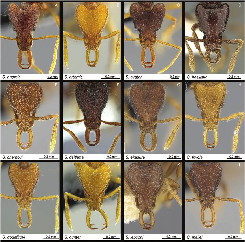
Insect Systematics and Diversity, 2019, Vol. 3, No. 6                                                                                              3

two were later synonymized with S. membranifera and S. tumida                Strumigenys suggests the long-mandible Fijian endemics are all des-
Bolton, respectively. A key to these six species was included and            cended from a single colonist ancestor (Liu et al. unpublished data).
the genus was briefly discussed with respect to the high number              The in situ evolution of such dramatic morphological disparity
of species on the archipelago. Wilson and Taylor (1967) described            strongly contradicts previous biogeographic hypotheses based on
S. mailei from Samoa, and designated as paratype a worker collected          morphological observation. Bolton assigned each of Fiji’s native and
from the uplands of Viti Levu by E.C. Zimmerman in 1938. Dlussky             endemic Strumigenys to species groups (Table 1). Given that none
(1993) described S. chernovi from Fiji in a study of the southwestern        of the listed groups or complexes are composed strictly of Fijian
Pacific Strumigenys. Bolton (2000) described eight new species of            species, Bolton’s assignments assume Fijian endemic Strumigenys are
Fijian Strumigenys in his global revision of Strumigenys (S. basiliska,      descended from no fewer than six independent colonization events.
S. daithma, S. ekasura, S. frivola, S. panaulax, S. praefecta, S. sulcata,       Although the meticulous global revision was explicit that many of
S. trauma (= Pyramica trauma) and provided S. wheeleri Mann with             the Fijian species did not neatly conform to their assigned groups—
the replacement name S. tumida.                                              and that the assignments more accurately reflected morphological
    Sarnat and Economo (2012) presented a diagnostic key, species            similarity than phylogenetic proximity—the extent of evolutionary
accounts, specimen photographs, and distribution maps for all the            convergence with distantly related Indoaustralian congeners was not
valid Strumigenys known from Fiji along with seven putatively un-            anticipated by Bolton (2000) or Sarnat and Economo (2012). That

                                                                                                                                                        Downloaded from https://academic.oup.com/isd/article/3/6/6/5610803 by guest on 31 December 2021
described species. After further review of these seven morphospecies         all long-mandible Fijian endemic Strumigenys are monophyletic is
discussed in Sarnat and Economo (2012) we propose here that five             remarkable not only because the morphology of S. nidifex Mann
represent new species (S. anorak n. sp., S. artemis n. sp., S. avatar n.     so closely parallels that of the distantly related szalayi-group, or be-
sp., S. gunter n. sp., and S. oasis n. sp.). Comparison with additional      cause the morphology of S. basiliska Bolton so closely parallels that
type material also suggests that the specimens treated in Sarnat and         of the distantly related biroi-group, but because these two disparate
Economo (2012) as S. panaulax Bolton constitute a distinct species,          species together with their endemic congeners—even the aberrant
described here as S. parzival n. sp.                                         S. oasis—all descended from a single common ancestor in the puta-
    The present work is an ongoing effort (Sarnat 2006; Lucky and            tively recent past (Lucky and Sarnat 2010, Sarnat and Moreau 2011,
Sarnat 2008; Sarnat 2008; Sarnat and Economo 2012, 2013; Hita                Sarnat and Economo 2012).
Garcia et al. 2015; Fischer et al. 2016) to describe Fiji’s rich and
highly endemic ant fauna. Taxonomy is the foundation of organ-               Methods
ismal biology, and these morphological studies have prompted new
hypotheses concerning taxon cycles (Economo and Sarnat 2012,                 Species identification and delineation
Matos-Maraví et al. 2018a), island biogeography (Lucky and Sarnat            Strumigenys is relatively unique among ant genera with respect
2010, Sarnat and Moreau 2011, Clouse et al. 2015, Economo et al.             to its abundance of morphological characters—particularly hairs,
2017, Matos-Maraví et al. 2018b), and functional ecology (Sarnat             spongiform tissues, mandibles, and sculpture. Despite their minute
et al. 2017).                                                                size and rich taxonomic diversity, species of Strumigenys are thus
    The Fijian Strumigenys are a model group for studying rapid              readily discerned from one another even when few specimens are
morphological and ecological radiation of social insects on island           available for analysis. Initial specimen identification morphospecies
systems. Analysis of molecular data from a global sampling of                designation was conducted as part of Sarnat and Economo (2012).

Table 1. Fijian Strumigenys arranged by species name. Species group names and group-complex names to which Bolton (2000) assigned
Fijian Strumigenys are listed. Species assigned by Bolton (2000) to Pyramica are also noted

Species                                            Status                             Bolton group/genus                           Group complex

S. anorak, n. sp.                                  endemic                          —                                             —
S. artemis, n. sp.                                 endemic                          —                                             —
S. avatar, n. sp.                                  endemic                          —                                             —
S. basiliska Bolton                                endemic                          biroi                                         —
S. chernovi Dlussky                                endemic                          godeffroyi                                    smythiesii
S. daithma Bolton                                  endemic                          caniophanes                                   —
S. ekasura Bolton                                  endemic                          godeffroyi                                    smythiesii
S. frivola Bolton                                  endemic                          rofocala                                      —
S. godeffroyi Mayr                                 introduced                       godeffroyi                                    —
S. gunter, n. sp.                                  endemic                          —                                             —
S. jepsoni Mann                                    endemic                          godeffroyi                                    smythiesii
S. mailei Wilson & Taylor                          native                           godeffroyi                                    signeae
S. membranifera Emery                              introduced                       (Pyramica) membranifera                       —
S. nidifex Mann                                    endemic                          szalayi                                       —
S. oasis, n. sp.                                   endemic                          —                                             —
S. panaulax Bolton                                 endemic                          godeffroyi                                    smythiesii
S. parzival, n. sp.                                endemic                          —                                             —
S. praefecta Bolton                                endemic                          godeffroyi                                    signeae
S. rogeri Emery                                    introduced                       —                                             —
S. scelesta Mann                                   endemic                          godeffroyi                                    smythiesii
S. sulcata Bolton                                  endemic                          godeffroyi                                    signeae
S. trauma (Bolton)                                 endemic                          (Pyramica) capitata                           —
S. tumida Bolton                                   endemic                          godeffroyi                                    signeae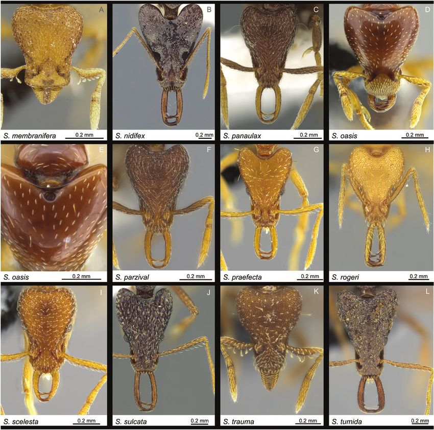
4 Insect Systematics and Diversity, 2019, Vol. 3, No. 6
Bolton’s (2000) authoritative global revision of Strumigenys was
Amperage
used as the primary identification reference together with examin-
(µA)
84
84
84
86
85
72
83
84
86
80
84
84
79
80
80
ation of type material for all available Fijian endemic Strumigenys.
Specimens which could not be identified using the aforementioned
references were compared with invasive and tramp Strumigenys
Voltage
before publishing them as morphospecies in Sarnat and Economo
(kV)
60
60
60
70
70
70
60
60
70
50
60
60
50
50
50
(2012). The species concepts presented here are also supported by
rigorous phylogenetic analysis of genome-wide SNP markers (Liu
et al. in review) that includes 18 of the putative 23.
Power
(W)
5
5
5
6
6
5
5
5
6
4
5
5
4
4
4
X-ray microtomography
Micro-CT scanning
We captured X-ray microtomographic scans of 15 Strumigenys spe-
cimens representing all species endemic to Fiji for which suitable
Detector distance
material was available. The specimens were left attached to their
Downloaded from https://academic.oup.com/isd/article/3/6/6/5610803 by guest on 31 December 2021
paper point, which was clamped to a holding stage. Scan settings
(mm)
11
30
11
19
25
30
22
25
27
13
25
25
22
13
13
were selected according to yield optimum scan quality and primarily
follow Hita Garcia et al. (2017a) and Hita Garcia et al. (2017b). In
contrast to the former study, we performed only full body scans and
refrained from including standardized scans of head, mesosoma, and
metasoma. All micro-CT scans were performed using a Zeiss Xradia
Source distance
510 Versa 3D X-ray microscope operated with the Zeiss Scout-and-
Scan Control System software (version 11.1.6411.17883). Specimen
(mm)
−15
−11
−25
11
11
11
11
−11
11
14
−11
−11
12
14
12
data and scanning parameters are provided in Table 2.
Virtual reconstruction and postprocessing of raw data
Three-dimensional reconstructions of the resulting scan projection
data were generated using the Zeiss Scout-and-Scan Control System
Exposure time
Reconstructor (version 11.1.6411.17883) and saved in DICOM file 0.5
1.0
1.0
0.5
0.6
1.0
0.6
1.0
0.7
0.7
1.0
1.0
1.2
0.8
0.8
format. Post-processing of DICOM raw data was performed with
(s)
Amira software (version 6.2). Virtual examinations of 3D surface
models were performed by using either the ‘volren’ or ‘volume ren-
dering’ functions. Desired volume renderings were generated by
adjusting color space range to a minimum to ensure the exterior
Voxel size
3.8979
1.8136
4.6916
2.4775
2.0646
1.8129
2.2523
1.9559
2.0649
2.3851
3.5032
3.2434
2.065
3.503
2.065
surface of specimens remained visible at the highest available quality.
Three-dimensional models were rotated and manipulated to allow a
complete virtual examination of the scanned specimens. Images of
shaded surface display volume renderings were made with the ‘snap-
shot’ function at the maximum resolution (approximately 1900 by
893 pixels).
30 June 2018
30 June 2018
30 June 2018
17 Dec. 2017
16 Dec. 2017
17 Dec. 2017
16 Dec. 2017
16 Dec. 2017
17 Dec. 2017
Table 2. Micro-CT scanning parameters arranged by species name
1 July 2018
1 July 2018
1 July 2018
1 July 2018
1 July 2018
1 July 2018
Date
Three-dimensional model development and
optimization for augmented reality applications
Computer generated 3D specimen models
The 3D mesh models presented here were generated using micro-CT
volumetric data. Each model is composed of large sets of connected
worker
worker
worker
worker
worker
worker
worker
worker
worker
worker
worker
worker
worker
worker
worker
Caste
vertices and faces (polygons) that can be rendered or streamed in
real time. The tradeoff between detail and performance is inherent to
3D modeling (Franco et al. 2004). Increasing model detail decreases
model performance: detail is measured by the number of polygons
CASENT0186900
CASENT0186982
CASENT0185902
CASENT0185655
CASENT0184909
CASENT0184622
CASENT0186834
CASENT0184984
CASENT0184653
CASENT0185548
CASENT0185751
CASENT0186960
CASENT0186943
CASENT0185705
CASENT0185699
(or vertices) in the model, performance is measured by the processing
Specimen code
time needed to render the model
Micro-CT is an effective method for capturing internal struc-
tures and reconstructing surface volumes. However, CT volumes
do not capture surface appearance such as colors and textures. To
create specimen models with both morphologically accurate surface
volumes and visually accurate colors and textures, we mapped 2D
specimen photographs onto the CT surface volume using the appli-
Taxon code
S. praefecta
S. basiliska
S. chernovi
S. parzival
S. ekasura
cation Zbrush.
S. artemis
S. tumida
S. anorak
S. nidifex
S. sulcata
S. frivola
S. gunter
S. avatar
S. mailei
S. oasis
We developed two versions of 3D specimen models for each of the
newly described Strumigenys species: maximum detail and optimized.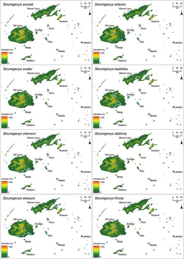
Insect Systematics and Diversity, 2019, Vol. 3, No. 6                                                                                           5

The maximum detail models were generated to preserve as much of            generated using Blender’s ‘Ocean’ modifier on a separate surface and
the original specimen surface topology as data allowed. All polygons       merged with the normal map for elevation dataw.
are preserved from the original micro-CT scans described in the pre-           Finally, the models were added onto the Sketchfab (Sketchfab,
ceding section. The target audiences of the maximum detail models          New York City, New York), a 3D content publishing platform to
are colleagues interested in using the models for scientific research.     provide a website interface for interactive manipulation of the
    The optimized models were generated to preserve the maximum            model and future presentation and annotation. Sketchfab is a plat-
level of detail that can be smoothly rendered by an iPhone 6s or equiva-   form to share and discover 3D, virtual reality, and augmented reality
lent mobile device. In order to determine a suitable tradeoff of detail    content. All models are available for viewing and manipulating
versus performance, we created four additional models of varying           at the following URL [https://sketchfab.com/arilab/collections/
polygon numbers (1m, 500k, 100k, 14k) derived from the maximum             strumigenys-species-from-fiji].
quality model (8.1m polygons) of the Strumigenys avatar holotype
(CASENT0185902). Of the optimization trials, the 500k polygon              Augmented reality app development
model was the highest-detail model that reliably rendered using the        We designed the mobile app ‘Insects3D’ for interacting with three-
augmented reality function in the Sketchfab app with an iPhone 6s (the     dimensional models of taxonomic content in augmented reality.
1m and 8.1m polygon models proved too large for rendering). Based          The experience was developed in Unity 2018.2.13f1 using the

                                                                                                                                                     Downloaded from https://academic.oup.com/isd/article/3/6/6/5610803 by guest on 31 December 2021
on the optimization trial results for S. avatar, we optimized the models   Vuforia 7.2.23 Unity package, which enabled AR in the application.
of the additional five new species to 500k polygons.                       Extended tracking, which enabled ground detection system and im-
    The first step of the optimization process was to use the Zbrush       proved tracking stability, was implemented using Apple ARKit. The
digital sculpting application to remove as many noninformative             experience was designed for Apple iPhones and Apple iPads with
polygons from the model as possible, specifically polygons repre-          AR-capable processing power (6S and above for iPhone).
senting the paper point and glue used for mounting the ant spe-
cimen. The number of polygons was reduced using the DynaMesh               Data availability
tool within the Zbrush application. Rather than distribute polygons        All specimens used in this study have been databased and the data
equally across the entire mesh model, priority was given to parts of       are freely accessible on AntWeb (http://www.antweb.org). Each spe-
the specimen that are morphologically complex, such as the head.           cimen can be traced by a unique specimen identifier attached to its
The mesh was prepared for texture mapping using the ZRemesher              pin. The cybertype/virtual datasets provided in this study consist of
tool of Zbrush which projects a 2D image to a 3D model surface.            the full micro-CT original volumetric datasets (in DICOM format).
The model was then imported into the application 3Ds Max and               All data have been archived and are freely available from the Dryad
unwrapped (separated and unfolded) for projecting the texture. The         Digital Repository (https://doi.org/10.5061/dryad.1f9d55b). In add-
images used for texture were the three standard 2D photographs             ition to the cybertype data at Dryad, we also provide freely access-
of each respective specimen. The Zbrush application was used for           ible 3D surface models of all treated species on Sketchfab in the
painting and applying the 2D photographs onto the 3D model. The            ‘Strumigenys species from Fiji’ collection .
final step in the process was to export the model to FBX and to up-
load together with the MTL file and texture files to Sketchfab.
                                                                           Material examined and type depositories
Map model                                                                  Museum abbreviations
The goal of three-dimensional modeling of geographic terrain is to         ANIC	Australian National Insect Collection (Canberra, Australia)
displace a textured 3D planar mesh vertically using the heightmap,         BPBM    Bernice Pauahi Bishop Museum (Honolulu, HI, USA)
smooth the mesh, simplify the mesh, and export the mesh as a 3D            MCZC	Museum of Comparative Zoology, Harvard, Cambridge
model. Landsat 8 imagery of the Fiji Islands pansharpened to 15                    (Boston, MA, USA)
meters was used to create a natural color texture overlay for a 3D         OIST	Okinawa Institute of Science and Technology (Okinawa,
elevation model. Multiple Landsat 8 images were color matched and                  Japan)
mosaicked in ENVI 5.1 and retouched and further color matched in           USNM	United States National Museum of Natural History
Adobe Photoshop to create a seamless cloud-free landscape. Date                    (Washington D.C., USA)
ranges for the imagery used are from 22 January 2014 to 20 March
2018 and were chosen from among 1,440 different images of mixed            Study specimens
seasons. 3D map elevation data were derived from the Shuttle Radar         The material examined in this study was collected from 2002 to
Topography Mission (SRTM) 1 arc-sec global dataset. Voids present          2007, and includes specimens collected as part of the Fiji Terrestrial
in the SRTM dataset were filled with the void-filled SRTM 3-arc-sec        Arthropod Survey (Evenhuis and Bickel 2005) and other collections
product. Aberrations and spikes in the SRTM data were smoothed             made by the authors (Sarnat and Economo 2012). The ant specimens
using the ‘Spot Healing Brush’ in Adobe Photoshop. The 3D eleva-           were collected and stored in ethanol before they were dry mounted
tion model was made using Blender 2.79b (www.blender.org). The             on paper tips and insect pins and are currently deposited in the ant
modified SRTM elevation raster, Landsat 8 imagery, and species loca-       collection in OIST, Okinawa, Japan. The holotype material desig-
tions were imported to Blender using the BlenderGIS plugin (https://       nated here will be deposited in the BPBM. Paratypes will also be
github.com/domlysz/BlenderGIS). The SRTM elevation raster was              deposited in MCZC and USNM. Readers are referred to the relevant
converted to a mesh using the ‘Displace’ modifier in Blender with a        species accounts in Sarnat and Economo (2012) for a comprehensive
strength of zero applied to a mesh of 19.2 million vertices. The final     list of all material examined. All specimens and their associated data
mesh was decimated and smoothed to simplify the geometry and               can be accessed by using the advanced search option in Antweb.org.
then combined with a normal map generated from the high-density            Additional images along with additional specimen, collection and
mesh to recover detail and remain performant. Ocean waves were             locality data for all species treated here are available on Antweb.org.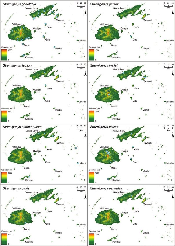
6                                                                                       Insect Systematics and Diversity, 2019, Vol. 3, No. 6

Standard measurements and indices                                           biology. Insects3D is available as a free download from the Apple App
Measurements and terminology follow Bolton (2000). All measure-             Store      .
ments are expressed in millimeters. Measurements presented for all          A short video demonstrating the features and functionality of
previously described species, with the exception of S. trauma, are          Insects3D is available from the online version of this article (Supp.
taken from Bolton (2000). Measurements presented for S. trauma              Vid. S2) and on YouTube .
include original data taken from Fijian specimens.                              The Insects3D app allows users to visualize 3D specimen models
                                                                            of the six new Strumigenys species described here using two different
MsL
    Mesosoma length (= Weber’s Length, = Alitrunk length).
                                                                            modes (Fig. 1). In 3D mode, the models display on-screen and can
    The diagonal length of the mesosoma in profile from the
                                                                            be rotated along three axes using touch gestures. In AR mode, the
    point at which the pronotum meets the cervical shield to
                                                                            models are anchored either to a visual marker or to a user-selected
    the posterior basal angle of the metapleuron.
                                                                            surface. The computer-generated models display on the device’s
CI  Cephalic index. HW/HL × 100.
                                                                            camera screen and behave as if they were fixed objects in the room.
EL	Eye length. Maximum diameter of the eye measured in
                                                                            The user observes the models from different angles either by keeping
    lateral view.
                                                                            the mobile device stationary and moving the visual marker, or by
FI  Femur index. FL/HW × 100.
                                                                            keeping the marker stationary and moving the mobile device.

                                                                                                                                                     Downloaded from https://academic.oup.com/isd/article/3/6/6/5610803 by guest on 31 December 2021
FL	Metafemur length. Maximum length of hind femur meas-
                                                                                Users select which species to visualize using the taxon selector
    ured from junction with trochanter to junction with tibia.
                                                                            dropdown menu and toggle between specimen model and map
HL	Head length. The length of the head capsule excluding
                                                                            model by tapping the specimen/map icon. The following features are
    the mandibles, measured in full-face view in a straight
                                                                            available in both 3D and AR mode: a) pinch-to-resize [scales size of
    line from the mid-point of the anterior clypeal margin to
                                                                            specimen and map model], b) spin [rotates specimen model 360° on
    the mid-point of the occipital margin. In species where
                                                                            vertical axis], c) texture slider [increases/decreases opacity of spe-
    one or both of these margins is concave the measure-
                                                                            cimen model texture (e.g., surface color)].
    ment is taken from the mid-point of a transverse line that
                                                                                The app recognizes two classes of markers: universal markers
    spans the apices of the projecting portions.
                                                                            and species-specific markers. Visual markers are typically graphic
HW	Head width. The maximum width of the head in full-face
                                                                            symbols that can be recognized with machine vision and accessed
    view, excluding the eyes.
                                                                            using cameras in mobile devices (Zhou et al. 2008, Costanza and
MI  Mandibular index. MdL/HL × 100.
                                                                            Huang 2009, Alakärppä et al. 2017). The universal marker (Supp.
MdL
    Mandible length. The straight-line length of the mandible
                                                                            Fig. 1 [online only]) is a vector-based illustration of a Strumigenys
    at full closure, measured in the same plane for which the
                                                                            head in face-view. Once scanned with the device camera in AR mode
    HL measurement is taken (i.e., full-face view), from the
                                                                            of the app, the universal marker allows users to select across all
    mandibular apex to the anterior clypeal margin, or to
                                                                            six specimen models. The species-specific markers are the plates of
    the transverse line connecting the anteriormost points in
                                                                            shaded surface display volume renderings presented for each of the
    those taxa where the margin is concave medially.
                                                                            six new species presented here. Scanning micro-CT plates labeled
PI  Pronotal index. PW/HW × 100.
                                                                            ‘AR marker’ with the Insects3D app in AR mode will trigger the dis-
PW	Pronotal width. The maximum width of the pronotum in
                                                                            play of the respective specimen model.
    dorsal view.
SI  Scape index. SL/HW × 100.
                                                                            Texture mapping
SL	Scape length. The maximum straight-line length of the
                                                                            After evaluating our texture mapping results, we suggest several
    scape, excluding the basal constriction or neck that
                                                                            modifications to improve future methods. With respect to texture
    occurs just distal of the condylar bulb. (In taxa with a
                                                                            mapping, our primary objective in this study was to determine
    hypertrophied subbasal lobe on the scape SL is measured
                                                                            whether adequate results could be achieved using only the three
    from the tip of the subbasal lobe to the scape apex.)
                                                                            standard 2D specimen photographs (head, profile, dorsal). Although
TL	Total length. The total outstretched length of the ant
                                                                            the addition of color to the mesh models presented here may make
    from the mandibular apex to the gastral apex; when
                                                                            them more engaging for a nonscientific audience, they are not satis-
    measured in profile the sum of MdL + HL + MsL +
                                                                            factory for scientific research applications. Quality would most cer-
    lengths of waist segments + length of gaster.
                                                                            tainly increase to the extent photographs from additional angles are
                                                                            incorporated. Constraining our sources to the three standard images
Nomenclature
                                                                            limited the texture quality of the models. Much of the surface de-
This paper and the nomenclatural acts it contains have been regis-          tail preserved in the 500k polygon micro-CT model (e.g., individual
tered in Zoobank (www.zoobank.org), the official register of the            ommatidia) was masked by low-resolution texture imagery. As re-
International Commission on Zoological Nomenclature. The LSID               sult, comparable visual quality could be achieved by using lower
(Life Science Identifier) number of the publication is: urn:lsid:zoobank.   polycount and faster performing mesh models.
org:pub:F19C2E83-6D10-4EA8-BD17-A9C5C5ED73C3                                     Upon reviewing the nascent literature, we propose 360° image-
                                                                            based techniques, such as those used in photogrammetry, are more
                                                                            economical and will arguably yield higher quality texture results—
Results and Discussion
                                                                            as measured by verisimilitude and processing requirements—than
Three-dimensional model development and                                     those achieved by our use of only three standard images mapped
optimization for augmented reality applications                             onto CT-based mesh models. As caveat, CT-based techniques are
Insects3D: an augmented reality app for visualizing                         superior to image-based techniques for capturing internal and oc-
specimen models                                                             cluded structures, and new methods for combining the two are being
Insects3D is an iOS compatible app designed as a proof-of-concept           developed with results more impressive than those presented here
solution for integrating augmented reality content with systematic          (Ijiri et al. 2018).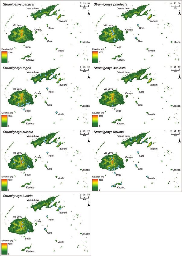
Insect Systematics and Diversity, 2019, Vol. 3, No. 6                                                                                                        7

                mode selector       map/specimen toggle            taxon selector

                 spin 360˚                             texture slider
                                                                                A                                                          C

                                                                                                                                                                  Downloaded from https://academic.oup.com/isd/article/3/6/6/5610803 by guest on 31 December 2021
                                                                                B                                                         D

                                                                                                                                          E

Fig. 1. Screenshots illustrating available features in the Insects3D app. The Insects3D app is available for free on the Apple App Store and is compatible with
iPhone and iPad devices. A video demonstrating the features and functionality of Insects3D is available from the online version of this article (Supp. Vid. S2)
and on YouTube . https://youtu.be/uSy1M7zPjIY. (A) 3D mode displaying specimen model of Strumigenys avatar. (B) 3D mode
displaying map model of S. avatar. (C) Augmented reality (AR) mode projecting specimen model of S. avatar using Fig 13 as the marker. (D) AR mode projecting
map model of S. artemis using the universal marker (Supp. Fig.1 [online only]). (E) 3D model of S. avatar projected onto the surface of a laboratory desk using
the 'place anywhere' function

Opportunities and challenges for AR enhanced biodiversity                           installed on your phone. You pull the Strumigenys avatar holotype
science                                                                             out from the drawer, place it in an empty unit tray or on a stage,
With respect to the study presented here, the current state of AR                   and use your app to scan the printed ‘CASENT0185902’ unique spe-
technology suggests its value to biodiversity education outstrips its               cimen identifier label. Without needing computer or microscope, you
value to taxonomic research. The models that worked best on our                     examine from all angles a 3D virtual model using your phone screen.
mobile devices with respect to loading time and rendering speed were                You view a 3D map of the collection locality to ascertain biogeo-
the ones with the lowest visual quality—and hence, most limited                     graphic factors. If the collection site was captured with 360° photo
taxonomic value. And even if the highest visual quality models were                 or video, you point your phone up to view the type locality’s canopy
loaded quickly and rendered smoothly, using a mobile device for                     cover, down for ground cover and substrate, straight ahead and all
model viewing is cumbersome and not necessarily conducive to a                      around for vegetation composition.
taxonomic description workflow that already includes microscopes,                       In contrast to AR, the micro-CT technology used to generate
keyboards, and computer screens.                                                    virtual specimen models added discernable value to our taxonomic
    While AR added limited value to the taxonomic study presented                   workflows. For example, incorporating 3D models into our species
here, it is worth discussing the potential of AR technology as it re-               description and diagnostic key workflows decreased the time and
lates to specimen-based research. A future—albeit a distant one—                    risk of handling type specimens. Unlike our phones, which were un-
is imagined in which a researcher surrounded by museum drawers                      able to smoothly render high polycount mesh models, our personal
accesses specimen data in AR using a mobile device. Consider, for                   computers had no difficulty processing the highest quality scans.
example, a specimen AR app similar to the one presented here is                     There were numerous occasions whereupon the character states such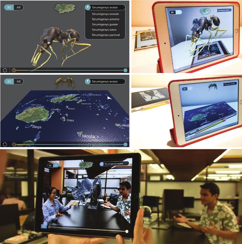
8                                                                                         Insect Systematics and Diversity, 2019, Vol. 3, No. 6

as shape and sculpture were rendered clearly enough in the 3D spe-           available to large audiences. Being able to interact with large-scale,
cimen models as to be used for species description without needing           3D and potentially natural-color models using one’s own phone or
to physically handle the type specimens. Likewise, viewing multiple          tablet is a more immersive experience than viewing a small, static, 2D
models simultaneously was useful for designing the diagnostic key.           photograph of the same specimen. In many ways, interacting with spe-
We found the 3D models to be preferable to 2D photographs for                cimen models using AR technologies offers a compelling alternative to
examining shape and sculpture, but they were inadequate for exam-            microscopes. Microscopes are expensive, limited in availability, and
ining pilosity—a result also discussed by Hita Garcia et al. (2017b).        difficult to use without prior training. With AR, an experience similar
    Although we advocate for increasing the use of 3D technolo-              to viewing minute invertebrates on a rotating stage under strong mag-
gies in taxonomic research, we caution that its inclusion—as with            nification can be achieved using the phone in one’s pocket.
molecular data—should not be prerequisite for the publication of
species descriptions. In our introduction, we argued that new im-            Taxonomic Analysis
aging technologies have increased the rate and clarity of species            Synoptic list of Strumigenys species known
descriptions. While we believe this claim true for focus-stacking            from Fiji
technologies used for 2D specimen photography, it is less applic-
able to 3D imagining—at least when microtomographic methods are              S. anorak, n. sp.

                                                                                                                                                       Downloaded from https://academic.oup.com/isd/article/3/6/6/5610803 by guest on 31 December 2021
used. With respect to taxonomic research, 3D microtomography is              S. artemis, n. sp.
arguably more similar to molecular data. Both add significant value          S. avatar, n. sp.
to taxonomy but also significant investments in time, training, and          S. basiliska Bolton
treasure. Given the enormous numbers of undescribed species rela-            S. chernovi Dlussky
tive to the few numbers of taxonomists and slow publication rate,            S. daithma Bolton
adding additional constraints is not necessarily a winning strategy          S. ekasura Bolton
for addressing the taxonomic impediment.                                     S. frivola Bolton
    Whereas the value of AR technology to taxonomic research is dis-         S. gunter, n. sp.
putable in its current state, AR has demonstrated broad value in edu-        S. godeffroyi Mayr
cational contexts (Wu et al. 2013, Akçayır and Akçayır 2017, Chen               = S. butteli Forel
et al. 2017)—and biodiversity education specifically (White et al.              = S. geococci Calilung
2006, Yeh et al. 2006, Barry et al. 2012, Chiang et al. 2014, Seltmann          = S. indica Forel
et al. 2017). The technology already exists for taxonomists to upload        S. jepsoni Mann
3D specimen models to online databases, and for end users to view            S. mailei Wilson & Taylor
models on their mobile devices using free iOS and Android apps such          S. membranifera Emery
as Sketchfab, Augment, and similar platforms. Student posters at sci-           = S. foochowensis Wheeler
entific conferences, curated exhibits at natural history museums, text-         = S. marioni Wheeler
books in classrooms could all use AR to make taxonomic subjects                 = S. santschii Forel

                                     head                                 mesosoma                            metasoma
                                                                                                                  spongiform
                                                                             propodeal                              tissue
                                                         humeral hair                        propodeal petiole
                                 upper scrobe                                 declivity        spine                postpetiole
                                   margin
                                                                                          lamella
                                                                                                                                  basigastral
         dorsal

                                                                                                                                   costulae
         ventral

                                                              apicoscrobal
                                                                  hair
                                                               mesopleuron

                                                                                                                          hind femur

                                                                                                                               hind tarsus

                                                                                                                                basitarsal
                                                                                                                                  hair

Fig. 2. Taxonomic characters in profile view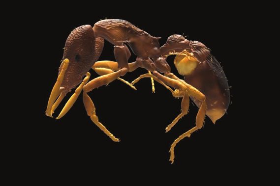
Insect Systematics and Diversity, 2019, Vol. 3, No. 6 9
= S. silvestriana Wheeler S. sulcata Bolton
= S. simillima Emery S. trauma (Bolton)
= S. vitiensis Mann S. tumida Bolton
= S. williamsi Wheeler
S. nidifex Mann
Diagnosis of Strumigenys worker caste among Fijian ants
S. oasis, n. sp.
Small to minute. Head shape triangular. Antennae 6-segmented.
S. panaulax Bolton
Antennal club 2-segmented. Eyes located on lower margin of an-
S. parzival, n. sp.
tennal scrobes. Mandibles either linear and tipped with apical fork
S. praefecta Bolton
or triangular and armed with numerous denticles. Propodeum armed
S. rogeri Emery
with spines or teeth. Waist 2-segmented. Spongiform tissue attached
= S. incisa Godfrey
to at least some portion of waist. Hairs appearing flagellate or spatu-
= S. sulfurea Santschi
late on at least some portion of head or body.
S. scelesta Mann
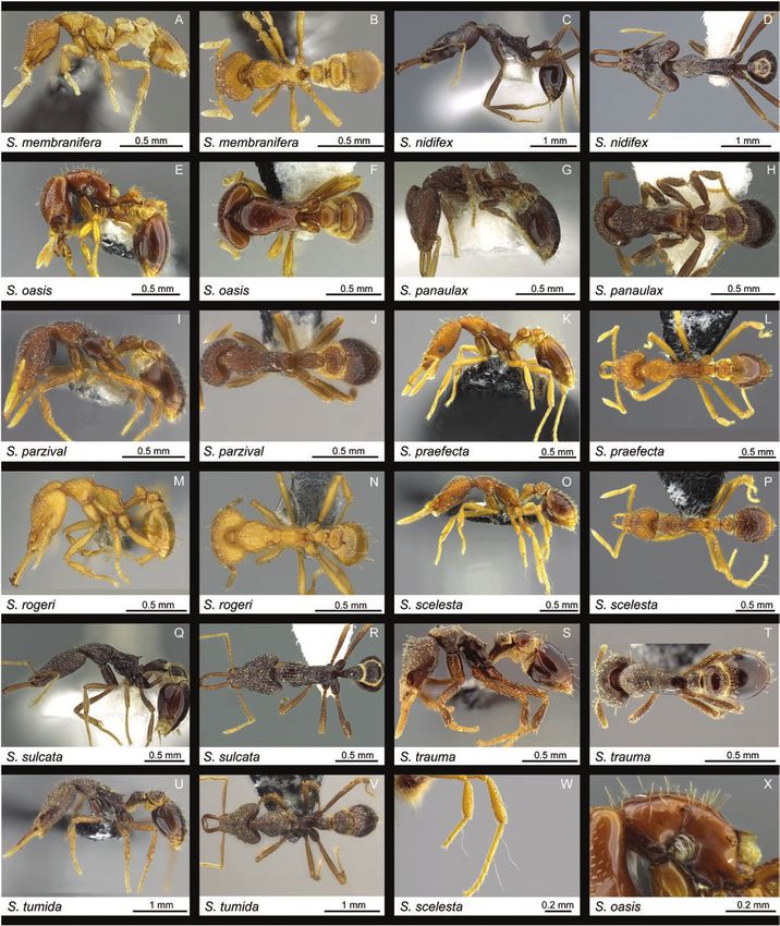
Key to worker caste of Fijian Strumigenys
Downloaded from https://academic.oup.com/isd/article/3/6/6/5610803 by guest on 31 December 2021
apiscrobal Refer to Figs. 2–5 for morphological terms used in the diagnostic
posterior
hair key. Specific character states referred to in the key are presented
in Fig. 6.
1. Mandibles triangular and armed with denticles (Fig. 6A)���������2
– Mandibles linear and armed with an apical fork (Fig. 6B–F)����3
2. Dorsal surface of head covered by short appressed spatulate
antennal scape hairs (Fig. 5B, Fig. 8K). Pronotal humeral hairs present (Fig.
6R, Fig. 10T). Propodeal declivity lacking a broad and con-
spicuous spongiform lamella (Fig. 6V, Fig. 10S). Dorsal surfaces
of mesosoma and petiole punctate (Fig. 10T) ����������� S. trauma
– Dorsal surface of head not covered by short appressed spatulate
hairs, hairs thin and simple (Fig. 5A, C, Fig. 8A). Pronotal hu-
mandible meral hairs absent (Fig. 6S). Propodeal declivity equipped with a
broad and conspicuous spongiform lamella (Fig. 6W, Fig. 10A).
Dorsal surfaces of mesosoma and petiole completely polished
anterior
(Fig. 10B)�������������������������������������S. membranifera (introduced)
3. Preapical dentition of each mandible with two preapical teeth
(Fig. 6F, Fig. 8H). Ventrolateral margin of head immediately in
front of the eye with an abrupt and very conspicuous preocular
preapical tooth notch or indentation (Fig. 6G)������������������ S. rogeri (introduced)
apical fork – Preapical dentition of each mandible either absent or with a
single tooth (Fig. 6E) or denticle (Fig. 6C). Ventrolateral margin
Fig. 3. Taxonomic characters in full face view of head immediately in front of the eye lacking an abrupt and
metafemur
basitarsal
hair
basigastral
apiscrobal
hair humeral costulae
hair spongiform
tissue
anterior posterior
Fig. 4. Taxonomic characters in dorsal view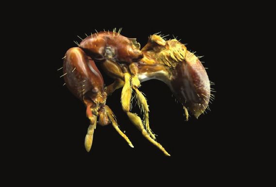
10                                                                                                                        Insect Systematics and Diversity, 2019, Vol. 3, No. 6

                                                                                                         –	With head in full-face view the upper scrobe margin usually
                                                                                                              with a single hair that freely projects laterally, in apicoscrobal
                                                                                                              position; this hair may be flagellate (Fig. 6K), filiform, or short
                                                                                                              and stiff (Fig. 6L); sometimes lacking a hair in this position
                                                                                                              (Fig. 6M) (dorsolateral margin of posterolateral lobe to apex
                                                                                                              of scrobe may have laterally projecting hairs)����������������������10
                                                                                                         9.	Freely projecting filamentous hairs present along entire lateral
                                                                                                              margin of head from posterolateral lobe to antennal insertions
                                                                                                              (Fig. 6I, Fig. 7A). Cephalic and pronotal surfaces smooth with
                                                                                                              large and deep irregular pits (Fig. 7A, Fig. 9B). First gastral
                                           B       C         D          E                                     segment lacking any sculpturing the entire length of the tergite
                            A                                                     F                           (Fig. 9B)��������������������������������������������������������������������S. anorak
                                                                                                         –	 Freely projecting filamentous hairs lacking between the
                                                                                                              apicoscrobal hair and eye level (Fig. 6J, Fig. 7B). Cephalic and
                                                                                                              pronotal surfaces reticulate-punctate, lacking large irregular

                                                                                                                                                                                                                Downloaded from https://academic.oup.com/isd/article/3/6/6/5610803 by guest on 31 December 2021
Fig. 5. Types of hairs (A–E); (A) appressed simple, (B) appressed spatulate,                                  pits (Fig. 7E, Fig. 9J). First gastral segment with basigastral
(C) erect simple, (D) erect spatulate, (E) filiform, (F) flagellate
                                                                                                              costulae distinctly longer than the length of the postpetiolar
                                                                                                              disc (Fig. 9J)����������������������������������������������������������� S. chernovi
   very conspicuous preocular notch or indentation (Fig. 6H)��������                                    10.	With mesosoma in profile dorsum of pronotum usually with
   �������������������������������������������������������������������������������������������������4        one additional pair of hairs equal to the length of the humeral
4. Dorsal (outer) surface of hind basitarsus with one or more freely                                          hair (Fig. 6X); rarely with more than one pair���������������������11
   projecting filiform (Fig. 5E) or flagellate (Fig. 5F) hairs that are                                  –	 With mesosoma in profile dorsum of pronotum lacking
   very long and suberect to erect (Fig. 6P); this specialized pilosity                                       additional hairs equal to the length of the humeral hair
   may also be present on the middle basitarsus and the middle and                                            (Fig. 6Y)�������������������������������������������������������������������������������12
   hind tibiae (Fig. 10W)��������������������������������������������������������������5                11. Eye composed of single facet (Fig. 9S)������������������������S. gunter
– Dorsal (outer) surface of the middle and hind basitarsi and tibiae                                     –    Eye composed of four or more facets (Fig. 9U)��������������������
   lacking freely projecting long filiform or flagellate hairs (Fig.                                          ��������������������������������������������������������������������������� S. jepsoni
   6Q); any pilosity present is simple to spatulate and usually de-                                      12. Apicoscrobal hair present and long, filiform or flagellate; this
   cumbent to appressed (Fig. 5A, B)������������������������������������������14                             hair very different in form and length from any other on the
5. First gastral tergite entirely covered with fine dense longitudinal                                        margin both anterior and posterior to it (Fig. 6K). In profile
   sulcate sculpture (Fig. 6Z); no other form of sculpture present on                                         dorsal surfaces of mesosoma, metasoma and gaster with long
   sclerite��������������������������������������������������������������������������������������6           flagellate hairs (Fig. 6BB)�������������������������������������S. godeffroyi
– First gastral tergite not entirely covered with longitudinal sul-                                      –	Apicoscrobal hair absent; entire margin with a dense row of
   cate sculpture; tergite usually with basigastral costulae that                                             uniformly shaped small curved hairs (Fig. 6M). In profile dorsal
   may extend up to half the length of the sclerite, which is usu-                                            surfaces of mesosoma, metasoma and gaster lacking long fla-
   ally unsculptured posteriorly (Fig. 6AA); occasionally tergite                                             gellate hairs (Fig. 6CC)������������������������������������������� S. scelesta
   entirely smooth or with another form of sculpture distal of the                                       13.	With head in full-face view the upper scrobe margin with two
   basigastral costulae������������������������������������������������������������������7                   or more flagellate or filiform hairs that freely project laterally
6. Dorsum of pronotum and petiole obliquely costulate-rugulose                                                (Fig. 7F); at least with one in apicoscrobal position and another
   (Fig. 10H)����������������������������������������������������������������S. panaulax                     anterior to this (dorsolateral margin of posterolateral lobe to
– Dorsum of pronotum and petiole longitudinally and irregularly                                               apex of scrobe may have additional laterally projecting hairs)
   rugoreticulate (Fig. 10J)��������������������������������������������� S. parzival                        �������������������������������������������������������������������������������S. daithma
7. With mesosoma in profile the propodeal declivity equipped with                                        –	With head in full-face view the upper scrobe margin usually
   a broad and conspicuous cuticular or spongiform lamella (Fig.                                              with a single flagellate hair that freely projects laterally; lat-
   6W); the propodeal tooth may be replaced by the lamella or                                                 erally projecting hairs also occur on dorsolateral margin of
   completely buried in the lamella, or the lamella may subtend the                                           posterolateral lobe to apex. Refer to labeled structures in Fig.
   ventral margin of the tooth for most or all of its length; posterior                                       2 for location of upper scrobe margin and apicoscrobal hair
   (free) margin of lamella may be convex, straight or irregular but                                          ��������������������������������������������������������������������������������S. ekasura
   it is not narrowly concave, nor is it close to and parallel with the                                  14. Fully closed mandible in full-face view comparatively short and
   edge of the declivity������������������������������������������������������������������8                  very broad proximally and strikingly tapered distally (Fig. 6D),
–	With mesosoma in profile the propodeal declivity equipped                                                  not linear or curvilinear. Outer margin of mandible flared out-
      with a simple carina or at most a narrow cuticular spongiform                                           wards or strongly convex prebasally, the mandible not straight,
      flange (Fig. 6V); carina or narrow flange does not subtend the                                          not evenly convex, not evenly bowed outwards�������������������15
      ventral margin of the tooth for most or all of its length; pos-                                    –    Fully closed mandible in full-face view comparatively long
      terior (free) margin of carina or narrow flange concave, close to                                       and usually obviously linear or curvilinear, sometimes slightly
      and parallel with the edge of the declivity����������������������������13                              increasing in width towards the base (Fig. 6B, C). Outer margin
8.	With head in full-face view the upper scrobe margin with two                                              of mandible not flared outwards or strongly convex prebasally,
      or more flagellate or filiform hairs that freely project laterally                                      mandible straight, evenly convex, or evenly bowed outwards
      (Fig. 6I, J); at least with one in apicoscrobal position and an-                                        ��������������������������������������������������������������������������������������������16
      other anterior to this (dorsolateral margin of posterolateral                                      15. Posteromedian margin of head with single deep circular punc-
      lobe to apex of scrobe may have additional laterally projecting                                         ture (Fig. 8D). With mesosoma in profile the propodeal de-
      hairs)�������������������������������������������������������������������������������������9           clivity equipped with a broad and conspicuous spongiform
Insect Systematics and Diversity, 2019, Vol. 3, No. 6 11
Downloaded from https://academic.oup.com/isd/article/3/6/6/5610803 by guest on 31 December 2021
Fig. 6. Taxonomic characters used in identification key (A–CC)12 Insect Systematics and Diversity, 2019, Vol. 3, No. 6
lamella (Fig. 6W). Dense tuft of white filamentous hairs arising Species accounts
from the lateral promesonotal border above the procoxae (Fig.
S. anorak n. sp.
10E). Dorsum of mesosoma with abundant long white erect
hairs (Fig. 10E)������������������������������������������������������������ S. oasis (Fig. 7A; Fig. 9A, B; Fig. 11; Fig. 26, Model 1)
– Posteromedian margin of head lacking single deep circular
puncture (Fig. 7D). Dense tuft of white filamentous hairs (Zoobank LSID: urn:lsid:zoobank.org:act:65D7817A-
lacking from the lateral promesonotal border above the EAA1-4A8F-9A87-992C2C141829)
procoxae (Fig. 9G). With mesosoma in profile the propodeal
HOLOTYPE. Fiji, Viti Levu, Ra Prov., 7.5 km NE Vunisea Village,
declivity lacking a conspicuous spongiform lamella (Fig. 6V).
2003-07-14, 300 m, −17.4833° 178.1430°, small forest fragment,
Dorsum of mesosoma lacking abundant long white erect hairs
sifted litter, A. Rakabula (worker, dry pinned, BPBM, specimen
(Fig. 9G)����������������������������������������������������������������S. basiliska
code CASENT0186900).
16. Pronotal humeral hair absent (Fig. 6S)���������������������������������17
= Strumigenys sp. FJ18 (Sarnat and Economo 2012: 136, pl. 140)
– Pronotal humeral hair present (Fig. 6R)�������������������������������18
17. Dorsum of pronotum coarsely and deeply longitudinally Cybertype. Volumetric raw data (in DICOM format) of the phys-
sulcate, with a ploughed appearance (Fig. 10R). Disc of ical holotype (CASENT0186900) is deposited at Dryad (https://doi.
Downloaded from https://academic.oup.com/isd/article/3/6/6/5610803 by guest on 31 December 2021
postpetiole longitudinally sulcate, sulci narrower and finer org/10.5061/dryad.1f9d55b/1) and can be freely accessed as vir-
than on pronotum (Fig. 10R). Cephalic dorsum coarsely longi- tual representation of the type. In addition to the cybertype data at
tudinally rugose (Fig. 8J). Ventral spongiform curtain of petiole Dryad, we also provide a freely accessible 3D surface model of the
narrow, at maximum only a fraction the depth of the peduncle holotype at Sketchfab (https://skfb.ly/6vsIH).
(Fig. 10Q)�����������������������������������������������������������������S. sulcata Worker. Measurements (n = 3): TL 3.18–3.40 mm, HL 0.79–
– Dorsum of pronotum densely reticulate-punctate (Fig. 10L). 0.87 mm, HW 0.54–0.59 mm, CI 68, MdL 0.35–0.36 mm, MI
Disc of postpetiole weakly rugulose (Fig. 10L). Cephalic 41–45, SL 0.51–0.57 mm, SI 96, PW 0.37–0.40 mm, PI 68, FL 0.62–
dorsum sharply and densely reticulate-punctate (Fig. 8G). 0.68 mm, FI 115, EL 0.08–0.09 mm. Color a polished reddish-brown
Ventral spongiform curtain of petiole deep, at maximum at with yellowish-brown appendages. Mandible long and linear with one
least equal to the depth of the peduncle (Fig. 10K)������������������� preapical tooth; preapical tooth approximately as long as maximum
����������������������������������������������������������������������������� S. praefecta mandible width. Dorsolateral head margin with abundant laterally
18. In full-face view, sides of head immediately in front of the eye projecting filiform and occasionally flagellate hairs. Apicoscrobal hair
with an abrupt and very conspicuous indentation (Fig. 6N). flagellate. Upper scrobe margin anterior to apicoscrobal hair with fili-
Large species (HW > 0.80 mm)��������������������������������������������19 form hairs. Cephalic dorsum irregularly reticulate-punctate. Ground
– In full-face view, sides of head immediately in front of the eye pilosity of cephalic dorsum simple and long. Pronotal humeral hair
lacking an abrupt and very conspicuous indentation (Fig. 6O). flagellate. Promesonotal dorsum with abundant projecting filiform
Small species (HW < 0.70 mm)��������������������������������������������20 apically curved hairs; mesonotum with at least one pair of longer fla-
19. Sculpture on first gastral tergite consisting of basigastral gellate hairs. Promesonotal dorsum and propodeal dorsum smooth
costulae restricted to less than a quarter of the length of the and marked by laterally by deep and occasionally overlapping spongi-
tergite, remainder the tergite strongly polished and shiny (Fig. form filled pits of irregular size and shape from small and circular to
10C, D)���������������������������������������������������������������������S. nidifex large and ovoid. Side of promesonotum smooth and marked with
– Sculpture on first gastral tergite consisting of basigastral pits similar to those of dorsal surface. Mesopleuron, metapleuron and
costulae restricted to basal half of the tergite and strongly side of propodeum glassy smooth. Propodeal tooth short, narrowly
reticulate-punctate ground sculpture covering the entire tergite triangular, subtended by a broad lamella with a convex posterior
(Fig. 9; Fig. 10E, F)����������������������������������������������������S. avatar margin. Dorsal and ventral surfaces of hind femur with abundant
20. Preapical dentition of mandible a stout tooth/denticle that is erect filiform and subflagellate hairs. Dorsal surface of hind tibia with
distinctly shorter than the width of the mandible at the point one filiform hair on basal portion and one subflagellate hair on apical
where it arises (Fig. 6C; Fig. 8L); preapical tooth never as long portion. Dorsal surface of hind tarsus with three flagellate hairs. With
as the maximum width of the mandible. Mesopleuron and petiole node in profile the dorsum of the node much longer than its
procoxa reticulate-punctate (Fig. 10U)��������������������� S. tumida short oblique anterior face; lateral spongiform lobe small; restricted
– Preapical dentition of mandible a slender spiniform tooth that to posterior margin of node. Dorsum of petiole node highly pol-
is always longer than the width of the mandible at the point ished with scattered irregular shaped pits. Disc of postpetiole glassy
where it arises (Fig. 6B); preapical tooth usually at least as long smooth. Ventral lobe of postpetiole spongiform. First gastral tergite
as the maximum width of the mandible. Mesopleuron and with erect flagellate hairs and long ground pilosity. Basigastral sculp-
procoxa smooth and shiny���������������������������������������������������21 ture entirely absent.
21. Apicoscrobal and pronotal humeri hairs flagellate (Fig. 7H; Strumigenys anorak is a relatively large and highly distinctive
Fig. 9O, P). Pronotal dorsum lacking erect hairs in addition to rich reddish brown species with abundant flagellate and filiform
the humeral pair (Fig. 6Y)����������������������������������������� S. frivola hairs on its dorsal surfaces, a unique sculpture characterized by a
– Apicoscrobal and pronotal humeri hairs simple. Pronotal smooth integument interrupted by deep irregularly sized and shaped
dorsum with erect hairs in addition to the humeral pair (Fig. pits, and the presence of a broad and conspicuous propodeal lamella.
6X)��������������������������������������������������������������������������������������22 Additional characters for separating S. anorak from similar Fijian
22. Hairs on dorsal surface of head, mesosoma and metasoma species are given in the notes for S. daithma. The species is repre-
short, thick and subdecumbent (Fig. 9C). In full face view eyes sented by three specimens, two from Gau and one from Viti Levu, all
mostly shielded by lateral head margins (Fig. 7B)��������������������� between 400 and 475 meters and collected from extracted leaf litter.
�������������������������������������������������������������������������������� S. artemis Etymology: Anorak refers to the online avatar of James Donovan
– Hairs on dorsal surface of head, mesosoma and metasoma Halliday, creator of the virtual reality world OASIS, in the fictional
long, fine and erect (Fig. 9W). In full face view eyes visible, not work Ready Player One (Cline 2011). The name is a noun in appos-
shielded by lateral head margins (Fig. 7L)������������������ S. mailei ition and thus invariable.You can also read